HAL Id: hal-03021301
https://hal.archives-ouvertes.fr/hal-03021301
Submitted on 25 Nov 2020
HAL is a multi-disciplinary open access
archive for the deposit and dissemination of
sci-entific research documents, whether they are
pub-lished or not. The documents may come from
teaching and research institutions in France or
abroad, or from public or private research centers.
L’archive ouverte pluridisciplinaire HAL, est
destinée au dépôt et à la diffusion de documents
scientifiques de niveau recherche, publiés ou non,
émanant des établissements d’enseignement et de
recherche français ou étrangers, des laboratoires
publics ou privés.
Cnidarian cell cryopreservation : A powerful tool for
cultivating and functional assays
Clara Fricano, Eric Röttinger, Paola Furla, Stephanie Barnay-Verdier
To cite this version:
Clara Fricano, Eric Röttinger, Paola Furla, Stephanie Barnay-Verdier. Cnidarian cell
cryopreserva-tion : A powerful tool for cultivating and funccryopreserva-tional assays. Cells, MDPI, In press. �hal-03021301�
Cells 2020, 9, x; doi: FOR PEER REVIEW www.mdpi.com/journal/cells Article
1
Cnidarian cell cryopreservation: A powerful tool for
2
cultivation and functional assays
3
Clara Fricano1, Eric Röttinger1, Paola Furla1,$ and Stéphanie Barnay-Verdier 1,2,$,*
4
1 te d’Azur, CNRS, INSERM, Institute for Research on Cancer and Aging (IRCAN), 28 avenue
5
de Valombrose, F-06107 Nice, France
6
2 Sorbonne Université, UFR 927, 4 place Jussieu, F-75252 Paris, France
7
* Correspondence: Corresponding author: stephanie.barnay-verdier@upmc.fr; Tel.: +33 4 93 37 77 39
8
$ These authors share the seniorship position
9
16-digit ORCID:10
Clara Fricano:0000-0001-8496-591511
Eric Röttinger:0000-0002-2938-677412
Paola Furla: 0000-0001-9899-942X13
Stéphanie Barnay-Verdier:0000-0002-4615-305914
Running Title: Cryopreservation of Cnidarian Cell Cultures
15
Abstract: Cnidarian primary cell cultures have a strong potential to become a universal tool to
16
assess stress-response mechanisms at the cellular level. However, primary cell cultures are
17
time-consuming regarding their establishment and maintenance. Cryopreservation is a commonly
18
used approach to provide stable cell stocks for experiments, but it is yet to be established for
19
Cnidarian cell cultures. The aim of this study was therefore to design a cryopreservation protocol
20
for primary cell cultures of the Cnidarian Anemonia viridis, using dimethyl sulfoxide (DMSO) as a
21
cryoprotectant, enriched or not with foetal bovine serum (FBS). We determined that DMSO 5%
22
with 25% FBS was an efficient cryosolution, resulting in 70% of post-thaw cell survival. The success
23
of this protocol was first confirmed by a constant post-thaw survival independently of the cell
24
culture age (up to 45 days old) and the storage period (up to 87 days). Finally, cryopreserved cells
25
displayed a long-term recovery with a maintenance of the primary cell culture parameters and
26
cellular functions: formation of cell aggregates, high viability and constant cell growth, and
27
unchanged intrinsic resistance to hyperthermal stress. These results will further bring new
28
opportunities for the scientific community interested in molecular, cellular and biochemical aspects
29
of cnidarian biology.
30
Keywords: primary cell culture; sea anemone; Anemonia viridis; DMSO; marine invertebrate;
31
post-thaw recovery
32
1. Introduction
33
In vitro cell cultures are important tools for research in many fields, including development,
34
virology, cancer research, toxicity testing, biotechnology, biomedicine as well as for environmental
35
research [1–3]. Mammalian cells lines are well established and commonly used since decades,
36
followed by other vertebrates (e.g. zebrafish; for review Vallone et al. 2007 [4]) and insect cell lines
37
(for review Lynn, 2001 [5]). Despite much effort devoted since the 1970s (see for reviews Rinkevich,
38
2005 [6], 2011 [7] and Cai and Zhang, 2014 [8]) marine invertebrate cell cultures are not as advanced.
39
While marine invertebrate cell lines (i.e. permanently established cell cultures) are yet to be
40
available, recent reports on the establishment of primary cell cultures are encouraging with
41
maintenance and/or growing from a couple of days to weeks [9–14].
42
Once protocols for reproducibly initiating primary cell cultures are established, the next
43
important obstacle to overcome is the development of preservation procedures in order to outreach
primary cell cultures limitations, notably their limited lifespan. Indeed, such a preservation tool will
45
reduce the frequency of primary cell culture establishment and will minimize wastage of a valuable
46
resource at reseedings by creating cell stocks for as long as a primary cell culture is healthy. Among
47
preservation procedures, cryopreservation is considered to be the optimal long-term storage method
48
for maintaining a variety of biological materials, including cell cultures, in a state of metabolic arrest
49
for considerable periods of time [16].
50
To date, for marine invertebrates, spermatozoa, oocytes, embryos and different larval stages
51
have been successfully cryopreserved mostly from Mollusk and Echinoderm species, but also from
52
Arthropod and Cnidarian species (see for reviews Odintsova and Boroda, 2012 [17] and Paredes,
53
2015 [18]). Other biomaterials have been studied in a cryopreservation context, such as coral
54
fragments [19] and primmorphs from sponge cells [20]. The tolerance to various cryoprotectants of
55
tissue balls from corals was also investigated by Feuillassier et al. (2015) [21]. However, considering
56
the limited advancements in marine invertebrate cell cultures, cryopreservation of marine
57
invertebrate dissociated cells is seldom reported [18]. If they are, they largely focus on Mollusks
58
[22–30], with some other studies conducted on Echinoderms (e.g. dissociated cells from sea urchins
59
larvae [24,28]), on sponge cells [31–34] and on coral cells dissociated from embryos or larvae [35,36].
60
Besides, none of these studies, except those on sponge cells, maintained cryopreserved cells in
61
cultures for more than a few days nor use cryopreserved cells for subsequent experiments.
62
One of the major factors that determines the success of a cryopreservation protocol is the type of
63
cryoprotecting agents (CPAs) used [16]. CPAs prevent damage to the cells from changes in osmotic
64
pressure and intracellular ice crystal formation. Among the various CPAs, dimethyl sulfoxide
65
(DMSO), a penetrating CPA, is the most common and widely used cryoprotectant to maintain frozen
66
cell lines. The precise mechanism by which it protects cells remains unclear; it has been suggested
67
that DMSO depresses the freezing point of cryosolutions [37], and that it can modulate the water
68
network hydrating the membrane hence reducing the stress induced by the volume changes of
69
water during freeze-thaw [38]. Penetrating CPAs could however induce some cytotoxicity due to the
70
disruption of intracellular signalling which results in cell death [39–41]. For marine invertebrate
71
studies, Paredes (2015) [18] reported in her review that DMSO was the most effective CPA for 70% of
72
the published work on germ cells, embryos and larvae compared to others CPAs such as glycerol.
73
This trend is also found for marine invertebrate dissociated cells [22,35,21,33]. In addition, in marine
74
invertebrate studies, DMSO was found as an efficient CPA on its own [22,29,35] but more frequently
75
in combination with other CPAs or with proteins, vitamins or sugar cocktails [18,24, 26,27,33].
76
Indeed, the preservative capacity of DMSO was long known to be increased when serum, such as
77
foetal bovine serum (containing cocktail of proteins), is added to the cryosolution [42,43].
78
We have previously reported the establishment of primary cell cultures of a soft-body
79
cnidarian, the temperate sea anemone Anemonia viridis [9]. The established cell culture protocol
80
resulted in the maintenance of primary cell cultures with gastrodermal signature [15]. These cell
81
cultures were successfully used to assess the cellular response (e.g. viability) to environmental stress
82
[15] thus creating new perspectives for further fundamental, environmental and biotechnological
83
questions. An efficient cryopreservation procedure would therefore be an essential and powerful
84
tool for facilitating research in deciphering molecular mechanisms and cellular events in cnidarian
85
cells.
86
The aim of this study was therefore to design a cryopreservation protocol for primary
87
gastrodermal A. viridis cell cultures in order to ensure a high post-thaw cell survival, preserving
88
long-term recovery: cell viability, cell growth and physiological responses. All these advances will
89
participate to raise the cnidarian cell cultures as a model system for marine invertebrate research
90
perspectives.
2. Material and Methods
92
2.1. Biological Material
93
Five individuals of Anemonia viridis (Forska l 1775) were collected (prefectural authorization
94
n°107 ; 02/28/2019) from ‘Plage des ondes’, Antibes, France, (43°33’17’’N, 7°07’17.7’’E), and
95
maintained in a closed-circuit aquarium with artificial seawater (ASW) at 36-38‰ w h P d b
96
Expert Reef Salt, at 18.0 ± 0.5 °C with weekly water changes. A LED bar (450 nm – Deckey LED
97
aqua um) p d d l gh a a c a a u a g ad a c f 100 μm l m−2s−1 (measured using a
98
special sensor QSL-100, Biospherical Instruments Inc.) on a 12h:12h (light:dark) photoperiod. Sea
99
anemones were fed once a week with oysters.
100
2.2. Primary cell cultures
101
From each A. viridis individual, an independent primary cell culture was obtained and
102
maintained as described in Ventura et al. (2018) [15]. Briefly, cell dissociation was performed
103
enzymatically with 0.15% collagenase type I (Sigma-Aldrich). Cells were cultured at 20.0 ± 0.5 °C and
104
in the dark, in an optimized culture medium (CM) consisted of : 20% GMIM (Gibco), 5% foetal
105
b um (FBS; PAA/GE H al hca ), 1% ka amyc (100 μg/ml, S gma-Aldrich), 1%
106
amph c B (2.5 μg/ml; I ch m), 1% a b c a myc c lu (Sigma-Aldrich), 1% L-
107
glutamate (Sigma-Aldrich) and 71% of filtered ASW. The CM was adapted in respect to the
108
Mediterranean Seawater characteristics (i.e. salinity 40 ppt and pH 8.1). From day 3, culture medium
109
was replaced weekly and cells were seeded at 250,000 cells/ml in 12 well-plates.
110
2.3. Cryopreservation protocol
111
As cryoprotectant, DMSO (Sigma-Aldrich) was tested at two concentrations in the final CPA
112
solution: 5% or 10% (following Munroe et al., 2018 [33]). DMSO was dissolved in the CM or in the
113
CM enriched with foetal bovine serum (FBS) at 25% final. Control conditions without DMSO were
114
also tested using CM enriched or not with FBS (i.e. ‘CM’ or ‘CM + 25% FBS’).
115
From day 17 after dissociation, the primary cell cultures were established with reliable cellular
116
parameters [15]. By consequence, the cultivated cells were cryopreserved at different time points,
117
from day 17 to 45 after cell dissociation. Each cryopreserved material contained 2 million cells that
118
were placed in a cryotube containing 1 ml of the tested solution. Cryotubes were directly placed in a
119
-80°C freezer (Ultra-Low Temperature VIP series, SANYO) and kept there for 8 to 87 days.
120
For thawing, cryotubes were removed from the -80°C freezer after the defined period and
121
immediately transferred for 1-2 min into a water bath, pre-warmed at 20°C.
122
For seeding the cryopreserved cells, the cryotubes were centrifuged for 5 min at 1500 rpm. The
123
supernatant was then removed, the cell pellets resuspended in the cell culture medium and seeded
124
at 250,000 cells/ml in 12 well-plates [15].
125
2.4. Cell survival, cell viability, cell growth rate and cell size assessment
126
Cell survival was measured right after thawing cryopreserved cells, before reseeding. It was
127
determined as the percentage of viable cells relative to the 2 million cells initially cryopreserved. To
128
assess the number of viable cells, a sub-sample (100 µl) of cryopreserved cells was harvested after
129
the thawing phase. Cell viability was assessed by evaluating the membrane integrity thanks to the
130
Evans blue method. Therefore, viable cells (unstained) and dead cells (stained) were identified and
131
counted. on a Neubauer improved haemocytometer (Sigma-Aldrich) using an optic microscope
132
(Zeiss Axio Imager Z1).
133
Cell viability was measured every week to monitor the cell culture health state overtime. A
134
sub-sample (100 µL) of cultivated cells was harvested weekly and using Evans blue method, viable
135
cells (unstained) and dead cells (stained) were identified and counted. The cell viability was defined
136
as the percentage of viable cells relative to total cells (i.e. viable and dead cells). In addition, two
137
complementary methods for cell viability assessment, i.e. overall enzymatic activity using the
fluorescein diacetate (FDA) staining combined with a non-vital dye (Hoechst) and cell metabolic
139
activity with 2-(4, 5-dimethyl-2-thiazolyl)-3, 5-diphenyl-2H tetrazolium bromide (MTT) assay, were
140
also conducted (see details in Supplementary Material and Methods).
141
Cell growth rate was also assessed every week using the previous viable cells counts with
142
Evans blue method. The following formula was then used to calculate the 7-day averaged daily
143
growth rate:
144
, (d= day)
145
Cell growth rate and cell viability were monitored for each cell culture before and after
146
cryopreservation. The monitoring of these factors for cryopreserved cells was done by considering
147
that the age of the cells at the thawing time is the same age they were at the freezing time.
148
Before and after cryopreservation, during the cell counts under optic microscope (objective
149
x20), cells were measured, and cell sizes were scored with Zeiss microscope software Zen 2 (blue
150
edition).
151
2.5. Hyperthermal stress experiment
152
In order to assess the maintenance of cryopreserved cells functionality, the response of
153
cryopreserved cells to a controlled stress experiment was investigated. The cultivated A. viridis cells
154
response to hyperthermal stress was assessed following the protocol published by Ventura et al.
155
(2018) [15]. Hyperthermal stress was induced in 12-well plates exposed to two different
156
temperatures: 20°C (control) and 28°C (hyperthermal condition), for 7 days. This experiment was
157
conducted either with non-cryopreserved or cryopreserved cells, 7 days after thawing. At least four
158
independent experiments were conducted from four primary cell cultures. For each assay, we
159
analyzed 3 wells as technical replicates.
160
2.6. Statistical analyses
161
All statistical analyses were conducted using the R v.3.6.0 software[44]. In order to assess the
162
effect of the cryosolutions on cell survival, to compare global viability and growth between
163
non-cryopreserved and cryopreserved cells, and to compare viability and growth rate values after
164
hyperthermal stress, either one-way ANOVA analyses were performed when parametric analyses
165
were possible (under normality and variance equality assumptions), or Kruskal-wallis when
166
non-parametric analyses were required. These analyses were followed, if necessary, by the
167
appropriate post-hoc, i.e. Tukey for ANOVA analyses, and Dunn for Kruskal-wallis analyses. Then,
168
to investigate the effect of the storage duration and the cell culture age on the cell survival,
169
correlation tests with linear regression model were conducted. Finally, repeated measures ANOVA
170
were conducted to compare cell viability and growth through time of non-cryopreserved and
171
cryopreserved cultures.
3. Results and Discussion
173
3.1. Success in set up of cryopreservation protocol on cell survival
174
The efficiency of the cryopreservation solutions was first evaluated with the percentage of cells
175
that survive a cryopreservation period of 10 ± 1 days and the subsequent thawing process. When
176
cells were cryopreserved in the culture medium (CM) or in CM with DMSO 5 or 10% the mean
177
percentage of cells that survived at -80°C, was around 7%. There were no significant differences
178
between these conditions (ANOVA; p>0.05) (Figure 1). However, when cells were cryopreserved in
179
the two conditions containing an FBS supplementation, we observed a significant higher percentage
180
of cell survival compared to non-enriched medium conditions (p<0.01 and p<0.0001 respectively for
181
the CM+25% FBS and for the DMSO 5% in the CM+ 25%FBS). Compared to FBS enriched CM alone
182
(± 45% survival rate), adding 5% of DMSO to the FBS enriched CM significantly enhanced the
183
survival rate of the cells to 67% (p<0.05; Figure 1). Thus, the latter constituted the optimal
184
cryosolution among those tested for cryopreserving cnidarian cells.
185
186
Figure 1. Percentage of cell survival following 10 ± 1 days of cryopreservation and thawing. For each
187
tested cryopreservation solution, the number of viable cells were compared to the 2 million cells
188
initially cryopreserved. Mean values and standard errors are represented, with n≥3 biological
189
replicates per condition. The ANOVA revealed significant differences between data (p=1.14.10-10) and
190
the results from the Tukey post-hoc analysis are represented with letters: a ≠ b (p<0.01), a ≠ c
191
(p<0.0001), b ≠ c (p<0.05).
192
As it was observed in most of marine invertebrate studies [18,24,26,27,33], DMSO was found to
193
be an efficient CPA for A. viridis cultivated cells when it combined with serum supplementation. The
194
reason could be that carbohydrates, lipids and proteins present in serums act as membrane
195
stabilizers, therefore they may help preventing membrane damage during the freezing process
196
[45–48].
197
The survival rate of the designed cryopreservation protocol for A. viridis cultivated cells with
198
the optimal cryosolution is comparable to those determined for dissociated cells [26,33] or other
199
biomaterials [18] from marine invertebrates, as well as the ones from vertebrate in vitro cells [49].
200
Since the optimal cryopreservation solution, among those tested in this study, was found to be
201
the DMSO 5% in FBS enriched culture medium, this solution was reused for different
202
cryopreservation durations in order to determine the influence of the cryopreservation time on cell
203
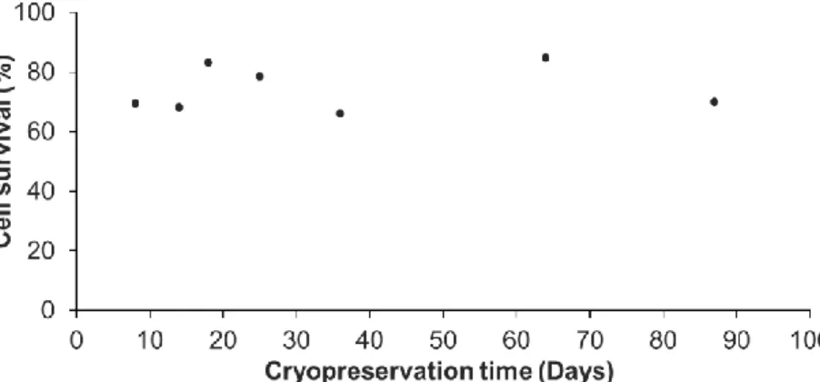
survival after thawing. The representative results for a A. viridis cell culture shown in Figure 2
204
demonstrated that there was no influence of the cryopreservation duration on the cell survival.
205
Indeed, from 8 to 87 days at -80°C, the percentage of cells that survived cryopreservation and
206
thawing, did not vary significantly (linear regression model; coefficient not statistically significantly
207
different from 0, p>0.05). The statistical analyses done on all available data (at least 3 biological
208
replicates) confirmed that there were no significant differences between the cryopreservation
209
durations (p>0.05; data not shown).
211
Figure 2. Percentage of cell survival following different cryopreservation durations and thawing. For
212
each tested cryopreservation duration, the number of viable cells were compared to the 2 million
213
initially cryopreserved cells. These representative results were obtained from one primary cell
214
culture cryopreserved at 24 days after initial seeding in 7 cryotubes, each cryotube being analyzed at
215
the end of a given cryopreservation duration (from 8 to 87 days). No significant differences between
216
times were found (linear regression model; r²=0.037; p=0.443).
217
Cells could therefore be cryopreserved for almost 3 months without any impact on the
218
post-thaw survival compared to short-term cryopreservation, further validating the efficiency of the
219
designed protocol. This is a major and significant progress for marine invertebrate cell
220
cryopreservation. In fact, although few studies evaluated the cryopreservation efficiency after a
221
storage of several weeks [22,24,25,28] and a maximum of 12 months [30], the majority only
222
cryopreserved cells for a few hours to a few days [23,26,27,35,36,29,33].
223
Cryopreservation of vertebrate cell lines is well-advanced, and protocols allow to keep
224
cryopreserved cells for years [16]. Therefore, additional experiments are required to assess if longer
225
cryopreservation durations are possible and/or to determine the maximal duration using the
226
protocol developed in this study. If a duration limitation were to be found, a long-term storage in
227
liquid nitrogen, or -150°C freezers following the initial -80°C freezing should be envisioned.
228
In addition, we also assessed the influence of the age (time after initial seeding) of the primary
229
cell culture on the survival at thawing. Representative results of one primary cell culture,
230
cryopreserved at five different ages (from day 17 to day 45, Figure 3) revealed no significant
231
differences (linear regression model; coefficient not statistically significantly different from 0,
232
p>0.05). The statistical analyses done on all available data (3 biological replicates) confirmed that
233
there was no influence of the culture age on the cell survival (p>0.05; data not shown). One primary
234
cell culture can therefore be cryopreserved at each reseeding, as long as the cellular parameters
235
p u ly d f d f “h al hy” A. viridis primary cell culture are maintained, i.e. cell aggregates
236
formation, high viability and constant growth rate [15]. Being able to do so, considerable amounts of
237
cell stocks for each cell culture could be created.
238
239
Figure 3. Percentage of cell survival after thawing in function of the age of the cell culture.
240
Representative results were obtained from one primary cell culture cryopreserved at different times
since its initial seeding (from day 17 to day 45). At each age tested, one cryotube was analyzed. No
242
significant differences between ages were found (linear regression model; r²= 0.15; p=0.512).
243
3.2. Absence of cryopreservation impact on cell recovery and functional parameters
244
To assess long-term cell recovery, we measured weekly, at each reseeding, the cell viability of
245
all primary cell cultures cryopreserved. Viability of cryopreserved cells was monitored for a period
246
going from 6 to 12 weeks after thawing and was compared to the viability of the corresponding
247
non-cryopreserved cell culture. Results of cell viability monitoring for 6 weeks after thawing for one
248
primary cell culture cryopreserved at day 24 for 8 or 36 days are presented in Figure 4. The data
249
show that cryopreserved cells were stably viable after thawing through time (>90% viability) with a
250
cell viability equivalent to that of the corresponding non-cryopreserved cell culture (repeated
251
measures ANOVA; p>0.05). Repeated measures ANOVA conducted on all biological replicates,
252
confirmed no differences in the cell viability over time between non-cryopreserved and
253
cryopreserved cells nor with the cryopreservation storage period (p>0.05; see Figure S1 and S2). The
254
mean of viability, over time, was maintained at 95 ± 1.9 % and at 96 ± 0.99 %, respectively in the
255
different non-cryopreserved and cryopreserved cell cultures monitored (ANOVA; p>0.05; see Figure
256
S3a). In addition, data obtained with the two complementary cell viability assays performed on
257
non-cryopreserved and cryopreserved cells at different time points of the kinetics confirmed all
258
these results, i.e. no differences in cell viability between non-cryopreserved and cryopreserved cells
259
and a maintenance over time of the cell viability (ANOVA ; p>0.05; see Figure S4).
260
261
Figure 4. Over time cell viability of a representative cell culture, comparing cryopreserved and
262
non-cryopreserved cells. Non-cryopreserved culture in grey dotted line, the same culture
263
cryopreserved at 24 days since initial seeding and thawed after 8 days of storage in grey solid line
264
and after 36 days in black solid line. The age of the cryopreserved cells at thawing is considered the
265
same as at the freezing time. Mean values of three technical replicates are shown with standard error
266
bars (although not visible because smaller than the data point symbols). Repeated measures ANOVA
267
revealed no significant differences in cell viability at each time between the non-cryopreserved
268
culture and the cryopreserved ones (p=0.582).
269
As a first functional parameter, the long-term cell growth was assessed. Indeed, we considered
270
the resumption of cell cycle after cryopreservation as an essential functional parameter to explore.
271
Non-cryopreserved and cryopreserved cells displayed a similar daily growth rate over time and
272
independently of the cryopreservation duration (8 or 36 days) as shown by a representative cell
273
culture in Figure 5 (repeated measures ANOVA; p>0.05).
274
The same result was obtained for all cell cultures tested in this study (see Figure S1 and S2).
275
Interestingly, the cryopreserved cells stored for 79 days (Figure S2d) displayed a low initial growth
276
rate one week after thawing and reseeding, suggesting that cryopreserved cells may need a longer
277
time (between one and two weeks after thawing) to fully recover after such cryopreservation storage
278
duration.
Furthermore, the mean of the daily growth rate was maintained at 1.78 ± 0.39 and at 1.73 ± 0.33,
280
respectively in the different non-cryopreserved cell cultures and cryopreserved cell cultures
281
monitored, with no significant differences between these two conditions (ANOVA; p>0.05; see
282
Figure S3b).
283
284
Figure 5. Over time daily growth rate of a representative cell culture, comparing cryopreserved and
285
non-cryopreserved cells. Non-cryopreserved cell culture in grey dotted line, the same culture
286
cryopreserved at 24 days and thawed after either 8 days of storage in grey solid line or after 36 days
287
in black solid line. Mean values of three technical replicates and standard error bars are shown.
288
Repeated measures ANOVA revealed no significant differences in cell growth trend between the
289
non-cryopreserved culture and the cryopreserved ones (p=0.722).
290
Long-term cell viability and growth rate monitoring of cryopreserved vs non-cryopreserved
291
cell cultures corroborated the healthy state of cryopreserved cells. Therefore, these analyses were
292
completed with weekly microscope observations in order to assess the cell culture behavior. In the
293
Figure 6, the comparison of a 31-day old non cryopreserved culture with the corresponding
294
cryopreserved culture (considering the thawing age is equal to the freezing age) showed that
295
cryopreserved cells, as the non-cryopreserved cells, form adherent cell aggregates, which is the
296
characteristic architecture of A. viridis primary cell cultures[9,15]. Besides, cryopreserved cells in
297
culture presented the same mean size (5.2 ± 0.49 µm) that non-cryopreserved cells (5.17 ± 0.54 µm;
298
ANOVA; p=0.957), suggesting no volume change after thawing (see Figure S5).
299
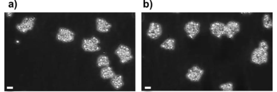
300
Figure 6. Observation of aggregates of A. viridis gastrodermal cells in culture before and after
301
cryopreservation; a) Cell culture at day 31 since its establishment, and b) the same culture
302
cryopreserved at day 24 for 79 days, 7 days after thawing. Phase contrast microscopy (objective x20),
303
scale bars = 10 µm.
304
Therefore, cryopreserved cells maintained through time identical viability, growth and shape to
305
the corresponding non-cryopreserved cell culture, and this independently of the cryopreservation
306
duration. These results indicate that the cryopreservation designed protocol stored cells in a healthy
307
state allowing them to fully recover after thawing and to behave like their origin culture. This
308
monitoring constitutes an essential part in validating the storage protocol for further use of
309
cryopreserved cells and represents a major strength of this study. Indeed, the reseeding of thawed
310
cells was rarely done on previous marine invertebrate studies, and cells are usually maintained only
for a few hours to a few days [22–24,28,25,27], with a maximum of 15 days for bivalve cells in Dessai
312
(2018) [30] and around 40 days for sponge cells [32,34].
313
As a second parameter of the cell functionality after cryopreservation, we investigated the
314
response of cryopreserved cells to a controlled stress experiment. In Cnidarians, and more
315
particularly in our research model, A. viridis, hyperthermal stress is well known to induce oxidative
316
damages and cell death, i.e. apoptosis [50,51]. Using A. viridis primary cell cultures, we previously
317
p d ha hyp h m a (+8° ) d d ’ duc a y x da damag mpac u al bu
318
provoked a drastic decrease of cell growth [15]. Thus, in this study, we compared the response of
319
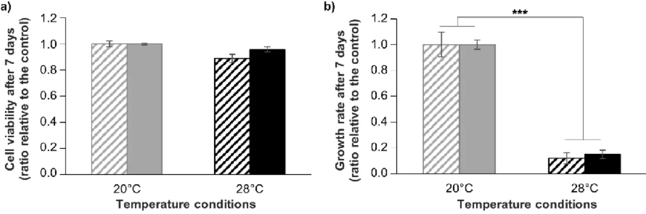
cryopreserved and non-cryopreserved cells submitted to the same hyperthermal stress, in terms of
320
ab l y a d g w h. Th ul h w ha hyp h mal d d ’ cha g ignificantly the cell
321
viability neither for non-cryopreserved cells nor for cryopreserved cells (ANOVA; p>0.05) (Figure
322
7a). Moreover, cell growth rate was drastically decreased by around 80% after 7 days at 28°C
323
compared to 20°C condition (Kruskal-wallis; p<0.001), without any significant differences between
324
non-cryopreserved and cryopreserved cells (Kruskal-wallis; p>0.05) (Figure 7b). These results are in
325
line with data from Ventura et al. (2018) [15] and show that cryopreserved cells displayed identical
326
resistance to non-cryopreserved cells, strongly corroborating the non-alteration of cell functionality.
327
Therefore, not only A. viridis cells in vitro can be successfully cryopreserved and reseeded, but they
328
can be reliably used for further experiments, like it is done for mammalian cells. Although functional
329
analyses are sometimes conducted for marine invertebrate cells, through metabolic and enzymatic
330
activities [23,26,24,28,25,30], only some sponge cells studies conducted experiments using
331
cryopreserved cells [31,34]. However, this is a fundamental assessment in order to validate the
332
cryopreservation protocol for creating reliable models.
333
334
Figure 7. Comparison of cryopreserved and non-cryopreserved cells in response to hyperthermia.
335
Assessment of cell viability (a) and growth (b) of A. viridis cells in response to a hyperthermal stress
336
of +8°C (black bars) for 7 days, for non-cryopreserved cultures (striped bars) and for cryopreserved
337
cultures (filled bars). Cell viability and growth are expressed relative to control condition (20°C –
338
g y ba ). M a alu w h a da d ba a h w , b l g cal pl ca ≥4. Th a k
339
represent the significant differences between control and stress conditions (*** Kruskall-Wallis:
340
p<0.001).
341
4. Conclusions
342
In this study we succeeded to design an easy and rapid cryopreservation procedure for
343
Anemonia viridis primary cell cultures. The established protocol enabled us to obtain high cell
344
survival after thawing and a full long-term recovery of the cell culture behavior. The development of
345
cryopreservation in cnidarian primary cell cultures enables us to preserve stable cell stocks available
346
shortly after thawing for experimental procedures and sharing with the scientific community. This
347
new tool will be an important asset to raise A. viridis primary cell cultures as a powerful model for
348
studying and understanding the cnidarian properties (i.e. symbiosis lifestyle, response to stress,
349
aging), difficult to study in most cnidarian models at the molecular and cellular levels.
Supplementary Materials
351
352
Supplementary Material and Methods
353
Complementary cell viability assays
354
FDA (Fluorescein Diacetate, 4 µg/mL; Sigma-Aldrich) and Hoechst 33342 (5 µg/mL; Sigma-Aldrich) were
355
added and incubated with cells during 15 minutes at 20°C in the dark. Viable cells (fluorescent in green)
356
and dead cells (fluorescent in blue) were identified and counted on a Neubauer improved
357
haemocytometer (Sigma-Aldrich) using a fluorescence microscope (Zeiss Axio Imager Z1). The cell
358
viability was defined as the percentage of viable cells relative to total cells (i.e. viable and dead cells).
359
MTT assay was performed following manufacturer instructions with slight modifications. Briefly, prior the
360
assay 60 000 cells were seeded in triplicate in 96-well plate in 100 µL of culture medium and incubated for 24h.
361
20 µL of 5mg/mL MTT solution (Sigma-Aldrich) is then added to each well and incubated 5h at 20°C in the
362
dark. Then, the supernatant is removed, and the yielded formazan was dissolved in the suitable detergent
363
(isopropanol) for 15 minutes. Subsequently, the plates' light absorption (OD) is read at wavelength 590 nm on
364
spectrofluorometer (SAFAS, Monaco). The cell viability was expressed as follow: Viability % = (cryopreserved
365
cells OD /non-cryopreserved cells OD) ×100
366
367
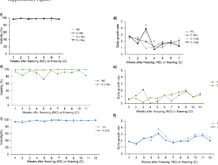
Supplementary Figures368
369
370
Figure S1. Over time viability (a, b, c) and daily growth rate (d, e, f) of three primary cell cultures,
371
comparing non-cryopreserved cells (NC) and cells cryopreserved for a short storage period (C). The
372
c y p a ag p d m d h l g d, g f m 8 day (‘8d’) 18 day
373
(‘18d’). Each graph per variable measured represent one primary cell culture and its corresponding
374
c y p d c ll cul u ( ). T m p a m d a “W k af f z g (N ) haw g
375
( ) m ” . . h m g p d h f h -cryopreserved cultures begins after the
376
freezing time, and the one for cryopreserved cells is done after thawing, thus considering the age at
377
that time is the same age as at the freezing time.
379
Figure S2. Over time viability (a, b) and daily growth rate (c, d) of two primary cell cultures, comparing
380
non-cryopreserved cells (NC) and cells cryopreserved for a long storage period (C). The cryopreservation
381
ag p d m d h l g d, g f m 25 day (‘25d’) 79 day (‘79d’). Each graph per
382
variable measured represent one primary cell culture and its corresponding cryopreserved cell culture(s).
383
T m p a m d a “W k af f z g (N ) haw g ( ) m ” . . h m g
384
presented here for the non-cryopreserved cultures begins after the freezing time, and the one for
385
cryopreserved cells is done after thawing, thus considering the age at that time is the same age as at the
386
freezing time.
388
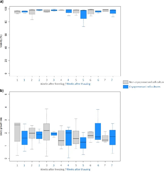
Figure S3. Box plots of over time viability (a) and daily growth rate (b) of all cell cultures monitored
389
(non-cryopreserved in gray and cryopreserved in blue). Time points are identical to the ones present
390
in Fig. S1 and S2 and are here going from 1 to 7 weeks, which represent the time points after freezing
391
(for non-cryopreserved cells) or after thawing (for cryopreserved cells) where at least 3 biological
392
replicates were monitored.
394
Figure S4. Comparison of cell viability between 38-day old non-cryopreserved cell cultures (‘N
395
38d’), a d c y p d c ll cul u , h 38-day ld (‘ 38d’) 80-day ld (‘ 80d’). ll
396
viability was measured with (a) FDA/Hoechst staining and with (b) MTT assay. Mean values with
397
standard error bars are shown ( ≥3). ANOVA analyses revealed no significant differences in the
398
viability values between non-cryopreserved and cryopreserved cells, and over time between
399
cryopreserved cells (p=0.45 for panel a; p=0.378 for panel b).
400
401
402
403
404
405
406
Figure S5. Observation, on Neubauer improved haemocytometer under optic microscope (objective
407
x20), of the same A. viridis gastrodermal cell culture (a) before and (b) after cryopreservation and
408
stained with Evans blue (i.e. the dead cells stained in blue) (scale bar = 10 µm).
409
410
Acknowledgments: Authors thank Thalassa Marine Research & Environmental Awareness for the sampling of
411
the sea anemones. Authors thank also Brigitte Poderini, Thamilla Zamoum and Maxence Burtin for aquaria
412
maintenance and taking care of the animals. Authors are grateful to Pauline Cotinat for her help in the cell
413
culture establishment.
414
Funding: This work was supported by the SATT Sud-Est (project number: 1082-SA-18-CNRS).
415
Author Contributions: C.F. did all the investigations (experimental work) and the statistical analyses. P.F. and
416
S.B-V. designed and supervised the research. C.F., P.F. and S.B-V. wrote the manuscript. Finally, E.R., P.F. and
417
S.B-V. reviewed the manuscript.
418
Conflicts of Interest: On behalf of all authors, the corresponding author states that there is no conflict of
419
interest.
420
References