HAL Id: hal-03231086
https://hal.univ-lorraine.fr/hal-03231086
Submitted on 20 May 2021
HAL is a multi-disciplinary open access archive for the deposit and dissemination of sci-entific research documents, whether they are pub-lished or not. The documents may come from teaching and research institutions in France or abroad, or from public or private research centers.
L’archive ouverte pluridisciplinaire HAL, est destinée au dépôt et à la diffusion de documents scientifiques de niveau recherche, publiés ou non, émanant des établissements d’enseignement et de recherche français ou étrangers, des laboratoires publics ou privés.
Clinical Outcome in Acute Ischemic Stroke Treated with
Mechanical Thrombectomy?
Imad Derraz, Romain Bourcier, Marc Soudant, Sébastien Soize, Wagih Ben
Hassen, Gabriella Hossu, Frederic Clarencon, Anne Laure Derelle, Marie
Tisserand, Helene Raoult, et al.
To cite this version:
Imad Derraz, Romain Bourcier, Marc Soudant, Sébastien Soize, Wagih Ben Hassen, et al.. Does Clot Burden Score on Baseline T2*-MRI Impact Clinical Outcome in Acute Ischemic Stroke Treated with Mechanical Thrombectomy?. Journal of Stroke, Korean Stroke Society, 2019, 21 (1), pp.91-100. �10.5853/jos.2018.01921�. �hal-03231086�
Copyright © 2019 Korean Stroke Society
This is an Open Access article distributed under the terms of the Creative Commons Attribution Non-Commercial License (http://creativecommons.org/licenses/by-nc/4.0/) which permits unrestricted non-commercial use, distribution, and reproduction in any medium, provided the original work is properly cited.
Original Article
Background and Purpose A long clot, defined by a low (0-6) clot burden score (CBS) assessed by T2*-MR sequence, is associated with worse clinical outcome after intravenous thrombolysis (IVT) for acute ischemic stroke than is a small clot (CBS, 7-10). The added benefit of mechanical thrombectomy (MT) might be higher in patients with long clot. The aim of this pre-specified post hoc analysis of the THRombectomie des Artères CErebrales (THRACE) trial was to assess the association between T2*-CBS, successful recanalization and clinical outcome.
Methods Of 414 patients randomized in the THRACE trial, 281 patients were included in this analysis. Associations between T2*-CBS and clinical outcome on the modified Rankin Scale (mRS) at 3 months were tested.
Results High T2*-CBS, i.e., small clot, was associated with a shift toward better outcome on the mRS; proportional odds ratio (POR) per point CBS was 1.19 (95% confidence interval [CI], 1.05 to 1.34) in the whole population, 1.34 (95% CI, 1.13 to 1.59) in IVT group, and 1.04 (95% CI, 0.87 to 1.23) in IVTMT group. After adjustment for baseline prognostic variables, the effect of the full scale T2*-CBS was not statistically significant in the whole population and for the IVTMT group but remains significant for the IVT group (POR, 1.32; 95% CI, 1.11 to 1.58).
Conclusions A small clot, as assessed using T2*-CBS, is associated with improved outcome and may be used as a prognostic marker. Despite the worst outcome with long clot, the relative benefit of MT over IVT seemed to increase with low T2*-CBS and longer clot.
KeyWords Ischemic stroke; Magnetic resonance imaging; Thrombosis; Endovascular recanalization
Does Clot Burden Score on Baseline T2*-MRI Impact
Clinical Outcome in Acute Ischemic Stroke Treated
with Mechanical Thrombectomy?
Imad Derraz,
aRomain Bourcier,
bMarc Soudant,
cS
ébastien Soize,
dWagih Ben Hassen,
eGabriella Hossu,
fFrederic Clarencon,
gAnne Laure Derelle,
aMarie Tisserand,
hHelene Raoult,
iLaurence Legrand,
eSerge Bracard,
aCatherine Oppenheim,
eOlivier Naggara,
eon behalf of the
THRACE Investigators
aDepartment of Neuroradiology, CHRU Nancy, INSERM, University of Lorraine, Nancy, France bDepartment of Neuroradiology, CHU Nantes, Nantes, France
cDepartment of Biostatistics, CHRU Nancy, INSERM, University of Lorraine, Nancy, France dDepartment of Neuroradiology, CHU Reims, Reims, France
eParis Descartes University, INSERM UMR 894 and Department of Neuroradiology, Sainte-Anne Hospital Center, Paris, France fDepartment of CIC1433 Innovative Technology, CHRU Nancy, INSERM, University of Lorraine, Nancy, France
gDepartment of Neuroradiology, Pitié-Salpêtrière Hospital, Paris, France hDepartment of Neuroradiology, Foch Hospital, Suresnes, France iDepartment of Neuroradiology, CHU Rennes, Rennes, France
Correspondence: Olivier Naggara
Paris Descartes University, INSERM UMR 894 and Department of Neuroradiology, Sainte-Anne Hospital Center, 1 rue Cabanis, 75014 Paris, France Tel: +33-1-4565-8574 Fax: +33-1-4565-8470 E-mail: [email protected] Received: July 4, 2018 Revised: September 8, 2018 Accepted: October 2, 2018
Introduction
Recanalization of the arterial occlusion is the cornerstone of treatment in acute ischemic stroke (AIS) patients. Several ran-domized clinical trials (RCTs)1-7 and an individual patient data
meta-analysis8 have recently shown that mechanical
throm-bectomy (MT) combined with standard treatment (including intravenous thrombolysis [IVT]) was superior to standard treat-ment alone, with higher rates of reperfusion, more frequent fa-vorable functional outcome, and lower 3 months mortality, for severe AIS caused by large-vessel occlusion (LVO) in the anteri-or circulation. The cumulative evidence from these studies re-sulted in the modification of practice guidelines and profound changes in worldwide stroke care organization.9
Despite its compelling efficacy, up to half of AIS-LVO patients do not regain functional independence after MT.8 This
unfavor-able outcome is largely attributunfavor-able to unsuccessful mechanical recanalization.1-4,7 Amongst factors influencing recanalization
success, clot length is an important determinant,10-12 that was
scarcely assessed in the recent RCTs. Indeed, if patients were in-cluded in case of LVO on computed tomography angiography (CTA), only few studies13,14 assessed clot length using the
CTA-de-fined clot burden score (CTA-CBS). These analyses demonstrated a direct link between clot length, likelihood of recanalization, fi-nal infarct volumes, and 3-month neurological outcome.
Brain magnetic resonance imaging (MRI), using the T2*-MRI sequence, is a powerful tool to identify thrombus in AIS pa-tients, based on the presence of a susceptibility vessel sign (SVS).15,16 Clot length is included in the T2*-CBS,17 in which a
lower score reflects longer thrombus. T2*-CBS was recently used after IVT or MT, as a predictor of recanalization and func-tional outcome.17,18 However, as for CTA-CBS,19 these studies
were mainly retrospective and no adjustment was performed for crucial baseline prognostic variables such as initial National Institutes of Health Stroke Scale (NIHSS) score or recanaliza-tion results.
With more than 300 patients included with pre-treatment MRI, the THRombectomie des Artères CErebrales (THRACE) trial (ClinicalTrials.gov, number NCT01062698) offers a unique op-portunity to study the associations between T2*-CBS, success-ful recanalization rate and functional independence. The pur-pose of this prespecified post hoc analysis of the THRACE trial was to determine the relation between T2*-CBS and the effect on endovascular treatment and neurological improvement in AIS-LVO patients.
Methods
Study design
THRACE was a randomized controlled trial done in 26 centers in France. Study design and protocol have been previously de-tailed.7 AIS-LVO patients were randomly assigned in a 1:1 ratio
to receive either IVT alone (IVT group) or IVT+MT (IVTMT group). IVT had to be started within 4 hours and MT within 5 hours of symptom onset. Occlusions had to be confirmed by CTA or magnetic resonance angiography (MRA). Before randomization, written informed consent was obtained from all patients or their legal representatives. The study protocol was approved by
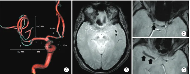
Figure 1. The T2*-clot burden score (T2*-CBS). (A) A score of 10 is normal, implying absence of susceptibility vessel sign (SVS) on T2*. Three points (as indicat-ed) are subtracted for SVS found in the supraclinoid internal carotid artery (ICA), 2 points for SVS in each of the proximal and distal halves of the middle cere-bral artery trunk (M1), 1 point for SVS in the A1–A4 segment and 2 points for SVS in the M2–M4 branches. A score of 0 implying complete multisegment vessel occlusion. (B) Patient 1, distal occlusion with T2*-CBS=9 (SVS in 1 left M2 branch). (C, D) Patient 2, proximal occlusion with T2*-CBS=3 (SVS in right supraclinoid ICA, proximal and distal halves of M1).
A B
C
the Comité de Protection des Personnes (CPP) III Nord Est Eth-ics Committee and the research boards of the participating centers. No data were reported on thrombus characteristics obtained on admission brain MRI.
Outcomes measures
The primary outcome in the THRACE trial was the proportion of patients with a score of 0–2 on the modified Rankin Scale (mRS), indicating functional independence, at 3 months after the intervention. Secondary outcomes nonexhaustively includ-ed, successful recanalization, defined as a modified Thromboly-sis in Cerebral Infarction (mTICI) score ≥2b20 in the IVTMT group,
and the NIHSS score at 24 hours in all patients. Clinical assess-ments were done by vascular neurologists who were not masked to the treatment to which the patients had been allo-cated.
Image analysis
MRI images and angiograms before and after MT were re-viewed by four experienced neuroradiologists, who were masked to randomization group and patient clinical outcome. Baseline examinations included the determination of the Al-berta Stroke Program Early CT Score (ASPECTS)21 on diffusion
weighted imaging (DWI) sequence and the location of the ar-terial occlusion by MRA. After initial training, two experienced observers from the core imaging committee searched for SVS, that is, a hypointense signal on T2* within a vascular cistern, exceeding the size of the homologous contralateral arterial di-ameter. If present, an appropriate T2*-CBS was assigned ac-cording to the methods of Legrand et al.17 T2*-CBS is a
10-point scoring system used to define the extent of thrombus in the anterior circulation (Figure 1). Because susceptibility ar-tifact at the skull base prevents evaluation of the infraclinoid internal carotid artery (ICA), this segment was not analyzed and 3 points were assigned to the supraclinoid ICA level, for consistency with the CTA-CBS. A score of 10 implies clot ab-sence. A score of 0 implies complete multisegment vessel oc-clusion by a long clot. T2*-CBS was subsequently dichotomized using a >6-point cut-off (0–6: long clot vs. 7–10: small clot), according to and for comparison with CTA-CBS studies.13
Statistical analysis
All primary outcome analyses were performed according to the intention-to-treat principle. For this study, the primary effect variable was the proportional adjusted common odds ratio for a shift in the direction of better outcome on the 3-month mRS. The association between full-scale or dichotomized T2*-CBS (0–6 vs. 7–10) with shift in the direction of better outcome on
the mRS was assessed using ordinal logistic regression respec-tively.
For all outcome parameters, two models were generated as previously used.13 Model A contained the T2*-CBS variable and
treatment. In model B, the main prognostic baseline variables were added: age, stroke severity (NIHSS score), glycemia, and ASPECTS score. An interaction term of treatment allocation with T2*-CBS was added to the both unadjusted and adjusted models to assess whether T2*-CBS was a treatment effect modifier. The models with and without added interaction term (nested models) were compared using the chi-square test.
Patient characteristics are reported for groups with and without SVS and patient with long (T2*-CBS 0–6) and small clot (T2*-CBS 7–10). Continuous variables were compared with Student t-test, a Mann-Whitney test, or Median test, as ap-propriate. Categorical variables were compared using chi-square or Fisher exact test, as appropriate.
For all statistical analyses, P<0.05 was considered statisti-cally significant. All statistical analyses were done with SAS/ STAT version 9.4 (SAS Institute Inc., Cary, NC, USA).
Results
Patient characteristics
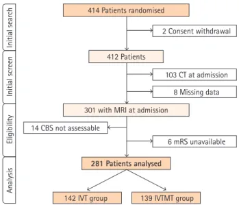
Among 414 patients randomized in the THRACE trial (Figure 2), initial imaging work-up was performed using MRI in 153 of 208 patients (74%) in the IVT group and 148 of 204 patients (73%) in the IVTMT group (P=0.83). Of these, 20 patients were excluded leaving 281 patients for analysis in the present study.
Figure 2. Flow chart study. CT, computerized tomography; MRI, magnetic resonance imaging; CBS indicates clot burden score; mRS, modified Rankin Scale; IVT, intravenous thrombolysis; IVTMT, intravenous thrombolysis me-chanical thrombectomy. 414 Patients randomised 2 Consent withdrawal 412 Patients 281 Patients analysed 14 CBS not assessable 103 CT at admission 8 Missing data 6 mRS unavailable
142 IVT group 139 IVTMT group 301 with MRI at admission
Initial search
Analysis
Initial screen
Baseline characteristics are summarized in Supplementary Table 1. Sociodemographic characteristics, comorbidities, and baseline NIHSS did not differ between IVT and IVTMT groups, nor delays from symptom onset to IVT initiation or to randomization.
Baseline characteristics according to T2*-CBS
group
Among 281 patients included in 19 centers, 234 (83.3%) demonstrated presence of SVS, 118 (50.4%) in the IVT group, and 116 (49.6%) in the IVTMT group. Overall, the median
T2*-Table 1. Baseline patient characteristics according to dichotomized T2*-CBS groups
Characteristic T2*-CBS (7–10) T2*-CBS (0–6) P
Number 170 ( 111 (
IVTMT randomization group 87 (51.2) 52 (46.8) 0.48*
Undergo IVTMT 57 (33.5) 43 (38.7) 0.37* Age (yr) 61.8±14.2 65.4±13.4 0.01† ≤70 109 (64.1) 50 (45.0) 0.002* >70 61 (35.9) 61 (55.0) Sex 0.32* Male 93 (54.7) 54 (48.6) Female 77 (45.3) 57 (51.4) Comorbidities Hypertension 84 (49.7) 57 (52.3) 0.67* Diabetes mellitus 20 (11.8) 5 (4.6) 0.04* History of stroke 9 (5.5) 9 (8.3) 0.37* Hypercholesterolemia 79 (53.0) 50 (50.5) 0.70*
Current tobacco use 43 (28.3) 14 (14.1) 0.009*
Coronary disease 20 (12.4) 20 (18.7) 0.16*
Etiology of cerebral infarction 0.41*
Large-artery atherosclerosis 25 (15.3) 13 (13.0)
Cardioembolism 64 (39.3) 51 (51.0)
Small-vessel occlusion 1 (0.6) 0 (0)
Other determined etiology 10 (6.1) 5 (5.0)
Undetermined etiology 63 (38.7) 31 (31.0)
Baseline NIHSS score 18 (13–21) 17 (14–20) 0.69‡
ASPECTS at baseline 7 (5–9) 7 (4–8) 0.72‡
ASPECTS (0–4) 25 (14.7) 28 (25.2) 0.04*
ASPECTS (5–7) 73 (42.9) 35 (31.5)
ASPECTS (8–10) 72 (42.4) 48 (43.2)
Baseline occlusion location (%) <0.001§
ICA 13 (7.6) 30 (27.0)
M1 155 (91.2) 81 (73.0)
M2 2 (1.2) 0 (0)
Workflow time (min)
From stroke onset to imaging 112 (88–135) 112 (89–142) 0.91‡
From stroke onset to IVT 146 (124–170) 154 (120–180) 0.49‡
From stroke onset to recanalization 239 (208–270) 261 (204–291) 0.44‡
ASPECTS at 24 hours 7 (4–8) 6 (3–8) 0.40‡
Values are presented as number (%), mean±standard deviation, or median (interquartile range).
CBS, clot burden score; IVTMT, intravenous thrombolysis mechanical thrombectomy; NIHSS, National Institutes of Health Stroke Scale; ASPECTS, Alberta Stroke Program Early CT Score; ICA, internal carotid artery; IVT, intravenous thrombolysis.
CBS was 7 (interquartile range, 6 to 8), similar in IVT and IVTMT groups (P=0.38). After dichotomizing the T2*-CBS, 111 patients (39.5%) had a long clot and 170 (60.5%) a small clot. Patients with a long clot were significantly older, had a lower DWI-AS-PECTS score, more often had diabetes mellitus or were active smokers at baseline (Table 1).
Primary outcome
The primary outcome was assessed in 281 patients (Table 2). At 3 months, 79 of 139 patients (56.8%) in the IVTMT group and 65 of 142 (45.8%) in the IVT group had functional independence. The primary outcome was not influenced by the presence of a SVS (120/144 in mRS 0–2 and 114/137 in mRS >2, P=0.98).
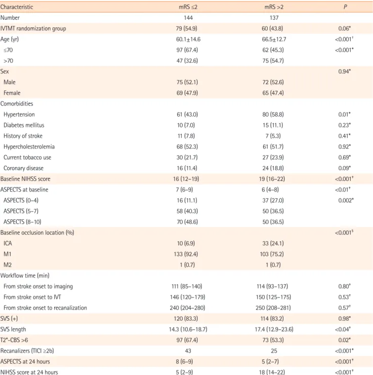
Table 2. Patient characteristics of dichotomized mRS groups
Characteristic mRS ≤2 mRS >2 P
Number 144 137
IVTMT randomization group 79 (54.9) 60 (43.8) 0.06*
Age (yr) 60.1±14.6 66.5±12.7 <0.001† ≤70 97 (67.4) 62 (45.3) <0.001* >70 47 (32.6) 75 (54.7) Sex 0.94* Male 75 (52.1) 72 (52.6) Female 69 (47.9) 65 (47.4) Comorbidities Hypertension 61 (43.0) 80 (58.8) 0.01* Diabetes mellitus 10 (7.0) 15 (11.1) 0.23* History of stroke 11 (7.8) 7 (5.3) 0.41* Hypercholesterolemia 68 (52.3) 61 (51.7) 0.92*
Current tobacco use 30 (21.7) 27 (23.9) 0.69*
Coronary disease 16 (11.4) 24 (18.8) 0.09*
Baseline NIHSS score 16 (12–19) 19 (16–22) <0.001‡
ASPECTS at baseline 7 (6–9) 6 (4–8) <0.01‡
ASPECTS (0–4) 16 (11.1) 37 (27.0) 0.002*
ASPECTS (5–7) 58 (40.3) 50 (36.5)
ASPECTS (8–10) 70 (48.6) 50 (36.5)
Baseline occlusion location (%) <0.001§
ICA 10 (6.9) 33 (24.1)
M1 133 (92.4) 103 (75.2)
M2 1 (0.7) 1 (0.7)
Workflow time (min)
From stroke onset to imaging 111 (85–140) 114 (93–137) 0.80‡
From stroke onset to IVT 146 (120–179) 150 (125–175) 0.53‡
From stroke onset to recanalization 240 (204–280) 250 (208–281) 0.57‡
SVS (+) 120 (83.3) 114 (83.2) 0.98*
SVS length 14.3 (10.6–18.7) 17.4 (12.9–23.6) <0.04‡
T2*-CBS >6 97 (67.4) 73 (53.3) 0.02*
Recanalizers (TICI ≥2b) 43 25 <0.001*
ASPECTS at 24 hours 8 (6–9) 5 (2–7) <0.001‡
NIHSS score at 24 hours 5 (2–9) 18 (14–22) <0.001‡
Values are presented as number (%), mean±standard deviation, or median (interquartile range).
mRS, modified Rankin Scale; IVTMT, intravenous thrombolysis mechanical thrombectomy; NIHSS, National Institutes of Health Stroke Scale; ASPECTS, Alberta Stroke Program Early CT Score; ICA, internal carotid artery; IVT, intravenous thrombolysis; SVS, susceptibility vessel sign; CBS, clot burden score; TICI, Throm-bolysis in Cerebral Infarction.
Proportion of small clot was significantly higher in patients with favorable (97/144 patients, 67.4%) than in patients with unfavorable outcome (73/137, 53.3%; OR, 1.81; 95% confi-dence interval [CI], 1.12 to 2.94; P=0.016). Small clot was as-sociated with a shift toward better outcome on the mRS; pro-portional odds ratio (POR) per each T2*-CBS point was, with randomization group adjustment: 1.19 (95% CI, 1.05 to 1.34), 1.34 (95% CI, 1.13 to 1.59), and 1.04 (0.87 to 1.23) (interac-tion, P=0.03) in the whole popula(interac-tion, in IVT and IVTMT groups, respectively. After adjustment, the effect of the full scale T2*-CBS was not statistically significant in the whole population (POR, 1.12; 95% CI, 0.99 to 1.27; P=0.1) and for the IVTMT group (POR, 0.94; 95% CI, 0.79 to 1.12; P=0.73) but remains
significant for the IVT group (POR, 1.32; 95% CI, 1.11 to 1.58). Considering the dichotomized T2*-CBS, similar results were found (Table 3 and Figure 3).
Secondary outcomes
Successful recanalization was observed in 66 of 91 patients (72.5%) in the IVTMT group who received MT treatment. There was no association between the presence of SVS and recanali-zation result (P=0.45) nor influence of the T2*-CBS, considered as a continuous variable (P=0.75) or dichotomized (7-10 vs. ≤6,
P=0.57) (Supplementary Table 2).
Table 3. Estimates of POR of T2*-CBS using ordinal logistic regressions explaining lower modified Rankin Scale score at 3 months Proportional odds ratio for POR
† APOR‡
IVT subgroup IVTMT subgroup All IVT subgroup IVTMT subgroup All Increase of 1 point of CBS 1.34 (1.13–1.59)* (0.87–1.23)1.04 (1.05–1.34)*1.19 (1.11–1.58)*1.32 (0.79–1.12)0.94 (0.99–1.27)1.12 CBS >6 vs. CBS ≤6 3.19 (1.75–5.82)* 0.93 (0.51–1.7) 1.73 (1.13–2.65)* 3.06 (1.63–5.74)* 0.71 (0.38–1.32) 1.48 (0.95–2.30) Adjustment on age, National Institutes of Health Stroke Scale (NIHSS), glycemia, and Alberta Stroke Program Early CT Score (ASPECTS).
POR, proportional odds ratio; CBS, clot burden score; APOR, adjusted proportional odds ratio; IVT, intravenous thrombolysis; IVTMT, intravenous thrombolysis mechanical thrombectomy.
*Statistically significant (P≤0.05); †POR of lower mRS for T2*-CBS (with 95% confidence interval) estimated by Model A, i.e., without adjustment; ‡APOR of lower mRS for T2*-CBS (with 95% confidence interval) estimated by Model B.
Figure 3. Modified Rankin Scale distribution for the intra-arterial treatment and control arms for T2*-CBS groups (0–6) and (7–10). CBS, clot burden score; IVT, intravenous thrombolysis; and IVTMT, intravenous thrombolysis mechanical thrombectomy.
IVT group T2*-CBS 0 1 2 3 4 5 6 (deceased) Modified Rankin 0% 20% 40% 60% 80% 100% 6 or less >6
Modified Rankin 0 1 2 3 4 5 6 (deceased)
IVTMT group
T2*-CBS
0% 20% 40% 60% 80% 100%
6 or less
Discussion
In this prespecified post hoc analysis of THRACE trial, longer clot as assessed using T2*-CBS was associated with a higher likelihood of unfavorable neurological outcome. In analysis of 3-month ordinal mRS, adjusted on randomization group, there was a 19% relative increase in the likelihood of a worse out-come with every point decrease in T2*-CBS. Despite the worst outcome with long clot, the relative benefit of IVTMT over IVT seemed to increase with longer thrombus.
We found a significant interaction between treatment alloca-tion and clot length as measured using T2*-CBS. In the recent RCTs,1-7 evaluation of intracranial thrombus burden was performed
on CTA. In the ESCAPE trial, Puetz et al.22 demonstrated an
in-crease in benefit from MT for lower CTA-CBS, i.e., longer thrombus, when compared with higher CTA-CBS. Similarly, in a post hoc analysis of 108 patients included in the THERAPY trial,14 longer
thrombi, as defined on CTA, were independently associated with worse clinical outcomes. Furthermore, in adjusted analyses of 90-day ordinal mRS, there was a 33% relative increase in the likeli-hood of a worse outcome with every 5-mm increase in thrombus length and the relative benefit of MT compared with IVT alone in-creased with thrombus length.14 Our results stand in apparent
contradiction with the post hoc analysis of the MR CLEAN trial, which did not establish thrombus length as a treatment effect modifier.13 In this latter study, the underestimation of the increased
benefit of MT over IVT in longer thrombi can be tentatively ex-plained by the fact that CTA may overestimate the extent of thrombus involvement. Indeed, if the collateral circulation is weak or with short delays between contrast injection and imaging ac-quisition,23 an overestimation of clot length is possible. In addition,
although CTA has been demonstrated to provide accurate throm-bus length measurement,24 it is less sensitive than MR
susceptibili-ty weighted sequences.25 However, there are no comparative
stud-ies of clot length imaged with both CT and MRI.
The greater beneficial effect in IVTMT group accounts for the paradoxical finding of increasing relative benefit of MT despite the overall worse outcome associated with longer thrombi. This finding illustrates the difference between prognostic and thera-peutic imaging biomarkers; in AIS-LVO patients, long clot is si-multaneously a negative prognostic biomarker and a positive therapeutic biomarker with regards to MT (i.e., clot length mod-ifies the differential treatment effect of MT or IVT). The detri-mental effect of low CBS on clinical outcome is most likely at-tributable to greater difficulty of recanalizing longer and/or multisegment thrombi.14,24 Other explanation of the worst
out-comes seen in lower T2*-CBS may be that longer clot were as-sociated with lower baseline ASPECTS. In case of longer clot,
there is a higher probability of occlusion of the lenticulostriate and insula perforators, for which collateral compensation is lim-ited. In addition, an inverse correlation between pial collaterals strength and clot length has been demonstrated.14 Weaker
col-laterals may contribute to both the extent of the thrombus and a reduction in cerebral blood flow to the ischemic penumbra, potentiating the extent and degree of injury, hence contributing to worse clinical outcomes. Taken together, worst outcome seen with long clot is likely the consequence of a synergistic effect of poor collateral strength, larger baseline infarct and longer thrombotic occlusion that is difficult to rapidly recanalize.
In line with THERAPY trial results,14 we did not demonstrate
any influence of the T2*-CBS on recanalization result, in the IVTMT group. As a difference, in our study, patients were not included based on clot length whereas in THERAPY trial, only patients harboring thrombi >8 mm were included. A non-ran-domized study of mostly stent-retriever (SR) MT also demon-strated no relationship between thrombus length, as measured on SVS, and successful recanalization.26 Our study reinforces
the idea that, if longer thrombi are unlikely to recanalize in IVT patients, length did not impact efficacy of MT.
Recently, randomized comparison of first-line MT with ADAPT technique versus SR did not result in an increased successful re-vascularization rate.27 However, in the ASTER trial, thrombus
length was short with mean values of 13 and 11.5 mm, in ADAPT and SR groups, respectively. Furthermore, in a recent post hoc analysis of the ASTER trial, the first-line MT strategy (aspiration vs. stent) did not result in an increased successful reperfusion rate in AIS-LVO patients according to the admission CBS.28
Iden-tifying the best MT method to address LVOs with long clot, i.e., T2*-CBS (0–6), will most likely be a challenge, notably because of the expected limited added benefit of a device association choice versus another, resulting in anticipated power issues. Knowledge of other thrombus characteristics, such as complex composition, tensile, compressive, rheological, and frictional properties, which might contribute to their relative resistance to clot removal during MT, may help in optimizing endovascular devices and strategies.29,30 Until more data are available, clot
length, assessed using T2*-CBS, is a reasonable approach to guide treatment strategies and select patients in future trials.
The strength of our study is that, beyond the demonstration of a LVO, no other imaging criteria was used to select patients in the THRACE trial. Hence, patients with unfavorable clinical and imaging profiles were included, resulting in generalizable findings. The present study was based on the largest to date AIS-LVO population initially included based on brain MRI data in a RCT and allows for a less biased assessment of the efficacy of baseline imaging prognostic biomarkers to select patients
for future studies.
A few points may require clarification. First, 59 patients ran-domized to the intervention arm did not receive MT. When eval-uating imaging biomarker of efficacy for MT, it could add to the sensitivity of the analysis to only include the patients who actu-ally received MT. A second limitation is that T2*-CBS likely un-derestimates full clot extent, given that the susceptibility effect depends on thrombus composition and age.15 An additional
limi-tation is the variability in MRI measurement.31 Indeed, the extent
of the SVS blooming artifact might vary with different magnetic field strengths and echo time. Future studies should examine the impact of imaging acquisition parameters on the T2*-CBS vari-ability. Finally, 47 of 281 patients did not demonstrate SVS and were included in the analysis as T2*-CBS=10. This may have un-derestimated the real clot length in these patients.
Conclusions
Clot length, as assessed using the MRI based T2*-CBS is inde-pendently associated with functional outcome in patients with AIS caused by a LVO, and may be used as prognostic biomarker. Despite the worst outcome with long clot, the relative benefit of MT over IVT seemed to increase with low T2*-CBS.
Supplementary materials
Supplementary materials related to this article can be found online at https://doi.org/10.5853/jos.2018.01921.
Disclosure
The authors have no financial conflicts of interest.
Acknowledgments
The funder of the study (French Ministry for Health) had no role in study design, data collection, data analysis, data inter-pretation, or writing of the report. The corresponding author had full access to all the data in the study and had final re-sponsibility for the decision to submit for publication.
Appendix
THRACE investigators: Alain Bonafé (Department of Neurora-diology, Gui de Chauliac Hospital, Montpellier, France), Xavier Leclerc (Department of Radiology, University Hospital of Lille, Lille, France), Nelly Agrinier (Department of Clinical Epidemiolo-gy INSERM CIC-EC 1433, University of Lorraine and University
Hospital of Nancy, Nancy, France), Serge Bakchine (Department of Neurology, University Hospital of Reims, Reims, France), Flore Baronnet (Stroke Unit, Pitié-Salpêtrière Hospital Group and Paris 6 University—Pierre et Marie Curie, Paris, France), Marine Beaumont (Department of INSERM CIC-IT, University of Lorraine and University Hospital of Nancy, Nancy, France), Yannick Bejot (Department of Neurology, University Hospital of Dijon, Dijon, France), Jerome Berge (Department of Interventional and Diag-nostic Neuroradiology, University Hospital of Bordeaux, Bor-deaux, France), Marc Bintner (Department of Neuroradiology Sud-Reunion Hospital Group, Saint Pierre, France), Romain Bourcier (Department of Interventional and Diagnostic Neurora-diology, University Hospital of Nantes, Nantes, France), Tae Hee Cho (Department of Neurology, University Hospital of Lyon, Lyon, France), Frédéric Clarencon (Department of Interventional Neuroradiology Pitié-Salpêtrière Hospital Group and Paris 6 University—Pierre et Marie Curie, Paris, France), Julien Cogez (Department of Neurology, University Hospital of Caen, Caen, France), Charlotte Cordonnier (Department of Neurology, Uni-versity Hospital of Lille, Lille, France), Christian Denier (Depart-ment of Neurology, University Hospital of Bicêtre, Le Krem-lin-Bicêtre, France), Anne Laure Derelle (Department of Diag-nostic and Interventional Neuroradiology, University Hospital of Nancy, Nancy, France), Olivier Detante (Department of Neurolo-gy, University Hospital of Grenoble, Grenoble, France), Anthony Faivre (Department of Neurology, Hôpital d’Instruction des Armées, Sainte Anne, Toulon, France), Anne Ferrier, (Department of Neurology, University Hospital Gabriel-Montpied, Cler-mont-Ferrand, France), Laetitia Gimenez (Department of Neu-rology, University Hospital of Limoges, Limoges, France), Sophie Godard (Department of Neurology, University Hospital of An-gers, AnAn-gers, France), Gregoire Boulouis (Department of Neu-roradiology, Sainte-Anne Hospital and Paris- Descartes Univer-sity, INSERM U894, Paris, France), Benoit Guillon (Department of Neurology, University Hospital of Nantes, Nantes, France), Emmanuel Houdart (Department of Neuroradiology, University Hospital Lariboisière, Paris, France), Bertrand Lapergue (Depart-ment of Neurology, Foch Hospital, Suresnes, France), Mariano Musacchio (Department of Neuroradiology, Pasteur Hospital, Colmar, France), Olivier Naggara (Department of Neuroradiolo-gy, Sainte-Anne Hospital and Paris-Descartes University, IN-SERM U894, Paris, France), Jean Philippe Neau (Department of Neurology, University Hospital of Poitiers, Poitiers, France), Mi-chael Obadia (Department of Neurology, Rothschild Ophthalmo-logical Foundation, Paris, France), Anne Pasco-Papon (Depart-ment of Radiology, University Hospital of Angers, Angers, France), Michel Piotin (Department of Interventional Neurora-diology [MP] Rothschild Ophthalmological Foundation, Paris,
France), Laurent Pierot (Department of Neuroradiology, Univer-sity Hospital of Reims, Reims, France), Helene Raoult (Depart-ment of Neuroradiology, University Hospital of Rennes, Rennes, France), Sébastien Richard (Department of Neurology University Hospital of Nancy, Nancy, France), Frederic Ricolfi (Department of Neuroradiology, University Hospital of Dijon, Dijon, France), Thomas Ronziere (Department of Neurology, University Hospital of Rennes, Rennes, France), Guillaume Saliou (Department of Neuroradiology, University Hospital of Bicêtre, Le Krem-lin-Bicêtre, France), Igor Sibon (Department of Neurology, Uni-versity Hospital of Bordeaux, Bordeaux, France), Sebastien Soize (Department of Neuroradiology, University Hospital of Reims, Reims, France), Jacques Sedat (Department of Radiology, Uni-versity Hospital of Nice, Nice, France), Christian Stapf (Depart-ment of Neurology, University Hospital Lariboisière, Paris, France), Laurent Suissa (Department of Neurology, University Hospital of Nice, Nice, France), Marie Tisserand (Department of Neuroradiology, Sainte-Anne Hospital and Paris- Descartes Uni-versity, INSERM U894, Paris, France), Francis Turjman (Depart-ment of Interventional Neuroradiology, University Hospital of Lyon, Lyon, France), and Stephane Velasco (Departments of Ra-diology, University Hospital of Poitiers, Poitiers, France).
References
1. Berkhemer OA, Fransen PS, Beumer D, van den Berg LA, Lings-ma HF, Yoo AJ, et al. A randomized trial of intraarterial treat-ment for acute ischemic stroke. N Engl J Med 2015;372:11-20. 2. Goyal M, Demchuk AM, Menon BK, Eesa M, Rempel JL,
Thorn-ton J, et al. Randomized assessment of rapid endovascular treat-ment of ischemic stroke. N Engl J Med 2015;372:1019-2030. 3. Campbell BC, Mitchell PJ, Kleinig TJ, Dewey HM, Churilov L, Yassi
N, et al. Endovascular therapy for ischemic stroke with perfu-sion-imaging selection. N Engl J Med 2015;372:1009-1018. 4. Saver JL, Goyal M, Bonafe A, Diener HC, Levy EI, Pereira VM,
et al. Stent-retriever thrombectomy after intravenous t-PA vs. t-PA alone in stroke. N Engl J Med 2015;372:2285-2295. 5. Jovin TG, Chamorro A, Cobo E, de Miquel MA, Molina CA,
Rovira A, et al. Thrombectomy within 8 hours after symptom onset in ischemic stroke. N Engl J Med 2015;372:2296-2306. 6. Mocco J, Zaidat OO, von Kummer R, Yoo AJ, Gupta R, Lopes D,
et al. Aspiration thrombectomy after intravenous alteplase ver-sus intravenous alteplase alone. Stroke 2016;47:2331-2338. 7. Bracard S, Ducrocq X, Mas JL, Soudant M, Oppenheim C,
Moulin T, et al. Mechanical thrombectomy after intravenous alteplase versus alteplase alone after stroke (THRACE): a ran-domised controlled trial. Lancet Neurol 2016;15:1138-1147. 8. Goyal M, Menon BK, van Zwam WH, Dippel DW, Mitchell PJ,
Demchuk AM, et al. Endovascular thrombectomy after large-vessel ischaemic stroke: a meta-analysis of individual patient data from five randomised trials. Lancet 2016;387:1723-1731. 9. Powers WJ, Derdeyn CP, Biller J, Coffey CS, Hoh BL, Jauch EC,
et al. 2015 American Heart Association/American Stroke As-sociation focused update of the 2013 guidelines for the early management of patients with acute ischemic stroke regard-ing endovascular treatment: a guideline for healthcare pro-fessionals from the American Heart Association/American Stroke Association. Stroke 2015;46:3020-3035.
10. Tan IY, Demchuk AM, Hopyan J, Zhang L, Gladstone D, Wong K, et al. CT angiography clot burden score and collateral score: correlation with clinical and radiologic outcomes in acute middle cerebral artery infarct. AJNR Am J Neuroradiol 2009;30:525-531.
11. Yan S, Chen Q, Xu M, Sun J, Liebeskind DS, Lou M. Thrombus length estimation on delayed gadolinium-enhanced T1. Stroke 2016;47:756-761.
12. Sillanpaa N, Saarinen JT, Rusanen H, Hakomaki J, Lahteela A, Numminen H, et al. The clot burden score, the Boston Acute Stroke Imaging Scale, the cerebral blood volume ASPECTS, and two novel imaging parameters in the prediction of clini-cal outcome of ischemic stroke patients receiving intravenous thrombolytic therapy. Neuroradiology 2012;54:663-672. 13. Treurniet KM, Yoo AJ, Berkhemer OA, Lingsma HF, Boers AM,
Fransen PS, et al. Clot burden score on baseline computer-ized tomographic angiography and intra-arterial treatment effect in acute ischemic stroke. Stroke 2016;47:2972-2978. 14. Yoo AJ, Khatri P, Mocco J, Zaidat OO, Gupta R, Frei D, et al.
Im-pact of thrombus length on outcomes after intra-arterial aspi-ration thrombectomy in the THERAPY trial. Stroke 2017;48: 1895-1900.
15. Flacke S, Urbach H, Keller E, Träber F, Hartmann A, Textor J, et al. Middle cerebral artery (MCA) susceptibility sign at sus-ceptibility-based perfusion MR imaging: clinical importance and comparison with hyperdense MCA sign at CT. Radiology 2000;215:476-482.
16. Naggara O, Raymond J, Domingo Ayllon M, Al-Shareef F, Touzé E, Chenoufi M, et al. T2* "susceptibility vessel sign" demonstrates clot location and length in acute ischemic stroke. PLoS One 2013;8:e76727.
17. Legrand L, Naggara O, Turc G, Mellerio C, Roca P, Calvet D, et al. Clot burden score on admission T2*-MRI predicts recana-lization in acute stroke. Stroke 2013;44:1878-1884. 18. Soize S, Batista AL, Rodriguez Regent C, Trystram D,
Tisser-and M, Turc G, et al. Susceptibility vessel sign on T2* mag-netic resonance imaging and recanalization results of me-chanical thrombectomy with stent retrievers: a multicentre
cohort study. Eur J Neurol 2015;22:967-972.
19. Heo JH, Kim K, Yoo J, Kim YD, Nam HS, Kim EY. Computed tomography-based thrombus imaging for the prediction of recanalization after reperfusion therapy in stroke. J Stroke 2017;19:40-49.
20. Zaidat OO, Yoo AJ, Khatri P, Tomsick TA, von Kummer R, Saver JL, et al. Recommendations on angiographic revasculariza-tion grading standards for acute ischemic stroke: a consen-sus statement. Stroke 2013;44:2650-2663.
21. Barber PA, Demchuk AM, Zhang J, Buchan AM. Validity and reliability of a quantitative computed tomography score in predicting outcome of hyperacute stroke before thrombolytic therapy. ASPECTS Study Group. Alberta Stroke Programme Early CT Score. Lancet 2000;355:1670-1674.
22. Puetz V, Barlinn K, Bodechtel U, Campbell B, Linn J, Gerber JC. Imaging-based selection for revascularization in acute ischemic stroke. Curr Opin Neurol 2016;29:20-29.
23. Frölich AM, Schrader D, Klotz E, Schramm R, Wasser K, Knauth M, et al. 4D CT angiography more closely defines in-tracranial thrombus burden than single-phase CT angiogra-phy. AJNR Am J Neuroradiol 2013;34:1908-1913.
24. Riedel CH, Jensen U, Rohr A, Tietke M, Alfke K, Ulmer S, et al. Assessment of thrombus in acute middle cerebral artery oc-clusion using thin-slice nonenhanced computed tomography reconstructions. Stroke 2010;41:1659-1664.
25. Gratz PP, Schroth G, Gralla J, Mattle HP, Fischer U, Jung S, et al. Whole-brain susceptibility-weighted thrombus imaging in stroke: fragmented thrombi predict worse outcome. AJNR
Am J Neuroradiol 2015;36:1277-1282.
26. Weisstanner C, Gratz PP, Schroth G, Verma RK, Köchl A, Jung S, et al. Thrombus imaging in acute stroke: correlation of throm-bus length on susceptibility-weighted imaging with endovas-cular reperfusion success. Eur Radiol 2014;24:1735-1741. 27. Lapergue B, Blanc R, Gory B, Labreuche J, Duhamel A,
Mar-nat G, et al. Effect of endovascular contact aspiration vs stent retriever on revascularization in patients with acute ischemic stroke and large vessel occlusion: the ASTER ran-domized clinical trial. JAMA 2017;318:443-452.
28. Zhu F, Lapergue B, Kyheng M, Blanc R, Labreuche J, Ben Machaa M, et al. Similar outcomes for contact aspiration and stent retriever use according to the admission clot bur-den score in ASTER. Stroke 2018;49:1669-1677.
29. Bourcier R, Brecheteau N, Costalat V, Daumas-Duport B, Guyomarch-Delasalle B, Desal H, et al. MRI quantitative T2* mapping on thrombus to predict recanalization after endo-vascular treatment for acute anterior ischemic stroke. J Neu-roradiol 2017;44:241-246.
30. Bourcier R, Mazighi M, Labreuche J, Fahed R, Blanc R, Gory B, et al. Susceptibility vessel sign in the ASTER trial: higher recanalization rate and more favourable clinical outcome af-ter first line stent retriever compared to contact aspiration. J Stroke 2018;20:268-276.
31. Bourcier R, Détraz L, Serfaty JM, Delasalle BG, Mirza M, Der-raz I, et al. MRI interscanner agreement of the association between the susceptibility vessel sign and histologic compo-sition of thrombi. J Neuroimaging 2017;27:577-582.
Supplementary Table 1. Baseline characteristics of stroke patients with intravenous thrombolysis alone or intravenous thrombolysis plus mechanical throm-bectomy
Sociodemographic characteristic IVT alone (n=142) IVTMT (n=139) P
Age (yr) 62.8±15.2 63.7±12.7 0.59* ≤70 79 (55.6) 80 (57.6) 0.75† >70 63 (44.4) 59 (42.4) Sex 0.08† Male 67 (47.2) 80 (57.6) Female 75 (52.8) 59 (42.4) Comorbidities Hypertension 77 (54.2) 64 (47.1) 0.23† Diabetes mellitus 17 (12.0) 8 (5.9) 0.08† History of stroke 9 (6.4) 9 (6.8) 0.89† Hypercholesterolemia 71 (55.0) 58 (48.7) 0.32†
Current or past tobacco use 52 (40.3) 55 (44.7) 0.48†
Coronary disease 20 (14.7) 20 (15.2) 0.92†
Baseline NIHSS score 18 (14–21) 18 (14–20) 0.99‡
ASPECTS at baseline 7 (5–9) 7 (6–8) 0.12‡
Workflow time (min)
From stroke onset to MR imaging 110 (90–138) 114 (84–138) 0.57‡
From stroke onset to IVT 149 (124–180) 150 (120–175) 0.94‡
From stroke onset to randomization 167 (137–197) 163 (142–192) 0.474‡
Baseline occlusion location (%) 0.047§
ICA 28 (19.7) 15 (10.8)
M1 114 (80.3) 124 (89.2)
Values are presented as mean±standard deviation, number (%), or median (interquartile range).
IVT, intravenous thrombolysis; IVTMT, intravenous thrombolysis mechanical thrombectomy; NIHSS, National Institutes of Health Stroke Scale; ASPECTS, Alber-ta Stroke Program Early CT Score; MR, magnetic resonance; ICA, internal carotid artery.
Supplementary Table 2. Comparison between recanalizers (TICI ≥2b) and non-recanalizers for demographic, clinical and imaging data Characteristic mRS ≤2 mRS >2 P Number 24 66 Age (yr) 64.9±12.4 62.0±13.7 0.33* ≤70 14 (58.3) 38 (57.6) 0.95† >70 10 (41.7) 28 (42.4) Sex 0.08† Male 17 (70.8) 33 (50.0) Female 7 (29.2) 33 (50.0) Comorbidities Hypertension 13 (54.2) 25 (39.1) 0.20† Diabetes mellitus 1 (4.2) 2 (3.1) 1‡ History of stroke 1 (4.2) 3 (4.8) 1‡ Hypercholesterolemia 8 (42.1) 25 (41.7) 0.97†
Current tobacco use 3 (15.0) 17 (28.8) 0.22†
Coronary disease 1 (4.3) 7 (11.1) 0.68‡
Baseline NIHSS score 19 (16–21) 18 (15–21) 0.34§
ASPECTS at baseline 6 (4–8) 7 (6–8) 0.39§
ASPECTS at 24 hours 4 (2–6) 7 (5–8) 0.002§
ASPECTS (0–4) 14 (60.9) 12 (18.2) <0.001‡
ASPECTS (5–7) 7 (30.4) 26 (39.4)
ASPECTS (8–10) 2 (8.7) 28 (42.4)
Baseline occlusion location (%) 0.154‡
ICA 5 (20.8) 6 (9.1)
M1 19 (79.2) 60 (90.9)
Workflow time (min)
From stroke onset to imaging 110 (94–129) 108 (85–135) 0.85§
From stroke onset to IVT 148 (134–170) 138 (117–170) 0.34§
From stroke onset to recanalization 250 (230–270) 234 (200–279) 0.29§
SVS (+) 20 (83.3) 60 (90.9) 0.45‡
T2*-CBS >6 14 (58.3) 34 (51.5) 0.57†
NIHSS score at 24 hours 18 (13–22) 6 (4–14) <0.001§
Values are presented as mean±standard deviation, number (%), or median (interquartile range).
TICI, Thrombolysis in Cerebral Infarction; mRs, modified Rankin Scale; NIHSS, National Institutes of Health Stroke Scale; ASPECTS, Alberta Stroke Program Ear-ly CT Score; ICA, internal carotid artery; IVT, intravenous thromboEar-lysis; SVS, susceptibility vessel sign; CBS, clot burden score.