Cell Death Modulation by Intravenous Immunoglobulin
Stephan von Gunten&Hans-Uwe SimonPublished online: 20 April 2010
# Springer Science+Business Media, LLC 2010
Abstract The induction of cell death in immune cells by naturally occurring antibodies specific for death receptors may present an important antiinflammatory mechanism of intravenous immunoglobulin (IVIG). Conversely, the protection of tissue cells from death receptor-mediated apoptosis by blocking antibodies is thought to contribute to the beneficial effects of IVIG in certain inflammatory disorders such as toxic epidermal necrolysis, also known as Lyell's syndrome. In this review, we focus on recent insights into the role of functional antibodies against Fas, sialic acid-binding immunoglobulin-like lectin (Siglec)-8, and Siglec-9 receptors in IVIG-mediated cell survival or death effects. In addition, we examine a variety of factors in inflammatory disease that may interplay with these cellular events and influence the therapeutic efficacy or potency of IVIG. These involve activation status of the target cell, cytokine microenvironment, pathogenesis and stage of dis-ease, individual genetic determinants, species characteristics, and batch-to-batch variations of IVIG preparations.
Keywords Intravenous immunoglobulin . IVIG .
autoimmunity . immunoregulation . Fas . Siglec-8 . Siglec-9
Introduction
Commercial intravenous immunoglobulin (IVIG) prepa-rations are being used increasingly as a high-dose therapy
for the treatment of inflammatory diseases. In addition to its licensed indications for Kawasaki disease, immune thrombocytopenic purpura, Guillain–Barré syndrome, and chronic inflammatory demyelinating polyneuropathy, IVIG is also used as an antiinflammatory drug for numerous off-label applications [1,2]. Its mode of action is complex. Although a variety of potential modes of action have been proposed, the exact mechanisms of the immunoregulatory effects of IVIG remain to be described, even for approved indications. Both F(ab)- and Fc-related mechanisms may act in a mutually nonexclusive fashion depending on the pathogenesis of the disease [1–3]. Indeed, the multiplicity of immunoregulatory mechanisms might explain the beneficial effects of IVIG in a broad range of pathogenetically heterogenous inflammatory disorders.
Derived from the pooled plasma of thousands of donors, IVIG contains the large repertoire of antibody specificities of the donor population [4,5]. In addition to antibodies with antiinfectious activity, a broad range of naturally occurring autoantibodies with the capacity to regulate important immune functions are present in IVIG. Natural antibodies are thought to be implicated in maintaining immune homeostasis in healthy individuals and to contribute to the antiinflammatory effects of IVIG [1,6,7].
Regulation of immune cells by cell death or survival is the key for the maintenance of immune homeostasis, the control of immune responses, and the resolution of inflammatory processes [8–13]. Functional antibodies that regulate the life span of inflammatory cells contained in commercial IVIG preparations might, therefore, play an important role for the therapeutic effects of IVIG in inflammatory disorders. The focus of this review will be on agonistic and antagonistic autoantibodies against the
S. von Gunten
:
H.-U. Simon (*)Institute of Pharmacology, University of Bern, Friedbühlstrasse 49,
CH-3010 Bern, Switzerland e-mail: [email protected]
receptors Fas, sialic acid-binding immunoglobulin-like lectin (Siglec)-8, and Siglec-9 contained in IVIG. Potential counteracting or facilitating mechanisms that control the activity of these effector molecules also will be discussed.
Fas
Programmed cell death of effector cells plays an important role in shutting down cellular and humoral immune responses [8–11]. In addition, apoptosis of activated inflammatory cells minimizes the collateral damage to healthy tissues that could be caused by immune effector molecules [14].
Fas (also called APO-1 or CD95), a death domain-containing member of the tumor necrosis factor receptor family, triggers apoptotic cell death in many cell systems. Interestingly, we found that in the short-living neutrophil granulocytes. Fas blocks cytokine-induced antiapoptotic signaling [15], which may prevent neutrophil accumulation at the site of inflammation through lack of clearance [16]. In mutant mouse strains, homozygous defects in the genes encoding Fas (Faslpr/lpr or Faslprcg/lprcg) [17] or its physio-logical ligand FasL (FasLgld/gld) [18] are associated with lymphadenopathy and autoimmune diseases that display symptoms characteristic of systemic lupus erythematosus. In addition, a large fraction of human autoimmune lymphoproliferative syndrome patients exhibit heterozy-gous inherited mutations in the Fas gene [19, 20]. These findings suggest an important role of Fas in controlling both the innate and the adaptive immune system.
Anti-Fas Autoantibodies in IVIG
Viard et al. (1998) reported that IVIG contains blocking antibodies with the capacity to inhibit Fas-mediated keratinocyte apoptosis in toxic epidermal necrolysis (TEN), also known as Lyell's syndrome [21]. In contrast, other groups reported that IVIG induces caspase-dependent apoptosis in lymphocytes and monocytes [22,23], gener-ated by agonistic anti-Fas antibodies contained in IVIG [22]. Using neutrophils as target cells, we were able to clarify this discrepancy, showing that both agonistic and antagonistic anti-Fas antibodies are present in IVIG and that the effect on Fas is concentration dependent [24]. The balance between Fas-stimulating and Fas-blocking anti-bodies has recently been shown to vary among different IVIG preparations [25]. Hence, the overall Fas-related effects of IVIG may depend on the sensitivity of the target cell toward Fas, the concentration of IVIG, and the ratio between agonistic and antagonistic anti-Fas autoantibodies in the preparation (Fig.1).
Siglecs
Siglecs represent a novel family of lectins that charac-teristically bind to sialic acid-containing carbohydrate structures (sialoglycans) [26–30]. Siglec receptors, which are predominantly expressed on immune cells, have received particular attention in light of their capacity to mediate cell death and antiproliferative effects and their ability to regulate cellular activities [27–30]. Whereas some Siglec members are evolutionary conserved in mammalians, the remaining members, especially those of the CD33-related subgroup, which includes Siglec-8 and Siglec-9, differ in structure or expression levels depending on the species [27–29,31]. The nomenclature reflects the structural differences between species: the remaining Siglecs in humans are numbered, but in mice, they are lettered [27]. It has been proposed that the occurrence of multiple changes in sialobiology might have resulted in rapid evolutionary adjustments of Siglecs [32]. The rapid evolution of CD33-like Siglecs may have been driven by pathogens that bind sialic acids in a process called the “Red Queen effect” (an unremitting evolutionary arms race between competing species, or between a pathogen and its host) [28,29].
Most, but not all, Siglecs possess one or more cytoplasmic tyrosine residues located within characteris-tic signaling-domain configurations, especially those involved in promoting inhibitory responses such as the immunoreceptor tyrosine-based inhibitory motifs (ITIMs). Upon ligation of the receptor, ITIMs are tyrosine phosphorylated and they recruit inhibitory phosphatases, such as Src homology domain 2 (SH2)-containing tyrosine phosphatase-1 (SHP-1), SHP-2, and SH2-containing inositol phosphatase, which eventually lead to inhibitory or death signals [13,27–29]. In Siglecs, increased tyrosine phosphorylation of these motifs also occurs within minutes after cytokine stimulation [33,34]. This phosphorylation event might contribute to the cytokine dependency of Siglecs, one element of a complex interplay between cytokine receptor and Siglec signaling pathways [13]. The cytokine sensitivity of Siglecs may explain their key role as regulators of immune responses in a cytokine-rich microenvironment, such as at an inflammation site [13].
Siglec-8
Siglec-8, which is highly expressed on human eosinophil granulocytes and mast cells and weakly expressed on basophils, may play an important role in allergic diseases [12, 27]. Although one isoform with an extremely short cytoplasmic domain without any known signaling motifs
has been described initially, the predominant form expressed by cells contains a longer cytoplasmic tail with a membrane-proximal ITIM and a membrane-distal ITIM-like domain, similar to that found in CD150 or signaling lymphocyte activation molecule [35]. The engagement of Siglec-8 on mast cells counteracts stimulatory signals delivered by FcεRI cross-linking, which inhibits inflammatory mediator release and calcium flux [36]. Little is known about the effect of Siglec-8 on basophils.
In eosinophils, ligation of Siglec-8 with monoclonal antibodies results in caspase, mitochondrial, and reactive oxygen species-dependent apoptosis [37, 38]. Intriguingly, Siglec-8-mediated eosinophil death is significantly enhanced in the presence of cytokines, such as interleukin (IL)-5 and granulocyte/macrophage colony-stimulating factor (GM-CSF), both of which are known eosinophil survival factors [38–40]. When eosinophils isolated from the lungs of allergen-challenged allergic subjects are exposed to cyto-kines in vivo, they also display enhanced susceptibility to Siglec-8-mediated death [27]. Evidence suggests that this increase of Siglec-8-induced eosinophil death in inflamma-tory cells, compared with normal, unstimulated eosinophils, involves the recruitment of additional, caspase-independent death pathways [13,38].
Eosinophil Death Mediated by Anti-Siglec-8 Autoantibodies in IVIG
Delayed eosinophil apoptosis has been observed in associ-ation with many chronic allergic and eosinophilic inflam-matory diseases. This mechanism may, in concert with chemotaxis, lead to tissue accumulation of eosinophils at the inflammation site and/or contribute to increased eosinophil numbers in the blood, a phenomenon called eosinophilia [10, 12, 41]. Intravenous immunoglobulin or F(ab′)2fragments of IVIG accelerate spontaneous eosinophil death in a time- and concentration-dependent manner [40]. Both the efficacy and potency of this cytotoxic effect are significantly increased in the presence of certain cytokines and eosinophil survival factors such as IL-5 and GM-CSF [40]. Indeed, eosinophils isolated from patients with hyper-eosinophilic syndrome who presumably have been exposed to IL-5 in vivo [42,43] demonstrate increased susceptibility for IVIG-mediated eosinophil death ex vivo [40]. Evidence from depletion and blocking experiments revealed that autoantibodies against Siglec-8 contained in IVIG are responsible for the increased eosinophil cytotoxicity of IVIG in the presence of cytokines [40]. The Siglec-8-mediated increase of efficacy and potency of IVIG in a Cell death
Species
IVIG contents
Patient
characteristics
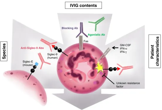
Fig. 1 Schematic representation of proposed factors influencing IVIG-mediated cell death of a neutrophilic granulocyte as a target cell. The efficacy and/or potency of the cytotoxic effects of IVIG depend on the preparation being used and on characteristics of the treated patient or species (experimental models). Engagement of Fas or Siglec-9 by agonistic autoantibodies contained in IVIG induces cell death in neutrophils. Batch-to-batch variations in titer and ratio of agonistic versus antagonistic anti-Fas autoantibodies result in
differ-ential effects on Fas, which are concentration dependent. The systemic or local cytokine profile of the patient may facilitate IVIG-induced neutrophil death, because Siglec-9-mediated cell death is enhanced in the presence of cytokines due to priming effects. For yet unknown reasons, transient resistance may block Siglec-9 death pathways in a subset of patients, depending on the stage of disease. Siglec-E, but not Siglec-9, is expressed in mouse neutrophils
cytokine-rich environment might explain local antiin-flammatory effects of systemically applied IVIG and ameliorate the situation in patients with hypereosino-philia, in whom eosinophils are exposed to survival factors and/or inflammatory cytokines [42, 43]. This mechanism might contribute to the beneficial effects of IVIG in patients with Churg–Strauss syndrome [44–46] and in other eosinophilic patients.
Siglec-9
Siglec-9 is highly expressed on human neutrophil granulocytes and monocytes; however, it is expressed at lower levels on subpopulations of lymphocytes [27]. In neutrophils and monocytes, as well as in transfected cell lines, ligation of Siglec-9 has been shown to result in inhibition of cellular activities or even cell death [33,47– 49]. Siglec-9, like Siglec-8, contains a membrane-proximal ITIM and a membrane-distal ITIM-like domain. The recruitment of the inhibitory phosphatases SHP-1 and SHP-2 as a result of tyrosine phosphorylation is thought to contribute to the inhibitory and death-promoting functions of Siglec-9 [13,33,34,47,48].
Immature human neutrophils gain Siglec-9 surface expression late in differentiation, appearing after the myelocyte stage but before the expression of CD16 [33]. In cultures of mature neutrophils in vitro, ligation of Siglec-9 by a monoclonal antibody induces caspase-dependent apoptosis at a rate similar to that of Fas [33, 50]. Compared with Siglec-8-mediated death of eosino-phils, Siglec-9-mediated neutrophil death is dramatically enhanced in inflammatory neutrophils, such as cells after priming with proinflammatory cytokines in vitro or activated cells ex vivo from inflammatory diseases like sepsis or rheumatoid arthritis [33]. The accelerated death that occurs in the presence of cytokines turns out to involve additional caspase-independent molecular path-ways that include reactive oxygen species [33].
Neutrophil Death Induced by Anti-Siglec-9 Autoantibodies Contained in IVIG
Paralleling the reported effects of Siglec-9 on eosinophils mentioned above, IVIG induces death of neutrophil granulocytes in a F(ab′)2-dependent manner. This effect dramatically accelerates in the presence of proinflammatory cytokines such as GM-CSF and interferon-γ [51]. Intrave-nous immunoglobulin-mediated death in the presence of cytokines involves reactive oxygen species and caspase-dependent and caspase-incaspase-dependent pathways. This process is comparable to that which occurs after ligation of Siglec-9
by a monoclonal antibody under similar conditions, as mentioned above [33, 51]. In depletion experiments, we showed that anti-Siglec-9 autoantibodies contained in IVIG are responsible for this cytokine-dependent cytotoxicity [33].
The increased neutrophil cytotoxicity of IVIG in a cytokine-rich environment may contribute to the antiin-flammatory effects of IVIG in the broad range of inflammatory diseases associated with causative neutrophil participation. However, we observed that septic shock patients exhibit different ex vivo death responses of blood neutrophils after Siglec-9 ligation early in shock [50]. Both the initial resistance and the increased susceptibility to Siglec-9-mediated neutrophil death were reversible and approached normal levels in the latter course of disease. These findings suggest that in disease, Siglec-9-dependent effects of IVIG may vary according to the disease stage and patient characteristics (Fig.1).
Factors Influencing IVIG-Mediated Cell Death Responses
The capacity of IVIG to induce or prevent cell death depends on multiple factors, determined by characteristics of the recipient and properties of the IVIG preparation used (Fig. 1). For instance, batch-to-batch variations of specific antibody titers and the ratio between agonistic and antagonistic antibodies in commercial IVIG preparations may result in dissimilar pharmacological effects [4,25,52]. Recently, we obtained experimental evidence that indicates the presence of antiidiotypic antibodies against immuno-regulatory autoantibodies in commercial IVIG preparations. These antiidiotypic antibodies, in concert with antagonistic antibodies, may counter-regulate the effects of agonistic antibodies directed against death receptors (unpublished results).
Patient-dependent factors that may influence the antiin-flammatory actions of IVIG include the pathogenesis, severity and stage of disease, the individual cytokine profile (either systemically in the blood or locally at the site of inflammation), and other genetic determinants. The obser-vation that the efficacy and potency of the cytotoxic effects of IVIG are enhanced on inflammatory granulocytes compared with normal granulocytes [40,51] indicates that the activation status of the target cell may significantly influence its response to IVIG. Resistance or susceptibility to immunoregulatory stimuli, as observed for Siglec-9 in different patient subpopulations and in various stages of disease [50], may greatly affect the response of immune cells following IVIG exposure. The reason for IVIG-induced neutropenia in some patients remains unknown [53–57]. It has been speculated that agonistic anti-Siglec-9
autoantibodies might cause neutropenia in patients with increased cytokine levels [58,59].
As discussed above, there are no exact Siglec-8 and Siglec-9 orthologs in the mouse [27–29]. The most likely paralogs of Siglec-8 and Siglec-9 in the mouse are Siglec-F and Siglec-E, respectively. However, these receptors exhibit no identical expression patterns on leukocyte subtypes [27]. Intravenous immunoglobulin-mediated effects on neutrophil and eosinophil granulocytes may therefore be underestimated in conventional animal studies, because human anti-Siglec-8 and anti-Siglec-9 autoantibodies are not able to exert specific functions in such experimental models (Fig.1).
Conclusions
The broad range of inflammatory disorders for which IVIG has shown therapeutic effects suggests that differential antiinflammatory mechanisms of action might more or less be effective, depending on the pathogenesis of a certain disease. It is possible that multiple mechanisms may act in concert and in a mutually nonexclusive manner [1]. For instance, blocking antibodies against Fas may interfere with FasL-induced keratinocyte apoptosis in TEN, whereas their agonistic counterparts might down-regulate immune responses by Fas-dependent apoptosis of immune effector cells. Although we have focused on the death-inducing effects of anti-Siglec and anti-Fas antibodies in receptor-expressing cells, it is possible to speculate that these functional antibodies may trigger additional immunoregu-latory functions [8,27–30].
The insights on IVIG-mediated cell death may have several implications for future studies on the mechanisms of action of IVIG. For instance, it was shown that cellular responses to IVIG might differ significantly between inflammatory and normal cells; this may be a consideration for future functional experiments. Furthermore, it is implicit that experimental evidence needs to be confirmed in a human system. Certain surface receptors or effector molecules of the immune system, such as the Siglecs, exhibit significant species-specific differences in terms of function, structure, or expression pattern. Although animal experiments may provide a powerful means of studying disease-specific mechanisms of IVIG in vivo, uncertainties remain about the use of xenogenic (human-pooled) immu-noglobulin in animals in terms of potential loss-of-function or gain-of-function effects.
In this review, we emphasized that several independent factors, including batch-to-batch variations of IVIG prepa-rations, pathogenesis, and stage of disease, and other patient characteristics may influence the efficacy of IVIG treatment. These factors may also explain the discrepancies
between certain clinical studies regarding the therapeutic effects of IVIG. Certainly, a better understanding of the mechanisms of action is required, which will help to improve IVIG therapy in inflammatory diseases and to identify the patient subpopulations that are likely to profit the most from this drug.
Acknowledgments This work was supported by grants from the Swiss National Science Foundation (310000-107526) and CSL Behring AG, Bern, Switzerland. We thank Aldona Liechti for the illustration.
References
1. Negi VS, Elluru S, Sibéril S, Graff-Dubois S, Mouthon LUC, Kazatchkine MD, et al. Intravenous immunoglobulin: an update on the clinical use and mechanisms of action. J Clin Immunol. 2007;27:233–45.
2. Nimmerjahn F, Ravetch JV. Anti-inflammatory actions of intrave-nous immunoglobulin. Annu Rev Immunol. 2008;26:513. 3. von Gunten S, Simon HU. Natural anti-Siglec autoantibodies
mediate potential immunoregulatory mechanisms: implications for the clinical use of intravenous immunoglobulins (IVIg). Autoimmun Rev. 2008;7:453–6.
4. Simon HU, Späth PJ. IVIG—mechanisms of action. Allergy. 2003;58:543–52.
5. von Gunten S, Smith D, Cummings R, Riedel S, Miescher S, Schaub A, et al. Intravenous immunoglobulin contains a broad repertoire of anti-carbohydrate antibodies that is not restricted to the IgG2 subclass. J Allergy Clin Immunol. 2009;123:1268–76. 6. Kazatchkine MD, Kaveri SV. Immunomodulation of autoimmune
and inflammatory diseases with intravenous immune globulin. N Engl J Med. 2001;345:747–55.
7. Vani J, Elluru S, Negi VS, Lacroix-Desmazes S, Kazatchkine MD, Bayary J, et al. IVIg perspective. Autoimmun Rev. 2008;7:440–4. 8. Strasser A, Jost PJ, Nagata S. The many roles of FAS receptor
signaling in the immune system. Immunity. 2009;30:180–92. 9. Simon HU. Neutrophil apoptosis pathways and their
modifica-tions in inflammation. Immunol Rev. 2003;193:101–10. 10. Simon HU. Regulation of eosinophil and neutrophil apoptosis—
similarities and differences. Immunol Rev. 2001;179:156–62. 11. Sprent J, Tough DF. T cell death and memory. Science.
2001;293:245–8.
12. von Gunten S, Bochner BS. Expression and function of Siglec-8 in human eosinophils, basophils, and mast cells. In: Pawankar R, Holgate ST, Rosenwasser LJ, editors. Allergy frontiers: classification and pathomechanisms. Tokyo, Japan: Springer; 2009. p. 297–231.
13. von Gunten S, Simon HU. Sialic acid binding immunoglobulin-like lectins may regulate innate immune responses by modulating the life span of granulocytes. FASEB J. 2006;29:601–5. 14. Savill J, Fadok V. Corpse clearance defines the meaning of cell
death. Nature. 2000;407:784–8.
15. Daigle I, Yousefi S, Colonna M, Green DR, Simon HU. Death receptors bind SHP-1 and block cytokine-induced anti-apoptotic signaling in neutrophils. Nat Med. 2002;8:61–7.
16. Conus S, Perozzo R, Reinheckel T, Peters C, Scapozza L, Yousefi S, et al. Caspase-8 is activated by cathepsin D initiating neutrophil apoptosis during the resolution of inflammation. J Exp Med. 2008;205:685–98.
17. Watanabe-Fukunaga R, Brannan CI, Copeland NG, Jenkins NA, Nagata S. Lymphoproliferation disorder in mice explained by
defects in Fas antigen that mediates apoptosis. Nature. 1992;356:314–7.
18. Takahashi T, Tanaka M, Brannan CI, Jenkins NA, Copeland NG, Suda T, et al. Generalized lymphoproliferative disease in mice, caused by a point mutation in the Fas ligand. Cell. 1994;76:969– 76.
19. Fisher GH, Rosenberg FJ, Straus SE, Dale JK, Middelton LA, Lin AY, et al. Dominant interfering Fas gene mutations impair apoptosis in a human autoimmune lymphoproliferative syndrome. Cell. 1995;81:935–46.
20. Rieux-Laucat F, Le Deist F, Hivroz C, Roberts IA, Debatin KM, Fischer A, et al. Mutations in Fas associated with human lymphoproliferative syndrome and autoimmunity. Science. 1995;268:1347–9.
21. Viard I, Wehrli P, Bullani R, Schneider P, Holler N, Salomon D, et al. Inhibition of toxic epidermal necrolysis by blockade of CD95 with human intravenous immunoglobulin. Science. 1998;282:490–3. 22. Prasad NK, Papoff G, Zeuner A, Bonnin E, Kazatchkine MD,
Ruberti G, et al. Therapeutic preparations of normal polyspecific IgG (IVIg) induce apoptosis in human lymphocytes and monocytes: a novel mechanism of action of IVIg involving the Fas apoptotic pathway. J Immunol. 1998;161:3781–90.
23. Sooryanarayana, Prasad N, Bonnin E, Pashov A, Ben Jilani K, Ameisen JC, et al. Phosphorylation of Bcl-2 and mitochondrial changes are associated with apoptosis of lymphoblastoid cells induced by normal immunoglobulin G. Biochem Biophys Res Commun. 1999;264:896–901.
24. Altznauer F, von Gunten S, Späth P, Simon HU. Concurrent presence of agonistic and antagonistic anti-CD95 autoantibodies in intravenous Ig preparations. J Allergy Clin Immunol. 2003;112:1185–90.
25. Reipert BM, Stellamor MT, Poell M, Ilas J, Sasgary M, Reipert S, et al. Variation of anti-Fas antibodies in different lots of intravenous immunoglobulin. Vox Sang. 2008;94:334–41. 26. Crocker PR, Clark EA, Filbin M, Gordon S, Jones Y, Kehrl JH, et
al. Siglecs: a family of sialic-acid binding lectins. Glycobiology. 1998;8:v–vi.
27. von Gunten S, Bochner BS. Basic and clinical immunology of Siglecs. Ann N Y Acad Sci. 2008;1143:61–82.
28. Crocker PR, Paulson JC, Varki A. Siglecs and their roles in the immune system. Nat Rev Immunol. 2007;7:255–66.
29. Varki A, Angata T. Siglecs—the major subfamily of I-type lectins. Glycobiology. 2006;16:1R–27R.
30. O'Reilly MK, Paulson JC. Siglecs as targets for therapy in immune-cell–mediated disease. Trends Pharmacol Sci. 2009;30:240–8.
31. Nguyen DH, Hurtado-Ziola N, Gagneux P, Varki A. Loss of Siglec expression on T lymphocytes during human evolution. PNAS. 2006;103:7765–70.
32. Varki A. Glycan-based interactions involving vertebrate sialic-acid–recognizing proteins. Nature. 2007;446:1023–9.
33. von Gunten S, Yousefi S, Seitz M, Jakob SM, Schaffner T, Seger R, et al. Siglec-9 transduces apoptotic and non-apoptotic death signals into neutrophils depending on the pro-inflammatory cytokine environment. Blood. 2005;106:1423–31.
34. Rashmi R, Bode BP, Panesar N, King SB, Rudloff JR, Gartner MR, et al. Siglec-9 and SHP-1 are differentially expressed in neonatal and adult neutrophils. Pediatr Res. 2009;66:266–71. 35. Guo JP, Nutku E, Yokoi H, Schnaar R, Zimmermann N, Bochner
BS. Siglec-8 and Siglec-F: inhibitory receptors on eosinophils and mast cells. Allergy Clin Immunol Int. 2007;19:54–9.
36. Yokoi H, Choi OH, Hubbard W, Lee HS, Canning BJ, Lee HH, et al. Inhibition of FcepsilonRI-dependent mediator release and calcium flux from human mast cells by sialic acid-binding immunoglobulin-like lectin 8 engagement. J Allergy Clin Immunol. 2008;121:499–505.
37. Nutku E, Aizawa H, Hudson SA, Bochner BS. Ligation of Siglec-8: a selective mechanism for induction of human eosinophil apoptosis. Blood. 2003;101:5014–20.
38. Nutku E, Hudson SA, Bochner BS. Mechanism of Siglec-8– induced human eosinophil apoptosis: role of caspases and mitochondrial injury. Biochem Biophys Res Commun. 2005;336:918–24.
39. Nutku-Bilir E, Hudson SA, Bochner BS. Interleukin-5 priming of human eosinophils alters Siglec-8 mediated apoptosis pathways. Am J Respir Cell Mol Biol. 2008;38:121–4.
40. Simon HU. Molecules involved in the regulation of eosinophil apoptosis. Chem Immunol Allergy. 2006;91:49–58.
41. von Gunten S, Vogel M, Schaub A, Stadler BM, Miescher S, Crocker PR, et al. Intravenous immunoglobulin preparations contain anti–Siglec-8 autoantibodies. J Allergy Clin Immunol. 2007;119:1005–11.
42. Vassina EM, Yousefi S, Simon D, Zwicky C, Conus S, Simon HU. cIAP-2 and survivin contribute to cytokine-mediated delayed eosinophil apoptosis. Eur J Immunol. 2006;36:1975–84. 43. Plötz SG, Simon HU, Darsow U, Simon D, Vassina E, Yousefi S, et
al. Use of an anti–interleukin-5 antibody in the hypereosinophilic syndrome with eosinophilic dermatitis. N Engl J Med. 2003;349:2334–9.
44. Klion AD, Bochner BS, Gleich GJ, Nutman TB, Rothenberg ME, Simon HU, et al. Approaches to the treatment of hypereosino-philic syndromes: a workshop summary report. J Allergy Clin Immunol. 2006;117:1292–302.
45. Takigawa N, Kawata N, Shibayama T, Tada A, Kimura G, Munemasa M, et al. Successful treatment of a patient with severe Churg–Strauss syndrome by a combination of pulse cortico-steroids, pulse cyclophosphamide, and high-dose intravenous immunoglobulin. J Asthma. 2005;42:639–41.
46. Tsurikisawa N, Taniguchi M, Saito H, Himeno H, Ishibashi A, Suzuki S, et al. Treatment of Churg–Strauss syndrome with high-dose intravenous immunoglobulin. Ann Allergy Asthma Immunol. 2004;92:80–7.
47. Ikehara Y, Ikehara SK, Paulson JC. Negative regulation of T cell receptor signaling by Siglec-7 (p70/AIRM) and Siglec-9. J Biol Chem. 2004;279:43117–25.
48. Avril T, Floyd H, Lopez F, Vivier E, Crocker PR. The membrane-proximal immunoreceptor tyrosine-based inhibitory motif is critical for the inhibitory signaling mediated by Siglecs-7 and -9, CD33-related Siglecs expressed on human monocytes and NK cells. J Immunol. 2004;173:6841–68419.
49. Carlin AF, Uchiyama S, Chang YC, Lewis AL, Nizet V, Varki A. Molecular mimicry of host sialylated glycans allows a bacterial pathogen to engage neutrophil Siglec-9 and dampen the innate immune response. Blood. 2009;113:3333–6.
50. von Gunten S, Jakob S, Geering B, Takala J, Simon HU. Different patterns of Siglec-9–mediated neutrophil death responses in septic shock. Shock. 2009;32:386–92.
51. von Gunten S, Schaub A, Vogel M, Stadler BM, Miescher S, Simon HU. Immunological and functional evidence for anti– Siglec-9 autoantibodies in intravenous immunoglobulin (IVIg) preparations. Blood. 2006;108:4255–9.
52. Gelfand EW, Winkelstein J. Are all IGIVs the same? J Allergy Clin Immunol. 2002;110:938.
53. Lassiter HA, Bibb KW, Bertolone SJ, Patel CC, Stroncek DF. Neonatal immune neutropenia following the administration of intravenous immune globulin. Am J Pediatr Hematol Oncol. 1993;15:120–3.
54. Tam DA, Morton LD, Stroncek DF, Leshner RT. Neutropenia in a patient receiving intravenous immune globulin. J Neuroimmunol. 1996;64:175–8.
55. Berkovitch M, Dolinski G, Tauber T, Aladjem M, Kaplinsky C. Neutropenia as a complication of intravenous immunoglobulin
(IVIG) therapy in children with immune thrombocytopenic purpura: common and non-alarming. Int J Immunopharmacol. 1999;21:411–5.
56. Niebanck AE, Kwiatkowski JL, Raffini LJ. Neutropenia following IVIG therapy in pediatric patients with immune-mediated throm-bocytopenia. J Pediatr Hematol Oncol. 2005;27:145–7.
57. Matsuda M, Hosoda W, Sekijima Y, Hoshi K, Hashimoto T, Itoh S, et al. Neutropenia as a complication of high-dose intravenous
immunoglobulin therapy in adult patients with neuroimmunologic disorders. Clin Neuropharmacol. 2003;26:306–11.
58. Buenz EJ, Howe CL. Appropriate use of intravenous immuno-globulin in neonatal neutropenia. J Perinatol. 2007;27:196–7. 59. Khan S, Dore PC, Sewell WAC. Both patient characteristics and
IVIG product-specific mechanisms may affect eosinophils in immunoglobulin-treated Kawasaki disease. Pediatr Allergy Immu-nol. 2008;19:186–7.