HAL Id: tel-03186843
https://tel.archives-ouvertes.fr/tel-03186843
Submitted on 31 Mar 2021
HAL is a multi-disciplinary open access archive for the deposit and dissemination of sci-entific research documents, whether they are pub-lished or not. The documents may come from teaching and research institutions in France or abroad, or from public or private research centers.
L’archive ouverte pluridisciplinaire HAL, est destinée au dépôt et à la diffusion de documents scientifiques de niveau recherche, publiés ou non, émanant des établissements d’enseignement et de recherche français ou étrangers, des laboratoires publics ou privés.
Transcription Inhibitor Lurbinectedin and Oncolytic
Peptide LTX-401 trigger Immunogenic Cell Death and
Synergize With Immune Checkpoint Blockade
Wei Xie
To cite this version:
Wei Xie. Transcription Inhibitor Lurbinectedin and Oncolytic Peptide LTX-401 trigger Immunogenic Cell Death and Synergize With Immune Checkpoint Blockade. Cancer. Université Paris-Saclay, 2020. English. �NNT : 2020UPASL072�. �tel-03186843�
INDEX
ABSTRACT ... 1
RESUME... 2
ORIGINAL PAPERS ... 3
ABBREVIATION ... 4
INTRODUCTION ... 7
Cancer and anticancer immunity ... 7
Cancer epidemiology ... 7
Cancer immunology ... 8
Immunogenic cell death in cancer therapy ...13
Major DAMPs of ICD ...16
Methods to detect ICD ...25
ICD inducers ...27
Induction of ICD sensitize cancers to immune checkpoint blockade ...31
Transcription inhibitor Lurbinectedin ...33
Oncolytic peptide LTX-401 ...36
AIMS OF THE THESIS ...38
RESULTS ...39
Lurbinectedin synergizes with immune checkpoint blockade to generate
anticancer immunity ...39
Tumor lysis with LTX-401 creates anticancer immunity ...41
CONCLUDING REMARKS ...42
ACKNOWLEDGEMENT ...45
REFERENCES ...46
ANNEX 1: SCIENTIFIC PUBLICATIONS ...66
ANNEX 2: PAPERS NOT INCLUDED IN THIS THESIS (INSERTED IN PDF)
...69
1
ABSTRACT
Cancer is the second leading cause of death worldwide, despite the existence of standard treatment, innovative therapeutic strategies and drugs are still in urgent demand. The combination of immunogenic cell death (ICD) inducing drugs and immune checkpoint blockade (ICB) seems to be a promising modality.
In this thesis, we demonstrated Lurbinectedin, a transcription inhibitor newly approved for relapsed lung cancer treatment, triggers hallmarks of ICD in four different human and murine cell line in vitro. Vaccinated with Lurbinectedin-treated fibrosarcoma cell protects immunocompetent mice from rechallenge of syngeneic tumours. Lurbinectedin restrains transplanted fibrosarcoma growth in an immune dependent manner. Both transplanted MCA205 cancer and hormone/carcinogen induced breast cancer were sensitized by Lurbinectedin to PD-1 and CTLA-4 double ICBs. Of note, long-term immunological memory was generated in cured mice.
Further, we evaluated the anticancer capacity of LTX-401, an oncolytic peptide designed for local immunotherapy. Sequential intratumoral injections of LTX-401 dramatically retards subcutaneous MCA205 and TC-1 tumour growth in immunocompetent host, yet shows limited therapeutic effect on abscopal syngeneic tumours. Single LTX-401 administration boosts the efficacy of anti-CTLA-4 or anti-PD-1/anti-CTLA-4 ICBs. Moreover, sequential LTX-401 treatment with double ICBs exhibits systemic antitumor immunity to both treated and abscopal tumour.
In conclusion, lurbinectedin and LTX-401 induce cancer cell immunogenic cell death and enhance the anticancer effects of immune checkpoint blockade. These results lay the experimental foundation of combination regiments and may facilitate the clinical trial designs.
Key words
: cancer, immunogenic cell death, immune checkpoint blockade, transcription, oncolytic peptide2
RESUME
Le cancer est la deuxième cause de mortalité dans le monde. Malgré l’existence des traitements standards, le développement et la recherche de stratégies thérapeutiques innovantes et de médicaments est toujours nécessaire. La combinaison des médicaments, induisant la mort cellulaire immunogène (ICD) et l’inhibition des points de contrôle immunitaire (ICB), semble être un protocole prometteur.
Dans cette thèse, nous avons démontré que la Lurbinectédine, un inhibiteur de la transcription nouvellement approuvé pour le traitement du cancer du poumon récidivant, déclenche les caractéristiques de l’ICD dans quatre différentes lignées cellulaires humaines et murines in vitro. Vaccinée par des cellules de fibrosarcome traitées par la lurbinectine, les souris immunocompétentes sont protégées lors du rechallenge des tumeurs syngéniques. La lurbinectédine limite la croissance du fibrosarcome transplanté d'une manière immunodépendante. Dans les souris, le fibrosarcome murin (MCA205) transplanté et le cancer du sein, induit par des hormones en combinaison avec des cancérigènes, ont été sensibilisés par la lurbinectédine aux deux ICB : PD-1 et CTLA-4. Il convient de noter que la mémoire immunologique à long terme a été générée chez des souris guéries.
En outre, nous avons évalué la capacité anticancéreuse de LTX-401, un peptide oncolytique conçu pour l'immunothérapie locale. Les injections intratumorales séquentielles de LTX-401 retardent considérablement la croissance des tumeurs sous-cutanées MCA205 et TC-1 chez un hôte immunocompétent, mais montrent un effet thérapeutique limité sur les tumeurs syngéniques abscopales. Une seule administration de LTX-401 augmente l'efficacité des ICB anti-CTLA-4 ou anti-PD-1 + anti-CTLA-4. De plus, le traitement séquentiel avec LTX-401 et les deux ICB présente une immunité antitumorale systémique à la fois contre la tumeur traitée et la tumeur abscopale.
En conclusion, la lurbinectédine et le LTX-401 induisent la mort cellulaire immunogène des cellules cancéreuses et renforcent les effets anticancéreux des inhibiteurs de points de contrôle immunitaires. Ces résultats jettent les bases expérimentales de traitements combinés et peuvent faciliter les conceptions d'essais cliniques.
Mots clés
: cancer, mort cellulaire immunogène, inhibiteur de point de contrôle immunitaire, transcription, peptide oncolytique3
ORIGINAL PAPERS
This thesis is based on the following papers:
Paper I. Lurbinectedin synergizes with immune checkpoint blockade to generate anticancer immunity.
Xie W, Forveille S, Iribarren K, Sauvat A, Senovilla L, Wang Y, Humeau J, Perez-Lanzon M, Zhou H, Martínez-Leal JF, Kroemer G, Kepp O.
Published in Oncoimmunology, 2019, VOL. 8, NO. 11, e1656502 (9 pages). Paper II. Tumor lysis with LTX-401 creates anticancer immunity.
Xie W, Mondragón L, Mauseth B, Wang Y, Pol J, Lévesque S, Zhou H, Yamazaki T, Eksteen JJ, Zitvogel L, Sveinbjørnsson B, Rekdal Ø, Kepp O, Kroemer G.
Published in Oncoimmunology, 2019, VOL. 8, NO. 7, e1594555 (8 pages).
(In agreement with the doctoral school, Paper I&II are inserted to replace chapter ‘Materials and methods’ and ‘Results’.)
4
ABBREVIATION
AIDS acquired immune deficiency syndrome ATF activating transcription factor
AML acute myeloid leukaemia
ATP adenosine triphosphate
ANXA1 annexin A1
APCs antigen-presenting cells AMPs antimicrobial peptides LfcinB bovine lactoferricin
BFA brefeldin A
CALR calreticulin
CRC colorectal cancer
CXCL10 CXC-chemokine ligand 10
CTLA-4 cytotoxic T-lymphocyte-associated protein 4 DAMPs damage-associated molecular patterns
DC dendritic cell
DMBA dimethylbenzantracene
DOXO doxorubicin
ER endoplasmic reticulum
ERAD ER-associated degradation
eIF2α eukaryotic translation initiation factor 2 FDA federal drug administration
FPR1 formyl peptide receptor 1
GCN2 general control nonderepressible 2 Hsp70 heat shock protein 70
HCC hepatocellular carcinoma
HMGB1 high-mobility group box 1 IFNGR1 IFN-γ receptor
ICB immune checkpoint blockade
5 IRE1α inositol-requiring enzyme 1α
IFN interferon
IFNAR1 interferon α/β-receptor subunit 1
IFN-γ interferon γ
IL interleukin
RIDD IRE1α-dependent decay
KS Kaposi's sarcoma
LPS lipopolysaccharides
LRP1 low density lipoprotein receptor-related protein 1
MPA medroxyprogesterone acetate
MTX mitoxantrone
MYD88 myeloid differentiation primary response 88
NK natural killer
NSCLC non-small cell lung cancers NF-κB nuclear factor-κB
ORR overall response rate
OS overall survival
PERK PKR-like ER-resident kinase PD-1 programmed cell death protein 1 PD-L1 programmed death-ligand 1
PKR protein kinase R
P2RX7 purinergic receptor P2X7 P2RY2 purinergic receptor P2Y2
ROS reactive oxygen species
RAGE receptor for advanced glycation end-products
RAG recombination Activating
ROCK1 Rho-associated, coiled-coil containing protein kinase 1 SCID severe combined immunodeficiency
STAT signal transducer and activator of transcription
SCLC small-cell lung cancer
6
7
INTRODUCTION
Cancer and anticancer immunity
Cancer epidemiology
Cancer, which was first recorded by Edwin Smith in 3000 B.C. and described in a scientific manner by Hippocrates in 400 B.C., refers to a series of diseases due to uncontrollably replicating cells. According to WORLD HEALTH STATISTICS:2018, cancer has become a serious global public health problem, approximately 9 million deaths were caused by cancer in 2016, which accounts for 15.8% of total 57 million deaths all over the world . As a fatal disease, cancer ranks as the 1st leading cause of premature death (30 to 69 years old) in 55 countries, including USA,
japan and the majority of European countries. In other 79 countries, cancer has been the second most important killer among premature populations. Specifically, more than 1.8 million Americans are estimated to be diagnosed as new invasive cancer cases in 2020, and nearly 600 000 deaths will occur in USA in the same year (Cancer statistics, 2020). In 2018, the top 3 common cancers in men are prostate cancer, lung cancer and colorectal cancer, which contributed almost 40% of total cancer incidence. Breast cancer (30%) is the most common cancer in women, followed by lung cancer (13%) and colorectal cancer (7%) (Cancer Facts & Figures 2018) (Figure 1).
Figure 1. Leading Sites of New Cancer Cases and Deaths – 2018 Estimated (Cancer Facts & Figures 2018)
8
Cancer immunology
The immune system has been defined as a host defence system which could recognise non-self-compartment and protect organism from a lot of diseases, especially for infectious disease. The immune system can be divided into two parts: innate immunity and adaptive immunity. More and more researches demonstrated the pivotal function of innate and adaptive immunity in anti-cancer immune response (Coca et al., 1997; DeNardo et al., 2011; Donadon et al., 2017; Fridman et al., 2012; Hildner et al., 2008; Ishigami et al., 2000; Keizman et al., 2012; Pylayeva-Gupta et al., 2016).
From cancer immunosurveillance to cancer immunoediting
In the early 1900s, Paul Ehrlich firstly hypothesized that immune system could recognises cancer cells as extrinsic components and eliminates them effectively (Burnet, 1970; Ehrlich, 1909). After around half centenary, the theory of ‘immunosurveillance’ was formally proposed by Macfarlane Burnet and Lewis Thomas based on the availability of inbred strain of mice and accumulated evidence of ‘tumour-specific antigen’, they speculated that the non-stop emerging transformed cells could provoke immune response and be recognised and eliminated by lymphocytes before it becomes clinically detectable (Burnet, 1957; Klein, 1966; Old and Boyse, 1964).
In the next dozens of years, the concept of immunosurveillance was in great debate. One of the representative studies which challenged this theory was conducted by Osias Stutman. Stutman observed the phenomenon that compared to genetically matched wild-type mice, carcinogen injection neither induced more tumours nor shorten tumour latency period in CBA/H strain nude mice (Stutman, 1974). Moreover, similar results obtained by different groups conclusively destroyed the bases of immunosurveillance theory and led to the abandonment of this hypothesis (Outzen et al., 1975; Rygaard and Povlsen, 1974a, b; Stutman, 1979; Thomas, 1982). Due to the solid data aforementioned, the concept of cancer immunosurveillance faded away since then.
The resuscitate of cancer immunosurveillance started from 1990s, due to the application of immunodeficiency mice on pure genetic backgrounds.
Between 1990 and 2000, the function of two fundamental immunological components, interferon γ (IFN-γ) and perforin, in controlling primary tumour formation were revealed. In the first finding, researchers observed that transplanted fibrosarcoma grows faster when host
9
endogenous IFN-γ was neutralized by blocking antibodies (Dighe et al., 1994). Also, sarcoma cells which express dominant negative IFN-γ receptors (IFNGR1) showed increased tumorigenicity when implanted into syngeneic immunocompetent mice (Dighe et al., 1994). Both IFN-γ receptor deficient mice and STAT1-/-mice in 129/SvEv background are much more sensitive to MCA
carcinogen-induced fibrosarcoma (Kaplan et al., 1998). Another study which employed different genetic background’s IFN-γ-/- mice, reinforced the protective function of endogenous IFN-γ in
restraining spontaneous tumour formation and transplanted tumour growth (Street et al., 2001). The next demonstrated key factor is perforin. Perforin is a glycoprotein which is mainly produced by natural killer (NK) cells and CD8+ T cells, upon interacting with target cell’s membrane,
channels and pores formed subsequently (Osinska et al., 2014; Russell and Ley, 2002). Similar to IFN-γ-/- mice, perforin-/- mice developed more tumours and showed shorter tumour latency time
post MCA incubation (Smyth et al., 2000; Street et al., 2001; van den Broek et al., 1996). In conclusion, these findings indicated the indispensable role of immunological components in subverting cancer development.
In 2001, a seminal study conducted by R.D. Schreiber’s group in Washington University renovated the cancer immunosurveillance concept (Shankaran et al., 2001). In this 5-pages short assay, firstly they showed inbred mice lacking either lymphocytes (RAG2–/–) or IFN-γ response (IFNGR1–/– /STAT1–/–) were prone to grow MCA-induced tumour. Second, all of the RAG2–/– or RAG2–/– /STAT1–/– mice developed spontaneous tumours in the age of 15-21 months, supported the theory of immunosurveillance. Next, they observed an interesting phenomenon that when transplanted MCA induced tumours into wild type naïve mice, all of the tumours (17/17) derived from immunocompetent mice grew normally. On the contrary, 40% of tumours (8/20) derived from RAG2–/– immunodeficient mice were rejected post transplantation. The tumour rejection effect was abrogated when RAG2–/– mice were used as recipients. This ingeniously designed experiment proved the qualitative differences between tumours from immunocompetent mice and immunodeficient mice, strongly supports the existence of tumour immunogenicity alteration during cancer development in immunocompetent hosts. In the last part, the authors proved that tumour rejection was T cell dependent. Consistent results were obtained by other group by using nude mice, SCID mice or TCR Jα281–/– mice (Engel et al., 1997; Smyth et al., 2000; Svane et al.,
10
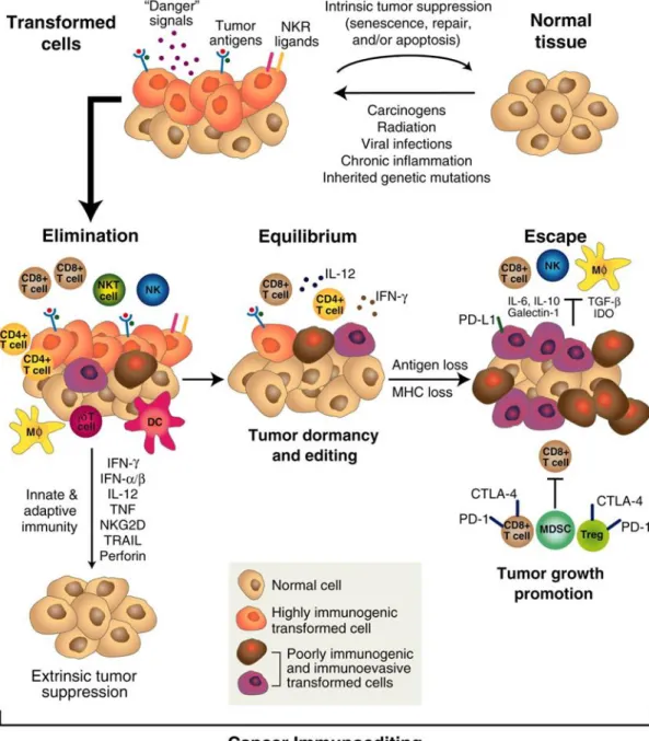
Given the fact that cancer occurs in the presence of immunosurveillance, and cancer cells with lower immunogenicity are more likely to survive compared to highly immunogenic ones, R.D. Schreiber and his colleagues proposed a more comprehensive immune theory named Immunoediting. This term was further elucidated as three different phases ‘elimination,’ ‘equilibrium,’ and ‘escape’ (Kim et al., 2007; Schreiber et al., 2011) (Figure 2).
The elimination phase could be regarded as updated immunosurveillance, in this phase transformed cells undergo stress and release danger signals such as reactive oxygen species and inflammatory cytokines (Kim et al., 2007; Matzinger, 1994). These compartments could be sensed by different types of innate immune cells like dendritic cell, macrophage and NK cell, subsequently promote their activation. The activated innate immune cells could either directly kill transformed cells or trigger adaptive immune response to achieve clearance. Of note, both innate immune system and adaptive immune system are indispensable in eliminating transformed cell. If early tumours are eliminated, this phase is the end of the whole process.
However, if a small fraction of transformed cells survived from elimination phase, the subsequent equilibrium phase starts. In this phase, tumour cells are in a special state named ‘functional dormancy’ and under consistent immune pressure. In this circumstance, cancer cells keep evolving to gain resistance to immune recognition or induce an immunosuppressive tumour microenvironment, resulting in the generation of less immunogenic tumour clones. Importantly, cytokine IL-12 and IL-23 were reported to play opposing roles in this process (Teng et al., 2012). This phase is a prolonged process, which may last from months to years, even to the end of host’s life. Anti-tumour immunity and tumour promoting factors are in dynamic balance. As a result, tumour growth is strictly controlled by adaptive immune system and tumour immunogenicity is sculpted in the meantime. Particularly, innate immune response seems inessential in this period (Kim et al., 2007; Mittal et al., 2014).
In the escape phase, evolved tumour variants evade immunological control and grow progressively, leading to visible tumours. The escaping mechanisms are extensively discussed but not fully elucidated, the balance is towards to tumour progression perhaps due to poor immunogenicity or immunologic cytotoxicity resistance of cancer cells, or by the gradually established immunosuppressive tumour microenvironment (Khong and Restifo, 2002; Radoja et al., 2000). The vast majority of clinically diagnosed cancers are in escape phase and immune
11
system can no longer work alone to control tumour growth, therefore appropriate therapeutic strategies should be taken to achieve favourable outcomes.
Figure 2. The cancer immunoediting concept. Cancer immunoediting is an extrinsic tumor suppressor mechanism that engages only after cellular transformation has occurred and intrinsic tumor suppressor mechanisms have failed. In its most complex form, cancer immunoediting consists of three sequential phases: elimination, equilibrium, and escape (Schreiber et al., 2011).
12
Cancer immunoediting in humans
To investigate the existence of cancer immunoediting in humans, cancer incidence in immunodeficient or immunosuppressive populations are studied. Compare to general population, acquired immune deficiency syndrome (AIDS) patients are prone to several types of cancer, especially for virally induced tumour like Kaposi's sarcoma (KS) (Mitsuyasu, 2009).
As a consequence of immune-suppressive treatment, patients who received transplant organs show higher risk of cancer. Birkeland analysed the cancer risk of 4178 patients who received renal transplantation between 1964 to 1986 in the Nordic countries and demonstrated an overall 3- to 5-fold increase in the combined cohort. Specifically, the incidence of lip cancer, colon and rectum cancer, non-melanoma skin cancer, melanoma, lung cancer, thyroid cancer, non-Hodgkin’s lymphoma elevated strikingly in both men and women (Birkeland et al., 1995). In another transplant recipients’ study, the malignancy incidence of 905 patients who received heart or/and lung between 1989 and 2004 were evaluated. Among which 102 patients were diagnosed with cancer, more than 7 times higher than general population. The report revealed that lymphoproliferative disorders, head and neck cancer and lung cancer rank the top three common cancer types in the cohort (Roithmaier et al., 2007). Of note, a review report from Cincinnati Transplant Tumour Registry showed two-fold higher melanoma incidence in transplant patients as compared with general population (Penn, 1996). Apart from that, other highly convincing results demonstrated the correlations between infiltrating immune cells and prognosis of patient, which strongly supported the principle of immunoediting theory (Fridman et al., 2012).
13
Immunogenic cell death in cancer therapy
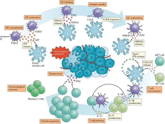
Depending on the capacity of activating an adaptive immune response, regulated cell death can be categorized as either immunogenic cell death (ICD) or non-immunogenic cell death (Galluzzi et al., 2018). As a host defence approach, cells infected with virus or intracellular bacterial undergo ICD release antigens and alarmins, which stimulates immune response to eliminate pathogens. The very first demonstrated ICD inducing drug was doxorubicin, other chemotherapeutic agents like oxaliplatin, cyclophosphamide, or some physical approaches (radiotherapy, photodynamic therapy, high hydrostatic pressure) were also proved to be able to induce ICD (Apetoh et al., 2007; Casares et al., 2005; Fucikova et al., 2014; Garg et al., 2012a; Obeid et al., 2007a; Tesniere et al., 2010; Vacchelli et al., 2014). These agents could trigger the emission of damage-associated molecular patterns (DAMPs) from cancer cells, resulting in innate and adaptive immune cell activation and immunological memory generation (Kepp et al., 2014; Lotze et al., 2007). The major DAMPs and cytokines involved in ICD include calreticulin (CALR), adenosine triphosphate (ATP), high‑mobility group box 1 (HMGB1), Type I interferon (IFN), CXC ‑ chemokine ligand 10 (CXCL10), annexin A1 (ANXA1) and so on. (Galluzzi et al., 2020b)(Table1). Of note, the same pathway in different forms of ICD may exhibits opposing effects (Garg et al., 2013).
In the context of ICD, CALR translocate from endoplasmic reticulum (ER) to cell surface, facilitating antigen uptake by dendritic cell (DC) (Gardai et al., 2005; Obeid et al., 2007b). ATP released by dying tumour cell could be sensed by DC through purinergic receptor P2Y2 (P2RY2) and purinergic receptor P2X7 (P2RX7), which mediate DC recruitment and activation respectively (Elliott et al., 2009; Ma et al., 2013; Michaud et al., 2011). HMGB1 emitted from cancer cell nuclear binds to Toll‑like receptor 4 (TLR4) to promotes DC maturation (Sims et al., 2010). Tumour cell secreted Type I IFN interacts with self-interferon α/β‑receptor subunit 1 (IFNAR1) to trigger autocrine and paracrine circuitries, leading to the release of CXCL10. As a chemoattractant, CXCL10 recruits both αβ and γδ T cells to tumour site (Harding et al., 2017; Mackenzie et al., 2017; Sistigu et al., 2014; Vanpouille-Box et al., 2017). Extra extracellular ANXA1 from cancer cell engages with formyl peptide receptor 1 (FPR1) on DC, resulting in the formation of corpse/DC synapses (Baracco et al., 2019; Vacchelli et al., 2015). Together, these
14
processes convert dying tumour cells into ‘cancer vaccines’, which activates DC and subsequently mediate T cell priming and memory T cell induction (Galluzzi et al., 2017)(Figure 3).
Table 1. Major immunostimulatory DAMPs and cytokines mechanistically linked to ICD in cancer. Adapted from (Galluzzi et al., 2020b).
ANXA1 Surface protein Directs APCs to dying cells FPR1
ATP Nucleotide Promotes the recruitment, maturation and
cross-presentation activity of APCs P2RX7 P2RY2
CALR ER chaperone Promotes the uptake of dying cells and type I IFN secretion
by APCs LRP1
CCL2 Cytokine Promotes T cell and neutrophil recruitment CCR2
CXCL1 Cytokine Promotes T cell and neutrophil recruitment CXCR2
CXCL10 Cytokine Promotes T cell and neutrophil recruitment CXCR3
Cytosolic
RNA Nucleic acid
Promotes the secretion of type I IFN and other
proinflammatory factors MDA5 RIG-I TLR3
Cytosolic
DNA Nucleic acid
Promotes the secretion of type I IFN and other
proinflammatory factors AIM2 CGAS ZBP1
ERp57 ER chaperone Promotes the uptake of dying cells by APCs LRP1 (?)
Extracellular
DNA Nucleic acid Promotes the recruitment and activation of neutrophils TLR9
F-actin Cytoskeletal component Promotes the uptake of dying cells by APCs CLEC9A
HMGB1 Nuclear DNA-binding protein Promotes the maturation and cross-presentation activity of
APCs AGER TLR2 TLR4
HSP70 ER chaperone Favors the uptake of dying cells by APCs LRP1
HSP90 ER chaperone Favors the uptake of dying cells by APCs LRP1
TFAM Transcription factor Promotes APC maturation and recruitment AGER
Type I IFN Cytokine Promotes APC maturation, cross-presentation, and T cell
recruitment IFNARs
Main receptor(s)
15
Figure 3. Mechanisms of chemotherapy-driven ICD. In response to inducers of immunogenic cell death (ICD), such as doxorubicin or oxaliplatin, malignant cells expose calreticulin (CALR) and other endoplasmic reticulum chaperones on their surface, secrete ATP, initiate a cell-intrinsic type I interferon (IFN) response culminating in the production of CXC-chemokine ligand 10 (CXCL10), and release high-mobility group box 1 (HMGB1) and annexin A1 (ANXA1). Upon binding to cognate receptors on the surface of myeloid or lymphoid cells, these damage-associated molecular patterns favour the uptake of cell corpses and debris thereof by antigen-presenting cells, including dendritic cells (DCs) in the context of robust immunostimulatory signals, eventually leading to the priming of an adaptive immune response involving both αβ and γδ T cells. In addition to being associated with the establishment of immunological memory, such a response has the potential to eradicate malignant cells that survive chemotherapy via an IFNγ-dependent mechanism. CXCR3, CXC-chemokine receptor 3; FPR1, formyl peptide receptor 1; IFNAR1; interferon α/β-receptor subunit 1; IL, interleukin; LRP1, LDL receptor related protein 1; P2RX7, purinergic receptor P2X7; P2RY2, purinergic receptor P2Y2; TLR4, Toll-like receptor 4 (Galluzzi et al., 2017).
16
Major DAMPs of ICD
Calreticulin exposure
As one of the most abundant proteins in ER, CALR plays fundamental role in calcium homeostasis and protein folding (Michalak et al., 1999). Upon ICD treatment, CALR translocate from ER to cell surface in cooperate with other chaperone proteins (Asadzadeh et al., 2020; Fucikova et al., 2011; Garg et al., 2012b; Panaretakis et al., 2008)(Figure 4). As an ‘eat me’ signal, cells exhibiting CALR on surface can be recognised by phagocytes through low density lipoprotein receptor-related protein 1 (LRP1; also known as CD91), resulting in antigen uptake and clearance of cellular components (Bergmann et al., 1992; Garg et al., 2012b). On the contrary, this process is impaired when CD47 (a don’t eat me signal) co-expressed on cancer cells (Chao et al., 2011). Also, CALR-CD91 conjunction on antigen-presenting cells (APCs) primes Th-17 cell response in an Interleukin 6 (IL-6) and tumor necrosis factor alpha (TNF-α) dependent manner (Pawaria and Binder, 2011). Of note, CALR ablation by siRNA or neutralising antibody abolish the immune response towards dying tumour cell in vivo, while incubation with recombinant CALR protein restores the immunogenicity of the cells (Liu et al., 2019; Obeid et al., 2007b). Moreover, the immunogenicity deficiency mediated by PERK, caspase-8 or SNAREs depletion in anthracyclines induced cell death can also be restored by absorbing recombinant CRT to the cell surface (Panaretakis et al., 2009). Altogether, these results definitely indicated that the immunogenicity of ICD is defined by cell surface CALR.
Importantly, CALR exposure is correlated with clinical outcomes. In human non-small cell lung cancers (NSCLC), high expression of CALR correlates with increased antitumor immune response and favourable prognosis (Fucikova et al., 2016a). CALR level cloud also be used as an independent prognostic factor in neuroblastoma (Hsu et al., 2005). Similarly, high level of CALR is associated with more infiltrated CD45RO+ cells in colon cancer, which correlates with a higher
5-year survival rate of patients (Peng et al., 2010). In acute myeloid leukaemia (AML) patients, CALR exposure on the membrane of malignant cells positively correlated with leukaemia specific immune response, further associated with both improved relapse-free survival and superior overall survival (Fucikova et al., 2016b). Moreover, low CALR level was associated with immunogenic chemotherapeutic resistance and poor survival rate in endometrial cancer patients (Xu et al., 2018).
17
Figure 4. Mechanism of CRT exposure. Several steps are involved in the exposure of CRT in response to immunogenic cell death inducers including activation of PERK, the phosphorylation of eIF2α, caspase-8-dependent cleavage of BAP31, and the activation of Bax and Bak. Finally, a pool of CRT that has transited the Golgi apparatus is secreted by SNARE-dependent exocytosis. Abbreviations: CRT; calreticulin, PERK; protein kinase R-like ER kinase, BAP31; B cell receptor–associated protein 31, Bax; BCL2-associated X protein, Bak; Bcl-2 homologous antagonist/killer, SNARE; soluble N-ethylmaleimide-sensitive fusion protein-attachment protein receptor (Asadzadeh et al., 2020).
ER stress
ER stress is considered to initiate CALR translocation in response to ICD induction. During homeostatic conditions, protein translation goes smoothly in ER, while the three unfolded protein
18
response (UPR) mediators are bind with heat shock protein 70 (Hsp70)-type chaperone protein Bip and stay in inactive state. In response to exogenous or endogenous ER stress, Bip preferably binds with misfolded proteins thus dissociates with these UPR mediators, leading to the activation of Inositol-requiring enzyme 1α (IRE1α), PKR-like ER-resident kinase (PERK) and activating transcription factor 6α (ATF6α). Upon Bip dissociation, 1) IRE1α forms a homodimer and undergo autophosphorylation, activating its C-terminal endoribonuclease activity. IRE1α splices the XBP-1 mRNA and facilitates XBP-XBP-1 expression, which further promotes the transcription of protein chaperones, glycosylase, and ER-associated degradation (ERAD) system (Lee et al., 2003). Additionally, IRE1α could directly modulate the abundance of ER-localized mRNAs through regulated IRE1α-dependent decay (RIDD) pathway; 2) As an ER-resident eukaryotic translation initiation factor 2 (eIF2α) protein kinase, PERK phosphorylates the translation initiation factor eIF2α under ER stress conditions, abolishing 5′-cap assembly-dependent translation (Harding et al., 2000; Harding et al., 1999). Meanwhile, the general translation inhibition enhances the activating transcription factor 4 (ATF4) production, driving the expression of downstream transcription factor CHOP (Ma et al., 2002; Vattem and Wek, 2004); 3) Post dissociation with Bip, ATF6α translocates into Golgi and activated by proteolytic cleavage, promoting the accumulation of ERAD components and chaperones (Bailey and O'Hare, 2007; Haze et al., 1999; Shen et al., 2002). Although these three sensors could coordinate to combat ER stress, the individual UPR signalling pathways varies in different circumstances (Bettigole and Glimcher, 2015; Shoulders et al., 2013).
Interestingly, ICD inducers like anthracyclines and oxaliplatin specifically induce PERK-dependent eIF2α phosphorylation, yet neither activating downstream ATF4 nor other two ER stress branches (Bezu et al., 2018a)(Figure 5). But the precise mechanism of ICD induced split ER stress response remains to be investigated. It’s demonstrated that PERK mediated eIF2α phosphorylation is mandatory for CALR exposure in response to ICD induction in several mouse and human cell lines (Bezu et al., 2018a; Panaretakis et al., 2009). However, it is protein kinase R (PKR) and general control nonderepressible 2(GCN2), rather than PERK, trigger eIF2α phosphorylation and CALR exposure in mitoxantrone (MTX) or doxorubicin (DOXO) treated melanoma cells (Giglio et al., 2018). Moreover, eIF2α phosphorylation and caspase-8 activation are dispensable in Hyp-PDT mediated CALR exposure, indicating that different ICD inducers may functions distinctively (Garg et al., 2012b). Of note, tumoral p-eIF2α and CALR expression
19
indicate positive prognostic value in NSCLC patients (Fucikova et al., 2016a). Altogether, p-eIF2α could be employed as a hallmark of ICD and promising biomarker in clinical prognosis prediction although exceptions exist.
Figure 5. eIF2α phosphorylation: A hallmark of immunogenic cell death. (A) The unfolded protein response of the endoplasmic reticulum in normal circumstances. Accumulation of misfolded proteins in the endoplasmic reticulum (ER) provokes the release of heat shock protein family A (Hsp70) member 5 (HSPA5, best known as binding immunoglobulin protein, BiP) and activates simultaneously the three arms of the unfolded protein response (UPR) ((i) EIF2AK3, (best known as PERK)-mediated phosphorylation of eukaryotic initiation factor α (eIF2α), which in turn halts general protein translation but favors the expression of activating transcription factor 4 (ATF4), (ii) the translocation of the ER transmembrane protein activating transcription factor 6 (ATF6) to the Golgi, proteolytic cleavage and the nuclear translocation of its cytosolic fraction (iii) the endoplasmic reticulum to nucleus signaling 1 (ERN, best known as IRE1α)-mediated splicing of X-box binding protein 1 (XBP1) to the XBP1s isoform. The signal transduction issued by the activation of the transcription factors ATF4, ATF6 and XBP1s aim at reestablishing ER homeostasis and cell survival (or the induction of cell death in conditions of enduring ER stress). (B) The unfolded protein response in the context of immunogenic cell death (ICD). First, the non-canonical activation of PERK and possibly other eIF2α kinases triggers the phosphorylation of eIF2α without BiP occupation by misfolded proteins and/or second, ICD-inducers inhibit UPR downstream signals such as ATF4 translation, ATF6 cleavage and the IRE1α−mediated XBP1 splicing (Bezu et al., 2018b).
20
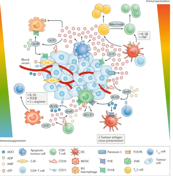
ATP release
As a multi-functional molecular expelled by dying cells in ICD event, the extracellular ATP plays an important role in immune response. Upon secretion, ATP binds to P2X7 and P2Y2 on APCs, promoting their maturation and facilitates chemotactic effect respectively (Elliott et al., 2009; Ghiringhelli et al., 2009; Michaud et al., 2011). In DCs, ATP triggers the aggregation of NLRP3-ASC–Casp-1 inflammasome and facilitates IL-1β release, favouring the prime of antigen-specific cytotoxic T cell and IL-17 producing-γδ T cell. Thus, deficiency of any of these genes (Nlrp, P2rx7, Casp1) attenuates the establishment of adaptive immune response in chemotherapeutic ICD (Ghiringhelli et al., 2009; Ma et al., 2011). Besides, ATP could modulate DC activity simultaneously by stimulating the expression of costimulatory molecules include CD40, CD80, and CD86 (Idzko et al., 2007).
The molecular mechanism of ATP secretion during ICD is often related to autophagy, a tightly regulated pathway mediates the recycle of cytosolic components in response to stress (Martins et al., 2014; Michaud et al., 2011). When treated with ICD inducer MTX or oxaliplatin, autophagy deficient cancer cells (by knock-down or knock-out of essential autophagy proteins or pharmacological inhibition) secrete much less ATP yet release similar amount of HMGB1 and expose equal level of CALR on cell surface . Immunization with MTX treated autophagy deficient CT26 or MCA205 cells failed to protect immunocompetent mice from re-challenging of the same cell type, while its immune stimulating capacity could be restored by co-injection of rIL-1β but not rCALR (Michaud et al., 2011). Moreover, autophagy-deficient tumour exhibits impaired tumour-infiltrating lymphocytes recruitment in response to immunogenic chemotherapy, but intra-tumoral administration of ecto-ATPase inhibitor regenerate therapeutic immune response in immunocompetent hosts (Chao et al., 2011). Accordingly, these results indicate that autophagy is required for ATP release during chemotherapy. Following research revealed a more complex machinery in regulating ATP secretion by dying cancer cell. Several relatively independent molecules include autophagy related proteins, caspases, Rho-associated, coiled-coil containing protein kinase 1 (ROCK1), PANX1 channels and LAMP1 are involved in ICD-associated ATP release. In other words, autophagy, lysosomal exocytosis, apoptosis, membrane blebbing and plasma membrane permeabilization are orchestrated in ATP secretion (Martins et al., 2014).
Apart from autophagy, the extracellular ATP level could also be regulated by CD39 and CD73 ectonucleotidase. CD39 and CD73 convert ATP to immunosuppressive adenosine, resulting in the
21
establishment of a tumour promoting microenvironment (Chalmin et al., 2012; Sun et al., 2010). Mice lacking CD73 shows resistance to MCA-induced fibrosarcoma and increased anti-tumour immunity, CD73 antibody treatment restrains prostate tumour growth and metastasis (Stagg et al., 2012)(Figure 6).
Breast cancer patients bearing the loss-of-function (Glu496Ala) P2RX7 allele are prone to cancer metastasis post anthracyclines treatment, comparing to P2RX7 normal counterpart (Ghiringhelli et al., 2009). In a cohort of colorectal cancer (CRC) patients, absence of Beclin-1 expression is associated with poor prognosis (Choi et al., 2014). Similarly, another CRC study shows that the LC3 level is positively correlated with long-term survival (Wu et al., 2015).
Figure 6. Modulation of immune cell responses by extracellular ATP in the tumour microenvironment (Di Virgilio et al., 2018).
22
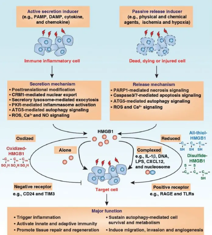
HMGB1 exodus
HMGB1 is the first identified HMGB family protein which locates in the nuclear as non-histone chromatin-binding protein in steady state (Palumbo et al., 2004). The function of HMGB1 mainly depends on its sublocation, namely nuclear, cytoplasm or extracellular milieu (Garg et al., 2010). Within nuclear, HMGB1 binds to chromatin to stabilize the structure and modulates transcription (Garg et al., 2010; Scaffidi et al., 2002). In cytoplasm, HMGB1 can interacts with Beclin-1 to affect autophagy (Huang et al., 2012; Tang et al., 2010). It’s reported that HMGB1 regulates the expression of various genes including p53 and nuclear factor-κB (NF-κB) (Krysko et al., 2012). Extracellular HMGB1 could directly interact with different surface receptors like RAGE (receptor for advanced glycation end-products), TLR2 and TLR4, to induce significant immune response in a cytokine-like manner (Sims et al., 2010). HMGB1 stimulates the production of a series of proinflammatory cytokines in human monocytes, but not lymphocytes. Accordingly, intraperitoneal administration of rHMGB1 increase TNF concentration in mice serum (Zhang et al., 2016). In the context of ICD, TLR4 seems to be the main HMGB1 receptor which contributes to tumour eradiation. HMGB1 inhibition and TLR4 deletion compromise the protective efficiency of dying tumour in vaccination experiment. Besides, the immune response to dying tumour cell is largely depends on TLR4 expression on DC. Moreover, both myeloid differentiation primary response 88 (MYD88) −/− mice and TLR4−/− mice failed to response to chemotherapy or radiotherapy, demonstrating the contribution of HMGB1/TLR4/MyD88 axis in anticancer immunity (Apetoh et al., 2007). It should be noticed that the function of HMGB1 is determined by its redox modification, HMGB1 acts as chemoattractant DAMP, pro-inflammatory cytokine-inducing DAMP or inactivated DAMP depending on actual reduced to oxidized forms (Venereau et al., 2012; Yang et al., 2012)(Figure 7).
HMGB1 can be released in two different fashions, named active secretion or passive liberation. Macrophages and monocytes secret HMGB1 through a cytokine secreting manner post IL-1β, TNF or lipopolysaccharides (LPS) stimulation (Garg et al., 2010; Muller et al., 2004). Primary necrosis cells could passively release large amount of HMGB1 to extracellular space (Scaffidi et al., 2002). Also, Cells undergo secondary necrosis discharge HMGB1. In particular, Z-VAD-fmk (a broad caspase inhibitor) treatment reduces the release of HMGB1 from ICD (Apetoh et al., 2007; Bell et al., 2006).
23
A study showed both TLR4 Asp299Gly and Thr399Ile polymorphisms were positively associated with cancer development and progression in CRC patients, and the expression of these mutations impact patients’ survival (Messaritakis et al., 2018). Also, Thr399Ile polymorphism was significantly associated with an elevated gastrointestinal cancer risk (Jing et al., 2012). Breast cancer patients bearing mutated TLR4 alleles relapse faster post anthracycline-based chemotherapy and local irradiation (Apetoh et al., 2007). However, controversial results showed no correlations between CRC risk and Asp299Gly or Thr399Ile alleles (Davoodi and Seow, 2011).
24
Figure 7. HMGB1 release and extracellular function. HMGB1 can be actively secreted by immune inflammatory cells or passively released by dead, dying or injured cells into the extracellular milieu by several different mechanisms as indicated. Extracellular HMGB1 acts as a DAMP molecule and plays a vital role in several pathophysiological processes. The activity of extracellular HMGB1 depends on redox state, receptors and their interactive partners (Chen et al., 2013).
25
Methods to detect ICD
As aforementioned, ICD inducers cause cancer cell death in parallel with the emission of DAMPs, which includes split ER stress induced CALR translocation, autophagy dependent ATP release, HMGB1 exodus and Type I IFN secretion. These DAMPs define its adjuvanticity and triggers adaptive immune response in company with antigenicity. One thing should be emphasized here is the precise mechanisms of ICD are not fully addressed, the immunogenicity of agents should be tested both in vitro and in vivo.
A drug discovery pipeline was constructed to identify ICD inducers from large libraries (Bezu et al., 2018a; Kepp et al., 2019)(Figure 8). This pipeline consists four procedures, 1) use artificial intelligence to pre-select drug candidates; 2) high content screening to find hits; 3) in vitro validation and 4) in vivo validation.
Figure 8. Discovery pipeline. (Kepp et al., 2019)
In vivo vaccination assay is employed as the ‘Gold Standard’ to evaluate immunogenicity. In brief, tumour cells were treated with cytotoxic ICD inducer candidates in vitro, then immunocompetent naïve mice were injected with these dying tumour cells without adjuvant in the flank subcutaneously, after one to two weeks these vaccinated mice were rechallenged by the syngeneic living tumour cell in the contralateral flank, tumour emergence and growth were monitored. The ‘real’ ICD inducer treated cells could protect mice from growing tumours, or at least delay tumour growth and extends lifespan compare to ‘PBS’ group and ‘non-ICD’ group (Humeau et al., 2019)(Figure 9). Given this in vivo vaccination experiment is only applicable for
26
mouse cancer cell line, the generation of humanized mice would be particularly useful in predicting ICD in human cancers.
Figure 9. Assessing the ability of a cytotoxicant to stimulate cancer ICD in vivo. (Humeau et al., 2019)
27
ICD inducers
Doxorubicin was the first identified ICD inducer in 2005, as tumour cells treated by DOXO could effectively elicit anticancer immunity in the absence of adjuvant (Casares et al., 2005). Since then, numerous ICD inducers were discovered by different research groups. ICD inducers could be categorized into four categories: chemotherapy, physical approach, immunotherapy and target therapy. As a well-studied ICD inducer cluster, chemotherapeutic drugs such as anthracyclines, platinum derivatives, alkylating agents and proteasome inhibitors are proved to trigger ICD potently (Casares et al., 2005; Panaretakis et al., 2009; Schiavoni et al., 2011; Spisek et al., 2007). Radio therapy and Photodynamic therapy are also effective approaches to induce ICD (Demaria et al., 2004; Formenti et al., 2018; Korbelik and Dougherty, 1999; Korbelik et al., 2011; Krosl et al., 1995; Paz-Ares et al., 2020). Some oncolytic virus and oncolytic peptides induce traits of ICD (Koks et al., 2015; Yamazaki et al., 2016; Zamarin et al., 2014; Zhou et al., 2016a; Zhou et al., 2018). Only specific target therapy drugs like Crizotinib and Cetuximab are capable to generate ICD (Liu et al., 2019; Pozzi et al., 2016). Confirmed ICD inducers are listed in Table 2 (Galluzzi et al., 2020a).
28
Agent P-eIF2α DAMPs Main cytokines Immune infiltrate Antagonized by Combination with ICIsa
7A7 Yes CALR ↑IFNγ ↑DCs, CD8+ CTLs, CD4+ T cells CD8+ T cell depletion ND
8-MOP PCT ND CALR, ATP, HMGB1 ↑Type I IFNs, IL-6, CCL2
↑DCs, monocytes,CD8+ CTLs, NK cells
CD8+, CD4+ or NK cell
depletion ND
Anthracyclines Yes CALR, ATP, HMGB1, ANXA1
↑1β, 6, 12, IL-17, CXCL10, type I IFNs, IFNγ ↑DCs, CD8+ CTLs, γδ TH17 cells; ↓Treg cells, MDSCs nu/nu genotype, CD8+ T cell depletion, IFNγ or IL-17 neutralization, IL-17 receptor inhibition Anti-PD-1, anti-CTLA4
Bleomycin Yes CALR, ATP, HMGB1 ↑IFNγ, TGFβ ↑CD8+ CTLs;↑Treg cells CD8+ T cell depletion, IFNγ neutralization ND
Bortezomib Yes CALR, HSP70 ↑IFNγ ↑DCs, CD8+ CTLs
nu/nu or Rag genotype, CD8+ T cell depletion
ND
Cardiac glycosides ND CALR, ATP, HMGB1 ↑IFNγ ↑CD8+ CTLs, CD4+ T cells, γδ TH17
cells nu/nu genotype ND
Cetuximab ND CALR, HMGB1 ND ↑DCs, CD8+ CTLs ND ND
Clostridium difficile
toxin B No CALR, HMGB1, ATP ND ND ND ND
Crizotinib Yes CALR, ATP, HMGB1 ↑Type I IFNs, IL-17 ↑DCs, CD8↓T + CTLs, NKT cells;
reg cells nu/nu genotype Anti-PD-1, anti-PD-L1
Cyclophosphamide ND CALR, HMGB1, ATP ↑Type I IFNs, IFNγ, IL-17
↑DCs, CD8+ CTLs, NK cellsb, NKT
cells; ↓MDSCs, Treg cells ND Anti-PD-1, anti-CTLA4
Dactinomycin Yes CALR, ATP, HMGB1 ↑Type I IFNs, IFNγ,
IL-17 ↑DCs, CD8+ CTLs, NK cells; ↓Treg cells
nu/nu genotype, CD8+ and CD4+ T cell depletion
29
Dinaciclib ND CALR, HMGB1, ATP ↑IFNγ ↑DCs, CD8+ CTLs, CD4+ T cells Rag-/- genotype Anti-PD-1
DTT-205 ND CALR, HMGB1 ↑Type I IFNs ND CD8+ and CD4+ T cell
depletion ND
DTT-304 ND CALR, HMGB1 ↑Type I IFNs ND CD8+ and CD4+ T cell
depletion ND
Electrical nanopulses ND CALR, ATP, HMGB1 ND ↑CD8+ CTLs ND ND
Hypericin-based PDT No CALR, ATP, HMGB1 ↑IL-1β ↑DCs ND ND
JQ1 Yes CALR, ATP, HMGB1 ND ↑DCs, CD8+ CTLs; ↓MDSCs nu/nu genotype ND
LTX-315 No CALR, ATP, HMGB1 ↑IL-1β, IL-6, type I IFNs
↑DCs, macrophages, CD8+ CTLs, TH1
CD4+ cells; ↓MDSCs, Treg cells ND Anti-PD-1, anti-CTLA4
LTX-401 No CALR, ATP, HMGB1 ↑Type I IFNs ↑ CD3+ T cells ND Anti-PD-1, anti-CTLA4
Lurbinectedin Yes CALR, ATP, HMGB1 ↑Type I IFNs ND CD8+ and CD4+ T cell
depletion Anti-PD-1, anti-CTLA4 Microwave thermal
ablation ND CALR, ATP, HMGB1 ↑IFNγ, TNF ↑CD8+ CTLs CD8+ T cell depletion ND
Newcastle disease
virus ND CALR, HMGB1 ↑IFNγ
↑DCs, ↑CD8+ CTLs, CD4+ T cells, NK and NKT cells;↓MDSCs
Rag2-/- genotype,
CD8+ T cell depletion Anti-CTLA4 Non-thermal plasma ND CALR, ATP, HMGB1 ↑ CXCL10, IFNγ, TNF;
↓TGFβ ↑DCs ND Anti-PD-L1
Oxaliplatin Yes CALR, ATP, HMGB1, HSP70
↑IL-1β, type I IFNs,
IFNγ ↑DCs, CD8+ CTLs, γδ TH17 cells nu/nu genotype, CD8+ T cell depletion, IFNγ or IL-17 neutralization, IL-17R blockage Anti-PD-1, anti-CTLA4
30
Table 2. Confirmed inducers of immunogenic cell death. All agents confer enduring immunological memory, excluding dactinomycin, electrical nanopulses, non-thermal plasma and redaporfin-based photodynamic therapy (PDT), for which evidence suggests only partial immunological memory. Cardiac glycosides and crizotinib confer immunological memory only in combination with cytotoxic agents. MOP, 8-methoxypsolaren; ANXA1, annexin A1; CALR, calreticulin; CTLA4, cytotoxic T lymphocyte-associated protein 4; CTL, cytotoxic T lymphocyte; DAMP, damage-associated molecular pattern; DC, dendritic cell; HMGB1, high mobility group box 1; HSP70, heat shock protein family A; ICI, immune checkpoint inhibitor; IFN, interferon; MDSC, myeloid-derived suppressor cell; ND, not determined; NK, natural killer; NKT, natural killer T; P-eIF2α, eIF2α phosphorylation; PCT, photochemotherapy; TH, T helper; Treg, regulatory T; UVC, ultraviolet C. aIn mouse models. bObserved in
some but not all models investigated. Adapted from (Galluzzi et al., 2020a).
Photofrin-based PDT ND CALR, HMGB1 ND ↑Monocytes, neutrophils, CD8+ CTLs,
NK cells CD8+ T cell depletion ND
PT-112 ND CALR, ATP, HMGB1 ND ↑DCsb, CD8+ CTLs; ↓macrophage
b,
Treg cellsb ND
Anti-PD-1, anti-PD-L1, anti-CTLA4
Radiotherapy ND CALR, ATP HMGB1 ↑IL-17, type I IFNs, IFNγ, TGFβ ↑DCs, CD8+ CTLs
nu/nu genotype, CD8+ T cell depletion, IFNAR1 blockage
Anti-PD-1, anti-PD-L1, anti-CTLA4
Redaporfin-based PDT Yes CALR, ATP, HMGB1 ND ND ND ND
Septacidin ND CALR, ATP, HMGB1 ND ND nu/nu genotype ND
Talimogene
laherparepvec ND CALR, ATP, HMGB1
↑IL-1β, type I IFNs,
TNF ↑CD8+ CTLs ND Anti-PD-1
Teniposide ND CALR, HMGB1 ↑IL-2, type I IFNs, IFNγ ↑DCs, CD8+ CTLs ND Anti-PD-1
UVC light ND CALRb, HMGB1b ↑IFNγ ↑DCs, CD8
+ CTLs ND ND
Wogonin Yes CALR, ATP, HMGB1,
31
Induction of ICD sensitize cancers to immune checkpoint blockade
Immunotherapy, particularly immune checkpoint blockade (ICB), is now largely applied to treat various cancers. But due to the immunosuppressive nature of tumour microenvironment, the objective response rates (ORRs) of standard ICB treatment in solid tumours are relatively low (Lim et al., 2016; Rosenberg et al., 2016; Topalian et al., 2012). ICD induction in tumour sites enables to recruit antigen presenting cells and CD8 T cells to the tumour bed, thus convert immunologically ‘cold’ tumours into ‘hot’ lesions. Given that ICB’s therapeutic function dedicated to the presence of CD8 T cells, pre-treatment with ICD inducers sensitize tumours to ICB in several studies (Liu et al., 2019; Pfirschke et al., 2016; Yamazaki et al., 2020).
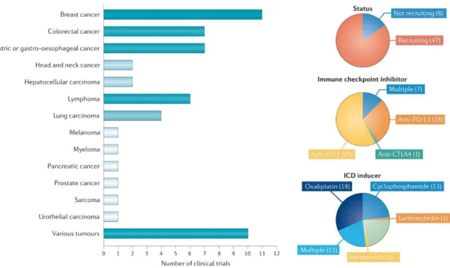
In a cohort of NSCLC patients, chemoradiotherapy plus programmed death-ligand 1 (PD-L1) blockade dramatically prolonged the overall survival of combination treatment group against placebo group (Antonia et al., 2018). In another study involves metastatic triple-negative breast cancer patients, doxorubicin treatment upregulates the expression of key genes which related to programmed cell death protein 1 (PD-1)/PD-L1 and T cell cytotoxicity pathways, thus evaluated the objective response rate to PD-1 blockade (Voorwerk et al., 2019). In addition, bunch of clinical trials that involves the combination of immunogenic chemotherapy and ICBs are still ongoing (Galluzzi et al., 2020a)(Figure 10).
Altogether, both preclinical and clinical studies reveal that ICD-inducing chemotherapy may bring benefits to cancer patients in some immunotherapy included settings. But the optimal therapeutic combination regiments remain to be critically evaluated.
32
Figure 10. Overview of ongoing clinical trials involving one or more immunogenic cell death inducers in combination with immune checkpoint inhibitors in patients with cancer. As of 1 March, 2020, ClinicalTrials.gov listed 55 ongoing (defined as those that are not completed, withdrawn, suspended, terminated or have unknown status) clinical trials in which patients are receiving chemotherapy plus agents with known immunogenic cell death (ICD)-inducing potential in combination with one or more FDA-approved immune checkpoint inhibitors in patients with cancer that were initiated after 1 January, 2019. CTLA4, cytotoxic T lymphocyte-associated protein (Galluzzi et al., 2020a).
33
Transcription inhibitor Lurbinectedin
Lurbinectedin (PM01183) is a novel synthetic marine-derived anticancer agent developed by PharmaMar Company. It was recently approved by Federal Drug Administration (FDA) for the salvage treatment of small-cell lung cancer (SCLC). As an alkaloid, lurbinectedin could binds to the DNA minor groove, induces double-strand DNA breaks, selectively blocks RNA polymerase II activity and promotes its degradation, disrupts mitosis and eventually induces cell death (Leal et al., 2010; Santamaria Nunez et al., 2016; Vidal et al., 2012). As an analogue of trabectedin, the structure of lurbinectedin is similar to trabectedin except the modification in C subunit (Leal et al., 2010). Thus, lurbinectedin gains different pharmacokinetic and pharmacodynamics properties from trabectedin (Bueren-Calabuig et al., 2011; Leal et al., 2010; Soares et al., 2011).
It’s reported that lurbinectedin has potent antiproliferative activity in numerous cancer cell lines in vitro, including cisplatin-sensitive and resistant cells (Leal et al., 2010; Santamaria Nunez et al., 2016; Soares et al., 2011). Single lurbinectedin treatment exhibits antitumor/antimetastatic activity in MNMCA1, M5076 murine reticulosarcoma, HOC8 and MNBPTX human ovarian carcinoma xenograft in vivo (Romano et al., 2013). In preclinical uterine cervical cancer models, lurbinectedin inhibits tumour growth by eliminating both cancer stem cell and non-cancer stem cell (Yokoi et al., 2019). Lurbinectedin inactivates oncoprotein EWS-FLI1 and synergized with irinotecan to retard Ewing sarcoma xenograft growth (Harlow et al., 2016). Also, lurbinectedin therapy overcomes cisplatin resistance in in epithelial ovarian cancer (Vidal et al., 2012). In particular, lurbinectedin depletes tumour-associated macrophages and modulates inflammatory microenvironment in both mouse fibrosarcoma tumour model and patient-derived (AVATAR) PDA xenografts (Belgiovine et al., 2017; Cespedes et al., 2016; Farago et al., 2019)(Figure 11).
34
Figure11. Chemical structure and schematized actions of lurbinectedin. (A) Chemical structure of lurbinectedin. (B) Schematic representation of lurbinectedin mechanism of action for binding DNA, inhibiting active transcription and inducing DNA breaks. (C) Schematic representation of lurbinectedin interactions with the tumor microenvironment. Lurb: Lurbinectedin (Farago et al., 2019).
The approval of lurbinectedin for second line SCLC treatment was based on several clinical trials. In a Phase I study enrolled 27 relapsed SCLC patients, the combination regiment of lurbinectedin and doxorubicin showed remarkable anticancer activity. As second-line treatment, 11 out of 12 patients with platinum-sensitive disease and 3 out of 9 patients with platinum-resistant disease were responded to the combination treatment (Calvo et al., 2017). In a single-arm, open-label phase II basket trial, 105 patients with relapsed SCLC were treated with lurbinectedin alone. The median overall survival (OS) was 9.3 months, 11.9 months, 5.0 months in the whole cohort, patients with chemotherapy sensitive or resistant disease respectively. Among all patients the
35
overall response rate (ORR) was 35.2%, whereas 45.0% in chemotherapy sensitive subgroup and 22.2% in chemotherapy resistant subgroup. Further, lurbinectedin was of good safety, no patient dead due to treatment related advise effects (Trigo et al., 2020). It’s notable that the combination therapy of lurbinectedin and atezolizumab (anti-PD-L1 antibody) in patients with progressed SCLC is under evaluation (ClinicalTrials.gov, NCT04253145). Apart from SCLC, lurbinectedin is in therapeutic trials for multiple cancer types, including breast cancer, ovarian cancer, sarcoma, acute leukaemia and different advanced solid tumours (Markham, 2020)(Table 3).
Table 3. Key clinical trials of lurbinectedin. mCRC metastatic colorectal cancer, NSCLC non-small cell lung cancer, SCLC small cell lung cancer (Markham, 2020).
36
Oncolytic peptide LTX-401
Antimicrobial peptides (AMPs) are a series of short cationic amphipathic peptides that protect hosts from various microorganisms (Fjell et al., 2011; Hancock and Sahl, 2006). More than 2000 kinds of natural AMPs were discovered currently, but no conserved motif or sequence has been identified yet (Wang et al., 2016). Apart from anti-microorganism activity, some AMPs exert anticancer property by directly lysing cancer cells (Al-Benna et al., 2011; Gaspar et al., 2013). Bovine lactoferricin (LfcinB) is the best studied natural AMP which is derived from milk protein. It’s reported that LfcinB could induce cancer cell necrosis and apoptosis by destabilise membranes (Eliassen et al., 2006; Eliassen et al., 2002; Mader et al., 2005; Yoo et al., 1997). Based on the structure of LfcinB, a large number of oncolytic peptides were designed and synthetized to treat cancer. One of these oncolytic peptides is LTX-401, an amphiphatic β (2,2)-amino acid derivate that is able to lysis different type of cells (Eike et al., 2016) (Figure 12).
Figure 12. Chemical structure of the small amphipathic β(2,2)-amino acid-derived antitumor molecule LTX-401 (MWnet = 367.53) (Eike et al., 2016).
Previous data demonstrated that LTX-401 preferentially enriches in Golgi apparatus and destroys its structure, subsequently inducing cell death. The presence of brefeldin A (BFA) reduces the cytotoxicity of LTX-401 not only due to the prophylactic dissociation of Golgi, but also through decreasing mitochondrial permeabilization. BAX/BAK deficient or mitochondrial depleted cells show partial resistance to LTX-401 mediated killing, while pan-caspase inhibitor Z-VAD-fmk doesn’t exhibit any protective effect (Zhou et al., 2016b). In addition, LTX-401 treatment impairs lysosomal integrity and induces the release of reactive oxygen species (ROS).
37
Massive DAMPs are released following LTX-401 treatment, including ATP, Type I IFN, HMGB1, cytochrome c and CALR exposure. Intra-tumour injection of LTX-401 causes focal necrosis and T lymphocytes infiltration (Eike et al., 2016; Mauseth et al., 2019; Zhou et al., 2016b).
The antitumor capacity of LTX-401 were verified in several cancer models. Local administration of LTX-401 in 3 consecutive days cured more than 80% of B16F1 melanoma (Eike et al., 2016). In subcutaneous JM1 tumour model, 7 out of 10 tumours were completed regressed post LTX-401 treatment. All cured animals were resistant to both subcutaneous and intrahepatic rechallenge of the same tumour cell. More convincing date was obtained from rat orthotopic JM1 liver cancer model, a preclinical model of human hepatocellular carcinoma (HCC). 5 out of 9 mice were cured from orthotopic JM1 liver tumour, by intrahepatic injection of LTX-401 on day 6 and day 8 post tumour incubation. Again, cured mice generated long-term antitumor immunity (Mauseth et al., 2019).
38
AIMS OF THE THESIS
Cancer cells undergo immunogenic cell death effectively provoke anticancer immune response in immunocompetent host. Therefore, ICD inducers could somehow convert dying tumour cells to therapeutic ‘vaccines’, attracting leukocytes to tumour beds and thus switching from the immunosuppressive tumour microenvironment to a proinflammatory context. In addition, both preclinical study and clinical trials support the hypothesis that pre-treatment with a panel of ICD inducers sensitize tumours to immune checkpoint blockade.
Based on this rationale, it’s of great value to evaluate the capacity of drugs/compounds in inducing ICD and potential benefits in combination with ICBs.
Lurbinectedin is a RNA polymerase II inhibitor which was newly approved by FDA for cancer treatment. The majority of previous studies investigated its anticancer ability in immunodeficient host, thus the immunogenicity of lurbinectedin induced cell death is still unknown.
As an oncolytic peptide, LTX-401 is designed for local administration. Past research shows that LTX-401 could selectively concentrate in and destroy Golgi apparatus, inducing all the hallmarks of ICD in vitro. But the capacity of LTX-401 to stimulate anticancer immune responses remains to be elucidated.
The specific aims of this thesis are:
To evaluate the capacity of lurbinectedin in stimulating the hallmarks of ICD.
To investigate the anticancer immune responses of lurbinectedin and LTX-401 in different experimental cancer models.
39
RESULTS
Lurbinectedin synergizes with immune checkpoint blockade
to generate anticancer immunity
Accumulating results indicate that chemotherapeutic drugs which efficiently inhibit transcription and promote eIF2α phosphorylation are potent ICD inducers, indicating the potential of lurbinectedin in inducing ICD. Both preclinical data and clinical trials demonstrate that pre-treatment with certain ICD inducers sensitizes to immune checkpoint blockade, it’s worthwhile to investigate the role of lurbinectedin in the era of immunotherapy.
To evaluate the immunogenicity of lurbinectedin, different parameters of ICD hallmarks were measured by robotized epifluorescence microscopy followed by automated image analysis. Lurbinectedin triggered CALR exposure, ATP release, HMGB1 exodus, Type I IFN release and cell death in a dose- and time-dependent manner in 4 human or mouse cancer cell lines. Also, lurbinectedin activated two ICD pathways named split ER stress and transcription inhibition, featured by elevated eIF2α phosphorylation level and decreased co-localization of nucleolin and fibrillarin. Thus, Lurbinectedin induced all traits of ICD in vitro. To further confirm its immunogenicity in vivo, we employed the vaccination experiment in immunocompetent mice. Lurbinectedin-treated cells significantly reduced tumour growth and led to increased overall survival.
Next, we investigated the therapeutic efficacy of lurbinectedin in immunocompetent and immunodeficient mice. Lurbinectedin significantly retarded MCA205 tumour growth and extended survival span in immunocompetent host but failed to do so in immunodeficient counterpart. Implying that the anticancer efficiency of lurbinectedin in MCA205 cancer depends on immune system.
Moreover, lurbinectedin, αCTLA-4 and αPD-1 triple combination regiment dramatically inhibited tumour progression and induced complete regression in 3 out of 8 animals. Cured mice exhibited systemic antitumor immunity to the same type of cancer cell. In particular, T lymphocyte depletion abolished the effect of lurbinectedin with αCTLA-4/αPD-1 dual checkpoint blockade.
40
Finally, we tested the therapeutic effect of lurbinectedin in MPA/DMBA-induced spontaneous breast cancer model. Both lurbinectedin alone and in combination with double ICBs significantly reduced tumour growth and increased overall survival.
ORIGINAL RESEARCH
Lurbinectedin synergizes with immune checkpoint blockade to generate anticancer
immunity
Wei Xiea,b,c,d*, Sabrina Forveillea,b,c,d*, Kristina Iribarren a,b,c,d*, Allan Sauvat a,b,c,d, Laura Senovillaa,b,c,d, Yan Wanga,b,c,d,
Juliette Humeaua,b,c,d, Maria Perez-Lanzon a,b,c,d, Heng Zhoua,b,c,d†
, Juan F. Martínez-Leale, Guido Kroemer a,b,c,d,f,g,h,
and Oliver Kepp a,b,c,d
aMetabolomics and Cell Biology Platforms, Gustave Roussy Comprehensive Cancer Institute, Villejuif, France;bEquipe 11 labellisée Ligue contre le Cancer, Centre de Recherche des Cordeliers, Paris, France;cUniversité Paris Descartes, Sorbonne Paris Cité, Paris, France;dUniversité Pierre et Marie Curie, Paris, France;ePharmaMar, Madrid, Spain;fSuzhou Institute for Systems Medicine, Chinese Academy of Medical Sciences, Suzhou, China; gDepartment of Women’s and Children’s Health, Karolinska Institutet, Stockholm, Sweden;hPôle de Biologie, Hôpital Européen Georges Pompidou, AP-HP, Paris, France
ABSTRACT
Systemic treatment with the active transcription inhibitor lurbinectedin aims at inducing tumor cell death in hyperproliferative neoplasms. Here we show that cell death induced by lurbinectedin reinstates and enhances systemic anticancer immune responses. Lurbinectedin treatment showed traits of immunogenic cell death, including the exposure of calreticulin, the release of ATP, the exodus of high mobility group box 1 (HMGB1) and type 1 interferon responses in vitro. Lurbinectedin treated cells induced antitumor immunity when injected into immunocompetent animals and treatment of transplanted fibrosarcomas reduced tumor growth in immunocompetent yet not in immunodeficient hosts. Anticancer effects resulting from lurbinec-tedin treatment were boosted in combination with PD-1 and CTLA-4 double immune checkpoint blockade (ICB), and lurbinectedin combined with double ICB exhibited strong antineoplastic effects. Cured animals exhibited long term immune memory effects that rendered them resistant to rechallenge with syngeneic tumors underlining the potency of combination therapy with lurbinectedin.
ARTICLE HISTORY Received 20 May 2019 Revised 29 July 2019 Accepted 13 August 2019 KEYWORDS Anticancer immunity; immunogenic cell death; checkpoint blockade; tumor clearance
Introduction
Primary or transplantable tumors react to anthracycline-based chemotherapy with durable response in syngeneic immunocom-petent mice yet fail to do so in immunodeficient hosts.1–3 Consistently, retrospective clinical studies in patients with solid tumors subjected to chemotherapy showed that severe lympho-penia negatively affects prognosis,4,5which points to the fact that chemotherapy-elicited anticancer immunity plays a critical role for the outcome of anticancer therapy.6,7 Based on these findings,1–3we introduced the hypothesis that some chemother-apeutic agents can induce immunogenic cell death (ICD) in tumors and convert them into a therapeutic vaccine, hence stimulating an immune response that can control residual cancer cells.
Selected chemotherapeutics such as anthracyclines and oxaliplatin are able to induce ICD1–3while many other anti-neoplastic agents including cisplatin and mitomycin C fail to do so. Cancer cells undergoing ICD can evoke anticancer immunity and protect against a subsequent challenge with living cells exhibiting the same antigenic profile in mice1–3 or elicit anticancer immune responses during chemotherapy in patients.8Distinctive properties of immunogenic cell death include the exposure of calreticulin (CALR) at the cytoplasmic surface,3,8,9the autophagy-dependent liberation of ATP from
stressed and dying cells,10,11the cell death-associated exodus of nuclear high mobility group box 1 (HMGB1)12,13and the stimulation of an autocrine or paracrine type-1 interferon response.14 CALR serves as a de novo uptake signal and stimulates the engulfment of dying cancer cells by dendritic cells (DCs).3 HMGB1 binds to toll-like receptor-4 (TLR4) entities on DC, eliciting MYD88-dependent signaling that facilitates tumor antigen processing.3,15ATP ligates purinergic receptors of the P2X type and thus activates the NLRP3 inflammasome to stimulate the production of interleukin-1β (IL-1β) by DC and eventually interferon-γ (IFNγ) by CD8+ cytotoxic T lymphocytes (CTL).10,16
The sum of danger associated molecular patterns (DAMP) emitted during ICD is necessary to generate anticancer immu-nity, thus tumors growing in Tlr4−/-, P2rx7−/-, Myd88−/-, Nlrp3 −/-, Il1r−/-, Ifnγ−/-, Ifnγr−/-, Fpr1−/-, athymic or CD8+T cell-depleted mice fail to respond to immunogenic chemotherapeutic regi-mens. Loss-of-function mutations of FPR1, P2RX7 or TLR4 in breast cancer are negatively correlated with clinical response to adjuvant chemotherapy with anthracyclines.3,10,13,14,17–19These results imply the obligate contribution of anticancer immune responses to the success of ICD-inducing chemotherapies.
Lurbinectedin is a selective inhibitor of active transcription of protein-coding genes20 that is currently undergoing clinical investigation and has recently gained orphan drug approval for
Guido Kroemer kroemer@orange.fr;CONTACTOliver Kepp captain.olsen@gmail.com Metabolomics and Cell Biology Platforms, Gustave Roussy Comprehensive Cancer Institute, Villejuif, France*contributed equally
†
current address: Institute of Modern Physics, Chinese Academy of Sciences, Lanzhou, China ONCOIMMUNOLOGY
2019, VOL. 8, NO. 11, e1656502 (9 pages) https://doi.org/10.1080/2162402X.2019.1656502