Effects of tumour necrosis factor-
α, interleukin-1 α, macrophage
colony stimulating factor and transforming growth factor
β on
trophoblastic matrix metalloproteinases
A.Meisser, D.Chardonnens, A.Campana and P.Bischof
1Department of Obstetrics and Gynaecology and WHO Collaborating Centre in Human Reproduction, University of Geneva, Switzerland
1To whom correspondence should be addressed at: Laboratoire d’Hormonologie Maternite´, 1211 Geneva 14, Switzerland The aim of this study was to determine the effects of tumour necrosis factorα (TNF), interleukin-1 α (IL-1α), macrophage colony-stimulating factor (MCSF) and transforming growth factor β (TGFβ) on the secretion of matrix metalloproteinases (MMP), human chorionic gonadotrophin (HCG) and fetal fibronectin (fFN) by purified first trimester cytotrophoblastic cells (CTB)
in vitro
. CTB were obtained from legal abortions and culturedin vitro
in the presence or absence of the different cytokines. Secreted gelatinases were analysed in the culture supernatants by zymography, by measurements of the total gelatinolytic activity and by enzyme immunoassays. HCG and fFN were measured by commercially available immunoassays. TNF increased the total gelatinolytic activity by increasing MMP-9 activity (P
J 0.025–0.0177) but decreased MMP-2 activity (P
< 0.03) and immunoreactivity (P
< 0.05), fFN (P
< 0.02) and HCG (P
< 0.01). IL-1α significantly increased the secretion of fFN (P
< 0.02), the activity (P
< 0.02) and immunoreactivity (P
< 0.05) of MMP-9 but had no effect on the other parameters. MCSF increased MMP-9 immunoreactivity (P
< 0.05) and moderately decreased HCG. TGFβ inhibited total gelatinolytic activity, MMP-9 activity and immunoreactivity, but was without effect on MMP-2 concentrations and activity. TGFβ decreased HCG (P
< 0.041) and increased fFN (P
< 0.042). Our results indicate that TGFβ, TNF and IL-1α are important regulators of trophoblastic MMP secretion.Key words:
cytokines/metalloproteinases/trophoblast invasionIntroduction
Implantation and placentation rely upon a fundamental bio-logical process: trophoblastic invasion (Cross et al., 1994; Bischof and Campana, 1996). The transiently invasive proper-ties of trophoblastic cells are related to their capacity of secreting proteolytic enzymes such as the metalloproteinases (MMP) and the serine proteinases. Gelatinase B also called matrix metalloproteinase-9 (MMP-9) is considered as the rate-limiting factor in extracellular matrix remodelling that takes place during trophoblastic invasion (Librach et al., 1991; Shimonovitz et al., 1994; Bischof et al., 1995a). In contrast to tumour invasion of a host tissue, trophoblastic invasion is stringently controlled both in space (it is limited to the endometrium and the proximal myometrium) and in time (it ends by about midgestation). The factors responsible for these important regulatory processes are unknown but in-vitro studies point to endometrial cytokines and growth factors as possible candidates. Graham and Lala (1991) reported that conditioned media from first trimester human decidua suppresses invasion of trophoblastic cells in the amnion invasion assay and that this effect is blocked by antibodies to transforming growth factor β (TGFβ) indicating that this cytokine could be a decidual regulator of trophoblast invasion. Other cytokines, e.g. leukaemia inhibitory factor (LIF) (Bischof et al., 1995b), epidermal growth factor (EGF) (Bass et al., 1994), interleukin-1β (IL-1β) (Simo´n et al., 1994a) and insulin-like growth factor binding protein-1 (Bischof et al., 1998) have also been
described as potential regulators of trophoblastic invasion (for review, see Tabibzadeh and Babaknia 1995; Hulboy et al, 1997). There is no reason to suppose that there are no other cytokines involved in this regulatory process. Indeed, tumour necrosis factor (TNFα) is produced by endometrial cells (Hunt
et al., 1992) and regulates trophoblastic MMP-1 (So et al.,
1992). Similarly, macrophage colony-stimulating factor (MCSF or CSF-1) is produced by decidualized endometrial cells (Azuma et al., 1990) and MCSF receptors have been found on extravillous but not on villous cytotrophoblastic cells, thus on the invasive trophoblast (Pampfer et al., 1992; Jokhi
et al., 1993).
Therefore, we studied the potential regulatory role of some of these cytokines (TGFβ, TNFα, IL-1α and MCSF) on the secretion of gelatinases (MMP-2 and MMP-9) particularly because MMP-9 is instrumental to trophoblast invasion (Librach et al., 1991) and because the effects of these cytokines on trophoblastic MMPs have not been investigated in primary cultures of first trimester cytotrophoblastic cells.
Materials and methods
Reagents
Roswell Park Memorial Institute (RPMI) medium and Dulbecco’s minimal essential medium (DMEM), gentamicin, amphoptericin-B, L-glutamin, microplates Maxisorb F16 (Nunc), fetal calf serum (FCS) and trypsin were from Life Technologies (Basel, Switzerland). Penicillin was from Hoechst-Pharma, Zu¨rich, Switzerland,
streptomy-cin from Gru¨nenthal, Sto¨lberg, Germany. Phorbol-12-myristate-13-acetate (PMA), lactalbumin hydrolysate, Brij 35, phenylmethylsul-phonyl fluoride (PMSF), biotin amidocaproate N-Hydrosuccinimide ester (activated biotin), Clostridium histolicum collagenase (EC 3.4.24.3, 330 IU/mg), HEPES, azide, Tween-20, bovine serum albu-min, Trypan Blue and dimethylsulphoxide (DMSO) were all from Sigma, Buchs, Switzerland. Gelatin–sepharose, concanavalin-A– sepharose, Percoll and high molecular weight standards were from Pharmacia Biotech (Du¨bendorf, Switzerland); Blotto Blocker in phosphate-buffered saline (PBS) was from Pierce (Socochim, Lausanne, Switzerland) whereas horseradish peroxidase (HRP) con-jugated to avidin, HRP-concon-jugated rabbit immunoglobulins against sheep immunoglobulins (RAS-PO), 1,2-phenylenediamine (OPD) were all from Dako Diagnostics AG (Zug, Switzerland). Methyl-α -D-mannopyrannoside, Triton X100 were from Fluka Chemika (Buchs, Switzerland). Sheep anti-MMP-2 (PC 158) and sheep anti-MMP-9 (PC 163) polyclonal immunoglobulin (Ig)G were from The Binding Site (Sodiag, Losone, Switzerland). Macrophage colony stimulating factor (MCSF), human interleukin 1-α(IL-1α), transforming growth factorβ-1 (TGFβ), and tumour necrosis factorα(TNFα) were from R&D systems, Bu¨hlmann Laboratories (Basel, Switzerland). The magnetic particles coated with anti-CD45 were from Dynal (Milian, Geneva, Switzerland).
Preparation of cytotrophoblastic cells (CTB) and culture conditions
CTB were isolated, purified and cultured as previously described (Bischof et al., 1991). Briefly, trophoblastic villi obtained from legal abortions (6–12 weeks pregnancy) were digested by trypsin. CTB were separated from blood cells and syncytia on a discontinuous Percoll gradient and the contaminating leukocytes removed by immunopurification with an antibody to CD45 coupled to magnetic particles. These CTB were counted in a Neubauer cell in presence of Trypan Blue and diluted to 106cells/ml.
Cells (23105/wells) were cultured overnight in DMEM containing 2 mML-glutamin, 4.2 mM magnesium sulphate, 2.5 mM HEPES, 1% gentamycin, 1% amphoptericin-B, 100µg /ml streptomycin and 100 IU/ml penicillin in presence of 10% FCS. The next morning (day 0), medium was changed to serum-free DMEM and the cells incubated in the presence or the absence of increasing concentrations of TNFα (1–100 ng/ml), TGFβ(0.01–10 ng/ml), MCSF (0.1–100 ng/ ml) or IL-1α(0.01–10 ng/ml). Incubation was performed under a 5% CO2and 95% air atmosphere in a humid incubator at 37°C. Medium was changed on day 2 and on day 4 and the culture was stopped on day 4. The supernatants were divided into aliquots and stored at – 20°C until assayed. The cells were lysed with 200µl Triton X-100 (25% in water) and stored at –20°C for total cell protein measurements. Each experiment was repeated at least three times with different CTB preparations and duplicates of each culture condition were used throughout the study.
Enzyme-linked immunosorbent assay (ELISA) for MMP-9 and MMP-2
In order to develop specific assays for MMP-2 and MMP-9 we purified MMP-9 from supernatants of the monocytic U937 cell line and produced a polyclonal anti-MMP-9 antiserum in rabbits (see below). The MMP-9 ELISA uses this polyclonal as the capturing antibody whereas the MMP-2 ELISA was constructed with a commer-cially available polyclonal antibody (see below). The MMP-9 standard was a pool of U937 cell supernatants calibrated against a supernatant from THP-1 cells (Prof J.M.Dayer, Department of Immunology, University of Geneva) whereas the MMP-2 standard was a pool of supernatants from gingival fibroblasts (a generous gift from Prof
P.Baehni, Department of Stomatology, University of Geneva) calibrated against recombinant human MMP-2 (a gift from Prof J.M.Foidart, Department of Obstetrics and Gynaecology, University of Liege).
Culture conditions of cell lines
U937 cells (a generous gift from Prof J.M.Dayer, Department of Immunology, University of Geneva) were grown in RPMI medium supplemented with antibiotics, 2.5µg/ml amphoptericin-B, 0.1 mg/ml gentamicin, 2 mML-glutamin and 10% of a pool of normal human serum (NHS). After centrifugation, the cells were resuspended at a concentration of 13106cells/ml in RPMI without NHS but with 20 ng/ml of PMA and 0.2% lactalbumin hydrolysate. After 48 h, the cell suspension was centrifuged, Brij 35 and PMSF were added to the supernatant at a final concentration of 0.05% and 2 mM respectively, to avoid degradation of MMPs. The supernatants were kept frozen at –80°C until purification. One pool of this medium was divided into aliquots and stored at –20°C to be used as a standard for MMP-9 ELISA.
Human gingival fibroblasts were grown in DMEM supplemented with antibiotics, 2.5µg/ml amphoptericin-B, 0.1 mg/ml gentamicin and 10% of fetal calf serum (FCS). The fibroblast conditioned medium was obtained when confluent cells (~106cells/ml) were made quiescent by alternated cycles of 48 h without FCS and 72 h with FCS. Fibroblast conditioned media without FCS were pooled, supplemented with 2 mM PMSF and 0.05% Brij 35 and was divided into aliquots and stored at –20°C to be used as a standard for MMP-2 ELISA. Purification of MMP-9
The purification procedure followed an already published protocol (Ward et al., 1991). Pooled U937 conditioned medium (4.8 l) to which 48 ml of 1 M Tris, pH 7.6 was added, was applied on a gelatin–sepharose column (532.5 cm), equilibrated in Tris 10 mM, NaCl 1 M, CaCl2 10 mM, 0.04% Brij 35, pH 7.6 (buffer A). The column was thoroughly washed with buffer A and eluted with 10% DMSO in buffer A. The presence of MMP-9 in the fractions was tested by gelatin zymography (see below). Fractions containing MMP-9 activity were pooled, dialysed against buffer A and applied to a concanavalin–A sepharose column (2.539 cm), equilibrated in buffer A. After washing, the column was eluted with 0.5 M methyl-α-D -manno-pyrannoside in buffer A. MMP-9 containing fractions were concentrated on a small gelatin–sepharose column (0.5310 cm). The pooled MMP-9 fractions were dialysed against Tris 0.01 M, NaCl 0.1 M, CaCl210 mM, Brij 35 0.04%, divided into aliquots and stored at –20°C.
Production of anti-MMP-9 polyclonal antibodies
Purified MMP-9 was dialysed against PBS, and ~40µg were injected s.c. in two New Zealand rabbits (medical faculty animal house). A second 20µg injection was performed 5 weeks later, and a third one 4 weeks after the second one. Titre was monitored by Ouchterlony’s double-immunodiffusion. Sera presenting a titreù1/32 were pooled. An IgG preparation was obtained by ammonium sulphate precipitation of these pooled rabbit sera. The IgG concentration (9.6 mg/ml) was estimated by measuring the OD at 280 nm.
Biotinylation of antibodies
Sheep anti-human MMP-2 (500µl, 13 mg/ml) was diluted 1:1 with bicarbonate buffer (0.1 M, pH 8.4) and dialysed against this buffer for 48 h at 4°C. Activated biotin, at a concentration of 10 mg/ml in DMSO, was added (110µl) and incubated for 2 h at room temperature. The preparation was then extensively dialysed against PBS containing 0.02% NaN3, and stored at 4°C.
MMP-2 ELISA
Microplates (96-well) were coated overnight at 4°C with 100µl of sheep anti-human MMP-2 (30µg/ml in Na-carbonate buffer, 50 mM, pH 9.6). Unbound sites were blocked for 2 h at room temperature with 250µl of 10% Blotto in PBS containing 0.02 % NaN3. Plates were then washed twice with PBS containing 0.1% Tween 20 (PBST, 250µl/well) and once with PBST1 10% Blotto (PBSTB).
Samples and standards were diluted in PBS containing 10% Blotto (PBSB), applied in duplicates (100µl/well) and incubated overnight at room temperature. After incubation, the plates were washed as previously described, and incubated with biotinylated anti-MMP-2 (100µl/well) for 2 h at room temperature on a rotating platform. Plates were then washed three times with PBST, and once with PBSTB and reincubated for 30 min at 20°C with avidin–peroxidase (1/4000 in PBSTB, 100µl/well).
After washing (four times) with PBST, the plates were incubated in the dark for 10 min with OPD and H2O230% (10 mg and 10µl/ 25 ml respectively in citrate-phosphate buffer 0.05 M, pH 5.0, 200µl/well). The reaction was stopped by the addition of sulphuric acid (3 M, 50µl/well ) and the absorbance measured at 492 nm in an ELISA plate reader (Labsystem Multiscan; BioConcept, Allschwill, Switzerland).
MMP-9 ELISA
Washing and incubation procedures are essentially the same as for the MMP-2 ELISA. Our rabbit anti-human MMP-9 IgG preparation was used for coating the plates (48µg/ml). The second antibody was a commercially available sheep anti-MMP-9, it was diluted 1/2000 in PBSTB. Peroxidase-labelled rabbit anti-sheep antibodies (100µl/ well) were incubated for 1 h at room temperature. Detection was the same as for MMP-2 ELISA.
The concentration of MMP-2 and MMP-9 were calculated by comparison to the respective standard curves expressed as log OD versus the log concentration of the MMPs. These calculations were performed on a Power Macintosh 7100/66 computer using a regression analysis from the StatView program (Abascus).
Gelatinolytic assays
Zymography was performed as previously described (Martelli et al., 1993). Zymograms were scanned in an ‘Apple Onescanner’ and the surface of the digestion bands measured by the NIH Image 1.60 program on the Power Macintosh 7100/66 computer. All zymograms were evaluated using the same pre-set standards.
Quantitative estimation of total (MMP-21 MMP-9) gelatinolytic activity was performed by measuring the degradation of heat-dena-tured [3H]-collagen type IV using a method already reported by us (Bischof et al, 1995c). The standard curve was built by using collagenase from Clostridium histolyticum and ranged from 0.8 to 50 ng/ml (0.26–16.5 IU/ml).
Hormone and protein assays
Total human chorionic gonadotrophin (HCG 1 free βHCG) was measured in the supernatants by a microparticle enzyme immunoassay with a sensitivity of 1 mIU/ml and a coefficient of variation of 3.6% (Abbott, Abbott Park, IL, USA). Fetal fibronectin (fFN) was measured by a commercially available enzyme immunoassay with a sensitivity of 50 ng/ml and a coefficient of variation of 7.5% (Adeza Biochemical; Sunnyvale, CA, USA). Total cell proteins were measured in the cell lysate by the Bio-Rad protein assay according to the manufacture’s instructions and using bovine serum albumin as the standard (Bio-Rad, Munich, Germany)
Statistical analysis
To evaluate the effects of the cytokines on the different trophoblastic parameters, the individual values were transformed into values per mg cell proteins and per day [(conc.day2/mg Prot)1 (conc.day4/mg Prot)]/4 and expressed as a percentage of the respective controls (CTB in absence of cytokines). All experiments were run in duplicates and repeated with three different preparations of CTB. Statistical analyses were performed by analysis of variance (ANOVA) using the StatView 4.5 program on the Power Macintosh 7100/66 computer.
Results
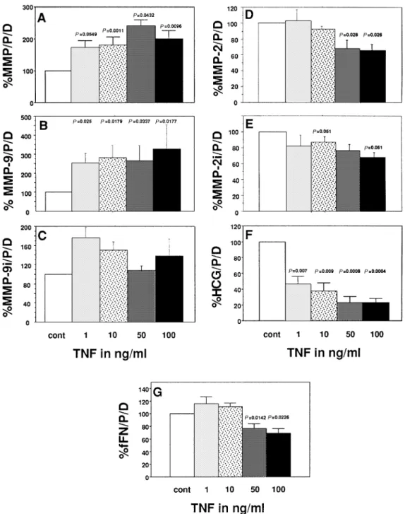
TNF significantly increased (P5 0.0432–0.0011) the total gelat-inolytic activity of CTB in a dose-dependent manner (Figure 1a). MMP-9 activity was also significantly increased (P5 0.025– 0.0177, Figure 1b), whereas its immunoreactivity remained unchanged (Figure 1c). In contrast, TNF significantly decreased (P5 0.028 and P 5 0.026 for 50 and 100 ng/ml respectively) MMP-2 activity and immunoreactivity (P 5 0.051 and P 5 0.061 for 10 and 100 ng/ml respectively, Figure 1d and 1e). The concentration of HCG was decreased in a dose-dependent and significant fashion (P5 0.0094–0.0004) by TNF (Figure 1f), whereas fFN was significantly inhibited but only by the highest concentrations of TNF (P5 0.0142 and P 5 0.0226 for 50 and 100 ng/ml respectively, Figure 1g).
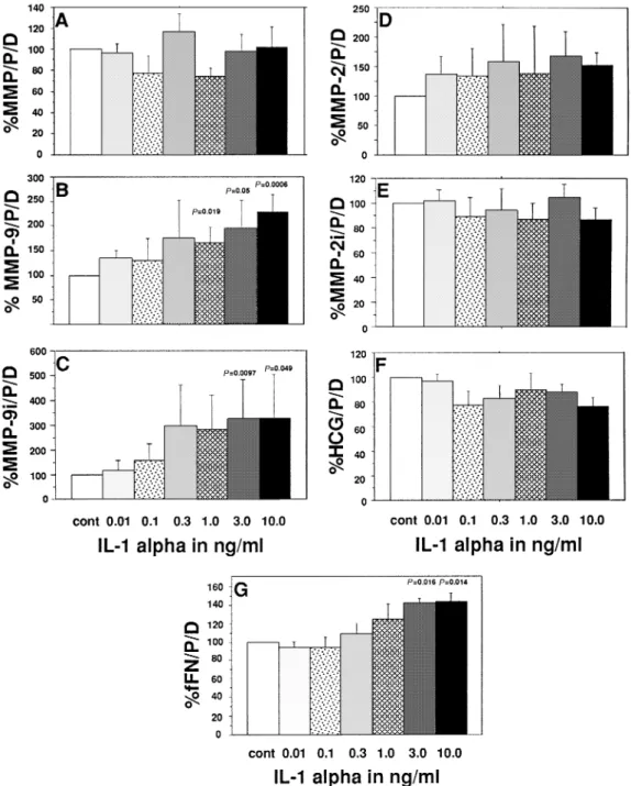
IL-1α significantly increased MMP-9 activity (P5 0.019– 0.006, 1.0–10.0 ng /ml, Figure 2b) and immunoreactivity (P5 0.0097, P5 0.049 for 3.0 and 10.0 ng/ml respectively, Figure 2c) as well as immunoreactive fFN (P5 0.016, P 5 0.014 for 3.0 and 10 ng /ml respectively, Figure 2g). None of the other parameters measured were statistically modified by this cytokine.
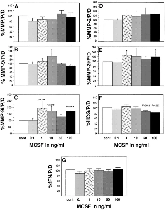
MCSF was inactive on most trophoblastic parameters measured except for an increased MMP-9 immunoreactivity (P 5 0.019–0.018 for 1–100 ng/ml respectively, Figure 3c) and for a significant inhibitory effect on HCG with the highest concentrations used (P 5 0.012, P 5 0.023 for 50 and 100 ng/ml respectively, Figure 3f).
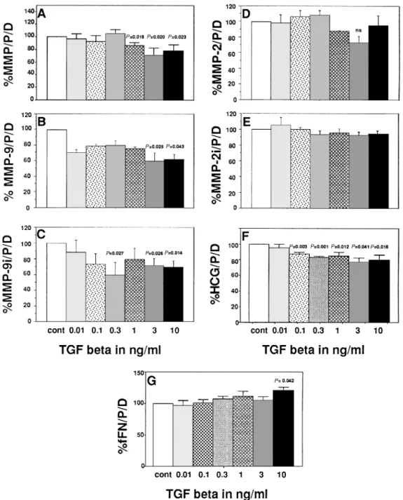
Figure 4 illustrates the effects of TGFβ. This cytokine inhibited the total gelatinolytic activity (P5 0.023–0.018, for 1.0–10 ng/ml, Figure 4a), the activity of MMP-9 (P5 0.025– 0.043 for 3 and 10 ng/ml respectively, Figure 4b) and MMP-9 immunoreactivity (P5 0.027 to P 5 0.014 for 0.3 to 10 ng/ ml respectively, Figure 4c) but had no effect on MMP-2 levels and activity (Figure 4d and e). In contrast, TGFβsignificantly decreased the concentration of HCG in dose-dependent manner (P 5 0.041–0.001 for 0.1–10 ng/ml, Figure 4f). Only the highest concentration of this cytokine (10 ng/ml) increased the secretion of fFN significantly (P5 0.042, Figure 4g).
Discussion
TNF, a potent apoptotic cytokine originally identified as a product of activated macrophages is now known to be produced by many types of cells including those in the female genital tract (Hunt, 1993). Protein and TNF transcripts were identified in villous and extravillous CTB (King et al., 1995), in syncytiotrophoblast (Haynes et al., 1993) as well as in endo-metrial large granular lymphocytes and CD 3 positive T cells
Figure 1. Effects of tumour necrosis factor (TNF) on (A) total gelatinolytic activity, activity of (B) matrix metalloproteinase (MMP)-9 and (C) MMP-2 and immunoreactivities of (D) MMP-9 and (E) MMP-2, and the concentration of (F) fetal fibronectin (fFN) and (G) human chorionic gonadotrophin (HCG) of cytotrophoblastic cells (CTB) cultured in vitro. Mean1 SEM, n 5 6. Values are expressed as mg/protein/day in percentage of controls (CTB in the absence of cytokines).
(Jokhi et al., 1994). Two types of receptors were described for TNF and both are present in endometrial epithelial cells (Tabibzadeh et al., 1995) and choriocarcinoma cell lines (Yang
et al., 1993). Therefore, this cytokine seems well positioned
to play a regulatory role in trophoblast invasiveness. Despite the fact that TNF receptors are secreted by CTB in the culture medium (Kno¨fler et al., 1998), TNF doubles the gelatinolytic activity of CTB. This effect seems to be due to an activation of proMMP-9 into MMP-9 since the activity of MMP-9 is increased by TNF but the levels of this gelatinase remain unchanged. The increased gelatinolytic activity of CTB cannot be attributed to MMP-2 since TNF decreases both the activity
and the levels of MMP-2 in these cells. It is however unclear if this TNF-induced proteolytic potential could favour trophoblast invasiveness in vivo since this cytokine does not increase CTB invasion in Matrigel (Bass et al., 1994). The inhibitory effect of TNF on HCG secretion observed here confirms previous observations (Ohashi et al., 1992) which clearly demonstrated that this inhibitory effect was not due to TNF cytotoxicity. In contrast to these results, Li et al., (1992) reported a stimulatory effect of TNF on HCG production using an interleukin-6 (IL-6) and IL-6-receptor-dependent system. This stimulatory effect was, however, observed when TNF was incubated for only 3 h with CTB.
Figure 2. Effects of interleukin-1α(IL-1α) on (A) total gelatinolytic activity; activities of (B) matrix metalloproteinase (MMP)-9 and (C) MMP-2; immunoreactivities of (D) MMP-9 and (E) MMP-2; and the concentrations of (F) fetal fibronectin (fFN) and (G) human chorionic gonadotrophin (HCG) of cytotrophoblastic cells (CTB) cultured in vitro. Mean1 SEM, n 5 6. Values are expressed as mg/protein/day in percentage of controls (CTB in the absence of cytokines).
IL-1 consists of two distinct but related peptides (IL-1 α andβ). IL-1, a known product of monocytes and macrophages is also produced by the tissues of the feto–maternal interface. In mice, IL-1 is an important mediator of implantation (Simo´n
et al., 1994b). In the human, IL-1 is similarly distributed both
at the protein and mRNA level (Romero et al, 1989; Kauma
et al., 1990; Simo´n et al., 1994a). Endometrial epithelial
cells and extravillous but not villous CTB have IL-1R-1. Interestingly, both CTB and decidualized stromal cells produce IL-1. IL-1 has been shown to stimulate the activity of MMP-1, MMP-3 and TIMP in human fibroblasts (Unemori et al., 1991) and MMP-9 in CTB (Librach et al., 1994). The present results confirm and extend this last observation since in our
hands IL-1 not only increases the activity of MMP-9 but also increases its immunoreactivity. We conclude that IL-1 increases both the synthesis and the activation of pro-MMP-9, a conclu-sion which is in line with the observation that IL-1 increases the mRNA of MMP-9 in first trimester CTB (Shimonovitz
et al., 1996). Masuhiro et al. (1991) showed that IL-1α
stimulates the secretion of HCG in first trimester CTB, this effect being dependent on trophoblastic IL-6 secretion and IL-6 receptor mediated signal transduction. Although we are using the same type of cells and similar concentrations of IL-1, we do not see any stimulatory effect of IL-1 on HCG secretion. This discrepancy could be due to the fact that Masuhiro et al. (1991) observed a maximal stimulation of
Figure 3. Effects of macrophage colony-stimulating factor (MCSF) on (A) total gelatinolytic activity; activities of (B) matrix metalloproteinase (MMP)-9 and (C) MMP-2; immunoreactivities of (D) MMP-9 and (E) MMP-2; and the concentrations of (F) fetal fibronectin (fFN) and (G) human chorionic gonadotrophin (HCG) of cytotrophoblastic cells (CTB) cultured in vitro. Mean1 SEM, n 5 6. Values are expressed as mg/protein/day in percentage of controls (CTB in the absence of cytokines).
HCG release 3 h after they had given IL-1 whereas we measured HCG only after 24 h of incubation. It is thus possible that IL-1 stimulates the secretion rather than the synthesis of HCG. The IL-1-induced trophoblastic fFN release observed here has not been reported previously.
MCSF null mutant mice are osteopetrotic and have a compromised reproductive potential (Pollard et al., 1991). It is thought that MCSF, a product of macrophages, is an important regulator of implantation in mice. In humans, MCSF mRNA was shown to be present in decidua (Kauma et al., 1991), villous CTB but not in extravillous CTB (King et al., 1995). In contrast, the MCSF receptor encoded by the proto-oncogene c-fms is present in the CTB columns of anchoring
villi (Jokhi et al., 1993) but not on villous CTB. In the present study, MCSF had virtually no effect except for a moderate stimulatory effect on immunoreactive MMP-9 and a slight inhibitory effect on HCG. According to a recent publication by Omigbodum et al. (1998), MCSF increases the trophoblastic mRNA of fFN and its receptor, the integrinα5β1at 24 and 72 h of exposure. In our hands, however MCSF had no effect at the fFN protein level. The reasons for this are obscure at the present time.
TGFβ is represented by five homodimeric polypeptides which share 70–80% structural homology. TGFβ 1, 2 and 3 are produced by many mammalian cells. TGFβ protein and mRNA have been localized in endometrial stromal, epithelial
Figure 4. Effects of transforming growth factorβ(TGFβ) on (A) total gelatinolytic activity; activities of (B) matrix metalloproteinase (MMP)-9 and (C) MMP-2; immunoreactivities of (D) MMP-9 and (E) MMP-2; and the concentrations of (F) fetal fibronectin (fFN) and (G) human chorionic gonadotrophin (HCG) of cytotrophoblastic cells (CTB) cultured in vitro. Mean1 SEM, n 5 6. Values are expressed as mg/protein/day in percentage of controls (CTB in the absence of cytokines).
and decidual cells, as well as in villous and extravillous CTB and in syncytium (Graham et al., 1992; Richards et al., 1993). CTB have three types of TGFβ receptors with differing affinities for TGFβ1 and TGFβ2 (Mitchell et al., 1992). In CTB or in human corneal fibroblasts, TGFβ stimulates the synthesis of matrix glycoproteins such as laminin, fibronectin and collagen (Ohji et al., 1993; Feinberg et al., 1994). In human fibroblasts, TGFβincreases MMP-2 and MMP-9 activity while it decreases TIMP (Overall et al., 1991). This, however, is not the case for CTB because the inhibitory effect that decidual cell conditioned medium exerts on the invasive behaviour of CTB seems to be due to TGFβ, since antibodies to this cytokine counteract the effect of decidual cell supernatants
(Graham and Lala 1991). TGFβexerts this anti-invasive effect by stimulating the TIMP secretion of CTB. Thus, TGFβcould well be an endometrial signal which controls trophoblast invasion during implantation and placentation. In the present study, TGFβ inhibits the gelatinolytic activity of CTB. This effect is attributable to a decrease in MMP-9 activity and immunoreactivity since TGFβhas no effect on MMP-2 activity and immunoreactivity. Despite the fact that TGFβ exerts inhibitory properties on the synthesis and activation of MMP-9 (an enzyme responsible for trophoblast invasion of Matrigel, Librach et al., 1991; Bischof et al., 1995a), it does not inhibit CTB invasion of Matrigel (Bass et al., 1994). The inhibitory effect that TGFβ exerts on HCG secretion is not a novel
finding and confirms data of a previous study (Song et al., 1996). Stimulation of fFN secretion as observed in the present study has also been reported previously (Feinberg et al., 1992). Although our data indicate that TNF, IL-1 and TGFβ are in-vitro regulators of MMP-9, a clear picture of the interactions of these and other modulators is far from being understood.
References
Azuma, C., Saji, F., Kimura, T. et al. (1990) Steroid hormones induce macrophage colony stimulating factor (MCSF) and MCSF receptor messenger RNAs in the human endometrium. J. Mol. Endocrinol., 5, 103–108.
Bass, K.E., Morrish, D., Roth, I. et al. (1994) Human cytotrophoblast invasion is up-regulated by epidermal growth factor: Evidence that paracrine factors modify this process. Dev. Biol., 164, 550–561.
Bischof, P., Friedli, E., Martelli, M. et al. (1991) Expression of extracellular matrix-degrading metalloproteinases by cultured human cytotrophoblast cells: Effects of cell adhesion and immunopurification. Am. J. Obstet. Gynecol., 65, 1791–1801.
Bischof, P., Martelli, M., Campana, A. et al. (1995a) Importance of metalloproteinases (MMP) in human trophoblast invasion. Early Pregn. Biol. Med., 1, 263–269.
Bischof, P., Haenggeli, L. and Campana, A. (1995b) Effect of leukaemia inhibitory factor on human cytotrophoblast differentiation along the invasive pathway. Am. J. Reprod. Immunol., 34, 225–230.
Bischof, P., Haenggeli, L. and Campana, A. (1995c) Gelatinase and oncofetal fibronectine secretion is dependent upon integrin expression on human cytotrophoblasts. Hum. Reprod., 10, 734–772.
Bischof, P. and Campana, A. (1996) Model for implantation of the human blastocyst and early placentation. Hum. Reprod. Update, 2, 262–270. Bischof, P., Meisser, A., Campana, A. et al. (1998) Effects of decidua
conditioned medium and insulin-like growth factor binding protein-1 on trophoblast matrix metalloproteinases and their inhibitors. Placenta, 19, 457–464.
Cross, J.C., Werb, Z. and Fisher, S.J. (1994) Implantation and the placenta: key pieces of the development puzzle. Science, 266, 1508–1518. Feinberg, R.F., Kliman, H.J. and Wang, C.L. (1994) Transforming growth
factor beta stimulates trophoblast oncofetal fibronectin synthesis in vitro: implications for trophoblast implantation in vivo. J. Clin. Endocrinol. Metab., 78, 1241–124.
Graham, C.H. and Lala, P.K. (1991) Mechanism of control of trophoblast invasion in situ. J. Cell Physiol., 148, 228–234.
Graham, C.H., Lysiak, J.J., Mc Crae, K.R. et al. (1992) Localisation of transforming growth factor at the human fetal–maternal interface: role in trophoblast growth and differentiation. Biol. Reprod., 4, 561–572. Haynes, M.K., Jackson, L.G., Tuan, R.S. et al. (1993) Cytokine production
in first trimester chorionic villi: detection of mRNAs and protein products in situ. Cell Immunol., 151, 300–308.
Hulboy, D.L., Rudolph, L.A. and Matrisian L.M. (1995). Matrix metalloproteinases as mediators of reproductive function. Mol. Hum. Reprod., 1, see Hum. Reprod., 10, 27–45.
Hunt, J.S. (1993) Expression and regulation of tumour necrosis factor alpha gene in the female reproductive tract. Reprod. Fertil. Dev., 5, 141–153. Hunt, J.S., Chen, H.L., Hu, X.L. et al. (1992) Tumor necrosis factor messenger
ribonucleic acid and protein in human endometrium. Biol. Reprod., 47, 141–147.
Jokhi, P.P., Chumbley, G., Gardner, L. et al. (1993) Expression of the colony stimulating factor-1 receptor (c-fms product) by cells at the human uteroplacental interface. Lab. Invest., 68, 308–320.
Jokhi, P.P., King, A., Sharkey, A.M. et al. (1994) Screening for cytokine messenger ribonucleic acids in purified human decidual lymphocyte populations by the reverse-transcriptase polymerase chain reaction. J. Immunol., 153, 4427–4435.
Kauma, S.W., Aukerman, S.L., Eierman, D. et al. (1991) Colony-stimulating factor-1 and c-fms expression in human endometrial tissues and placenta during the menstrual cycle and early pregnancy. J. Clin. Endocrinol. Metab.,
73, 746–751.
Kauma, S.W., Matt, D., Strom, S. et al. (1990) Interleukin 1 beta, human leukocyte antigen HLA-DR and transforming growth factor beta expression in endometrium, placenta and placental membranes. Am. J. Obstet. Gynecol.,
163, 1430–1437.
King, A., Jokhi, P.P., Smith, S.K et al. (1995) Screening for cytokine mRNA in human villous and extravillous trophoblasts using the reverse transcriptase–polymerase chain reaction (RT–PCR) Cytokine, 7, 364–371. Kno¨fler, M., Stenzel, M. and Husslein, P. (1998) Shedding of tumour necrosis
factor receptor from purified villous term trophoblast and cytotrophoblastic BeWo cells. Hum. Reprod., 13, 2308–2316.
Li, Y., Matsuzaki, N., Masuhiro, K. et al. (1992) Trophoblast-derived tumor necrosis factor-alpha induces release of human chorionic gonadotropin using interleukin-6 (IL-6) and IL-6-receptor dependent system in normal human trophoblasts. J. Clin. Endocrinol. Metab., 74, 184–191.
Librach, C.L., Feigenbaum, S.L., Bass, K.E. et al. (1994) Interleukin-1 beta regulates human cytotrophoblast metalloproteinase activity and invasion in vitro. J. Biol. Chem., 269, 17125–17131.
Librach, C.L., Werb, Z., Fitzgerald, M.L. et al. (1991) 92 kDa type IV collagenase mediates invasion of human cytotrophoblasts. J. Cell Biol.,
113, 437–449.
Martelli, M., Campana, A. and Bischof, P. (1993) Secretion of matrix metalloproteinase by human endometrial cells in vitro. J. Reprod. Fertil.,
98, 67–76.
Masuhiro, K., Matzusaki, N., Nishino, E. et al. (1991) Trophoblast-derived interleukin-1 (IL-1) stimulates the release of human chorionic gonadotropin by activating IL-6 and IL-6-receptor system in first trimester human trophoblasts. J. Clin. Endocrinol. Metab., 72, 594–601.
Mitchell, E.J., Fitz-Gibbon, L., O’Connor, M.C. et al. (1992) Subtype of betaglycan and of type I and type II transforming growth factor (TGF-) receptors with different affinities for TGF-1 and TGF-2 are exhibited by human placental trophoblast cells. J. Cell. Physiol., 150, 334–343. Ohashi, K., Saji, F., Kato, M. et al. (1992) Tumour necrosis factor-alpha
inhibits human chorionic gonadotropin secretion. J. Clin. Endocrinol. Metab., 74, 130–134.
Ohji, M., Sundar-Raj, N. and Thoft, R.A. (1993) Transforming growth factor beta stimulates collagen and fibronectin synthesis by human corneal stromal fibroblasts in vitro. Curr. Eye Res., 12, 703–709.
Omigbodun, A., Coukos, G., Ziolkiewicz, P. et al. (1998) Macrophage-colony stimulating factor (M-CSF) regulates the expression of fibronectin and its alpha5 integrin receptor in human trophoblasts. Endocrinology, 139, 2190–2193.
Overall, C.M., Wrana, J.L. and Sodek, J. (1991) Transcriptional and post transcriptional regulation of 72 kDa gelatinase type IV collagenase by transforming growth factorα1 in human fibroblasts. J. Biol. Chem., 266, 14064–14071.
Pampfer, S., Daiter, E., Barad, D. et al. (1992) Expression of colony-stimulating factor-1 receptor (c-fms proto-oncogene product) in the human uterus and placenta. Biol. Reprod., 46, 48–57.
Pollard, J.W., Hunt, J.S., Wiktor-Jedrzejczak, W. et al. (1991) A pregnancy defect in osteopetrotic (op/op) mouse demonstrates the requirement for CSF-1 in female fertility. Dev. Biol., 148, 273–283.
Richards, C.D., Shoyab, M., Brown, T.J. et al. (1993) Selective regulation of metalloproteinase inhibitor (TIMP-1) by oncostatin M in fibroblast culture. J. Immunol., 150, 5596–5603.
Romero, R., Wu, Y.K., Brody, D.T. et al. (1989) Human decidua: a source of interleukin-1. Obstet. Gynecol., 73, 31–34.
Shimonovitz, S., Hurwitz, A., Barak, V. et al. (1996) Cytokine-mediated regulation of type IV collagenase expression and production in human trophoblast cells. J. Clin. Endocrinol. Metab., 81, 3091–3096.
Shimonovitz, S., Huriwitz, A., Dushnik, M. et al. (1994) Developmental regulation of the expression of 72 and 92 kD type IV collagenase in the human trophoblasts: a possible mechanism for control of trophoblast invasion. Am. J. Obstet. Gynecol., 171, 832–838.
Simo´n, C., Frances, A., Piquette, G.N. et al. (1994a) Interleukin-1 system in the materno–trophoblast unit in human implantation: immunohistochemical evidence for autocrine/paracrine function. J. Clin. Endocrinol. Metab., 78, 847–854.
Simo´n, C., Frances, A., Piquette, G.N. et al. (1994b) Embryonic implantation in mice is blocked by interleukin-1 receptor antagonist. Endocrinology,
134, 521–528.
So, T., Ito, A., Sato, T. et al. (1992) Tumour necrosis factor stimulates the biosynthesis of matrix metalloproteinases and plasminogen activator in cultured human chorionic cells. Biol. Reprod., 46, 772–778.
Song, Y., Keelan, J. and France, J.T. (1996) Activin-A stimulates, while transforming growth factor beta 1 inhibits, chorionic gonadotrophin production and aromatase activity in cultured human placental trophoblasts. Placenta, 17, 603–610.
involved in implantation, a symbiotic interaction between blastocyst and endometrium involving adhesion and tissue invasion. Mol. Hum. Reprod.,
1, see Hum. Reprod., 10, 1579–1602.
Tabibzadeh, S., Zupi, E., Babaknia, A. et al. (1995) Site and menstrual cycle-dependent expression of proteins of the tumour necrosis factor (TNF) receptor family, and BCL-2 oncoprotein and phase-specific production. Hum. Reprod., 10, 277–286.
Unemori, E.N., Bair, M.J., Bauer, E.A. et al. (1991) Stromelysin expression regulates collagenase activation in human fibroblast. J. Biol. Chem., 266, 23477–23482.
Ward, R.V., Hembry, M., Reynolds, J.J. et al. (1991) The purification of tissue inhibitor of metalloproteinases-2 from its 72 kDa progelatinase complex. Biochem. J., 278, 179–187.
Yang, Y., Yelavarthi, K.K., Chen, H.L. et al. (1993) Molecular biochemical and functional characteristics of tumor necrosis factor alpha produced by human placental cytotrophoblastic cells. J. Immunol., 150, 5614–5624. Received on September 7, 1998; accepted on December 18, 1998