Université de Montréa]
Neuroprotection and Regeneration of Adu]t Lesioned Retinal
Ganglion Celis by the Modulation of Fibroblast Growth Factor-2
and Receptor Tyrosine Phosphatase-Sigma
par
PRZEMYSLAW SAPIEHA
Département de pathologie et bio]ogïe cellulaire Faculté de Médecine
Thèse présentée à la faculté des études supérieures en vue de l’obtention du grade de Philosophi Doctor (Ph.D.)
en neurocytologie moléculaire
Mars 2005
w
o
QOC
Université
de Montré al
Direction des bibliothèques
AVIS
L’auteur a autorisé l’Université de Montréal à reproduire et diffuser, en totalité ou en partie, par quelque moyen que ce soit et sur quelque support que ce soit, et exclusivement à des fins non lucratives d’enseignement et de recherche, des copies de ce mémoire ou de cette thèse.
L’auteur et les coauteurs le cas échéant conservent la propriété du droit d’auteur et des droits moraux qui protègent ce document. Ni la thèse ou le mémoire, ni des extraits substantiels de ce document, ne doivent être imprimés ou autrement reproduits sans l’autorisation de l’auteur.
Afin de se conformer à la Loi canadienne sur la protection des renseignements personnels, quelques formulaires secondaires, coordonnées ou signatures intégrées au texte ont pu être enlevés de ce document. Bien que cela ait pu affecter la pagination, il n’y a aucun contenu manquant. NOTICE
The author of this thesis or dissertation has granted a nonexclusive license allowing Université de Montréal to reproduce and publish the document, in part or in whole, and in any format, solely for noncommercial educational and research purposes.
The author and co-authors if applicable retain copyright ownership and moral rights in this document. Neither the whole thesis or dissertation, nor substantial extracts from it, may be printed or otherwise reproduced without the author’s permission.
In compliance with the Canadian Ptivacy Act some supporting forms, contact information or signatures may have been removed from the document. While this may affect the document page count, it does flot represent any loss of content from the document.
n
Université de Montréal Faculté des études supérieures
Cette thèse de doctorat intitulée
«Neuroprotection and Regeneration of Aduit Lesioned Retinal Ganglion Celis by the Modulation of Fibroblast Growth Factor-2
and Receptor Tyrosine Phosphatase-Sigma »
présentée par:
PRZEMYSLAW SAPIEHA
a été évaluée par un jury composé des personnes suivantes:
président-rapporteur
directeur de recherche membre du jury
Sam
?.iJh?
examinateur externe
111
SOMMAIRE
La capacité du système nerveux central (SNC) à survivre et à se régénérer par suite de lésions traumatiques ou lors des maladies dégénératives est fortement compromise chez les vertébrés supérieurs adultes. Le manque de survie découle de la perte du support trophique provenant des cellules environnantes et des cellules cibles (Burek and Oppenheim. 1996; Jacobson et al., 1997), ainsi que d’une perte générale de l’habilité à répondre aux signaux trophiques (Shen et al., 1999). De son coté, la régénération limitée est attribuable à un environnement hautement inhibiteur et une perte intrinsèque de la capacité de croître. Les deux scénarios aboutissent finalement à la mort neuronal par apoptose.
Les stratégies décrites dans cette thèse tentent de stimuler la régénération et la survie des cellules ganglionnaires de la rétine (CGR) adulte en réactivant leurs programmes intrinsèques de croissance et de survie. Plus spécifiquement, nous avons testé l’hypothèse selon laquelle certains gènes impliqués dans le développement ordonné du système nerveux peuvent être modulés chez l’adulte dans le but de stimuler la croissance axonale et de provoquer la survie.
À
cette fin, nous avons premièrement augmenté le niveau endogène du facteur de Croissance des f ibroblastes-2 (FGF-2). Cette molécule s’avère être un puissant stimulateur de croissance axonale chez les CGR lors du développement (Dingwell et al., 2000). En utilisant des adéno-virus associés recombinants (AAV.FGf-2), nous avons introduit le gène codant pour f Gf-2 dans les CGR matures, leurs fournissant ainsi des niveaux élevés de ce facteur. Nous démontrons ici que, par suite d’une blessure du nerf optique, cette approche augmente deiv
façon significative la repousse axonale des COR et engendre une neuroprotection robuste mais transitoire.
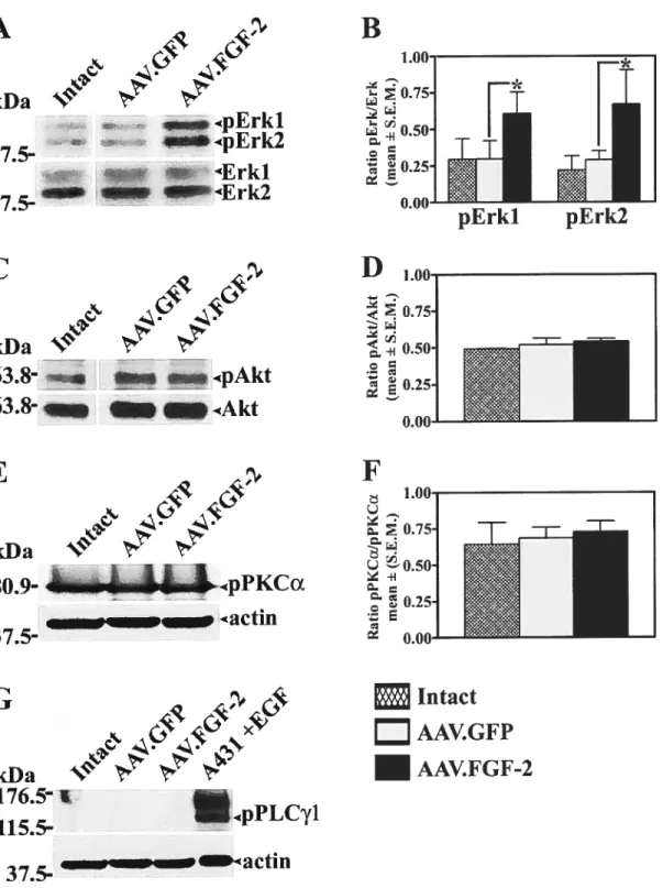
Ultérieurement, nous avons cherché à identifier les mécanismes moléculaires responsables de la croissance axonale induite par FGF-2. Nous avons d’abord déterminé le niveau d’activation de certaines voies de signalisation susceptibles d’être stimulées parle récepteur de FGF (FGfR). Nos résultats ont démontré que l’activation de la kinase «extracellular-signal regulated kinases 1/2 » (Erkl/2) est amplifiée dans les rétines traitées avec l’AAV.f 0f-2. Au contraire, des voies de signalisations comme celles de la phospholipase Cy, de la «phosphoinositide-3-kinase > et de la protéine kinase C n’ont pas été stimulées. De plus. l’inhibition pharmacologique de Erkl/2 amène une réduction d’environ 80% du nombre d’axones de COR pouvant se régénérer. Ceci établit un rôle important pour Erkl/2 dans la croissance axonale provoquée parf Gf-2.
En raison de l’importance de la phosphorylation dans la survie et la régénération neuronale, nous avons étudié le rôle du récepteur à protéine lyrosine phosphatase Sigma (RPTPa): un régulateur de la phosphorylation intracellulaire. L’implication de RPTPY dans le développement du SNC est illustrée par son haut niveau d’expression dans le tissu nerveux embryonnaire et par le fait que son absence chez des souris transgéniques entraîne une réduction globale de la taille de leur cerveau (Elchebly et al., 1999; Wallace et al.. 1999). En démontrant une hausse significative de la régénération des CGR lésées chez des souris RPTPr (-/-), nous proposons que cette phosphatase inhibe la croissance axonale chez l’adulte. Nous avons également détecté une forte activation endogène de Erkl/2 et de Aki à chez les souris RPTPa (-/-), suggérant que ces voies peuvent être impliquées dans l’augmentation de la repousse axonale observée chez ces animaux.
V
Les données présentées dans cette thèse suggèrent que la modulation des gènes impliqués dans le développement du SNC offre une stratégie prometteuse pour favoriser la croissance du SNC adulte lésé.
Mots clefs: Système Nerveux Central, facteur de Croissance de fibroblastes-2, Récepteur à Protéine Tyrosine Phosphatase-Sigma, Régénération Axonale, Survie Neuronale, Thérapie Génique, Cellule Ganglionnaire de la Rétine, Erkl/2.
vi
SUMMARY
The ability of the central nervous system (CNS) to regenerate and survive
following traumatic injury or disease is severely compromised in aduit higher vertebrates. Current data ascribes this lack of CNS regrowth to both a highly growth-prohibitive environrnent ami to the neurons own loss of intrinsic growth capacity. The lack of neuronal survival following CNS injury is mostly attributed to inadequate trophic support from neighboring and target ceils (Burek and Oppenheim, 1996; Jacobson et al., 1997) as well as the neuron’s overail loss of trophic responsiveness (Shen et al., 1999); both scenarios ultimately resulting in neuronal apoptosis.
The strategies described in this thesis attempt to promote the regeneration and the survival of aduit lesioned retinal ganglion celis (RGCs) by reactivating their intrinsic growth and anti-apoptotic programs. More specifically, we tested the bypothesis that genes involved in the order]y development of the nervous system can 5e modulated in the adu]t to ensure adequate neuroprotection and stimulate axonal regrowth. In this aim, we first chose to increase levels of fibroblast growth factor-2 (f 0f-2), a potent stimulator of ROC axon growth during development (Dingwell et al., 2000). Using recombinant adeno-associated virus (AAV), we introduced the gene coding for f Gf-2 into mature ROCs, thus ensuring a sustained upregulation of this neurotrophic factor. We provide evidence that this approach significantly enhances ROC axonal regrowth following acute optic nerve injury and promotes robust but transient neuroprotection.
vii
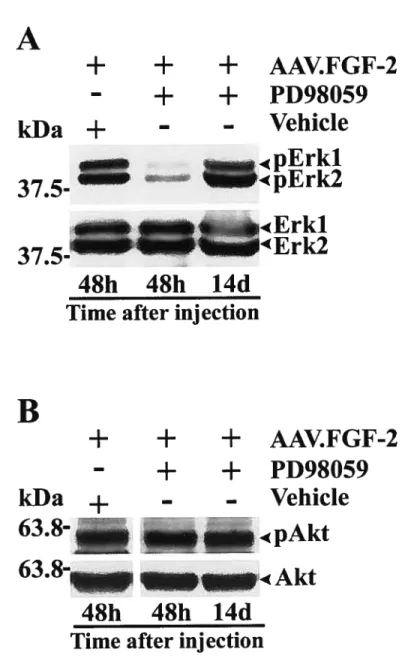
We subsequently souglit to elucidate the molecular signaling mechanisms that regulate fGF-2-induced RGC axon groÏh. To address this question. we first proceeded to screen candidate pathways known to be recruited upon stimulation of FGF receptor (f GfR). Our data demonstrate that the extracellular-signal regulated kinases 1/2 (Erk 1/2) were stimulated in AAV.fGf-2 treated retinas, but flot other pathways such as phosphoinositide 3-kinase, phospholipase Cy or protein kinase C. Inhibition of Erk 1/2 with the pharmacological inhibitor PD98059 led to a 8O ¾ reduction in the number of regenerating RGC fibers and thus established Erk 1/2 as an important signaling pathway in f Gf-2 mediated axonal re-growth.
As both neuronal survivai and regenerative signais rely heaviiy on phosphorylation events. we investigated the role of Receptor Protein Tyrosine Phosphatase-Sigma (RPTP-a). a regulator of intracellular phosphorylation. RPTP a’s involvement in CNS development is illustrated both by its high levels of expression in the embiyonic nervous system and by an overali reduction in brain size in transgenic animais lacking this protein (Elchebly et al., 1999; WalÏace et al., 1999). By demonstrating that RPTP-a
(-/-)
mice show a significant increase in RGC axon regrowth following acute injury when compared to wild type littermate controls, we provide evidence that RPTP-a hinders CNS regeneration in the aduit. This increased growth cannot be attributed to developmental defects as we found that knockout animais have normal retinal and optic nerve histologies and show identical time-courses of ROC death following lesion. Lastly. we show high levels of Erk 1/2 and Akt activation in RPTP-a(-/-)
mice. suggesting that these pathways may be implicated in the increased axonal growth observed in these animais.vin
The data presented in this thesis suggest that the modulation of genes involved in the sound development of the CNS is an adequate strategy to promote regenerative growth of aduit injured CNS neurons.
Kev words: Central Nervous System. fibroblast Growth Factor-2, Receptor Tyrosine Phosphatase-Sigma. Axonal Regeneration. Neuronal Survival, Gene Transfer. Retinal Ganglion Celis. MAPK
ix
TABLE 0f CONTENTS
SUMMARY iii
SOMMAIRE vi
TABLE 0F CONTENTS ix
LIST 0F TABLES xix
LIST 0F FIGURES xx
LIST 0F ABBREVIATIONS xxiv
ACKNOWLEDGEMENTS xxvii
PREFACE xxviii
CHAPTER 1.
GENERAL INTRODUCTION 1
1.1 CENTRAL NERVOUS SYSTEM REGENERATION: A HISTORICAL
PERSPECTIVE 2
1.2 AXON GROWTH IN TUE CNS 4
1.2.1 A Complex Neural Network 4
1.2.2 Growth Cone Dynamics 5
1.2.3 Growth Cone Remodeling 7
X
1.3111E GENESIS 0F RGCs AND AXONAL GROWTH FROM THE
RETINA TO THE MIDBRAIN 11
1.3.1 Origins ofthe Eye and Cellular Determination in the Developing Retina..12
1.3.2 Detemiinants ofRGC Differentiation 13
J. 3.2.] Sonic Hedge Hog and RGC Generation 13 1.3.2.2 fibroblast Growth Factor-2 andRGC Generation 14
1.3.3 Initiation ofAxon Growth 15
1.3.4 Molecular Determinants of Intraretinal Axonal Growth 17
1.3.4.1 Laminins and Integrins 17
1.3.4.2 CelliiïarAdhesion MoÏecides 1$
1.3.4.3 Receptor Protein Tyrosine Phosphatases 20 1.3.4.4 fibi-obÏast Growth factors and RGC Growth 21
1.3.5 Entering the Optic Nerve 22
1.3.6 Decisions at the Optic Chiasm 25
1.3.7 Innervation ofthe Superior Colliculus 26
1.4 OPTIC NERVE AXOTOMY 28
1.4.1 Acute Optic Nerve Injury and RGC Death 29
1.4.2 Pattem ofRGC Ceil Death in the Axotomy Model 29
1.5 APOPTOSIS 30
1.5.1 The Bd-2 Family 31
xi
1.5.3 Signaling and Apoptosis.36
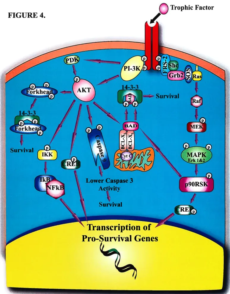
1.5.3.] FI-3K/AkiPathway 38
1.5.3.2 The MEK/M4FK Pathway 42
1.5.4 RGC Axotomy and Trophic Deprivation 42
1.6 AXON REGENERATION IN THE CNS 45
1.6.1 The Inhibitoiy CNS Environment 46
1.6.1.1 CN$MyeÏin 46
1.6.1.2 The Guai Scar 47
1 .6.2 Intrinsic Regenerative Ability of RGCs 49
1.6.3 Survival versus Regeneration ofLesioned CNS Neurons 51
1.7 NEUROTROPHIC SUPPLEMENTATION FOR LESIONED NEURONS 52
1.7.1 f Gf-2 and Retinallnjury 53
1.7.2 FGF Signaling 54
1.7.2.1 TheSNT-]/FRS2Pathway 55
1.7.2.2 The PLC7 Fathway 56
1.8. GENE MODULATION FOR THE INJURED CNS, PART I: ADDING A
GENE 57
1.8.1 Gene Transfer Techriology 57
xii
1.8.3 Viral Vectors for Retinal Gene Transfer 60
1.8.3.1 Adenovirus (Ad) 61
1.8.3.1.] A d Tropisrn in the Retina 61
].8.3.2Adeno-associated Virus (AAV, 63
1.8.3.2.1 A4 V Tropism in the Retina 64
1.8.3.3 Lentivirus (‘LJ’ 65
1.8.3.3.1 CelÏîtlar Tropism ofL V I7ectors 66
1.9 GENE MODULATION FOR THE INJURED CNS, PART II:
REMOVING A GENE 67
1.9.1 RNAlnterference 6$
1.9.2 Antisense Oligonucleotides 69
1.9.3 Ribozymes 70
1.9.4 Smail Moiecule Inhibitors 70
1.9.5 Transgenic Knockout Animais 71
1.10 OBJECTIVES 0f THE THESIS 73
CHAPTER 2.
FIRST ARTICLE 74
2.1 ABSTRACT 76
2.2 INTRODUCTION 77
xiii
2.3.1 Preparation of Recombinant AAV Serotype 2 Vectors 79
2.3.2 Surgical Procedures $0
I) Intravitreal In/ections 80
ii) AxonaÏ Growïh 81
iii, NeïtronaÏ SïtrvivaÏ $2
iv,.) Ccli Pro flferation $3
2.3.3 Retinal Immunocytochemistry $3
2.3.4 Optic Nerve Immunostaining and Quantification ofAxonal Growth 84
2.3.5 Quantification of Neuronal Survival $6
2.4 RESULTS 87
2.4.1 AAV-Mediated f Gf-2 Protein is Expressed by Aduit RGCs $7 2.4.2 ROCs Express the High Affinity f 0f-2 Receptor, f GfR-1 88 2.4.3 AAV.f0f-2 Promotes Regeneration of Injured ROC Axons $9 2.4.4 Selective Upregulation off Gf-2 in ROCs, but Not in Optic Nerve
Glia, is Required for Axonal Growth 92
2.4.5 f Gf-2 Gene Delivery Confers Transient Protection to Axotomized
RGCs 93 2.5.DISCUSSION 94 2.6 REFERENCES 101 2.7 FIGURES 114 CHAPTER 3. SECOND ARTICLE 131
xiv
3.1 ABSTRACT .133
3.2 INTRODUCTION 134
3.3 EXPERIMENTAL PROCEDURES 136
3.3.1 Preparation ofRecombinant AAV Serotype 2 Vectors 136
3.3.2 Intravitreal Injection of Viral Vectors 136
3.3.3 Retrograde ROC Labeling and Retinal Imrnunohistochernistry 137 3.3.4 Micro-crush Lesion ofthe Optic Nerve and Anterograde Axon
Labelling 138
3.3.5 Optic Neiwe Imrnunohistochemistry and Quantification of Axon
Growth 139
3.3.6 Western Blot Analysis 140
3.4 RESULTS 141
3.4.1 AAV Serotype 2 Directs f 0f-2 Transgene Expression in Aduit RGCs...141 3.4.2 FGF-2 Gene Transfer Promotes Regrowth of ROC Axons Afier Acute
Optic Nerve Injury 141
3.4.3 AAV.FGF-2 Stimulates Retinal Erkl/2 Activation 144 3.4.4 Erkl/2 Mediate ROC Axon Orowth Induced by f 0F-2 145
3.5 DISCUSSION 147
3.6 REFERENCES 150
3.7 FIGURES 160
CHAPTER 4.
xv 4.1 ABSTRACT .174 4.2 INTRODUCTION 175 4.3 EXPERIMENTAL METHODS 178 4.3.1 Animais 17$ 4.3.2 In Situ Hybridization 178
4.3.3 Retinal and Optic Nenie Histology 179
4.3.4 Surgical Procedures 180
i) Axonal Growth 180
ii) Neuronal Survival 181
4.3.5 Quantification ofAxonal Growth and Neuronal Survival 181 4.3.6 Western Blot Analysis and Immunocytochemistry 183
4.4 RESULTS 185
4.4.1 RPTPa mRNA is Expressed by Aduit Mouse RGCs 185 4.4.2 Lack of RPTPa Does Not Alter Retinai or Optic Nerve Histoiogy 186 4.4.3 Absence ofRPTPa Does Not Influence RGC Survivai Afier Optic
Nerve Transection 187
4.4.4 RPTPG (-I-) Mice Display Enhanced RGC Axon Regrowth Afler
Acute Optic Nerve Lesion 189
4.4.5 Retinal MAPK and Akt are Activated in Aduit RPTPa (-I-) Mice 190
4.5 DISCUSSION 191
4.6 REFERENCES 197
xvi
CHAPTER 5.
GENERAL DISCUSSION 221
5.1 SEARCHING FOR GENES INVOLVED IN REGENERATION 222
5.2 SUSTAINED UPREGULATION 0F FGF-2 IN INJURED ADULT
RGCS 224
5.3 NEUROPROTECTION AND FGFS 225
5.3.1 FGF-2, FGF-5 and f Gf-1$ Prornote Transient Survival of
Axotomized RGCs 225
5.3.2 fGfR1 Expression in the Injured Retina 225
5.4 SOMATIC EXPRESSION 0F FGF-2 IS REQUIRED TO
STIMULATE REGENERATION 227
5.5 RPTP-a HINDERS RGC REGENERATION 228
5.6 RPTP-a SIGNALING 232
5.6.1 How Does Ligand Binding Influence RPTP-a Signaling9 232
5.6.2 Ligand-induced Inactivation ofRPTPs 232
xvii
5.7 RPTPS AND CR0WTH YNAMICS.236
5.7.1 Cytosolic Effectors ofRPTP Signaling 236
5.7.2 RPTPs and Rho GTPases 237
5.8 ERK1/2 ACTIVATION AND RGC REGENERATION 23$
5.8.1 MAPKJErk1/2 may Require a Co-signal to Promote Axon Growth 239 5.8.2 Adequate Levels ofErkl/2 are Required for Axonal Growth 240
5.8.3 FGf-2 and Erkl/2 Activation 240
5.8.4 RPTP-a and Erkl/2 Activation 241
5.10 GENERAL CONCLUSIONS 243
REFERENCES 245
APPENDIX A
TrkB GENE TRANSFER PROTECTS RETINAL GANGLION CELLS
FROM AXOTOMY-INDUCED DEATU IN VIVO 292
APPENDIX B
HARNESSING THE GENE: STRATEGIES FOR RETINAL
xviii
APPENDIX C
AAV.FGF-2, AAV.FGF-5 OR AAV.FGF-l8 PROMOTES TRANSIENT
SURVIVAL 0F AXOTOMIZED RGCS 366
APPENDIX D
FGFR1 IS UPREGULATED IN PHOTORECEPTOR CELLS FOLLOWING
OPTIC NERVE TRANSECTION 367
APPENDIX E
TECHNICAL LIMITATIONS WHEN INHIBITING ERK 1/2 AND AKT IN
THE MOUSE RETINA 369
APPENDIX F
xix
LIST 0F TABLES CHAPTER II
Table]. Survival ofAxotomized RGCs following FGf-2 In Vivo
Geneiransfer 130
APPENDIX A
xx
LIST 0F FIGURES CHAPTER I
f igure 1. The organization of the neuronal growth cone and Rho GlPases
and growth cone dynamics 9
Figure 2. Guidance eues in the developing retino-collicular system 24 Figure 3. Bcl-XL prevents the loss ofmitochondrial cytochrome C 32 f igure 4. Neuronal survival induced by growth factors 41
CHAPTER II
figure 1. Experimental protocols used to test the effect of AAV.fGF-2 on
RGC axon growth. survival or pro liferation 114 Figure 2. AAV mediates f GF-2 transgene expression in adult RGCs 116 f igure 3. fGFR- 1 and heparan sulphate (HS) are expressed by adult RGCs 118 Figure 4. Micro-crush lesion resuits in complete transection ofthe optic nerve
and a well-defined injury site 120
f igure 5. f Gf-2 gene transfer promotes RGC axon growth afler micro-crush
lesion ofthe optic nerve 122
figure 6. AAV.fGf-2-induced RGC axon regeneration within the injured optic nerve: quantitative and immunocytochemical analysis of
growing axons 124
figure 7. Selective upregulation offGf-2 in RGCs, but not in optic nerve glia,
xxi
f igure 8. f Gf-2 gene transfer promotes transient survival of axotornized
ROCs 128
CHAPTER III
figure 1. AAV serotvpe 2 directs fGF-2 transgene expression in aduit ROCs 160 figure 2. Experimental protocol used to investigate the effect ofAAV.F0F-2
on RGC axon growth 162
figure 3. FGF-2 gene transfer promotes regrowth of ROC axons after acute
optic nerve injury 164
Figure 4. AAV.F0F-2 stimulates retinal Erkl/2 activation 166 figure 5. Selectivein vivo inhibition ofAAV.FGf-2-induced activation of
retinal Erkl/2 168
figure 6. Erkl/2 mediate ROC axon growth induced by F 0F-2 170
CHAPTER IV
F igure 1. RPTPa is expressed by aduit ROCs and optic nerve glia 207 figure 2. RPTPYdeficiency does flot alter retinal structure 209 figure 3. Lack ofRPTPc does flot affect optic nerve structure 211 figure 4. Retinal RPTPa protein continues to be expressed after optic nerve
injury but does flot regulate ROC survival 213 figure 5. Micro-crush (MC) lesion of the mouse optic nerve resuits in
complete transection of ROC axons 215
xxii
Figure 7. MAPK and Akt kinase pathways are constitutively active in
RPTPcY (-I-) mice retinas 219
CHAPTER V
f igure 5-1. The working model and general summary 231 F igure 5-2. Model for RPTP-a-mediated activation ofErkl/2 242
APPENDIX A
Figure 1. TrkB mRNA in intact and injured aduit rat retinas 322 Figure 2. AAV-mediated TrkB gene product expression in aduit RGCs 324 Figure 3. Effect of TrkB gene transfer on the protection of axotomized RGCs in
vivo 326
Figure 4. Correlation of RGC survival and AAV-mediated TrkB expression
in injured retinas 32$
Figure 5. In vivo activation ofretinal components ofthe BDNF/TrkB signaling pathway following TrkB gene transfer to RGCs 330 figure 6. Selective inhibition of AAV.TrkB-induced activation of retinal MAPK
andAkt 332
Figure 7. TrkB uses MAPK activation to promote the survival of aduit RGCs in
xxiii
APPENDIX C
f igure 1. AAV.FGF-2, AAV.FGF-5 or AAV.FGf-18 promotes transient survival of
axotomized RGCs 366
APPENDIX D
figure 1. fGfR1 is upregulated inphotoreceptor celis following optic nerve
transection 368
APPENDIX E
f igure 1. Technical limitations when inhibiting Erk 1/2 and Akt in the mouse
xxiv
LIST 0F ABBREVIATIONS
AAV adeno associated virus
ANT adenosine nucleotide transiocator
Ad Adenovirus
bHLH basic helix-loop-helix BRB blood-retinal banier
BDNF brain derived neurotrophic factor CaMMI Ca27ca1modulin kinase II
CREB cAMPresponse element binding protein CAM ceil adhesion molecule
CNS central nervous system
CSPG chondroitin-sulfate proteoglycans CNTF ciliary neurotrophic factor
cAMP cyclic adenosine monophosphate
cyt C cytochrome C
DCC deleted in colorectal cancer DNA deoxyribonucleic acid DSP dual specificity phosphatase
E13 embryonic day 13
ECM extracellular matrix
Erk extracellular regulated-signal kinase FGF fibroblast growth factor
xxv
FAK focal adhesion kinase GCL ganglion cell layer
GDNF guai celi line-derived neurotrophic factor GAPs GTPase activating proteins
GEF guanine nucieotide exchange factor HSV herpes simplex virus
1g immunoglobulin
1P3 inositol triphosphate INL inner nuclear layer
IGf- 1 insulin-like growth factor- Ï ITR inverted tenninal repeats
LN laminin
mm millimeters
MAPK mitogen activated protein kinase
MEK MAP kinase! extracellular signal-regulated kinase (ERK) kinase MAG myelin-associated glycoprotein
NGF nerve growth factor
NT-3 neurotrophin-3
NT-4 neurotrophin-4
NgR Nogo receptor
NLS nuclear localization signal
Omgp oligodendrocyte-myelin glycoprotein
xxvi
ONL outer nuclear layer
PDK 3’-phosphoinositide-dependent protein kinase
PLC phospholipase C
PKC protein kinase C
PNS peripheral nervous system PTK protein tyrosine kinase
RPTP receptor protein tyrosine phosphatase RTK receptor tyrosine kinase
RGM repulsive guidance molecule ROC retinal ganglion celi
RPE retinal pigment epithelium
RNA ribonucleic acid
RSK ribosomal S6 kinase
Robo roundabout
Shh Sonic Hedgehog
TGF- transforming growth factor-Beta VDAC voltage dependant anion channel
xxvii
ACKNOWLEDGEMENTS
b ail ihose who made my doctoralyears u stimulaling and memorable experience. I wish to thank my thesis supervisor Dr. Adriana Di Polo for giving me the opportunity to work on a series of exciting projects as well as for here support and guidance throughout my studies.
I would like to sincerely express my gratefulness to ail the co-authors of my papers and everyone else who bas contributed to the advancement of my projects, in particular Drs. Noriko Uetani, Laure Duplan. Sandrine Joiy, Timothy Kennedy, Michel Tremblay, William Hauswirth as well as Margaret Attiweil and Ken Watkins.
I would furthermore like to thank the members of my thesis jury for accepting to evaluate my work.
Many thanks to the members of the Di Polo Ïab, past and present. Thank you Vincent for the insightful conversations on ail subjects. Many thanks to Philippe. Fred, Yu. Laure and Marilyne for the atmosphere they bring to the work place. Also. I wouid like to express my gratitude to Philippe Bourgeois for the outstanding technical support.
I wouid further like to thank the professors of the Department of Pathology and Ceil Bioiogy for their availability. suggestions, teachings and support, in particular Drs. Nicole Leclerc, Laurent Descarries. Guy Doucet and Lisa McKerracher.
I thank the fond de Recherche en Santé du Québec for their financial support. Many thanks to my friends and coiieagues in the Department of Pathology and Ceil Biology and a speciai thanks to Monique and Céline for making every departmentai event unforgettable.
I wish to express my unbound gratitude to my parents for their ongoing advice, encouragement, direction and witty perspective that have been instrumental to my achievements.
Lastly, I would like to give special thanks to Cindy for the patience, the understanding, the motivation and the happiness that she brings to every day.
xxviii
PREFACE
The principal focus of this thesis was to study the invoïvement of f Gf-2 and RPTP-a in the regeneration and survival of retinal ganglion celis (RGCs), a prototypic central nervous system (CNS) neuronal population. In order to familiarize the reader with the subject at hand, this thesis includes a thorough introduction on axonal growth by RGCs, the consequences of acute axonal injury (both on survival and regeneration) and a review of current strategies that may be employed to modulate gene expression to stimulate neuronal survival and axon regrowth. The remainder of the thesis is manuscript-based: it comprises three original research articles that have been published in (or submitted to) peer reviewed journals, followed by a general discussion covering key topics pertaining to the articles.
xxix
Consider rhat at the core of the mmd is wiÏ/power. Whether there is success or faihire, ifone enrusts himsclfto the straightness ofthe path at the core ofi’he mmd. he wiÏÏ attain right-mindedness in either case. Severing oneseiffrom desire and being Ïike arock or a tree, nothingwiÏÏ ever be achieved. Not departingfroin desire, but realL-ing a desireÏess right-mindedness- this is the Way”
- TAKUAN SOHO, THE CLEAR SOUND 0F JE WELS
b my parents Jola and Slawek, for their wisdom. guidance and support and to Cindy, for sharing the highs and lows.
Chanter I
7
1.1 CENTRAL NERVOUS SYSTEM REGENERATION: A HISTORICAL PERSPECTIVE
The regeneration of the injured CNS lias been a daunting task since the dawn of modem neurobiology. CNS neurons severed from tlieir targets are incapable of surviving, regrowing axons and restoring function to affected structures. For these reasons, traumatic injuries and diseases of the CNS are of an irreversible nature. Although tlie intrinsic inability of lesioned CNS neurons to re-establisli function was documented as far back as Ancient Egypt (Jackowski, 1995), it was only in the early 1 900s, with the advent of histological and electropliysiological tools. that CNS regeneration began to be thoroughly explored.
In the late 1 $OOs, neuroscientists first described that rather than being completely incapable of regeneration, transected CNS axons undergo a phase of abortive sprouting. It was however Santiago Ramén y Cajal, the father of modem neurobiology, using the novel reduced silver nitrate staining method, who confirmed without a doubt the existence of a phase of regenerative sprouting in the lesioned optic nerve, spinal cord and cerebral cortex (Ramon y Cajal, 192$). These attempts at regrowth were characterized by club-like terminais, hypertrophic varicosities and irregular branched growth; all suggesting that the sprouting events were flot to yield full biown axonal growth. Nevertheless, these “frustrated regenerative acts” as they were named, gave the first indication that the CNS was capable ofmounting a response to counter injury.
In the peripheral nervous system (PNS), Cajal noticed that regenerating fibers were oriented towards and associated with Schwann cells. This lead Cajal to postulate that these guai ceils were a source of both trophic and tropic factors for the growing
3
axons. He went on to hypothesize that regenerative failure in the CNS was due to the absence of Schwanri ceils and the growth-promoting factors they secrete. These conclusions prompted Cajal and lis student Tello to examine the outcome of conferring a PNS environment to transected CNS axons. To this effect, Tello transplanted peripheral nerve segments to the lesioned cortex of a rabbit (Tello, 1911). Thirty days later. he was able to observe growth of central cortical axons through these grafts. Tello pursued this work by graffing sciatic nerves to transected optic nerve stumps and noted a similar regenerative phenomenon. The main conclusion of these studies was that CNS neurons were intrinsically capable of regenerating their axons when exposed to a permissive PNS environment and that this response was supported by growth promoting factors secreted by Schwann cells in the degenerating grafis (Ramon y Cajal, 192$). Nevertheless, there was skepticism as to the exact origin of the neurons growing into the PN grafis. Were they CNS axons or axons originating from peripheral bundies associated with the nerve or surrounding muscles?
It was not until the early 19$Os that Albert Aguayo and colleagues managed to unequivocally trace and confirm the origins of the regenerating axons. Using the techniques of retrograde tracing with horseradish peroxidase and fluorescent dyes, they were able to label celI bodies and axons of regenerating CNS neurons from spinal cord (Richardson et al.. 1980; David and Aguayo, 1981; Richardson et al., 1982), brain (Benfey and Aguayo, 1982), cerebellum (Dooley and Aguayo, 1982) and retina (So and Aguayo, 1985).
These studies were seminal to our understanding of the regenerative potential of CNS neurons and paved the way to designing strategies for CNS repair. This work also
4
established a proof of principie for future investigations into the molecular basis of CNS survival and regeneration.
1.2 AXON CR0WTH IN TUE CNS
In order to better understand the dynamics required for axonai growth during aduit CNS regeneration, I wiil first describe this process in the developing nervous system.
1.2.1 A Complex Neura] Network
The nervous system is an intricate structure that receives sensory input from the environment, integrates the significance of the stimulus and consequently produces an adequate physiological response. It is made up of two structurally distinct yet interconnected components. the PNS and the CNS. The CNS (the brain and spinal cord in vertebrates) acts as the main processing unit of the nervous system. It receives motor and sensory information from a subset of PNS neurons and then uses other PNS fleurai components to generate a behaviorai motor response.
The extreme complexity of these systems in humans is illustrated by the networks of hundreds of billions of neurons, each of which communicates with thousands of other neurons. It is thought that a singie cubic centimeter of human brain contains over 50 million nerve cells (Kandel and Siegelbaum, 2000). b establish appropriate functional synaptic connections, a neuron must extend its axon over long distances into the maze of chemical signais and physical barriers and ensure that this axon arrives in proximity of the adequate target and generates a working contact. This
5
growth process relies essentially on the axons ability to read and interpret molecular growth and guidance cues.
1.22 Growth Cone Dynamics
«I had the good fortune to behold for the first time that fantastic ending of the growing axon. In mv sections of the spinal cord of three day chick embryo. this ending appeared as a concentration of protoplasm of conical forrn, endowed with amoeboid movements. h could be compared with a living battering ram, soft and flexible, which advances. pushing aside mechanically the obstacles which it finds in its path, until it reaches the region of its peripheral termination. This curious terminal club, I christened the growth cone. »
Santiago Ramon y Cajal. 1890, Recollections ofMy Life
The oracular Cajal divulged this first accurate description of the neuronal growth cone over a century ago. Roughly two years later, while studying retino-tectal projections. Cajal was inspired by the leukocyte chemotaxis phenomenon and proposed that the growth cone is guided by “chemical flows” which aÏlow it to find the secretory target (Ramon y Cajal. 1893). Another century woutd pass before these ‘chemicals flows” were identified and the mechanisms by which they act were elucidated.
As put forth by Cajal, the growth cone is a locomotory and sensory structure that transduces environmental cues and assures sound axonal growth and accurate connections. It comprises three principal regions: filopodia which are long finger-like
6
extensions of bundled fibriilary (F)-actin, the fan-like lameliipodia rich in cross-linked F-actin and a central core that contains mitochondria, microtubules and other organelles (f igure 1. A. p.9) (Dent and Gertler, 2003; Huber et al.. 2003).
The filopodia are the major sensory elements of the growth cone. These highly motile protrusions probe the environment and transduce extracellular signais via a network of receptors on their outer membrane. The guidance cues encountered by the growth cone aiong its trajectory can be either attractive or repulsive, acting over iong distances (chemoattraction and chemorepuision) or short distances (contact-dependent attraction and repuision) (Huber et al., 2003). Once the receptors on the growth cone corne in contact with a given cue. it can respond by either advancing, stalling, turning or retracting. Axonal pathfinding is therefore dictated by the growth cones response to a series of attractive and repuisive cues which draw out the advancing axons trajectory.
The progression or the retraction of the growth cone is controlied by a balance of actin polymerization and depoiymerization at the leading edge of both the ifiopodia and larnellipodia (Mailavarapu and Mitchison, 1999). The bundied f-actin in the periphery of the fiiopodia is connected to the microtubuie network in the central core (f igure 1, p.9). Upon contact of growth cone receptors to an adhesive substrate, an attractive molecule will stimuiate the fiiopodia to contract and pull the growth cone forward. Actin then polymerizes at the leading edge (+) of the filopodia and lamellipodia and disassernbles at the trailing edge
(-).
The force generated by the addition of actin to the (+) end drives the filopodium forward. New membrane and receptors are then added at the leading edge to ensure that the growth/guidance process is adequately maintained. Microtubules subsequently assemble and extend from the axon shafi in the centrai core7
towards the growth cone and thus leave behind a new segment of axon (Dent and Gertler, 2003). When subjected to repulsive cues, actin at the leading edge (+) depolymerizes resuhing in the retrograde flow of f-actin and thereby causing the growth cone to stop, tum or retract.
1.2.3 Growth Cone Remodeting
Appropriate axonal navigation necessitates that the motor components of the growth cone function in response to the sensory apparatus of the filopodia. The coordinated function of these two elements implies that the receptors on the growth cone membrane not only mediate adhesion, but are also responsible for transducing an intracellular response via a network of secondary messengers capable of modifying the axonal cytoskeleton (Huber et al., 2003).
A possible mechanism linking adhesion molecule and growth factor signaling to the growth cone actin cytoskeleton involves Rho signaling pathways (Hall. 1998). The Rho family of smaÏl GTP-binding proteins influences actin filament assembly and disassembly as well as other growth cone remodeling events such as myosin-dependent retrograde flow of actin (Dickson, 2001). Rho GTPases are controlled via the state of their bound guanine nucleotide. When bound to GTP, they are able to recruit effector proteins and perform their tasks. Once the GTP is hydrolyzed to GDP by an intrinsic GiPase. the effectors are released and they are rendered inactive.
The best characterized members of the Rho familv are Rho A. Cdc42 and Rac. Activation of Cdc42 or Rac in neuronal celis induces formation of filopodia and lamellipodia, respectively. and promotes neurite extension (Hall, 199$). Conversely,
$
activation of Rho A in neuronal ceils provokes growth cone collapse and neurite retraction.
The equilibrium between activated GTP bound and inactivated GDP bound Rho is directed by the adverse activities of guanine nucleotide exchange factors (GEFs) and GTPase activating proteins (GAPs). These effectors can be activated directly or lateraily by growth cone receptors goveming the growthlguidance process. This mechanism therefore allows the receptors to control the cytosekeletal rearrangements necessary to ensure proper and directed axonal growth. Based on these findings, a simplified model of Rho GTPase dependent growth cone dynamics invoives attractive guidance cues that activate Cdc42 and Rac whiie growth inhibitory and repuisive signais activate Rho A (figure 1. B, p.9).
FIGURE 1.
A)
Leading Edgef-actin)
The organization of the neuronal growth cone.
The growth cone is composed offan-like structures called lamellipodia and finger-like protrusions named filopodia. Lamellipodia are rich in cross-linked F-actin filaments while the filopodia are cornposed of bundled F-actin. The actin network in the periphery is linked to microtubules found in the central core of the growth cone and axon shaft. Once the growth cone advances, the actin cyto skeleton supports the transiocation and polymerization of microtubules into more peripheral regions of the growth cone. Actin polymerization at the pltis (+) end (teading edge of the growth cone) and depolymerization at the minus (-) end (the central growth cone) govern growth cone
progression and retraction.
o
B)
(GEFS)
AP)
L
.1
L
Filopodia
Lamellipodia Growth cone
Formation
Formation
-Coliapse
Rho GTPases and growth cone dynamïcs.
Rho-like GlPases (e.g. Cdc 42, Rac I and Rho A) are controlled by the opposing activities of Rho GTPase activating proteins (GAPs) and Rho GTPase nucleotide exchange factors (GEfs). GAPs turn Rho GlPases off by activating endogenous GTPases whilc GEf s tum them on by facilitat ing the exchange of GDP to GTP. Once in an activated state, Cdc 42 provokes the formation of filopodia, Rac stirnulates the formation of lameilipodia and Rho A causes Microtubules
Core
10
1.2.4 The Retino-Collicular Pathway as a Model to Study CNS Repair
The visual system bas a rich history as the model pathway for the CNS. From the early pioneering work of Santiago Ramon y Cajal b the modem experiments of Aibert Aguayo, the retino-collicular pathway’s experimental accessibility (outside the brain proper) and compartmentalized layout has made it a system of choice for the study of the CNS. 0f ail the cellular populations within the retina, retinai ganglion cells (RGCs) lend themselves particularly well to study neuroprotective and regenerative strategies. Their somata lie within the eye, while their axons project into the optic nerve and synapse with sub-cortical regions of the brain. This anatomical design permits for easy, minimally intrusive access as well as for selective labeling and manipulation of these ceils.
Following transection of the optic nerve, RGCs die by apoptosis (Semkova and Krieglstein, 1999; Sofroniew et al., 2001) (described in section 1.4). The pattem of ce!! death following optic nerve transection is well documented and can be easily evaluated, thus facilitating the assessment of survival enhancing strategies (Berkelaar et al., 1994). The optic nerve itself is a convenient mode! for the study of CNS regeneration (Vidai Sanz et al.. 1987; Chierzi et al., 1999; Lebmann et al., 1999; Leon et ai., 2000; Sapieha et al., 2003). Lesion paradigms such as the micro-crush lesion, offer a neatly defined injury site with minimal cavitation (Selles-Navarro et al., 2001); a phenomenon seen in conventionai crush and cutting lesions (Gifiochristos and David, 1988). These advantages allow for an accurate measurement of axonaÏ regrowth into the CNS environment and thus make the optic nerve a choice system for investigation.
11
In this thesis, the rodent retino-collicular system was employed as model of the CNS. Using the optic nerve micro-crush lesion modeÏ, we studied the role offGF-2 and RPTPa in the regeneration of acutely injured ROC axons. Altematively, with the optic nerve axotomy injury paradigm, we investigated the role of these two proteins in the neuroprotection of RGCs. The following sections describe the events leading to ROC generation during development and their axonal growth within the optic nerve.
1.3 THE GENESIS 0F RGCs AND AXONAL GROWTH FROM THE RETINA TO THE MIDBRAIN
One of the most studied and best understood examples of axonal growth and guidance in the CNS is that of RGCs. The orderly wiring of RGCs to their midbrain targets is a truly elegant example of the awesome complexity of axonal growth and navigation. RGC axons are the only anatomical and functional pathway between the neural retina and the brain; appropriate axonal targeting to the mesencephalon, diencephalon and telencephalon is essential for vision. To ensure accurate targeting, ROC axons must travel within the eye, reach the optic disk, extend through the optic nerve, grow across the optic chiasm into the optic tract and connect with appropriate targets in the midbrain. This whole process is made up of several distinct steps: RGC generation, axon initiation, axon outgrowth and navigation and finally target recognition and innervation. These developmental stages of ROC axon growth are discussed bellow.
12
1.3.1 Origins ofthc Eye and Cellular Determination in the Developing Retina The eye and its fleurai retina originate from evaginations of the embryonic diencephalic wafl and remain connected to the brain by the optic staik. This transitory bridge serves as the initial substrate for RGC axonal growth to the brain (Isenmann et al.. 2003). A series of invagination events in the optic vesicle and optic stalk resuit in the formation of an opening termed the choroid fissure. In addition to being the site of formation of the retinal artery, it wiil be through this canai that the first RGC axons travel.
The retina is derived from a sheet of neuroepitheiiai ceils that robustly express Pax-6; a criticai gene for eye deveiopment in species as distantly related as humans and flues (Ziman et ai., 2001). Aithough the exact mechanism is flot ciearly understood, the generation of the various retinai ccli types occurs in a polar fashion across the thickness of the neuroepitheiium. It is specuiated that asymmetric segregation of mRNAs coding for ccli fate determination genes as well as the segregation of proteins such as Numb, which inhibit Notch signaling and control neural celi fate. may be involved (Cayouette and Raff, 2002). The positional information may appear during gastrulation and neurulation when positionai patteming genes are activated. Differentiation of retinai precursor celis starts in the center of the muer layer of the optic cup and radiates outwards towards the periphery (Prada et al., 1991; Hoit and Harris, 1993). The positionai information of precursor ceils dictates celluiar specificity and consequently influences vision acuity, sensitivity and movement anaiysis. RGCs are the first retinai ceil types to differentiate, followed by horizontal celis, cone photoreceptors, amacrine celis, rod photoreceptors, bipolar ceils and lastly Millier glia (Cepko et al., 1996).
1—,
I-,
1.3.2 Determinants of RGC Differentiation
The developrnent of RGCs is driven by several protein factors including Sonic Hedgehog (Shh) and fibroblast growth factor-2 (FGF-2). These molecules and their roles in RGC generation are described in the following sections.
1.3.2.1 Sonic Hedge Hog trnd RGC Generation
Shh is a member of the hedgehog family of signaling proteins known to play multiple roles in CNS development. For example. Shh influences the proliferation of neuronal precursors, controls axon growth and induces oligodendrocyte formation (Ingham and McMahon. 2001). It is synthesized as a 45 kDa protein that is later proteolytically cleaved into a 19 kDa N-terminal secreted protein and a 25 kDa C-terminal secreted protein. It is the 19 kDa fragment that exerts all known biological function. Shh exerts its biological activity by binding to Patched, a 12-pass transmembrane protein found on the ceil surface. This in tum relieves Smoothened, a 7-pass transmembrane G-protein-coupled receptor, of its normal inhibited state. Smoothened, considered as signaling arm of Shh, consequently either activates or represses gene expression (Ingham and McMahon, 2001).
In the developing vertebrate retina, Shh acts as a mitogen (Jensen and Wallace, 1997). It exerts its function early in the process of determining the RGC phenotype by activating Pax genes such as Pax-6. The latter stimulates basic helix-loop-helix (bHLH) transcription factors such as Math5 in mice (Xath 5 in Xenopus, atonal in drosophila). These bHLH genes are expressed in the early stages of retinal development (E 11-E 15 in mice). They activate the transcription factor Bm3b, necessary for RGC differentiation
14
after the initial stage of commitment to this ceflular fate (Gan et al.. 1999). Shh is then expressed and secreted by already differentiated RGCs and stimulates its own production. thereby ensuring the concentric differentiation of retinal precursor ceils towards a ROC phenotype (Zhang and Yang, 2001).
1.3.2.2 Fibroblast Growth factor-2 and RGC Generation
The FGF family of neurotrophic factors plays multiple roles in the development and maintenance of the CNS and retina (reviewed in Eckenstein, 1994 and Hicks. 199$). 0f the 23 currently identified members of this family, fGf-2 (basic FGF; bfGF) is the best characterized. f 0f-2 was discovered in 1984 and, as its name suggests, was described as a mitogenic factor for fibroblasts (Thomas et al., 1984). It exerts its biological function by binding its high affinity FGF receptors (f0f R) and activating an array of signaling pathways such as mitogen activated protein kinase (MAPK) Erk 1/2, PI-3K, PKC or PLCy (Boilly et al., 2000) (described in section 1.7.2).
f 0F-2 has been isolated in large quantities from the CNS (Eckenstein et al., 1991 a; Eckenstein et al., 1991 b). In the retina, f GF-2 immunoreactivity is present in the outer and inner nuclear layers throughout life and associated with ROCs between E14 and El 8, a period during early development when ROCs are forming (de Iongh and McAvoy, 1993). At this time, FGF receptors (FGFR) are expressed throughout the whole of the neuroepithelium (Wanaka et al., 1991). Moreover, FGF-2 is expressed in the embryonic retinal pigment epithelium (RPE) and has been shown to enhance retinal growth and differentiation (Hicks, 199$). When FGf-2 is added to naive retinal explants from rats, an increased rate of ROC differentiation is observed. Conversely, treatment
15
with antibodies against FGf-2 slows their appearance (Zhao and Barnstable, 1996). Furthermore. overexpression of f Gf2 in Xenopus laevis retinal precursors. favours RGC production while reducing the number of Millier guai celis (Patel and McFarlane. 2000). These data suggest that FGf-2 plays an important role in stimulating the differentiation of RGCs in the neural retina.
1.3.3 Initiation ofAxon Growth
The first RGCs originate in proximity to the optic fissure. The initial navigational assignment of ROC axons is to extend to the optic nerve head; thus, the first ROCs only travel short distances before entering the optic nerve. The process of axonogenesis is morphologicaiiy characterized by a thickening of the cytoplasm on the vitreai side of RGCs (HoIt, 1989). Initial ROC axon growth within the retina occurs in contact with endfeet neuroepithelial ceils which are sirnultaneousiy differentiating into Millier ceils. The first axons to grow, terrned the pioneering axons. extend directiy to the fissure (Silver and Sidman, 1980). In rats, the first axons exit the retina at E13. These initial ROC axons must reiy entireiy on growth cone guidance cues in the neuroepithelium. More peripheral and therefore younger ROCs, arise in an outwardiy concentric manner and elaborate their axons inward. To reach the optic nerve head, these newly formed RGC axons (the mai ority of retinai axons) use the established pioneer axons as guides in addition to the growth promoting Millier endfeet (Siiver and Sidman, 1980; Hoit, 1989; Wiliiams et ai., 1991). They corne in contact and fasciculate with the existing axons by interacting with growth promoting moiecules on the axons themseives. for example, Li, a member of the immunoglobulin superfamiiy, is present at points of contact between
16
new and elaborated axons and is thouglit to encourage axonai fasciculation on the way to the optic nerve head (Brittis and Siiver. 1995). Inhibition of Li causes retinai axons to stray and grow within the retina instead of heading directly for the optic nerve head (Brittis et al., 1995).
In addition to the cues on the axons themselves, the moiecular environrnent of RGCs exerts a great influence on axonal outgrowth and pathfinding. for example, RGC axons grow along the vitreai surface of the retinal neuroepithelium while dendrites arborize on the opposite side. When RGC axons contact Millier celi endfeet, they tum
900 and head towards the fissure (Hoit. 1989). Interestingly, in expiant cultures, the
axonai growth promoting properties of Millier ceils remain confined to the endfeet on which robust axonai growth can be supported. Upon mechanicai rernovai of the guai endfeet, however, RGC growth is suppressed (Stier and Schiosshauer, 1995). furthermore, RGCs cuitured on a bed of Millier ceii somata eiaborate dendritic processes (Bauch et ai., 1998). RGCs plated next to Millier ceiis were abie to grow axons, aithough growth cones coiiapse when they corne in contact with the giia. The exact repuisive signai that mediates the Mifiier-somata induced repuision of RGC axons has yet to be identified; however. these observations suggest that Millier ceii bodies are inhibitory towards ROC axons whiie their endfeet constitute a permissive substrate.
Another inhibitory mechanism that prevents axonai extension into the retinai periphery invoives a ring of chondroitin-sulfate proteogiycans (CSPGs), found in the extraceiiuiar matrix (ECM) adjacent to nascent RGCs (Brittis et al., 1992). When this ring is experimentaiiy degraded during the period of ROC deveiopment, axons project abnormally into the periphery. The moiecuiar environment ensures that nascent RGC
17
axons remain confined within the central retina to a zone delimited by ROC ceil bodies on one side and Millier endfeet and vitreal basal lamina on the other. The mechanism described here illustrate a well coordinated molecular environment that ensures proper growth of RGC axons and safeguards against disordered axon growth within the retina.
1.3.4 Mo]ecular Determinants of Intraretinal Axona] Growtb
Millier celi endfeet are distributed in a continuous layer across the vitreal side of the retina (Holt. 1989), interspersed with basal lamina (Easter et al., 1984; Ledig et al., 1 999b). This disposition permits receptors on ROC growth cones to come in contact with several ECM molecules such as laminin, fibronectin and nidogin, heparan sulfate proteoglycans such as agrin and perlecan as well as other CSPGs (Halfier, 1998). Proper ROC navigation is dependent on an adequate balance of attractive and repulsive directional cues present along its pathway. In the process of axonal elongation. growth cone adhesion to its substrate is an obligatory step to generate the necessary forces needed for axon outgrowth. In addition. the activation of intracellular signaling cascades is essentiai to link the extracellular cues to the axonal cytoskeleton. The following sections describe the receptors and ligands known to influence ROC growth. A general summary of these moiecules found along the retino-collicular pathway is outlined in figure 2 (p.24).
1.3.4.1 Laminins and Integrins
Laminin (LN) is an abundant growth promoting ECM molecule. LN receptors, termed integrins, are heterodimers of Πand
f3
subunits which iink the axonai plasma1$
membrane to the ECM. Integrins are important constituents of focaL adhesion sites: macromolecular signaling complexes that transmit extraceliular signais to the cytoskeleton (Hynes. 1992: Aota and Yamada, 1997). At these sites, the cytoplasmic tyrosine kinases pp6Orc and ppl25 FAK (focal adhesion kinase) transduce an array of
signais from integrins and growth factor receptors. fAKs are normally expressed in ROC filopodium: however. when tyrosine kinase activity is inhibited. axonal growth is blocked and the FAKs translocate to central regions of the growth cone (Worley and Hoit, 1996). pp60CrC is also expressed in ROC growth cones and plays a role in neurite outgrowth (Maness et al., 198$; Bixby and Jhabvala, 1993). The interplay between these two kinases is illustrated by the requirement of FAK activity in order for pp6Oc to phosphorylate its substrates (Schaller et al.. 1999). Therefore. following integrin binding. fAK is activated and recruits pp60rctO focal adhesion sites, allowing the latter
to phosphorylate the cytoskeletal proteins required to ensure axonal growth.
1.3.4.2 Cetlular Adhesion Molecutes
Developing RGC axons as well as the substrates on which they grow abound with celi adhesion molecules (CAMs). The immunoglobulin (1g) super family is a subfamily of adhesion molecules containing one to seventeen Ig-like domains (Huber et aÏ., 2003). Members of this famiÏy are widely expressed on the surface of MLiller ceil endfeet, basal lamina and on previously elongated pioneer axons, where they act as growth cone guiding cues. The most studied Ig-superfamily CAMs are the neural CAMs (NCAMs) and the closely related Li. They are expressed on elongating axons of vertebrates as distantly related as fish and mammals. When NCAM function is inhibited
19
in chick embryos, RGC axons extend to the contra-lateral side and fail to exit the retina (Pollerberg and Beck-Sickinger, 1993). Similarly, blockage of Li in rats promotes axons to grow at right angles to the pre-laid fascicles (Brittis et al., 1995).
Another large family of CAMs is the cadherins (calcium-dependent adherent proteins). The Neural (N)-cadherins, for example, are important in early axon fasciculation and growth and help stabilize retinal axons at their synaptic targets (Ranscht. 2000). Cadherins are linked to the actin cytoskeleton by their cytoplasmic domain via catenins. This interaction with the cytoskeleton, however, is flot the driving force behind cadherin-induced axonal growth. Instead. a signaling mechanism, thought to be mediated by the cytosolic protein 120Ctfl
must be triggered in addition to ceil-ceil adhesion for cadherins to stimulate axonal outgrowth (Shapiro and Colman, 199$; Provost and Rimm, 1999). pi2O a member of the catenin family and originaÏly described as a substrate ofpp60c.src binds to the juxtamembrane region of N-cadherin. Overexpression of 120Ctfl
resuits in long filopodial-like structures in fibroblasts (Reynolds et al., 1996), thereby making it an interesting effector of N-cadherin-induced axonal elongation.
The interest in CAMs as modulators of axonal growth stems from their capacity to act both as adhesion molecules and intracellular signal transducers (Walsh and Doherty, 1997). Although the exact mechanisms goveming CAM-induced growth remain ambiguous, one possibility lies in that stimulation of either N-CAM, N-cadherin or Li can lead to the phosphorylation of FGFR. Once activated, FGFR will recruit its downstream signaling cascades including MAPK and PLCy, both shown to play a role in axonal growth (Doherty and Walsh, 1996). This coupling of f GfR and CAMs during
20
axonal growth was demonstrated when the expression of a dominant-negative FGFR blocked NCAM mediated FGFR activation and consequently impaired axonal outgrowth (Saffeil et al., 1997). Thus, FGFR can be a signaling partner of CAMs and an important mediator of axonal initiation and elongation.
1.3.4.3 Receptor Protein Tyrosine Phosphatases
Axonal growth and guidance require the presence of many receptor types at the growth cone (e.g. integrins. CAMs. growth factor receptors) to sense, interpret and transmit extraceilular signais to intraceilular effectors. The mechanisms underiying the integration of extraceilular cues is flot fully understood; however, a large number of these signais are relayed via tyrosine phosphorylation. Protein tyrosine kinases (PTKs) piay important roles in signai transduction and it is now ciear that protein tyrosine phosphatases (PTPs) are equally crucial. These two groups of enzymes exert opposite activities, yet act in concert to ensure adequate cellular functioning (Ostman and Bohmer, 2001).
PTPs are largeiy classified in two categories: the transmembrane and non transmembrane. The former are termed receptor PTPs (RPTPs) because they contain a iigand-binding site on their extraceliular domain. 0f the five major classes of RPTPs, there is increasing evidence for the role of type lia RPTPs in axon guidance and growth (Stoker, 2001). Type lia RPTPs are CAM-iike proteins that have large extraceliuiar domains made of 1g and fibronectin type III (FNIII) repeats, a structure akin to NCAMs and thus placing them in the 1g superfamily. On the cytosolic side, type lia RPTPs are
21
composed of two phosphatase domains giving them the ability to modulate intracellular phosphoiylation events.
One member of this family that is of particular interest in RGC axonal growth is RPTP-o The avian orthologue of RPTPG, CRYPŒ, is strongly expressed in ROC axons during development (Stoker et al., 1995; Ledig et al., 1999b) and bas been shown to influence axonal growth in developing Xenopus (Johnson et al., 2001) and chick retinas (Ledig et al., 1 999a). Expression of dominant negative CRYPŒ increases the length of RGC neurites by 50% in cultured Xenopus retinal explants (Jobnson et al.. 2001). Moreover, the ligands for RPTPG have recently been identified as the heparan sulfate proteoglycans (HSPG) agrin and collagen XVIII (Aricescu et al., 2002). These molecules are expressed along the basal lamina and glial endfeet of the retina, two areas where ROC axon grow during development. These findings make RPTPcr an interesting target for modulation to enhance RGC axon growth.
1.3.4.4 fibroblast Growth factors and RGC Growth
RGC development and axonal growth require stimulation by growth factors. One of the key families of neurotrophic factors involved in the development and modeling of the retina are the FGFs. As mentioned above, there is ample evidence that FGF s are influential in the differentiation of ROC progenitors (Park and Hollenberg, 1989; Pittack et al., 1991; Guillemot and Cepko, 1992). A role of f 0f s in axonal growth is suggested by the observation that these growth factors are abundantly expressed in developing RGCs and throughout the entire optic pathway (de Iongh and McAvoy, 1993; McFarlane et al., 1995), while their receptors are expressed in the growth cones of extending ROC
22
axons (Cirillo et al., 1990). Moreover, f 0f-2 has been shown to be a potent stimulator of RGC axonal outgrowth in Xenopus (Mcfarlane et al.. 1995) and expression of a dominant negative. kinase defective FGFR resuits in a 40% reduction in the rate of ROC outgrowth.
1.3.5 Entering the Optic Nerve
Growth cones undergo a variety of morphological changes along their trajectory. Their shape varies from a simple club-like structure to a highly elaborated cone with multiple filopodia (Holt, 1989). Interestingly, these structural changes depend on the growth cones position along its path; the more complex the steering decisions, the more complex the growth cone. for example. cones tend to be simple throughout the retinal surface and in the optic nerve where limited navigational decisions need to be taken. In contrast, they become much more structured in Pvo zones along the pathway where crucial steering choices are made. These growth crossroads are the optic nerve head and the optic chiasm.
The exit of RGC axons from the retina is regulated by netrin-1 (Deiner et al., 1997), an axon guidance cue expressed by neuroepithelial celis at the optic nerve head (figure 2, p.24). In vitro, RGC axons are attracted to artificially formed gradients of netrin- 1. This tuming is accompanied by a profound transformation in growth cone morphology similar to that seen in vivo at the optic nerve head (Holt, 1989; de la Torre et al., 1997). The attraction of ROC growth cones seems to be govemed by the Deleted in Colorectal Cancer (DCC) receptor for netrin- 1, which is expressed in ROC axons and when stimulated can increase cAMP levels (de la Torre et al., 1997). In vivo, the
attractive nature of netrin- 1 is most likely contact-dependent, as homozygote knockout mice for netrin-1 are able to reach the optic nerve head, but then instead of entering, continue to grow errantly within the retina (Deiner et al., 1997).
The attraction of RGCs by netrin-1 can be transformed into a repulsive signal by altering intracellular cAMP levels or inhibiting protein kinase A (PKA) (Ming et al., 1 997b). Elevated levels of cAMP provoke actin polymerization while reduced levels lead to depolymerization. This conversion is primordial for the orderly exit of axons from the optic nerve head. For example. laminin. which has been shown to reduce the netrin-1-DCC-mediated increase in cAMP levels and therefore transform growth cone attraction to repulsion (Hopker et al., 1999), is abundant on the vitreal surface of the retina. Therefore, when axons reach the optic nerve head, the presence of netrin-1 will establish lower cAMP Jevels on the vitreal side ofthe growth cone. Conversely, the high levels of netrin-1 in the optic nerve head will increase cAMP concentrations in that region of the growth cone. This gradient will favor growth cone actin polymerization on the optic nerve head side and consequently leads to axonal prolongation into the optic nerve, whereas depolymerization is likely to occur on the vitreal side (Dingwell et al., 2000). Once out of the eye, a sheath of semaphorines wrapped around the optic nerve prevents defasciculation through chemorepulsive forces (Figure 2, p.24).
FIGURE 2.
A)
Eye-ONH Netrin Temporal Projections (Ipsi]ateral side) Nasal Projections (Contralateral side)B)
Low HigliGuidance cues in the developing retino-collicular system.
A) A vast number of guidance cues are expressed along the developing retino-coilicular system. For example, a ring ofnetrins found at the optic nerve head (ONH) helps RGC axons leave the eye and enter the optic nerve. Once axons exit the retina, proper axonal fasciculation is maintained through chemorepulsive forces exerted by a sheet of semaphorins around the optic nerve. At the optic chiasm, siits prevent the premature midline crossing of RGC axons. To properly interpret their environment, RGC axons express numerous receptors and adhesion moiecules (inset) including the receptor for FGF s, fibroblast growth factor receptor (fGFR), the sut receptor Roundabout (Robo), the netrin receptor deletedincolorectal cancer (DCC), the ephrin receptor (Eph), receptor protein tyrosine phosphatase-Sigma (RPTP), neuronal ceil adhe sion molecule (NCAM), Li, and integrins. (B) The topographical innervation of the superior coliiculus is dictated by concentrations ofephrins (and repulsive guidance molecule (RGM)) expressed along an anterior (A) -posterior (P) gradient and the conesponding nasal (N) - temporal (T) gradient of Eph receptors.
RGC
Semapliorins’
1
Slit/
.—Optic Chiasm
Retina
Superior Colliculus
Epli concentration Eplirin concentration RGM concentration
25
1.3.6 Decisions at the Optic Chiasm
In primates, RGC axons from the temporal haif of the retina project onto ipsilateral subcortical regions of the brain, while axons originating from the nasal haif cross-over at the optic chiasm to the contralateral side of the brain (Figure 2. p.24). In albino rats. 99% of ROC axons cross-over at the chiasrn and only 1 % project to the ipsilateral side (Isenmann et al., I 999b).
The navigational decisions made at the chiasm are important for the proper topographical wiring of ROC axons yet the molecular factors mediating these events are poorly understood. Although local guidance cues contribute to the final path taken by the axon, it is also possible that the destiny of ROC axons, is determined prior to arriving at the chiasrn. For example. axons originating in the ventro-temporal retina persistently form ipsilateral projections. Interestingly, it was found that the Zic2 zinc finger transcription factor is expressed excïusively in the ventro-temporal retina at a time when ipsilateral projections are being formed (Herrera et al., 2003). Moreover, both gain of function and loss of function experirnents dernonstrate that Zic2 by itself can redirect contralateral projections to ipsilateral ones. Another example of a change in neuronal biochemistry that can affect ROC decisions at the chiasm cornes frorn mice deficient in OAP-43. Here, the knockouts showed randomized ROC crossing and abnormal ipsilateral turning (Sretavan and Kruger, 199$). Furtherrnore. as mentioned above, albino animals have a greatly reduced number of ipsilateral projections (Isenmann et al., 1 999b). This has been attributed to a defect in the lyrosinase gene; a finding that was confirrned in animals with eye-specific albinism (Oa-1 knockout mice) (Incerti et al.,
26
2000). These examples provide evidence that the fate of axonal projections at the chiasm, may be govemed by the biochemical make-up ofRGCs themselves.
As RGC axons segregate at the optic chiasm it is important that nerve integrity be maintained. This is ensured by a family of inhibitory proteins known as the Siits. These proteins are structurally similar to other ECM molecules that contain EGF repeats. leucine-rich repeats and a laminin G domain. When sut proteins bind to their receptor Roundabout (Robo), they inactivate Cdc-42 and provoke growth cone collapse (Huber et al., 2003). Siits are expressed in the anterior and posterior regions of the developing optic chiasm where they prevent premature midiine crossing of RGC axons. The spatial expression pattern of Siits 1 and 2 in the visual system indicates that they form an inhibitory wall, funneling growing axons along appropriate traj ectories at the chiasm. They help maintain adequate fasciculation through chemorepulsive forces and their layout contributes to the formation of the optic chiasm (PÏump et al., 2002) (Figure 2, p.26). In transgenic mice deficient in Siits 1 and 2, certain RGC axons cross prior and form a secondary chiasm. This phenotype does flot involve ah RGC axons (Plump et al., 2002), most likely due to variable expression of the Robo receptor, and therefore indicates that other directional cues are required.
1.3.7 Innervation of the Superior Colliculus
Retinal innervation ofthe superior colliculus (one ofthe major subcortical targets of RGC axons) occurs along a specific anterior/posterior and dorsah/ventral arrangement. Axons projecting from the temporal retina innervate the anterior superior colliculus, while axons from the nasal side target posterior regions. In addition, axons originating
27
from the dorsal retina reach the ventral superior colliculus, while those from the ventral side project to dorsal regions. Retinal projections are highly organized in that adjacent RGCs in the retina project to adjacent midbrain targets while maintaining their spatial organization. In order to accurately preserve this orderly distribution, guidance cues are distributed in a gradient like pattem. A gradient of guidance molecules requires far fewer proteins than the colossal task of assigning a distinct receptor-ligand to each axon target combination and is therefore a considerably more efficient targeting mechanism (Sperry 1963).
Evidence for a chemical gradient that directs axonal growth came from experiments where RGC axons were collected from the posterior (temporal) or anterior (nasal) retina of a chick and plated on the membranes collected from the posterior or anterior tectum (avian superior colliculus) (Bonhoeffer and Huf, 1985). It was noticed that ROCs harvested from the posterior retina would only grow on the anterior tectal substrate whereas RGCs from the anterior retina would grow on both anterior and posterior tectal membranes. These observations indicated that a repulsive molecule was present in the posterior tectum and led to the identification of a vast family of receptor tyrosine kinases, the Eph kinases, and their membrane bound ligands: the ephrins (Bonhoeffer and Huf, 1985). It was later found, in both chicks and mice, that specific ephrins are expressed down an antero—posterior gradient in the superior colliculus, whereas a conesponding naso-temporal gradient of Eph receptors is present in the retina. Thus, axons traveling from the temporal retina, where Eph receptors abound, tend to avoid the inhibitory signals from ephrins expressed in the posterior colliculus. In the
![FIGURE 2. A) Eye-ONH Netrin Temporal Projections (Ipsi]ateral side) Nasal Projections (Contralateral side) B) Low Higli](https://thumb-eu.123doks.com/thumbv2/123doknet/2051154.5395/55.918.114.758.88.841/figure-netrin-temporal-projections-ateral-nasal-projections-contralateral.webp)