Restriction of Measles Virus Gene Expression in
Measles Inclusion Body Encephalitis
Knut Baczko, Uwe G. Liebert, Roberto Cattaneo, Martin A. Billeter, Raymond P. Roos,
and Volker ter Meulen
From the Institut fiir Virologie und lmmunbiologie, Universitiit Wurzburg, Wurzburg, Federal Republic of Germany; the lnstitut fiir MolekularbiologieI, Universitiit Ziirich, Ziirich, Switzer/and; and the Department of Neurology, University of Chicago, Chicago, l//inois Measles virus (MV) infection in brain tissue of a patient with measles inclusion body
encephalitis was characterized by immunologic and biochemicaltechniques. Of the five major structural proteins of MV, only nucleocapsid (N) protein and phosphoprotein (P protein) were consistently detected in diseased brain areas. In contrast, hemagglutinin protein was seen only occasionally, and no membrane and fusion proteins were found in any of the sections studied. Messenger RNAs (mRNAs) specific for these five viral proteins weredetected in all brain extracts examined; however, the mRNAs for the enve-lope proteins were clearly underrepresented in comparison with lytically infected cells. Only the mRNAs for Nand P proteins appeared active in in vitro translations. These findings indicate quantitative differences in the pattern of mRNA expression in brain tissue and a restricted expression of MV envelope proteins in infected cells as observed in subacute sclerosing panencephalitis.
Measles inclusion body encephalitis (MIBE) is an opportunistic infection occurring as a severe com-plication of acute measles in immunodeficient indi-viduals [1]. Frequently associated with malignancy of the lymphatic or reticuloendothelial system, this CNS disease has several similarities to subacute sclerosing panencephalitis (SSPE). Neuropatholog-icallesions found in MIBE consist of Cowdry type A inclusion bodies in neurons and glial cells, nerve cell loss, gliosis, diffuse and nodular proliferation of microglia, and perivascular round cell infiltra-tions. By electron microscopy, nucleocapsid (N) structures of paramyxoviruses can be seen located mainly in the cytoplasm of infected cells. No bud-ding viruses or released particles of virus have been observed, and only occasionally has infectious vi-rus been isolated [2]. However, several clinical and immunologic differences exist between MIBE and SSPE. The incubation period between acute
mea-Received for publication 30 November 1987.
This work was supported by funds from the Deutsche For-schungsgemeinschaft and the Schweizer Nationalfonds.
We thank Drs. E. Norrby and T. A. Sato for providing anti-bodies to measles virus, Jeanette Roller for technical assistance, Fritz Ochsenbein for preparing the photographs, and Helga Kriesinger for typing the manuscript.
Please address requests for reprints to Dr. Volker ter Meulen, Institut fur Virologie und Immunobiologie, Untversitat Wurz-burg, Versbacher Strasse 7,8700 WiirzWurz-burg, Federal Republic of Germany.
144
sles and the onset of MIBE is much shorter than that in SSPE. MIBE has a course of a few days to a few weeks, whereas SSPE usually lasts for many years. Subjects with MIBE demonstrate a reduced antibody response to measles virus (MV), in contrast to the hyperimmune reaction in SSPE.
In view of the immunodeficient state, it is surpris-ing that this opportunistic infection of the CNS does not lead to acute encephalitis with the synthesis of infectious MV, as is seen in experimental infections [3, 4]. Conceivably the replication of MV in brain tissue may be defective, as in SSPE. This interpreta-tion is supported by the observainterpreta-tion that in some cases of MIBE no antibodies to MV membrane (M) protein are found, whereas the humoral immune re-sponse to other viral structural proteins can be demonstrated.
In this study we characterized the virus-cell inter-action in brain tissue from a patient with MIBE. RNA was isolated from the brain and analyzed by both northern blot hybridization with strand-specific riboprobes derived from parts of the six MV genes and by the ability of the messenger RNAs (mRNAs) to direct the synthesis of viral proteins in vitro. In addition, the presence of five major structural pro-teins of MV was assessed in infected brain tissue with a panel of monoclonal antibodies by using immuno-histological techniques. The observed defects in repli-cation of MV virus in MIBE were compared with those described in SSPE [5-8].
MV Gene Expression in MIBE
Materials and Methods
Patient. The clinical course and the pathologi-cal findings have been described in detail [9]. A 22-mo-old girl was diagnosed as having acute lymphoblastic leukemia by bone marrow aspiration. Treatment with various immunosuppressive drugs in-duced remission of the malignancy, but the further course was complicated by development of acute measles at the age of 41 mo. Two months later a CNS disease with seizures developed, which progressed to a comatose state and status epilepticus. The girl died four months later. Pathological examination of the brain revealed severe cortical necrosis with few surviving neurons and massive infiltration of mac-rophages. Eosinophilic inclusion bodies, which con-sisted of paramyxovirus N structures, were detect-able. There was no evidence of budding virus. In numerous attempts, isolation of virus was unsuccess-ful. The patient's serum contained antibodies to MV hemagglutinin (H), fusion (F), and N proteins. In the CSF, only antibodies to N protein were found.
Tissueprocessing. Brain tissue from our patient was obtained shortly after death, immediately fro-zen, and stored - 70 C.
Immunofluorescence. For evaluation of in vivo synthesis of MV proteins and determination of the relative frequency of the MV proteins present in brain cells, a double-labeling indirect immunofluorescence technique was used [8]. Serial, 6-llm-thick, frozen sections were cut from several brain regions, fixed in acetone for 20 min at - 20 C, and then incubated with pooled monoclonal antibodies to different epi-topes of MV structural proteins: phosphoprotein (P protein), M, F, H, and biotinylated antibodies to N. The sources of the antibodies were our laboratory [10,11], Dr.E.Norrby (Karolinska Institute, Stock-holm) [12, 13], and Dr. T.A. Sato (National Insti-tute of Health, Tokyo) [14]. FITC-conjugated anti-bodies to mouse immunoglobulins and avidin-Texas red were subsequently applied. Sections were washed and mounted in glycerol. The number of cells ex-hibiting specific labeling with the different mono-clonal antibodies and their relative frequency were determined by using a fluorescence microscope (Leitz, Wetzlar, FRG).
Extraction of RNA. RNA was extracted from frozen brain tissue [6]. Samples from different brain regions were disrupted mechanically and homoge-nized in guanidinium isothiocyanate buffer, and the RNA was pelleted by centrifugation through cesium
145
chloride. The pellet was dissolved in guanidinium hy-drochloride, reprecipitated in ethanol, and finally dissolved in 100mMNaCl, 1 mMEDTA, and10mM
HEPES (pH 7.0). The mRNAs were selected by oligo-dthymidine chromatography.
Northern blots. MV-specific RNAs were ana-lyzed as described previously [6, IS]. Ten micrograms of total RNAs isolated from infected HeLa cells or MIBE brain tissue was loaded onto a broad slot of a 1.2070 agarose-formaldehyde gel together with a mixture of standard RNAs synthesized in vitro con-taining segments of MV mRNA species [16]. After electrophoresis, the RNA was blotted onto nitrocel-lulose paper, which was cut into strips. Sets with one strip of each blot and one strip from a blot contain-ing 4 fmol of each standard RNA exclusively (exter-nal standards) were then hybridized in the same bag with a 32P-labeled RNA probe of one of the MV genes [16]. After autoradiography of the strips, the bands corresponding to the MV mRNAs and to the internal and external standards were cut out, and the amount of radioactivity bound was determined.
In vitro translation and immunoprecipitation.
The ability of the different MV mRNAs to direct the synthesis of the respective proteins was inves-tigated using an in vitro translation system [6]. Prod-ucts made in vitro were immunoprecipitated using a polyclonal rabbit antiserum to MV and monospe-cific antisera to P, H, N, and M proteins. The total translate and the immunoprecipitated proteins were analyzed by10070SOS-PAGE [5]. Attempts to iden-tify F protein were unsuccessful, because none of the available monospecific or monoclonal antibod-ies to F protein were able to detect it in in vitro trans-lation reactions using mRNAs from Vero cells lyti-cally infected with MV.
Results
Detection of MV structural proteins in infected brain tissue by immunohistology. To assess the presence of MV structural proteins in infected brain cells, we incubated cryostat-cut brain sections with a panel of monoclonal antibodies to five structural proteins (N, P, M, F, and H) of MV. The antibody used reacted with different epitopes of each struc-tural protein. Nand P proteins were easily visual-ized in brain cells, whereas H protein could only oc-casionally be seen (table 1). In contrast, M and F proteins could not be detected in any area or section
Table 1. Detection of MV structural proteins in brain cells by double-labeling immunofluorescence.
NOTE. Monoclonal antibodies reacting with the respective proteins were pooled as described previously [8]. Positive cells were counted in 10 visual fields from three different brain regions - cerebral cortex, white matter, and cerebellum - using a x 40 objective. The percentage of cells positive is given rela-tive to reactivity for N protein.
tested, independent of the monoclonal antibody used.
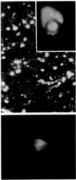
The fluorescence patterns of the three detectable MV structural proteins differred. Monoclonal anti-bodies to MV N protein gave the most widespread fluorescent staining in the cytoplasm of the cellular processes of the cell bodies, as well as within inclu-sion bodies of oligodendrocytes, astrocytes, and neu-rons (figure I, top). Monoclonal antibodies to P protein gave a strong fluorescent staining that was similar in appearance to the staining for N protein, except that no nuclear inclusion bodies were stained (data not shown). In contrast, monoclonal antibod-ies to H protein resulted in a weak, diffuse fluores-cence of the cytoplasm (figure I, bottom).
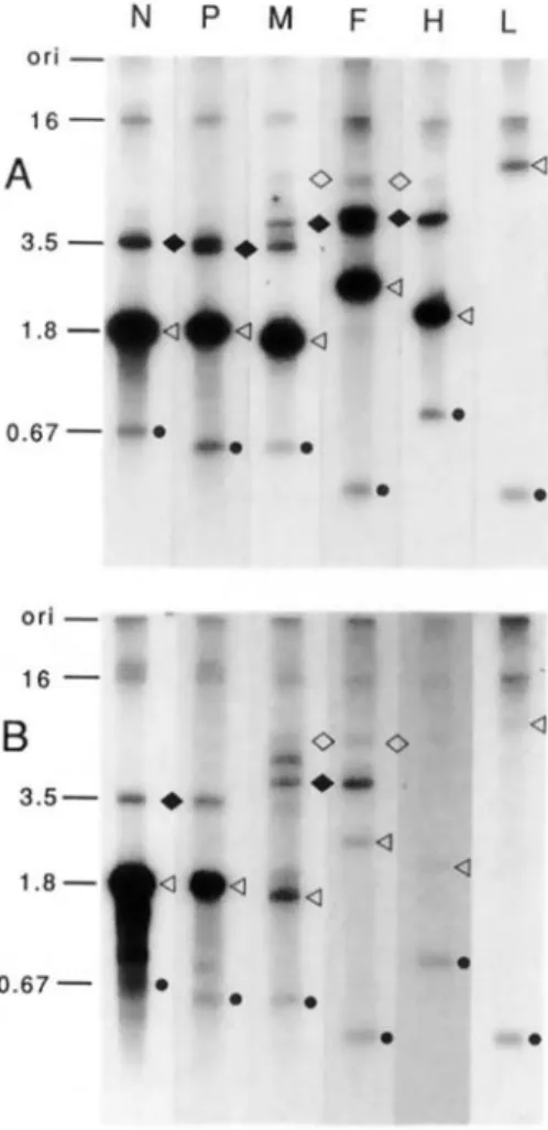
Analysis ofMV mRNAs in brain tissue by north-ern blots. Total RNA derived from brain tissue of the patient with MIBE and MV-infected HeLa cells was analyzed for the presence of virus-specific RNAs by northern blots using riboprobes of negative-strand polarity derived from five MV genes [16]. Lyti-cally infected HeLa cells contained all six MV mRNAs (figure 2A). Those for N, P, M, F, and H appear as strong bands. The bands of weaker inten-sity represent bicistronic and tricistronic transcripts of adjacent genes [6]. The two bands in figure 2A, lane M - one above the M mRNA and the other be-tween the M-F and M-F-H polycistronic RNAs-result from cross-hybridization of the GC-rich probe with 18Sand 28S ribosomal RNA [16]. Owing to the longer exposure, the intensity of these bands is stronger in figure 2B. Total RNA isolated from brain tissue of the patient with MIBE reacted strongly with the Nand P probes (figure 2B). However, the amounts of the M, F, and H mRNAs are significantly
reduced in comparison with Iytically infected cells. In addition, a band of weak intensity migrating more slowly than the 16-kilobase genome, is detected in figure 2B, lanes N, P, and M. This band could rep-resent a "copy-back" defective genome, containing only the N, P, and M genes, that migrates slowly be-cause of an extensive double helical structure. The amount of the M-F bicistronic transcript is increased (figure 2B, lanes M and F). This increment is accom-Figure 1. Indirect immunofluorescence using pooled monoclonal antibodies to Nand H proteins of MV.Top,
numerous cells stain for N protein. Cell processes reveal granular staining(bar
=
30 11m); and inclusion bodies (in-set)are also strongly positive (bar = 11 11m). Bottom,fluorescence for the H protein is weak and diffuse, re-stricted to single cells(bar = 20 11m).
438 (100) 416 (95) 17 (4)
o
o
No.(010) of cells positive Antibodyto protein N P H F MMV Gene Expression in MIBE
Figure 2. Northern blotanalysis of six MV-specific tran-scripts:(A)with total RNAderived from lytically infected HeLacells and(B)withtotal RNAderived frombraintis-suefroma patientwithMIBE. In both cases 10 ug of total RNA was loaded onto a broad slot of a 1.2% agarose-formaldehyde gel together with a mixture of standard RNAs [16] synthesized in vitro (1.0 fmol of each stan-dard inA and0.1frnol forB) and containing segments ofMV mRNA species(e).Afterelectrophoresis, the RNA was blottedonto nitrocellulose, which was cut into strips. Strips were thenhybridized withdifferent 32P-Iabeled RNA probesderived from the same plasmids as the unlabeled standards [16]. The monocistronic mRNAs(<1),the bicis-tronicRNAs (. ), and the tricistronic RNAs(0) areindi-cated. The lengths of the N internal standard (0.67 kilo-bases), the N mRNA (1.8 kilokilo-bases), the N-P bicistronic RNA (3.5 kilobases), and the genome (16 kilobases) are indicated to the left oflaneN. Ori
=
origin.panied, as expected, by a reduction in the amount of P-M and M-F bicistronic transcripts (not detect-able) and by enhancement of the M-F-H tricistronic transcript.
To quantitate the data presented in figure 2B, we
147
Table 2. Copy numbers of MV transcripts per 10 pg of total RNA.
MIBE
Transcript HeLa cells brain tissue
N 30000 4500 P 9000 1400 M 9500 180 F 5500 60 H 4000 30 L 500 <;;;15 M-F 600 150 Standard 600 60
NOTE. After exposure of the northern blots, bands cor-responding to theMYtranscripts and to theinternal standards were cut out, and the amount of radioactivity bound was deter-mined. The quantities ofMYtranscripts were related to10pg of total RNA, which isthe RNA content of anaverage cultured HeLa cell [16].
determined the amount of radioactivity of the differ-ent mRNAs in relation to the internal standard in-cluded in the assay (seethe lower bands, marked with a dot, in each track of figure 2). The number ofMV-specific transcripts, in brain tissue, involving mainly M, F, and H genes drops drastically in comparison with lytically infected HeLa cells (table 2). The amount of M-F bicistronic RNA is relatively high, equaling that of M mRNA and exceeding that of F mRNA.
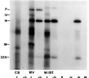
In vitro translation reactions of isolated brain mRNAs. To test the biologic activity of the MV mRNAs present in the brain and to determine their ability to direct the synthesis of the corresponding proteins, we used an in vitro translation procedure to detect such synthesis. Only Nand P proteins are synthesized, whereas M protein is not made, despite the presence of its mRNA (figure 3). In addition, no H protein is synthesized in the in vitro transla-tion reactransla-tion; this lack of synthesis may result from the low concentration of mRNA, as observed in the northern blot. Itis not possible to make any state-ment about the F protein because we could not de-tect it in control in vitro translation experiments using mRNAs from lyticallyinfected cells[6].
Discussion
Our immunohistochemical and biochemical results indicate that MV persistence in brain cells from an individual with MIBE is characterized by the restric-tion of expression of three viral genes. In infected brain cells, only the Nand P proteins of MV are
con-Figure 3. In vitro translation of mRNAs derived from control brain(CB),lytic MY(MV),or brain tissue from a patient with MIBE(MIBE).The products synthesized were either analyzed directly (I) or after immunoprecipi-tation in 10070 SDS-PAGE.rb,Immunoprecipitation with rabbit hyperimmune antiserum to MY;P,H, N,andM,
immunoprecipitations with specific antisera.
sistently detected, and only the mRNAs of these two proteins are biologically active in an in vitro trans-lation system. The expression of the H protein is re-duced, whereas M and F proteins were not detect-able in brain cells, despite the presence of their corresponding mRNAs. In part, this situation could be the result of the low concentrations of the mRNAs, particularly those for F and H proteins, which are significantly reduced when compared with values in lytically infected cells. However, because M protein is not formed either in vivo or in vitro, although M mRNA is present in greater amounts than H mRNA, it seems likely that M mRNA is non-functional. A relatively large amount of M-F bicis-tronic RNA is found, an observation suggesting that the polymerase often does not recognize correctly the processing signal at the M-F intercistronic region. These findings are similar to previous observations made in SSPE. In this slow virus disease the synthe-sis of MV structural proteins in infected brain cells is also highly variable. As in MIBE, Nand P pro-teins are consistently detected in each area of brain examined from individuals with SSPE, whereas H, F, and M proteins were either reduced in amount or absent [8,12]. The results of the biochemical analy-sis of MV gene expression in brains showing SSPE are rather complex[5-7, 15].Generally the mRNAs
coding for MV N, P, and M proteins are easily de-tectable in brain tissues, whereas those for Hand F proteins are strongly reduced in amount when com-pared with values in
lytically
infected cells. Although the amounts of viral mRNA within different parts of the same brain varied, the ratio of individual mRNAs remained constant. However, only the mRNAs for the N protein consistently directed syn-thesis of this polypeptide in in vitro translation sys-tems, whereas P protein synthesis was detected in 750/0 ofthe subjects studied, and electrophoretic mo-bility shifts were encountered. Attempts to detect H protein in in vitro translations by using mRNA iso-lated from infected brain tissue were unsuccessful, either as a result of the relatively low concentration of this mRNA or of functional defects of the tran-script. Of particular interest are the results obtained for M protein. Despite the presence of intermediate levels of M mRNA in infected brain tissue in most of the cases studied, no in vitro product of this mRNA could be detected [6]. This translation de-fect of the M mRNA was further analyzed in one case. The sequence analysis of the M gene revealed several differences from the Edmonston strain M se-quence [17]. In particular, one base substitution was observed that created a stop codon at position 12 of the M-gene reading frame, an observation explain-ing why the M protein could not be synthesized[17]. The lack of synthesis of MV envelope proteins could explain the failure to observe budding parti-cles of virus and to isolate infectious virus from brains of individuals with MIBE. There is apparently no need for production of infectious particles of vi-rus to perpetuate the infectious process in MIBE as in SSPE, because the MV nucleocapsid alone is in-fectious. In tissue culture experiments, isolated N structures - consisting of viral RNA and three viral structural proteins, L, P, and N - represent the rep-licative complex and are sufficient for maintaining viral replication in transfection experiments [18]. Therefore restriction of the expression of the MV envelope gene is not strictly deleterious to the sur-vival of the persisting virus in brain tissue and may actually enable the virus to escape immune surveil-lance. For effective elmination of virus-infected cells, immune effector cells must recognize the viral struc-tural proteins on the cell surface. In SSPE and MIBE, most infected brain cells do not demonstrate viral envelope proteins because these proteins are synthe-sized at very low levels if at all. Therefore infected brain cells can escape immune surveillance, and theMV Gene Expression in MIBE
replicative complex of MV can be spread in the CNS by means of cell-to-cell transport via cell processes. Although our findings shed some light on the mechanisms of MV persistence in human brain tis-sue, they do not delineate which factors are involved in the establishment of persistence and how the ob-served replication defects develop. Antibody-induced antigenic modulation of MV-infected cultured cells leads to persistence and affects viral replication [19]. Antibodies to MV remove viral glycoproteins from the surface of infected cells and reduce expression of some intracellular viral polypeptides. A marked perturbation of levels of the P and M proteins was observed when polyclonal antibody to MV was added to infected cultures. This mechanism may con-tribute to establishment of MV persistence in SSPE, where an immune defect has not been detected [20]. In MIBE, however, MV infection of brain tissue oc-curs in individuals with severe immunodeficiency, who are unable to respond properly to the infectious agent. In this disease, additional factors obviously interfere with MV replication in CNS tissue.
We observed recently that levels of the envelope proteins of MV are significantly reduced in infected brain cells of suckling Lewis rats at the onset of acute measles encephalitis [3]. This reduction in synthesis of MV proteins points to a rapid development of a defective multiplication cyclefor the virus in rat brain in the absence of a measurable immune response. This observation suggests that factors independent of the immune system, probably specific for CNS cells, interfere with viral replication. Little is known about such factors, but in recent in vitro studies with papaverine, transcription of MV RNAs was in-hibited, and a selectivesuppression of M protein syn-thesis occurred in neural cells [21, 22]. Many factors are clearly involved in controlling MV replication in brain tissue, and these factors determine whether an adventitious infection without further consequences ensues or a threat to the host with expression of al-tered viral genes and initiation of a pathological pro-cess is triggered.
References
I. Johnson RT. Chronic inflammatory and demyelinating dis-eases. In: Viral infections of the nervous system. New York: Raven Press, 1982:237-70
2. Ohuchi M, Ohuchi R, Mifune K, Ishihara T, Ogawa T. Char-acterization of the measles virus isolated from the brain of a patient with immunosuppressive measles encephali-tis. J Infect Dis 1987;156:436-41
149
3. Liebert UG, ter Meulen V. Virological aspects of measles vi-rus-induced encephalomyelitis in Lewisand BN rats. J Gen Virol 1987;68:1715-22
4. Morgan EM, RappF.Measles virus and its associated dis-eases. Bacteriological Reviews 1977;41:636-66
5. Baczko K, Carter MJ, Billeter M, ter Meulen V. Measles vi-rus gene expression in subacute sclerosing panencephali-tis. Virus Res 1984;1:585-95
6. Baczko K, Liebert UG, Billeter M, Cattaneo R, Budka H, ter Meulen V. Expression of defective measles virus genes in brain tissues of patients with subacute sclerosing panen-cephal itis. J Virol 1986;59:472-8
7. Haase AT,Gantz D, Eble B, Walker D, Stowring L, Ventura P, Blum H, Wietgrefe S, Zupancic M, Tourtellotte W, Gibbs CJ Jr, Norrby E, Rozenblatt S. Natural history of restricted synthesis and expression of measles virus genes in subacute sclerosing panencephalitis. Proc Natl Acad Sci USA 1985;82:3020-4
8. Liebert UG, Baczko K, Budka H, ter Meulen V. Restricted expression of measles virus proteins in brains from cases of subacute sclerosing panencephalitis. J Gen Viro11986; 67:2435-44
9. Roos RP, Graves MC, Wollmann RL, Chilcote RR, Nixon J. Immunologic and virologic studies of measles inclusion body encephalitis in an immunosuppressed host: the rela-tionship to subacute sclerosing panencephalitis. Neurol-ogy 1981;31:1263-70
10. Carter MJ, Willcocks MM, LofflerS, ter Meulen V. Rela-tionships between monoclonal antibody-binding sites on the measles virus haemagglutinin. J Gen Virol 1982; 63:113-20
II. ter Meulen V, Leffler S, Carter MJ, Stephenson JR. Anti-genic characterization of measles and SSPE virus haemag-glutinin by monoclonal antibodies. J Gen Virol 1981; 57:357-64
12. Norrby E, Kristensson K, Brzosko WJ, Kapsenberg JG. Mea-sles virus matrix protein detected by immune fluorescence with monoclonal antibodies in the brain of patients with subacute sclerosing panencephalitis. J ViroI1985;56:337-40 13. Sheshberadaran H, Chen SoN, Norrby E. Monoclonal
anti-bodies against five structural components of measles vi-rus.I.Characterization of antigenic determinants on nine strains of measles virus. Virology 1983;128:3451-53 14. Sato TA, Fukuda A, Sugiura A. Characterization of major
structural proteins of measles virus with monoclonal an-tibodies. J Gen Virol 1985;66:1397-409
15. Cattaneo R, Rebmann G, Baczko K, ter Meulen V, Billeter MA. Altered ratios of measles virus transcripts in diseased human brains. Virology 1987;160:523-6
16. Cattaneo R, RebmannG,Schmid A, Baczko K, ter Meulen V, Billeter MA. Altered transcription of a defective mea-sles virus genome derived from a diseased human brain. EMBO J 1987;6:681-8
17. Cattaneo R, Schmid A, Rebmann G, Baczko K, ter Meulen V, Bellini WJ, Rozenblatt S, Billeter MA. Accumulated measles virus mutations in a case of subacute sclerosing panencephalitis: interrupted matrix protein reading frame and transcription alteration. Virology 1986;154:97-107 18. Rozenblatt S, Koch T, Pinhasi0, Bratosin S. Infective
sub-structures of measles virus from acutely and persistently infected cells. J Virol 1979;32:329-33
19. Fujinami RS, Oldstone MBA. Antibody initiates virus per-sistence: immune modulation and measles virus infection. In: Notkins AL, Oldstone MBA, eds. Concepts in viral pathogenesis. New York: Springer-Verlag, 1984:187-93 20. ter Meulen V, Stephenson JR, Kreth HW, Subacute
sclero-sing panencephalitis. In: Fraenkel-Conrat H, Wagner RR, eds. Comprehensive virology. Vol.18. Virus-host interac-tions. Receptors, persistence, and neurological diseases. New York: Plenum Press, 1983:105-59
21. Miller CA, Carrigan DR. Reversible repression and activa-tion of measles virus infecactiva-tion in neural cells. Proc Natl Acad Sci USA1982;79:1629-33
22. YoshikawaY, Yamanouchi K. Effect of papaverine treatment on replication of measles virus in human neural and non-neural cells. J Virol1984;50:489-96