Active Manipulation of ECM Stiffness
The MIT Faculty has made this article openly available.
Please share
how this access benefits you. Your story matters.
Citation
Chen, Peter C. Y., et al. “Active Manipulation of ECM Stiffness.”
ASME 2011 Summer Bioengineering Conference, Parts A and B,
22-25 June, 2011, Farmington, Pennsylvania, ASME, 2011, p. 869. ©
2011 by ASME
As Published
http://dx.doi.org/10.1115/SBC2011-53090
Publisher
American Society of Mechanical Engineers
Version
Final published version
Citable link
http://hdl.handle.net/1721.1/118916
Terms of Use
Article is made available in accordance with the publisher's
policy and may be subject to US copyright law. Please refer to the
publisher's site for terms of use.
Copyright © 2011 by ASME 1
INTRODUCTION
The mechanical properties of the microstructures surrounding cells influence the behavior of cells in differentiation, proliferation, and apoptosis, etc. The stiffness of the extra- cellular microenvironment has been shown to be one such mechanical property [1][2]. Studies reported in the literature concerning the stiffness of the extracellular microenvironment mainly sought to understand the scientific principles and mechanisms underlying its effect on cell-environment interaction [3]. This paper describes an approach that achieves such manipulation, and reports experimental results that demonstrate the effectiveness of this proposed approach.
MATERIAL AND METHODS
Superparamagnetic particles are embedded in the extracellular matrix (ECM) and manipulated with an external magnetic field to achieve active manipulation of the stiffness of the extracellular microenvironment.
Bead-ECM bio-conjugation
To have the beads bound onto the collagen fibers, a specific coating is necessary. This coating should have a good affinity with the collagen fibers such that it forms a strong attachment between the magnetic beads and the collagen fibers. We used beads coated with streptavidin since streptavidin will affix to areas dense with collagen. Streptavidin contains an Arg-Tyr- Asp (RYD) amino acid sequence that mimics the Arg-Gly-Asp (RGD) receptor domain of fibronectin. This form of bio-conjugation involves four different types, namely, hydrophobic, van der Waals, hydrogen bonding network, and covalent bonds. The complementary shapes, charges, polarity, and hydrophobicity of the streptavidin and the collagen fibers permit multiple weak interactions which in combination produce a tight binding [7][8].
ECM sample preparation
The collagen was prepared according to the recipes listed
in Table I. Both samples (containing 2.0mg/ml of Rat Tail Collagen, Type 1) were obtained from BD Biosciences. The streptavidin-coated magnetic beads BM551 were procured from Bangs Laboratory. Two types of samples were prepared: one with magnetic beads and one without. For samples with magnetic beads, a concentration of 0.5mg/ml of the beads is added. The collagen (or the collagen with beads mixture) was thoroughly vortexed for two minutes until a homogeneous solution was formed and all the components of the mixture were spread throughout the entire volume. The mixture was then pipette into the mold carefully so that no air cavities would be formed. Fibrillogenesis was usually done in an external incubator at 37◦C and 5% CO2. To achieve self- assembly of collagen molecules into fibers and binding of beads to the collagen fibers, the samples were placed in an incubator for at least 22 hours to ensure that gellation occurred throughout the entire collagen strip.
Evaluation of ECM stiffness
The micro-force tester has a load cell rated at 10N with a 0.001N resolution. Stretch tests were carried out on different sets of ECM samples at the fixed rate of 10 mm/min. While performing the stretch tests, a certain portion of these samples was exposed to a static magnetic field produced by a permanent magnet. This permanent magnet is made of an alloy of neodymium, iron and boron (NdFeB) and it is capable of producing a magnetic field of 2T. Two magnets form an aligned configuration such that the ECM sample is located between the magnets with a gap of 1.5 cm between one side of the ECM and a magnet, allowing the magnetic field to pass though the magnetic beads embedded in the ECM.
ACTIVE MANIPULATION OF ECM STIFFNESS
Peter C.Y. Chen1, Sahan C. B. Herath1, Dong-an Wang2, Kai Su2, Kin Liao2 and Harry Asada3
1. Department of Mechanical Engineering, National University of Singapore,
Singapore
2. Division of Bioengineering Nanyang Technological University,
Singapore 3. Department of Mechanical Engineering,
Massachusetts Institute of Technology, USA
Proceedings of the ASME 2011 Summer Bioengineering Conference SBC2011 June 22-25, 2011, Farmington, Pennsylvania, USA
SBC2011-53090
Copyright © 2011 by ASME 2
RESULTS
Six sets of tests (involving four ECM samples in each set) were conducted under the conditions as summarized in Table I.
TABLE I TESTS
Set No. No. of tests Bead concentration Magnetic field
1 4 0 Off 2 4 0 On 3 4 0.1 mg/ml Off 4 4 0.1 mg/ml On 5 4 0.5 mg/ml Off 6 4 0.5 mg/ml On
Stiffness of pure ECM
The slopes of the average of Set 1 and Set 2 differ by 5x10⁻⁴ in magnitude. To demonstrate that this difference in stiffness was due to sample variations and that the presence of the magnetic field did not affect the stiffness of pure ECM samples, the eight sets of data are randomly divided into two groups by picking two curves each from Set 1 and Set 2. The slopes of these two randomly divided sets differ by 5x10⁻⁴ in magnitude.
Change in ECM stiffness due to 0.5 mg/ml concentration of embedded beads
To see the effect of the embedded beads on the stiffness of the overall ECM, the linear approximations of the force-displacement relationships of ECM samples with and without beads and under the condition that the magnetic field was absent. The plots show that embedding 0.5 mg/ml of beads in the pure ECM slightly increases its stiffness. The difference (in magnitude) in the slopes of the linear approximations is 1 x10⁻³.
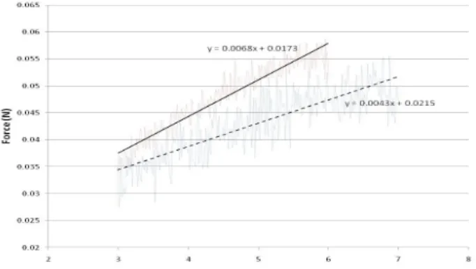
Change in ECM stiffness due to magnetic manipulation of 0.5mg/ml concentration of embedded beads
There is a clear separation of the two set of curves from set 5 and 6 in the elastic region before sample rupture occurs. The set of four curves exhibits a higher slope (as compared to the other four curves) in the force-displacement relationship that corresponds to Set 6. Figure 1 shows the linear approximations of the force-displacement relationships from Set 5 and Set 6.
Fig. 1. Force-displacement relationships from Set 5 (dash line) and Set 6 (solid line).
Change in ECM stiffness due to 0.1 mg/ml concentration of embedded beads
To see the effect of the embedded beads on the stiffness of the overall ECM the stretch results are obtained from carrying out experiments according to set 3 and 4. The plots show that embedding
0.1 mg/ml of beads in the pure ECM reduces the difference in stiffness between pure collagen and magnetic bead embedded collagen. The difference (in magnitude) in the slopes of the linear approximations is 9 x10⁻³.
CONCLUSION
The experimental results reported in this paper demonstrate that, by embedding magnetic beads in a ECM through bio- conjugation between the Streptavidin-coated beads and the collagen fibers, the stiffness of the ECM can be actively manipulated by the application of an external magnetic field. Such active manipulation can produce significant change in the stiffness of the ECM. The effect of a number of variables on the stiffness of the bead-embedded ECM remains to be explored; these variables include bead concentration, size and placement of the magnets and their distance to the ECM sample, and strength of the magnetic field, etc. The experiments described in this paper only concern manipulation of the ECM along a single axis. To study how ECM stiffness affects cell behavior in a realistic in vitro environment requires the ability to manipulate the stiffness gradient in the ECM in 3D. We are currently pursuing our research in this direction.
ACKNOWLEDGEMENT
The authors would like to thank the BioSystem and Micromechanics Interdisciplinary Research Group under the Singapore-MIT Alliance for Research and Technology Program for their financial support. The second author also would like to acknowledge the financial support provided by the National University of Singapore.
REFERENCES
1. Robert J. Pelham, Jr., and Yu-Li Wang, 1997, “Cell locomotion and focal adhesions are regulated by substrate flexibility,” Cell Biology, 94, pp. 13661-13665.
2. Richard K. Assoian and Eric A. Klein, 2008, “Growth control by intracellular tension and extracellular stiffness,” Trends in Cell Biology, 18, pp. 347-352.
.3. Dennis P. McDaniel, Gordon A. Shaw, John T. Elliott, Kiran Bhadriraju, Curt Meuse, Koo-Hyun Chung, and Anne L. Plant, 2007, “The stiffness of collagen fibrils influences vascular smooth muscle cell phenotype,” Biophysical Journal, 92, pp. 1759-1769. 4. Uday Chippada, Bernard Yurke, Penelope C. Georges and Noshir
A.Langrana, 2009, “A nonintrusive method of measuring the local mechanical properties of soft hydrogels using magnetic
microneedles,” Journal of Biomechanical Engineering, 131, pp. 021014-021025.
5. Francis J. Alenghat, Ben Fabry, Kenneth Y. Tsai, Wolfgang H. Goldmann, and Donald E. Ingber, 2000, “Analysis of cell mechanics in single vinculin-deficient cells using a magnetic tweezer,” Biochemical and Biophysical Research
Communications, 277, pp. 93-99.
6. Andreas R. Bausch, Florian Ziemann, Alexei A. Boulbitch, Ken Jacobson and Erich Sackmann, 1998, “Local measurements of viscoelastic parameters of adherent cell surfaces by magnetic bead microrheometry,” Biophysical Journal, 75, pp. 2038-2049. 7. S. Freitag, I. Le Trong, L. Klumb, P.S. Stayton, R.E. Stenkamp,
1997, “Structural studies of the Streptavidin binding loop,” Protein Science, 6, pp. 1157-1166.
8. Harvey Lodish, Chris A. Kaiser, Arnold Berk, Monty Krieger, Paul Matsudaira, Matthew P. Scott, 2008, “Molecular Cell Biology,” W. H. Freeman.