HAL Id: hal-03083047
https://hal.archives-ouvertes.fr/hal-03083047
Submitted on 4 Jan 2021
HAL is a multi-disciplinary open access
archive for the deposit and dissemination of sci-entific research documents, whether they are pub-lished or not. The documents may come from teaching and research institutions in France or abroad, or from public or private research centers.
L’archive ouverte pluridisciplinaire HAL, est destinée au dépôt et à la diffusion de documents scientifiques de niveau recherche, publiés ou non, émanant des établissements d’enseignement et de recherche français ou étrangers, des laboratoires publics ou privés.
MALDI-TOF MS as a promising tool to assess potential
virulence of Vibrio tapetis isolates
Alexandra Rahmani, Maaike Vercauteren, Katleen Vranckx, Filip Boyen,
Adeline Bidault, Vianney Pichereau, Annemie Decostere, Christine Paillard,
Koen Chiers
To cite this version:
Alexandra Rahmani, Maaike Vercauteren, Katleen Vranckx, Filip Boyen, Adeline Bidault, et al.. MALDI-TOF MS as a promising tool to assess potential virulence of Vibrio tapetis isolates. Aqua-culture, Elsevier, 2021, 530, pp.735729. �10.1016/j.aquaculture.2020.735729�. �hal-03083047�
MALDI-TOF MS as a promising tool to assess potential virulence of Vibrio tapetis isolates
1
Alexandra Rahmani1†, Maaike Vercauteren2†, Katleen Vranckx3, Filip Boyen2, Adeline Bidault1, Vianney
2
Pichereau1, Annemie Decostere2, Christine Paillard1, ** and Koen Chiers2**
3
1 Univ Brest, CNRS, IRD, Ifremer, UMR 6539 LEMAR, F-29280 Plouzane, France
4
2 Department of Pathology, Bacteriology and Avian Diseases, Faculty of Veterinary Medicine, Ghent
5
University, Salisburylaan 133, 9820 Merelbeke, Belgium 6
3 Applied Maths NV, Sint-Martens-Latem, Belgium
7
† Shared first authors
8
** Shared last authors 9
Alexandra Rahmani: Univ Brest, CNRS, IRD, Ifremer, UMR 6539 LEMAR, F-29280 Plouzane, France; 10
Alexandra.rahmani@univ-brest.fr; https://orcid.org/0000-0001-8888-1145 ; +33298498743 11
Maaike Vercauteren: Department of Pathology, Bacteriology and Avian Diseases, Faculty of Veterinary 12
Medicine, Ghent University, Salisburylaan 133, 9820 Merelbeke, Belgium; Maaike.vercauteren@ugent.be; 13
https://orcid.org/0000-0002-7618-143X 14
Katleen Vranckx: Applied Maths NV (bioMérieux SA), Sint-Martens-Latem, Belgium; 15
Katleen.vranckx@biomerieux.com; https://orcid.org/0000-0002-1980-9809 16
Filip Boyen: Department of Pathology, Bacteriology and Avian Diseases, Faculty of Veterinary Medicine, 17
Ghent University, Salisburylaan 133, 9820 Merelbeke, Belgium; filip.boyen@ugent.be ; 18
https://orcid.org/0000-0002-4777-6880 19
Adeline Bidault: Univ Brest, CNRS, IRD, Ifremer, UMR 6539 LEMAR, F-29280 Plouzane, France; 20
adeline.bidault@univ-brest.fr 21
Vianney Pichereau: Univ Brest, CNRS, IRD, Ifremer, UMR 6539 LEMAR, F-29280 Plouzane, France; 22
Vianney.pichereau@univ-brest.fr , https://orcid.org/0000-0003-1078-9407 23
Annemie Decostere: Department of Pathology, Bacteriology and Avian Diseases, Faculty of Veterinary 24
Medicine, Ghent University, Salisburylaan 133, 9820 Merelbeke, Belgium; annemie.decostere@ugent.be; 25
https://orcid.org/0000-0001-5150-555X 26
Christine Paillard: Univ Brest, CNRS, IRD, Ifremer, UMR 6539 LEMAR, F-29280 Plouzane, France; 27
Christine.paillard@univ-brest.fr; https://orcid.org/0000-0002-8429-918X 28
Koen Chiers: Department of Pathology, Bacteriology and Avian Diseases, Faculty of Veterinary Medicine, 29
Ghent University, Salisburylaan 133, 9820 Merelbeke, Belgium; koen.chiers@ugent.be; 30 https://orcid.org/0000-0002-7449-1047 31 32 33 34 35 36 37 38 39 40 41 42
ABSTRACT
43
Vibrio tapetis, the etiological agent of Brown Ring Disease, mainly affects the Manila clam Ruditapes 44
philippinarum. Although this bacterium is mainly known as a clam pathogen, it has been isolated from 45
several fish species. The main aim of the present study was to further explore the variability of 27 V. tapetis 46
isolates from bivalves and fish, considering three different aspects; in vitro virulence based on the loss of 47
clam hemocyte adhesion properties, detection of the gene virB4 encoding for an essential component of 48
the Type IV Secretion System, and MALDI-TOF MS characterization based on whole cell extracts. Finally, 49
these approaches were compared and evaluated for their ability to differentiate the potential 50
pathogenicity of the 27 isolates against the Manila clams. Among the 11 V. tapetis isolates from the 51
common dab isolated in 2018 in Belgium, only one (2BB) showed intermediate in vitro virulence against 52
the Manila clam, and seven carried the virB4 gene while none of the V. tapetis previously isolated from 53
fish in 2003 showed the presence of this particular gene. Finally, the peak protein profiles generated with 54
MALDI-TOF MS analysis from all 27 V. tapetis strains showed a clear clustering of clam pathogenic and 55
nonpathogenic isolates suggesting that a new isolate of V. tapetis that would cluster within the clam 56
pathogenic isolates could be potentially pathogenic to the Manila clam. Thereupon, MALDI-TOF MS typing 57
allows rapid and cost-efficient identification of V. tapetis isolates and can be defined as a complementary 58
method of the traditional qPCR that opens new perspectives to study the virulence of V. tapetis isolates 59
but also to perform environmental monitoring in order to prevent outbreaks. 60
KEYWORDS: Vibrio tapetis, MALDI-TOF MS, virB4, hemocyte cytotoxicity assay, Manila clam, fish
61
ABBREVIATIONS
62
BRD: Brown ring Disease 63
FSSW: Filtered Sterilized Sea Water 64
HPLC: High performance liquid chromatography 65
MALDI-TOF MS: Matrix-assisted laser desorption/ionization time-of-flight mass spectrometry 66
MLSA: Multilocus sequence analysis 67
RAPD-PCR: Randomly Amplified Polymorphic DNA PCR 68
T4SS: Type IV secretion system 69
TSA: Tryptic Soy Agar 70
TSB: Tryptic Soy Broth 71
72 73
1. INTRODUCTION
74
Vibrio tapetis is the etiological agent of Brown Ring Disease (BRD), mainly affecting Manila clam Ruditapes 75
philippinarum, and responsible for mass mortalities in cultured clams (Maes and Paillard, 1992; Paillard et 76
al., 1994). Clams are amongst the most heavily traded species in the global aquaculture market with Manila 77
clam being the second major cultured bivalve in the world with a yearly production of over 4.4 million ton 78
(FAO, 2018; Smits et al., 2020). BRD is characterized by a brown organic, conchiolin deposit, between the 79
pallial line and the edge of the shell (Paillard and Maes, 1995). The reference strain V. tapetis CECT4600, 80
first isolated from a cultured Manila clam exhibiting BRD in France, has been well characterized (Borrego 81
et al., 1996; Paillard and Maes, 1990). Since 1990, V. tapetis was identified in BRD of various bivalve hosts 82
such as common cockle Cerastoderma edule (Novoa et al., 1998; Paillard and Maes, 1990), rayed artemis 83
Dosinia exoleta (Paillard, 2004a), pink clam Polititapes rhomboïdes (Paillard, 2004a), grooved carpet shell 84
R. decussatus (Maes and Paillard, 1992; Novoa et al., 1998) and Venerupis aurea (Maes and Paillard, 1992), 85
This suggests that V. tapetis is able to cross species barriers (Paillard, 2016). 86
Since 2003, some studies described the isolation of V. tapetis from cultivated or captive held aquatic 87
vertebrates such as corkwing wrasse Symphodus melops ((Jensen et al., 2003), Atlantic halibut 88
Hippoglossus hippoglossus (Reid et al., 2003), Dover sole Solea solea (Declercq et al., 2015), fine flounder 89
Paralichthys adspersus and red conger eel Genypterus chilensis (Levican et al., 2017). V. tapetis was also 90
pinpointed as causative agent of ulcerative skin lesions in the wild-caught common dab Limanda limanda 91
(Vercauteren et al., 2019). 92
In the past, V. tapetis has been described as a homogenous taxon based on traditional methods such as 93
bio- or serotyping (such as indole production, growth in NaCl and production and utilization of different 94
nutrients) (Allam et al., 1996; Castro et al., 1996; Figueras et al., 1996; Paillard, 2004b). Later studies, using 95
advanced genetic or experimental techniques revealed that this species is more heterogeneous. To date, 96
three distinct subspecies of V. tapetis are described, i.e. subsp. tapetis (Balboa and Romalde, 2013), subsp. 97
britannicus (Balboa and Romalde, 2013) and subsp. quintayensis (Levican et al., 2017). However, using 98
genotyping methods, such as Randomly Amplified Polymorphic DNA (RAPD) PCR (Romalde et al., 2002) 99
and multilocus sequence analysis (MLSA) (Gulla et al., 2017) different clusters were defined between V. 100
tapetis subspecies. This indicates that the phylogeny of V. tapetis is not yet completely unraveled. 101
From an epidemical point of view, differences between V. tapetis isolates might provide valuable 102
information regarding the diagnosis of BRD outbreaks and detection of virulent isolates. Furthermore, 103
typing of isolates can also help examining the geographical and host distributions supporting a more 104
ecological approach for studying host-pathogen-environment interactions (Paillard, 2016; Rodríguez et al., 105
2006). 106
Several studies have indeed pointed towards differences between clam pathogenic and non-pathogenic 107
V. tapetis strains. In vivo infection into the pallial cavity of the clams revealed that none of the tested 108
strains derived from fish are able to induce BRD in manila clam (Bidault et al., 2015; Choquet, 2004; Dias 109
et al., 2018). In addition, these last authors have pointed out that virB4, an essential gene coding for 110
nucleoside triphosphatase of the Type IV Secretion System (T4SS), was only present in strains pathogenic 111
to the Manila clam and therefore suggested to be a discriminative tool to differentiate between 112
pathogenic and non-pathogenic strains (Dias et al., 2018). Furthermore, the virB4 real-time PCR assay 113
offers a method for quantification of V. tapetis in the extrapallial fluids of the Manila clam (Bidault et al., 114
2015). 115
The main aim of the present study was to further explore the variability between the 27 V. tapetis isolates 116
from bivalves and fish, considering three different aspects; in vitro virulence based on the loss of hemocyte 117
adhesion properties after contact with V. tapetis (Choquet et al., 2003), detection of the virB4 gene using 118
the TaqMan qPCR (Bidault et al., 2015), and protein-based matrix-assisted laser desorption/ionization 119
time-of-flight mass spectrometry (MALDI-TOF MS) characterization. Finally, these approaches were 120
compared and evaluated for their ability to discriminate the potential virulence of the 27 isolates against 121
the Manila clams. 122
2. MATERIALS AND METHODS 123
2.1. Bacterial isolates and cultivation 124
The V. tapetis isolates used in this study were isolated from nine different host species (five bivalves and 125
four fish species) in various countries between 1988 and 2017. Table 1 summarizes the information of all 126
isolates with the year, place and host of first isolation and a synthesis of the existing knowledge about 127
their virulence. After isolation, all isolates were frozen at -80 °C for further analysis 128
For the cytotoxicity bioassay, only the isolates derived from common dab and the CECT4600 reference 129
strain were used. All isolates were cultivated on Tryptic Soy Agar (TSA; DifcoTM) supplemented with 1.5 %
130
NaCl and were allowed to grow for two days at 16 ± 1 °C (fish isolates) or 18 ± 1 °C (reference clam isolates). 131
For the virB4 gene detection only common dab isolates were used. All isolates were cultured in TSA 132
supplemented with 1.6 % NaCl for PCR and Tryptic Soy Broth (TSB; DifcoTM) supplemented with 1.6 % NaCl
133
for qPCR. For MALDI-TOF analysis, all 27 isolates were cultivated in triplicate (biological replicates, 134
Supplementary file 1) on TSA supplemented with 1.5 % NaCl. Isolates were grown for minimally two days 135
at 16 ± 1 °C (fish isolates) or 18 ± 1 °C (clam isolates). We chose to use the optimal or common 136
temperatures for the strains to ensure optimal growth of the isolates. 137
2.2. In vitro hemocyte cytotoxicity bioassay 138
Virulence of V. tapetis isolates derived from common dab was tested using the standardized in vitro 139
hemocyte cytotoxicity bioassay (Choquet et al., 2003). This test is based on the ability of V. tapetis virulent 140
strains to induce a rounding phenotype on infected hemocytes, increasing the number of non-adherent as 141
compared to the negative control. V. tapetis induces during its phagocytosis, a process of cytoskeletal 142
inhibition and cell rounding. Thus, this adhesion test provides not only morphological but also functional 143
characterization. The type strain CECT4600 was used as the positive control, and filter-sterilized seawater 144
(FSSW) as negative control. Animals used in this study were Manila clams (4 cm Manila clam, February 145
2018) from the SATMAR shellfish aquaculture site in Landeda (Finistère, France). Clams were allowed to 146
acclimate in oxygenated seawater at 14°C for 14 days. Hemolymph was harvested from the adductor 147
muscle, pooled after quality check and the hemocytes were enumerated using a Malassez counting grid. 148
100 μL of hemolymph (5. 105 hemocyte/mL) was added in 24-well plates and kept for a few minutes to let
149
hemocytes adhere to the bottom of the plate. Then, 100 μl of bacterial suspension prepared with FSSW, 150
was added at a bacteria/hemocyte ratio of 25/1. Hemocyte exposure to each of the bacterial isolates was 151
performed in triplicate. In negative control samples, 100 μl of FSSW was added to the hemolymph. After 152
3 h of incubation, the number of non-adherent hemocytes was measured (used as a proxy of V.tapetis 153
cytotoxicity) using flow cytometry after addition of 4 μl of SYBR–Green solution in DMSO (nucleic acid gel 154
stain, dilution 1 : 10 000, Life Technology, USA) as already described (Choquet et al., 2003). A ratio was 155
calculated by dividing the number of non-adherent hemocytes in samples exposed to bacteria by the 156
number of non-adherent cells in negative controls. Statistical analyses were performed using a pairwise 157
Student test to determine significant differences in non-adherent cell ratios between bacterial suspensions 158
and both positive and negative control samples as already performed by Choquet et al. (2003). 159
2.3. VirB4 gene detection 160
A PCR assay was performed with common dab isolates, in which a fragment of 173 bp of the virB4 gene 161
was amplified using primers 170513 (5’-TTAAAAGTGGCGGAGGAATG-3’) and 170514 (5’- 162
AAGCTCTGCATCGGTTAGGA-3’) and GoTAQ polymerase. Subsequently, a Taqman real-time qPCR 163
quantification was performed in triplicate according to the standardized method previously described 164
(Bidault et al., 2015). The positive control used for both of these experiments was the strain V. tapetis 165
CECT4600. 166
2.4. MALDI-TOF MS characterization 167
An ethanol formic acid extraction was performed, based on MALDI Biotyper protocol (Bruker Daltonics, 168
Bremen Germany). Briefly, bacterial cultures were suspended in nuclease-free water aliquoted in 1.5 mL 169
Eppendorf tubes. Ethanol (70 %) dissolved in high performance liquid chromatography (HPLC) grade water 170
was added to the suspension and tubes were centrifuged twice (20 000 x g, 2 min) upon which the ethanol 171
was removed. Thereafter, 20 µL of 70% formic acid (in HPLC grade water) was added to the pellet. To finish 172
the extraction, 20 µL of acetonitrile was added. One µL of each extract was spotted eight-fold (technical 173
replicates) on a MALDI target plate (Bruker Daltonics, Bremen, Germany), air dried and covered with 1 µL 174
of alpha-cyano-4-hydroxycinnamic acid matrix (Bruker Daltonics, Bremen, Germany). All spots were 175
processed in triplicate (technical replicates, Supplementary file 1) with an Autoflex III Smartbeam MALDI-176
TOF MS, recording masses ranging from 2 000 to 20 000 Da using standard settings (flexControl 1.4, version 177
3.4, Bruker Daltonic, Bremen, Germany). The obtained raw spectra were imported in BioNumerics 7.6.3 178
(Applied Maths NV, Sint-Martens-Latem, Belgium) for data analysis. Preprocessing of the data was 179
performed according to (Giacometti et al., 2018). After preprocessing, peaks were detected using the 180
continuous wavelet transform method with a signal-to-noise threshold of two. The spectra were 181
summarized in an average spectrum per biological replicate (Supplementary file 1) and all replicates with 182
less than 95% similarity to this summary spectrum were removed from the analysis. Of V. tapetis 2BB, 183
RD0705 and RP2.3, only two biological replicates were implemented in the analyses due to inconsistencies 184
in the data or low similarity (< 50%) with other replicates of the same isolate. The resulting summary 185
spectra were used to construct an UPGMA dendrogram using a Pearson similarity coefficient. 186
3. RESULTS AND DISCUSSION
187
Infectious diseases remain one of the main limiting factors in the aquaculture of clams, therefore 188
increasing the value of research on the variability of those infectious agents (Smits et al., 2020). The wide 189
host (at least fourteen different host species) and geographic distribution (at least seven different 190
countries) of V. tapetis and linked questions on possible variability of isolates have been discussed in 191
previous research with a deep interest in finding a discriminative test for assessing pathogenicity of V. 192
tapetis isolates against clams (Bidault et al., 2015). Furthermore, the discovery of new isolates from skin 193
ulcerations in the common dab (Vercauteren et al., 2018) might urge for further exploration of pathogenic 194
markers using available techniques. 195
3.1. Most of the V. tapetis isolates from the common dab are unable to induce hemocyte toxicity. 196
To gain insight into the pathogenicity of V. tapetis to the Manila clam, an in vitro cytotoxicity assay was 197
to V. tapetis (Choquet et al., 2003). This bioassay was previously performed for many V. tapetis strains 199
pathogenic to bivalves (Table 1), whereby strains IS9 and CECT4600 induced the highest loss of adhesion 200
of the hemocytes (i.e. in vitro cytotoxicity). Most of the other strains from clams displayed intermediate 201
cytotoxicity and only a few showed no cytotoxicity (Table 1). Remarkably, the GDE and GTR-I strains, both 202
isolated from bivalves but not from the Manila clam, showed no cytotoxicity in vitro. Complete genome 203
analyses of these two strains revealed strong differences with clam isolates (Dias et al., 2018) and these 204
two strains were genetically closer to the LP2 strain derived from corkwing wrasse. This results further 205
substantiates the observed difference in cytotoxicity towards Manila clam hemocytes (Dias et al., 2018). 206
The common dab isolates, 2BG, 2AE, 2AC and 2BW tested in this study, showed non-adherent cells ratios 207
after bacterial exposure fluctuating around 1. The 2BU isolate displayed a ratio of 0.85 ± 0.14 and remained 208
clearly below 1 in all replicates. The ratios found with isolates 2BC, 2BT, 2AU, and 2BA ranged between 1 209
and 1.5. All isolates showed similar ratios with the FSSW control, therefore indicating negative cytotoxicity 210
(i.e. causing no additional loss of adherence of hemocytes) (pvalue > 0.05 ; Fig 1). The isolate 2BB showed 211
the highest cytotoxic activity with an average ratio of 2.27 ± 0.26 (Fig 1). The cytotoxicity of 2BB was 212
evaluated to be intermediate since the amount of non-adherent hemocytes was significantly higher as 213
compared to the FSSW control (pvalue: 0.0005) but was intermediate considering the positive control 214
strain (pvalue: 0.31) These results are consistent with previous ones obtained with strains isolated from 215
fish (e.g. LP2 and HH6087) which mostly showed negative cytotoxicity towards clam hemocytes (Dias et 216
al., 2018). 217
Comparison between different in vitro cytotoxicity bioassays is complicated since the non-adherent 218
hemocyte ratio might vary depending on the susceptibility of hemolymph to V. tapetis. This is illustrated 219
with the strain LP2, which showed intermediate cytotoxicity in Choquet et al. (2003) and negative 220
cytotoxicity in Dias et al. (2018). In the present study, this bias risk was reduced, by using one pool of 221
hemocytes and using a standardized analysis of the data comparing the results to both a positive 222
(CECT4600) and negative control (FSSW), as already described in Choquet et al. (2003). 223
3.2. First detection of the virB4 gene in non-cytotoxic fish isolates 224
Recently, a rapid and accurate Taqman real-time PCR assay for detection and quantification of V. tapetis 225
in extrapallial fluids of the Manila clam has been developed, based on the presence of the virB4 gene, 226
which encodes a component of the T4SS (Bidault et al., 2015). Since the T4SS has been described to be 227
essential for virulence in other pathogenic species such as Helicobacter pylori and Legionella pneumophila 228
(Voth et al., 2012) ,this PCR analysis was suggested as a possible screening tool to differentiate between 229
clam pathogenic and non-pathogenic isolates (Dias et al., 2018). Among 17 fully sequenced V. tapetis 230
genomes, in a previous study, the T4SS genes cluster was identified only in the genomes of isolates virulent 231
to the Manila clam (based on in vivo assays), thus proving that these strains do not only carry the virB4 232
gene but the entire T4SS gene cluster and also suggesting a role for this T4SS system in V. tapetis 233
pathogenicity towards clams (Dias et al., 2018). It should be noted that FPC1121, an isolate from Manila 234
clam in Japan, does show in vivo virulence towards Manila clam, even though the virB4 gene is not present. 235
Surprisingly, in the isolates from common dab, the virB4 gene was found to be present in seven out of the 236
11 isolates (2BM, 2AU, 2BA, 2BT, 2BU, 2AC and 2BG). This study is therefore the first to provide evidence 237
that V. tapetis isolated from fish can carry the virB4 gene, in contrast with previously reported results (Dias 238
et al., 2018). It needs however to be elucidated if these virB4 positive isolates carry the entire cluster 239
coding for T4SS and/or are able to induce BRD during in vivo experiments. Therefore, it seems that it was 240
not the only gene that determines virulence, as expected. 241
242
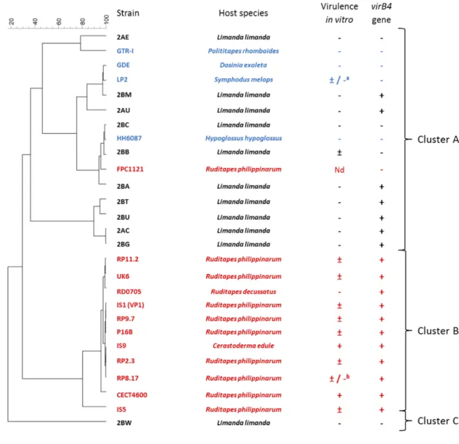
3.3. MALDI-TOF MS analysis reveals 3 clusters of V. tapetis isolates 243
All 27 V. tapetis isolates included in this study were analyzed using the MALDI-TOF MS method. This 244
method allows sensitive and rapid identification of microorganisms and is now widely used in different 245
fields such as clinical microbiology, epidemiological studies and water or food borne pathogens (Maier et 246
al., 2006; Singhal et al., 2015). The technique is broadly used to identify microorganisms at the species 247
level, but has recently been shown valuable for strain typing (Sandrin et al., 2013). 248
The main peaks generated by the MALDI-TOF MS were found between 2000 and 7500 Da. Based on the 249
peak profiles, the constructed dendrogram (Fig 2) of the investigated spectra revealed some clearly 250
delineated clusters. In total three distinct clusters (named A, B and C) were defined, based on differences 251
in protein profiles (Fig 2). All strains that were derived from BRD outbreaks in cultured Manila clam 252
between 1988 - 1996 clustered together in cluster B with only a 29.6 % similarity with other isolates. The 253
strains derived from the common cockle (IS9) and from the carpet shell clam, R. decussatus (RD0705) were 254
also included in this cluster. All these strains were collected during the BRD emergence in Europe, which 255
might explain the limited variability between the isolates. The two strains that were isolated from the 256
rayed artemis (GDE) and pink clam (GTR-I) were clustered together with isolates from fish, in cluster A. 257
The latter cluster could be further divided in four sub-clusters (Fig 2). Interestingly, one isolate (2BW) 258
demonstrated different protein profiles and was included in a separate cluster C. This bacterium was 259
isolated as a co-culture with Pseudoalteromonas sp. and Psychrobacer submarinus from a skin ulcer in dab. 260
In vitro assays demonstrated the absence of the virB4 gene and hemocyte cytotoxicity. Therefore, it is 261
possible that this isolate represents a non-pathogenic strain for clams and/or dab. The virulence of this 262
isolate should be characterized towards clams and fish to elucidate this clustering. Increasing the number 263
of isolates (from different hosts and geographic origin) would corroborate our results and fortify the 264
importance of MALDI-TOF MS analysis for differentiate between clam pathogenic and non-pathogenic V. 265
tapetis isolates. 266
The MALDI-TOF MS clustering in the present study showed some similarities with previously reported data 267
based on genome analyses (Dias et al., 2018). The latter have demonstrated that the two strains isolated 268
from the rayed artemis (GDE) and pink clam (GTR-I) clustered together with LP2 isolated from corkwing 269
wrasse, and were genetically distant from V. tapetis strains isolated from clams. 270
FPC1121 is the only clam pathogenic strain that was clustered together with fish isolates, based on the 271
MALDI-TOF MS profiling. FPC1121 was isolated from cultivated Manila clam in Japan (Table 1, Matsuyama 272
et al., 2010). The original report clearly described the brown deposit and mass mortalities of Manila clam 273
due to this strain, which was confirmed by in vivo virulence assays (Matsuyama et al., 2010). This clustering 274
of FPC1121 is an interesting result; detailed genetic analysis would be interesting to explore the 275
phylogenetic position of this strain and the linked genetic differences. 276
3.4. Value of the assays to differentiate isolates according to their host-species (Fig 3) 277
In vivo virulence tests are necessary to determine the ability for an isolate to induce BRD. However, several 278
tests have been developed to characterize the in vitro virulence regarding Manila clam. 279
The in vitro cytotoxicity assay has been used as a tool to evaluate the pathogenicity of V. tapetis strains 280
for clams (Dias et al., 2018; Bidault et al., 2015). This assay is based on the correlation between the 281
cytotoxic activity of bacteria to clam hemocytes and the in vivo pathogenicity. As demonstrated in Table 282
1, such a correlation was not found for strain RD0705. Indeed, this strain causes no hemocytes cytotoxicity 283
in vitro although it can cause BRD in vivo in the Manila clam (Table 1) (Dias et al., 2018; Novoa et al., 1998). 284
Since RD0705 was isolated from another clam species (i.e. the grooved carpet shell), it is tempting to 285
speculate that this inconsistency might be related to host specificity. However, it could also suggest that 286
cytotoxicity towards hemocytes is not the only virulence factor involved in the development of BRD. Beside 287
this exception, it should be recalled that the analysis of the virulence profiles of many V. tapetis strains 288
isolated from clam revealed a good correlation between the cytotoxic activity to clam hemocytes and the 289
in vivo pathogenicity. 290
Another assay commonly used to detect V. tapetis isolates pathogenic to clams is the search for the virB4 291
gene encoding part of the T4SS (Dias et al., 2018). In our study, we have demonstrated for the first time 292
that the virB4 gene can also be present in fish isolates. Since these isolates did not display in vitro virulence 293
to clam hemocytes and they clustered separately based on their protein profile, it could be possible that 294
they are not pathogenic for clams. In vivo studies should be performed to confirm the pathogenicity 295
towards clams and if so, this might question the use of virB4 detection as a pertinent marker for 296
pathogenicity to clams. 297
The MALDI-TOF dendrogram showed a good clustering of V. tapetis from different origins. However, this 298
clustering was not correlated with the presence of the virB4 gene. In fact, the clustering was more 299
correlated with the host species from which they were isolated. Based on the results of the different assays 300
in the present study, it could be hypothesized that MALDI-TOF clustering could differentiate between clam 301
pathogenic and nonpathogenic isolates. 302
3.5. Implications in the context of virulent strain detection 303
Based on these results, MALDI-TOF MS analysis seems to be a promising tool to indirectly evaluate the 304
pathogenicity of a V. tapetis isolate towards the Manila clam. Nevertheless, although the presented 305
MALDI-TOF MS isolate typing might be a rapid, cost-effective and powerful tool in identifying V. tapetis 306
isolates, the Taqman real-time qPCR remains necessary to quantify the load of V. tapetis during infection. 307
Although some inconsistencies exist, the in vitro assay is also believed to give a good indication of virulence 308
towards Manila clam in some cases. 309
Regarding the value of each test described in this study to determine V. tapetis isolates pathogenicity to 310
the Manila clam, we can consider the MALDI-TOF MS as a new promising screening tool which can be 311
complementary to the tools already used, increasing reliability of the screening. 312
4. CONCLUSION
313
In conclusion, the discovery of new V. tapetis isolates derived from common dab was linked with the need 314
for a further exploration of variability of V. tapetis isolates. The currently used assays (toxicity to 315
hemocytes and search of the virB4 gene), have previously been shown to be interesting to differentiate 316
between V. tapetis pathogenic and non-pathogenic clam isolates. Nevertheless, in this study, V. tapetis 317
isolates from dab showed inconsistencies with results of previously pinpointed techniques questioning 318
their discriminative power. In contrast, the MALDI-TOF MS analysis proved to be a promising tool with the 319
possible ability to differentiate between pathogenic and non-pathogenic clam isolates. This can be used 320
as a complementary discriminative test for virulence of clam isolates. 321
This approach allows rapid and cost-efficient identification of V. tapetis species and opens new 322
perspectives to study the virulence of V. tapetis isolates but also to perform environmental monitoring in 323
order to prevent outbreaks. 324
REFERENCES
325
Allam, B., Paillard, C., Auffret, M., 2000. Alterations in hemolymph and extrapallial fluid parameters in 326
the Manila clam, Ruditapes philippinarum, challenged with the pathogen Vibrio tapetis. J. Invertebr. 327
Pathol. 76, 63–69. https://doi.org/10.1006/jipa.2000.494 328
Allam, B., Paillard, C., Ford, S.E., 2002. Pathogenicity of Vibrio tapetis, the etiological agent of brown ring 329
disease in clams. Dis. Aquat. Organ. 48, 221–231. 330
Allam, B., Paillard, C., Maes, P., 1996. Localization of the pathogen Vibrio P1 in clams affected by Brown 331
Ring Disease. Dis. Aquat. Organ. 27, 149–155. https://doi.org/10.3354/dao027149 332
Balboa, S., Romalde, J.L., 2013. Multilocus sequence analysis of Vibrio tapetis, the causative agent of 333
Brown Ring Disease: Description of Vibrio tapetis subsp. britannicus subsp. nov. Syst. Appl. Microbiol. 36, 334
183–187. https://doi.org/10.1016/j.syapm.2012.12.004 335
Bidault, A., Richard, G.G., Le Bris, C., Paillard, C., 2015. Development of a Taqman real-time PCR assay for 336
rapid detection and quantification of Vibrio tapetis in extrapallial fluids of clams. PeerJ 3, e1484. 337
https://doi.org/10.7717/peerj.1484 338
Borrego, J.J., Castro, D., Luque, A., Paillard, C., Maes, P., Garcia, M.T., Ventosa, A., 1996. Vibrio tapetis sp. 339
nov., the causative agent of the brown ring disease affecting cultured clams. Int. J. Syst. Evol. Microbiol. 340
46, 480–484. 341
Castro, D., Santamaria, J.A., Luque, A., Martinez-Manzanares, E., Borrego, J.J., 1996. Antigenic 342
characterization of the etiological agent of the brown ring disease affecting manila clams. Syst. Appl. 343
Microbiol. 19, 231–239. https://doi.org/10.1016/S0723-2020(96)80049-X 344
Choquet, G., 2004. Caractérisation et pathogénie des isolats de Vibrio tapetis, bactérie responsable de la 345
maladie de l’anneau brun chez la palourde japonaise. Brest. 346
Choquet, G., Soudant, P., Lambert, C., Nicolas, J.-L., Paillard, C., 2003. Reduction of adhesion properties 347
of Ruditapes philippinarum hemocytes exposed to Vibrio tapetis. Dis. Aquat. Organ. 57, 109–116. 348
Declercq, A.M., Chiers, K., Soetaert, M., Lasa, A., Romalde, J.L., Polet, H., Haesebrouck, F., Decostere, A., 349
2015. Vibrio tapetis isolated from vesicular skin lesions in Dover sole Solea solea. Dis. Aquat. Organ. 115, 350
81–86. 351
Dias, G.M., Bidault, A., Le Chevalier, P., Choquet, G., Der Sarkissian, C., Orlando, L., Medigue, C., Barbe, 352
V., Mangenot, S., Thompson, C.C., Thompson, F.L., Jacq, A., Pichereau, V., Paillard, C., 2018. Vibrio tapetis 353
Displays an Original Type IV Secretion System in Strains Pathogenic for Bivalve Molluscs. Front. Microbiol. 354
9, 227. 355
FAO (Ed.), 2018. Meeting the sustainable development goals, The state of world fisheries and 356
aquaculture. Rome. 357
Figueras, A., Robledo, J.A.F., Novoa, B., 1996. Brown ring disease and parasites in clams (Ruditapes 358
decussatus and R. philippinarum) from Spain and Portugal. J. Shellfish Res. 15, 363–368. 359
Giacometti, F., Piva, S., Vranckx, K., De Bruyne, K., Drigo, I., Lucchi, A., Manfreda, G., Serraino, A., 2018. 360
Application of MALDI-TOF MS for the subtyping of Arcobacter butzleri strains and comparison with their 361
MLST and PFGE types. Int. J. Food Microbiol. 277, 50–57. 362
https://doi.org/10.1016/j.ijfoodmicro.2018.04.026 363
Gulla, S., Rønneseth, A., Sørum, H., Vågnes, Ø., Balboa, S., Romalde, J.L., Colquhoun, D.J., 2017. Vibrio 364
tapetis from wrasse used for ectoparasite bio-control in salmon farming: phylogenetic analysis and 365
serotyping. Dis. Aquat. Organ. 125, 189–197. https://doi.org/10.3354/dao03140 366
Jensen, Samuelsen, O., Andersen, K., Torkildsen, L., Lambert, C., Choquet, G., Paillard, C., Bergh, Ø., 2003. 367
Characterization of strains of Vibrio splendidus and V. tapetis isolated from corkwing wrasse Symphodus 368
melops suffering vibriosis. Dis. Aquat. Organ. 53, 25–31. https://doi.org/10.3354/dao053025 369
Levican, A., Lasa, A., Irgang, R., Romalde, J.L., Poblete-Morales, M., Avendaño-Herrera, R., 2017. Isolation 370
of Vibrio tapetis from two native fish species (Genypterus chilensis and Paralichthys adspersus) reared in 371
Chile and description of Vibrio tapetis subsp. quintayensis subsp. nov. Int. J. Syst. Evol. Microbiol. 67, 372
716–723. https://doi.org/10.1099/ijsem.0.001705 373
Maes, P., Paillard, C., 1992. Effect du Vibrio P1, pathogene de Ruditapes philippinarum, sur d’autres 374
espèces de bivalves, in: Les Mollusques Marins: Biologie et Aquaculture, Brest (France), 9 Nov 1990. 375
Maier, T., Klepel, S., Renner, U., Kostrzewa, M., 2006. Fast and reliable MALDI-TOF MS–based 376
microorganism identification. Nat. Methods 3, i–ii. https://doi.org/10.1038/nmeth870 377
Matsuyama, T., Sakai, T., Kiryu, I., Yuasa, K., Yasunobu, H., Kawamura, Y., Sano, M., 2010. First Isolation 378
of Vibrio tapetis, the Etiological Agent of Brown Ring Disease (BRD), in Manila Clam Ruditapes 379
philippinarum in Japan. Fish Pathol. 45, 77–79. https://doi.org/10.3147/jsfp.45.77 380
Novoa, B., Luque, A., Castro, D., Borrego, J.J., Figueras, A., 1998. Characterization and Infectivity of Four 381
Bacterial Strains Isolated from Brown Ring Disease-Affected Clams. J. Invertebr. Pathol. 71, 34–41. 382
https://doi.org/10.1006/jipa.1997.4704 383
Paillard, C., 2016. An ecological approach to understanding host-pathogen-environment interactions: the 384
case of Brown Ring Disease in clams, in: Oysters and Clams: Cultivation, Habitat Threats and Ecological 385
Impact. ed. Jesus L. Romalde Microbiology research advances, Nova Science Publishers, p. 228. 386
Paillard, C., 2004a. Rôle de l’environnement dans les interactions hôtes-pathogènes; développement 387
d’un modèle de vibriose chez les bivalves. Habilit. À Dir. Rech. HDR Univ. Bretagne Occident. Brest. 388
https://hal.archives-ouvertes.fr/tel-02866182 389
Paillard, C., 2004b. A short-review of brown ring disease, a vibriosis affecting clams, Ruditapes 390
philippinarum and Ruditapes decussatus. Aquat. Living Resour. 17, 467–475. 391
https://doi.org/10.1051/alr:2004053 392
Paillard, C., Maes, P., 1995. The brown ring disease in the Manila clam, Ruditapes philippinarum: I. 393
Ultrastructural alterations of the periostracal lamina. J. Invertebr. Pathol. 65, 91–100. 394
Paillard, C., Maes, P., 1990. Aetiology of brown ring disease in Tapes philippinarum: pathogenicity of a 395
Vibrio sp. Comptes Rendus Académie Sci. Ser. 3 Sci. Vie 310, 15–20. 396
https://doi.org/10.1016/0959-8030(94)90030-2 398
Reid, H.I., Duncan, H.L., Laidler, L.A., Hunter, D., Birkbeck, T.H., 2003. Isolation of Vibrio tapetis from 399
cultivated Atlantic halibut (Hippoglossus hippoglossus L.). Aquaculture 221, 65–74. 400
Rodríguez, J.M., López-Romalde, S., Beaz, R., Alonso, M.C., Castro, D., Romalde, J.L., 2006. Molecular 401
fingerprinting of Vibrio tapetis strains using three PCR-based methods: ERIC-PCR, REP-PCR and RAPD. Dis. 402
Aquat. Organ. 69, 175–183. 403
Romalde, J.L., Castro, D., Magariños, B., Lopez-Cortes, L., Borrego, J.J., 2002. Comparison of ribotyping, 404
randomly amplified polymorphic DNA, and pulsed-field gel electrophoresis for molecular typing of Vibrio 405
tapetis. Syst. Appl. Microbiol. 25, 544–550. 406
Sandrin, T.R., Goldstein, J.E., Schumaker, S., 2013. MALDI TOF MS profiling of bacteria at the strain level: 407
A review. Mass Spectrom. Rev. 32, 188–217. https://doi.org/10.1002/mas.21359 408
Singhal, N., Kumar, M., Kanaujia, P.K., Virdi, J.S., 2015. MALDI-TOF mass spectrometry: an emerging 409
technology for microbial identification and diagnosis. Front. Microbiol. 6. 410
https://doi.org/10.3389/fmicb.2015.00791 411
Smits, M., Artigaud, S., Bernay, B., Pichereau, V., Bargelloni, L., Paillard, C., 2020. A proteomic study of 412
resistance to Brown Ring disease in the Manila clam, Ruditapes philippinarum. Fish Shellfish Immunol. 413
99, 641–653. https://doi.org/10.1016/j.fsi.2020.02.002 414
Vercauteren, M., De Swaef, E., Declercq, A.M., Polet, H., Aerts, J., Ampe, B., Romalde, J.L., Haesebrouck, 415
F., Devriese, L., Decostere, A., Chiers, K., 2019. Scrutinizing the triad of Vibrio tapetis, the skin barrier and 416
pigmentation as determining factors in the development of skin ulcerations in wild common dab 417
(Limanda limanda). Vet. Res. 50, 41. https://doi.org/10.1186/s13567-019-0659-6 418
Vercauteren, M., Swaef, E.D., Declercq, A., Bosseler, L., Gulla, S., Balboa, S., Romalde, J.L., Devriese, L., 419
Polet, H., Boyen, F., Chiers, K., Decostere, A., 2018. First isolation of Vibrio tapetis and an atypical strain 420
of Aeromonas salmonicida from skin ulcerations in common dab (Limanda limanda) in the North Sea. J. 421
Fish Dis. 41, 329–335. https://doi.org/10.1111/jfd.12729 422
Voth, D.E., Broederdorf, L.J., Graham, J.G., 2012. Bacterial Type IV secretion systems: versatile virulence 423
machines. Future Microbiol. 7, 241–257. 424
425 426
ACKNOWLEDGEMENTS 427
We warmly thank Annelies M. Declercq that provides us the first isolates of V. tapetis at the beginning of 428
this collaboration. We would like to acknowledge Serge Verbanck for the great work with the MALDI-TOF. 429
AUTHORS CONTRIBUTIONS 430
The collaboration has been initiated by AD, KC, CP and VP. In vitro virulence assays were performed by MV 431
and AR. virB4 detection was performed by MV, AR and AB. MALDI TOF MS experiments and analyses were 432
performed by MV, FB and KV. The article was written by MV, AR, CP, VP, KC and AD. 433
CONFLICTS OF INTEREST 434
Alexandra Rahmani declares that she has no conflict of interest. 435
Maaike Vercauteren declares that she has no conflict of interest. 436
Katleen Vranckx is an employee of Applied Maths NV (bioMérieux SA). 437
Filip Boyen declares that he has no conflict of interest. 438
Adeline Bidault declares that she has no conflict of interest. 439
Vianney Pichereau declares that he has no conflict of interest. 440
Annemie Decostere declares that she has no conflict of interest. 441
Christine Paillard declares that she has no conflict of interest. 442
Koen Chiers declares that he has no conflict of interest. 443
FUNDING 444
The research was funded by the European Fisheries Fund (EVF – project VIS/15/A03/DIV), the Flemish 445
Government and the Research Foundation – Flanders (FWO). This work makes use of the resources, 446
facilities and/or services provided by UGent and Flanders Marine Institute as part of the Belgian 447
contribution to EMBRC-ERIC. This project received grants from the H2020 European project “VIVALDI” 448
(grant agreement N°678589). This work was also supported by the “Université de Bretagne Occidentale” 449
(UBO, France), and the “investment for the future” programs LabexMER (ANR-10-LABX-19) and ISblue 450
(ANR-17-EURE-0015). The MALDI-TOF mass spectrometer was financed by the Research Foundation 451
Flanders (FWO-Vlaanderen) as Hercules project G0H2516N (AUGE/15/05). The funding bodies had no role 452
in the study design, data collection, analysis or the writing process of the manuscript. 453
Compliance with Ethical Standards 454
All applicable international, national, and/or institutional guidelines for the care and use of animals were 455
followed. 456
This article does not contain any studies with human participants performed by any of the authors. 457
TABLES
458
Table 1: Information on the V. tapetis isolates of clams, other bivalves and fish with host species, common name of the host species, description of the health
459
of the host species, year and location of isolation and a reference for each isolate. Furthermore, a synthesis is provided of the existing knowledge on (1) in 460
vivo virulence, estimating the possibility of the isolate to cause Brown Ring Disease in Manila clam following experimental infection. The isolate indicated with 461
an asterisk is able to cause Brown Ring Disease in pink clam but not in Manila clam; (2) in vitro cytotoxicity against hemocytes of the Manila clam ((-): no 462
cytotoxicity, (±) intermediate cytotoxicity, (+):cytotoxicity
.
a: negative in Dias et al, 2018 and intermediate in Choquet, 2004. b: negative in Dias et al, 2018 463and intermediate in Choquet et al, 2003 And (3) presence of the virB4 gene from Bidault et al., 2015; (+) : presence, (-): absence. References for the virulence 464
analyses are: Bidault et al., 2015; Dias et al., 2018; Novoa et al, 1998; Choquet et al, 2004; Matsuyama et al., 2010. Nd: Not determined; *: Determined in this 465 study. 466 467 468 469 470 471
Isolates Host species Common name Isolated from Year Location Isolate reference In vivo virulence (1) In vitro cytotoxicity (2) virB4 gene detection (3) CLAMS
RD0705 Ruditapes decussatus Grooved carpet
shell
Animals with BRD signs 1992 Spain, Galice (Novoa et al., 1998) + - +
CECT4600 Ruditapes philippinarum Manila clam Animals with BRD signs 1990 France, Landeda (Borrego et al., 1996;
Paillard and Maes, 1995)
+ + +
FPC1121 Ruditapes philippinarum Manila clam Animals with BRD signs 2008 Japan (Matsuyama et al., 2010) + Nd -
IS1 (VP1) Ruditapes philippinarum Manila clam Animals with BRD signs 1988 France, Landeda (Paillard and Maes, 1990) + ± +
IS5 Ruditapes philippinarum Manila clam Animals with BRD signs 1991 France, Landeda (Borrego et al., 1996) + ± +
P16B Ruditapes philippinarum Manila clam Animals with BRD signs 1995 France, Golfe du Morbihan (Allam et al., 2002) + ± +
RP11.2 Ruditapes philippinarum Manila clam Animals with BRD signs 1990 France, Landeda (Borrego et al., 1996) + ± +
RP2.3. Ruditapes philippinarum Manila clam Animals with BRD signs 1990 France, Landeda (Borrego et al., 1996) + ± +
RP8.17 Ruditapes philippinarum Manila clam Animals with BRD signs 1990 France, Landeda (Borrego et al., 1996) + ±/-a +
RP9.7 Ruditapes philippinarum Manila clam Animals with BRD signs 1990 France, Landeda (Borrego et al., 1996) + ± +
UK6 Ruditapes philippinarum Manila clam Animals with BRD signs 1996 Great Britain (Allam et al., 2000) + ± +
OTHER BIVALVES
IS9 Cerastoderma edule Common cockle Healthy animals 1990 France, Quiberon (Borrego et al., 1996) + + +
GTR-I Polititapes rhomboïdes Pink clam Animals with BRD signs 2008 France, Glénan (Dias et al., 2018) -* - -
GDE Dosinia exoleta Rayed artemis Animals with BRD signs 2003 France, Glénan (Dias et al., 2018) - - -
LP2 Symphodus melops Corkwing wrasse Kidney of fish suffering vibriosis 1999 Norway, Bergen (Jensen et al., 2003) - ±/-b -
HH6087 (CECT8161) Hipoglossus hipoglossus Atlantic halibut Kidney of moribund fish 2002 Great Britain, Glasgow (Reid et al., 2003) - - -
2AC Limanda limanda Common dab Skin ulceration 2017 Belgium (North sea) (Vercauteren et al., 2018) Nd -* +*
2AE Limanda limanda Common dab Skin ulceration 2017 Belgium (North sea) (Vercauteren et al., 2018) Nd -* -*
2AU Limanda limanda Common dab Skin ulceration 2017 Belgium (North sea) (Vercauteren et al., 2018) Nd -* +*
2BA Limanda limanda Common dab Skin ulceration 2017 Belgium (North sea) (Vercauteren et al., 2018) Nd -* +*
2BB Limanda limanda Common dab Skin ulceration 2017 Belgium (North sea) (Vercauteren et al., 2018) Nd ±* -*
2BC Limanda limanda Common dab Skin ulceration 2017 Belgium (North sea) (Vercauteren et al., 2018) Nd -* -*
2BG Limanda limanda Common dab Skin ulceration 2017 Belgium (North sea) (Vercauteren et al., 2018) Nd -* +*
2BM Limanda limanda Common dab Skin ulceration 2017 Belgium (North sea) (Vercauteren et al., 2018) Nd -* +*
2BT Limanda limanda Common dab Skin ulceration 2017 Belgium (North sea) (Vercauteren et al., 2018) Nd -* +*
2BU Limanda limanda Common dab Skin ulceration 2017 Belgium (North sea) (Vercauteren et al., 2018) Nd -* +*
2BW Limanda limanda Common dab Skin ulceration 2017 Belgium (North sea) (Vercauteren et al., 2018) Nd -* -*
472 473
FIGURES
474
475
Fig 1 Effect of isolates of V. tapetis from common dab on non-adherent cell ratio. Incubation time: 3h.
476
Results are presented by a mean ratio of non-adherent hemocytes (i.e. round hemocytes) in presence of 477
bacteria to the number of non-adherent cells after incubation with filtered sterile seawater (FSSW). Letters 478
depict significant differences (Pairwise Student test). Error bar = Standard Deviation/ √n (n = 3 replicates) 479
480
Fig 2 Dendrogram, based on the MALDI-TOF peak lists, of all V. tapetis isolates based on the complete
481
spectra. Peak intensity is represented in the heat map using different colors ranging from blue (low 482
intensity), over green (low intermediate) to yellow (intermediate), orange (high intermediate) and red 483
(high intensity). 484
485
Fig 3 Dendrogram, based on the MALDI-TOF peak lists, of V. tapetis isolates derived from bivalves and fish
486
combined with in vitro virulence (hemocyte cytotoxicity assay) and presence of the virB4 gene. In red: 487
isolates that can induce Brown Ring Disease in vivo after injection in the pallial cavity. In blue: isolates that 488
cannot induce Brown Ring Disease after injection in the pallial cavity. In black: isolates where no in vivo 489
assay has been performed against the Manila clam. References of the virulence profiles can be found in 490
Table 1. Cytotoxicity in vitro is depicted as follows: (-): no cytotoxicity, (±) intermediate cytotoxicity, (+): 491
comparable cytotoxicity as the reference strain (CECT4600). a: negative in Dias et al, 2018 but intermediate
492
in Choquet et al, 2003, b: negative in Dias et al, 2018 but intermediate in Choquet et al, 2004. Presence (+)
493
or absence (-) of the virB4 gene as assessed using the TaqMan PCR method is also included. Nd: Not 494
determined 495