HAL Id: hal-01494735
https://hal.archives-ouvertes.fr/hal-01494735
Submitted on 23 Mar 2017
HAL is a multi-disciplinary open access
archive for the deposit and dissemination of
sci-entific research documents, whether they are
pub-lished or not. The documents may come from
teaching and research institutions in France or
abroad, or from public or private research centers.
L’archive ouverte pluridisciplinaire HAL, est
destinée au dépôt et à la diffusion de documents
scientifiques de niveau recherche, publiés ou non,
émanant des établissements d’enseignement et de
recherche français ou étrangers, des laboratoires
publics ou privés.
Distributed under a Creative Commons Attribution - ShareAlike| 4.0 International
License
Emilie Dordet-Frisoni, Brigitte Gaillard-Martinie, Régine Talon, Sabine Leroy
To cite this version:
Emilie Dordet-Frisoni, Brigitte Gaillard-Martinie, Régine Talon, Sabine Leroy. Surface migration of
Staphylococcus xylosus on low-agar media. Microbiological Research, Elsevier, 2008, 159 (4),
pp.263-269. �10.1016/j.resmic.2008.02.003�. �hal-01494735�
Surface migration of Staphylococcus xylosus on low-agar media
Emilie Dordet-Frisoni, Brigitte Gaillard-Martinie, Re´gine Talon, Sabine Leroy
*
INRA, UR 454 Microbiologie, F-63122 Saint-Gene`s Champanelle, FranceReceived 7 August 2007; accepted 8 February 2008 Available online 10 March 2008
Abstract
Staphylococcus xylosus is a commensal species commonly found on the skin of mammals, but also currently used as starter culture for meat fermentation. Most strains of this species colonize by forming a biofilm on abiotic surfaces. We show here that the majority of S. xylosus strains also exhibit extensive colony spreading on the surface of soft agar media. This phenomenon seemed to be independent of biofilm-forming ability. It occurred in different culture media and was dependent on temperature. Formation of a giant S. xylosus colony did not involve a biosurfactant. Microscopic observation showed that the front of the giant colony comprised a single layer of spacing cells with more packed cells in the median area. Supplementation of the soft media with DNase I increased S. xylosus colony spreading, indicating that extracellular DNA may be involved in limiting the phenomenon. The ability of S. xylosus to spread on semi-solid surfaces may constitute an advantage for surface colonization. ! 2008 Elsevier Masson SAS. All rights reserved.
Keywords: Staphylococcus xylosus; Motility; Giant colony
1. Introduction
It is increasingly believed that communities of bacteria act together rather than as a group of individual cells. It would be interesting to understand how these communities are formed and colonize a variety of natural habitats. Differing invasive behaviors are used by bacteria for the acquisition of nutrients, and motility constitutes a benefit. Many bacteria translocate by the propeller function of flagella[24]. Some bacteria, such as mycobacteria and streptococci, can spread on semi-solid sur-faces without locomotor organelles [2,18]. A mechanism called sliding is generally used by these bacteria[9]. It is pro-vided by the expansive forces of a growing culture in combi-nation with special surface properties of the cells, resulting in reduced friction between the cell and its substrate[8]. Sliding motility results in a uniform sheet of closely packed cells in a single layer[9]. A phenomenon called darting has been ob-served in some staphylococcal species [9] and involves slow
surface translocation produced by expansive forces developed in an aggregate of cells. Micromorphologically, cells or aggre-gates of cells are distributed randomly with empty areas in be-tween[9]. In other Firmicutes, spreading motility independent of flagella was observed. In Bacillus subtilis, a sliding type of surface motility was detected in a non-flagellated mutant[13]. Secreted surfactin and extracellular potassium ions are re-quired for spreading of B. subtilis[14]. Recently, rapid Staph-ylococcus aureus colony spreading on soft agar medium was reported [12]. Surface translocation of S. aureus differed from the darting of Staphylococcus epidermidis, involving slow surface translocation on solid agar medium, as described by Henrichsen[9].
Staphylococcus xylosus is a non-motile Gram-positive coccus belonging to the Staphylococcus saprophyticus group. S. xylosus is a common inhabitant of the skin of a variety of mammals and occasionally of humans [16]. This species is usually isolated from dairy and meat products [10] and
[3,17,21] is commonly used as starter culture for meat prod-ucts [27]. It is also isolated from environmental sources such as surfaces in food processing plants or those of medical equipment[1,4,5]. It probably colonizes the skin and surfaces by forming a biofilm [23]. Surface translocation could also
* Corresponding author.
E-mail addresses:[email protected](E. Dordet-Frisoni),brigitte. [email protected] (B. Gaillard-Martinie), talon@clermont. inra.fr(R. Talon),[email protected](S. Leroy).
0923-2508/$ - see front matter! 2008 Elsevier Masson SAS. All rights reserved. doi:10.1016/j.resmic.2008.02.003
was first reported by Kloss et al.[15]. In the present study, we characterized the ability of S. xylosus to migrate widely on semi-solid surfaces, and we suggest an important role for ex-tracellular DNA in cellecell aggregation.
2. Materials and methods 2.1. Strains and media
The S. xylosus strains used in this study are listed inTable 1. Bacteria were routinely grown in BHI (brain-heart infusion) broth (BD Becton, Dickinson and Company, Sparks, MD, USA) at 30!C and with orbital shaking of 200 rpm.
2.2. Spreading experiments
Colony spreading was performed in BHI medium solidified with 0.4% w/v agar (Difco, Detroit, MI, USA). In some exper-iments, agar content varied between 0.3 and 1.5% w/v. Colony spreading was also tested in two other media, LuriaeBertani (LB) medium containing 10 g/L tryptone peptone (Difco), 5 g/L yeast extract (Difco), 5 g/L NaCl (pH 7.0), and a minimal medium, MX medium developed specifically for the S. xylosus C2a strain by Fiegler and Bru¨ckner[6]and containing (per L): 1 g Na3-citrate $ 2H2O, 7 g Na2HPO4$ 2H2O, 3 g KH2PO4,
1 g NaCl, 1 g KCl, 4 g (NH4)2SO4, 0.5 g MgSO4$ 7H2O,
0.0147 g CaCl2$ 2H2O, three vitamins, 0.012 mg biotin,
4.6 mg nicotinic acid, 2 mg thiamine hydrochloride and trace elements, 1.5 mg FeCl2$ 4H2O, 0.07 mg ZnCl2, 0.1 mg
MnCl2$ 4H2O, 0.006 mg boric acid, 0.19 mg CoCl2,
0.002 mg CuCl2$ H2O, 0.024 mg NiCl2$ 6H2O and
0.036 mg Na2MoO4$ 2H2O. Different carbon sources were
added to MX medium: 5 g/L of glucose, fructose or lactose. Media were autoclaved and 20 mL or 65 mL were poured into plates of 8.5 or 14 cm diameter, respectively. Plates con-taining agar medium were freshly prepared before inoculation and dried, open for 15 min, in a laminar flow chamber. For standard tests, the centers of agar plates were inoculated in triplicate with 2 ml of overnight culture containing approxi-mately 109CFU/mL. After inoculation, open plates were dried in a laminar flow chamber for 10 min. The initial inoculum formed a spot with a diameter of 6 mm. Unless indicated oth-erwise, plates were incubated at 25!C for up to 96 h before
a negative reaction was recorded. Plates were photographed using a digital camera (Kodak EasyShare DX6490). In some cases, plates were inoculated from dilutions of overnight cul-ture containing approximately 107, 105 or 103CFU/mL, or from 10" or 100" concentrated bacterial suspension obtained after centrifugation of overnight culture. In other experiments, the BHI and MX media were supplemented with different substances: 0.025% Tween 80, 5 mg/mL surfactin from B. sub-tilis (Sigma-Aldrich, Saint Louis, MO, USA), 50 or 250 mg/ mL trypsin (Sigma-Aldrich), 90 units/mL DNase I (Roche, Mannheim, Germany) or 1 mg/mL DNase-free RNase A (Sigma-Aldrich). To measure spreading rates, the diameter
of the growth zone radiating from the point of inoculation on triplicate plates was measured each hour after surface mi-gration commenced.
2.3. Biofilm evaluation with crystal violet staining
This assay is based on the colorimetric measure of crystal violet incorporated by sessile cells [22]. For each strain,
form biofilm on microplate test
Strain Origin Spreading Biofilm
DSM20266 Human skin # þ DSM20267 Human skin # þ C2a DSM20267 cured # þ 839 Meat starter þþ # 840 Meat starter # þ S01002 Meat starter þþ # S01003 Meat starter þþ þ S01004 Meat starter # # S01006 Meat starter þþþ þ S01007 Meat starter þþþ þ S01008 Meat starter # þ S04002 Meat starter þþþ þþþ S01001 Dairy starter þ þ S04003 Dairy starter þþ þþ S03187 Sausage þþþ þþ S03191 Sausage þ # S06173 Raw milk þþ þ S06171 Cheese þþ þ S06175 Cheese þþþ þþ S06176 Cheese þ þþþ S06178 Cheese # þ
S00290 Environment of food plant þþ þ S06167 Environment of food plant þþþ þþ S06179 Environment of food plant þþ þ S06186 Environment of food plant # þþ S04009 Cow mastitis þþþ þþþ S04010 Cow mastitis þþþ þ S04012 Cow mastitis þþ þ S04013 Cow mastitis þþþ # S04016 Cow mastitis # þ S04017 Cow mastitis # # S04018 Cow mastitis þ þþ S04011 Goat mastitis þþ þþ S04014 Goat mastitis # þ S04019 Goat mastitis þþþ þ S04020 Goat mastitis þþþ þþ S04021 Goat mastitis þ þ 00-1747 Mouse dermatitis # þþ S07002 Mouse þ* þþ S07003 Mouse # þ S07001 Poultry feces þþ þþ S07010 Dog feces # # S07011 Rabbit feces þ þ
S06222 Clinical, prosthetic heart valve þþþ þþþ S07005 Clinical, endocarditis þ* þþ S06223 Clinical, liquid þþ þþ S06224 Clinical, urine þþ þþ S07006 Clinical, cerebrospinal fluid þþ þþ S07007 Clinical, blood culture þþ þþ S07008 Clinical, blood culture þ þþþ S06225 Clinical, blood culture þþ þþ *Strains spreading after 48 h.
200 ml of an overnight culture adjusted to OD600 0.01 was
loaded into a 96-well polystyrene microtiter plate. As a con-trol, 200 ml of sterile BHI medium was used. The ‘‘non-bio-film-forming’’ strain Staphylococcus carnosus UT TM300 was employed as a negative control[7]. After 24 h of incuba-tion at 30!C, the medium was removed and wells were
washed once with 200 ml sterile demineralized water to re-move non-adherent bacteria. Two hundred ml per well of a 0.1% (v/v) crystal violet solution in water (Merck, Fonte-nay-sous-bois, France) were added for 10 min. After the stain-ing step of adhered cells, the wells were washed two times with 200 ml of sterile demineralized water to remove excess stain. The dye incorporated by the adherent cells was solubi-lized with 200 ml of 33% (v/v) glacial acetic acid (Sigma-Aldricht, St Quentin Fallavier, France). One hundred and fifty ml of the solubilized solution or adapted dilution were trans-ferred to a new microtiter plate. The OD of each well was measured at 595 nm using a microtiter plate reader (iEMS, Thermo Electron Corporation, Courtaboeuf, France). Absor-bance was measured from the assay OD595 minus control
OD595. The assay was performed at least in triplicate with
four repeats for each strain. 2.4. Microscopy observation
One strain used as a model for spreading ability was grown for 16 h on 0.4% agar BHI medium. A giant colony on a plate was transferred directly to a glass coverslip. Optical microscopic analyses were performed using a phase-contrast Zeiss Axioplan 2E. For electron microscopic analyses, the cells on glass coverslips were fixed in 3% glu-taraldehyde in 0.1 M sodium cacodylate buffer (pH 7.4) at 4!C for 1 h, dehydrated using a graded ethanol and acetone
series, placed on stubs and coated with gold in an EM-SCOPE SC500, and observed with a SEM 505 Philips scan-ning microscope.
3. Results
3.1. Screening of spreading phenomenon in S. xylosus strains
Fifty-one S. xylosus strains described inTable 1 were in-oculated on BHI low-agar medium plates at 30!C.
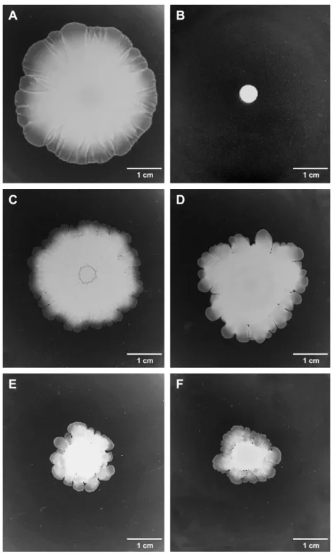
Thirty-seven S. xylosus colonized the surface of the soft medium, after 16 h of incubation (Table 1andFig. 1). Fourteen strains did not spread from the point of inoculation and showed a 6 mm colony diameter, which was the size of the spotted area before incubation (Fig. 1B). Efficacy of spreading and shape of the resultant giant colonies were distinct for the dif-ferent S. xylosus strains (Table 1andFig. 1). The majority of efficiently spreading strains formed a uniform surface film growth (Fig. 1A). Other morphologies were observed show-ing a fractal edge of migration (Fig. 1C) or protrusions (Fig. 1DeF). The less efficient strains usually spread with the formation of protrusions (Fig. 1E,F). The giant colonies varied in thickness. No correspondence between ecological
niches of S. xylosus strains and their spreading capacity was noticed.
3.2. Culture conditions for S. xylosus spreading
Four efficiently spreading S. xylosus strains, S04002, S04009, S06175 and S06222, were chosen to characterize col-ony spreading on semi-solid media. Four non-spreading S. xy-losus strains on agar 0.4% at 30!C in BHI medium, C2a,
S04016, S07003 and S07010, were also tested in different con-ditions of growth. These selected S. xylosus strains were from various ecological niches (Table 1). Different agar concentra-tions were tested ranging from 0.3 to 1.5% of agar (w/v) in BHI and LB media. S. xylosus strains S04002, S04009, S06175 and S06222 were spread on either LB or BHI medium. For these four strains, colony spreading was observed from 0.3 to 0.9% of agar, but above 0.5% of agar, a significant decrease in the size of surface migration was noticed. Optimal ing was observed on 0.3e0.4% agar (wt/vol). Colony spread-ing was observed for S. xylosus S04009 and S04020 strains in MX medium containing glucose, lactose and fructose as car-bon sources with 0.4% agar. The S06222 strain was not able to growth in MX medium containing fructose as carbon source, but spread on glucose and lactose MX medium. S. xy-losus S06175 did not grow in MX medium. S. xyxy-losus strains which are able to spread on the MX medium spread less effi-ciently on this medium than on BHI or LB media due to a low rate of growth (data not shown). It is important to note that whatever the media, colony spreading decreased when the pe-riod of plate drying before inoculation was prolonged (data not shown). The surface migration of the four spreading S. xylosus strains was observed at different temperatures ranging from 10!C to 42!C. The strains showed good growth at all tested
temperatures (data not shown). For all four strains, surface mi-gration was observed at temperatures ranging from 20!C to
35!C, except for strain S04009 which spread up to 37!C,
with an optimum between 25!C to 30!C. At 10!C, cellular
translocation was slow and was observed after almost 48 h of incubation.
Non-spreading S. xylosus strains C2a, S04016, S07003 and S07010 were not able to spread whatever the agar concentra-tion, media or temperature of growth used.
Kinetic measurements under optimal conditions for S. xylo-sus spreading (BHI medium solidified with 0.4% of agar, 25!C) indicated that the diameters of the growth zone did
not increase between 0 and 6 h for the four spreading strains, after which the growth zone diameter increased continuously and rapidly, reaching speeds of 126, 97, 144 and 63 mm/min for S04002, S04009, S06175 and S06222 strains, respectively. To investigate whether initial cell density influenced sur-face migration, we tested different bacterial inocula. The over-night culture of S04002, S04009, S06175 and S06222 was 100- or 10,000-fold diluted in BHI broth, or 10- or 100-fold concentrated. Two ml of these suspensions or pellets were spotted. No differences in colony spreading were observed af-ter inoculation of the dense cell suspensions. The start of spreading was delayed with the diluted suspensions.
3.3. Absence of a biosurfactant
To examine the mechanism of surface migration, we tested for production of a surfactant that could increase the wettabil-ity of the agar surface. We were unable to detect surfactant in S. xylosus cultures of strains S04002, S04009, S06175 and S06222 by the drop collapsing test of Jain et al.[11]. More-over, 10 ml water drops applied to the surface of the agar
medium near the edge of the giant colonies did not cause water spotting, confirming the absence of a surfactant. The addition of Tween 80, an anionic surfactant, resulted in disruption of surface film growth and formation of some protrusions of the spreading strains (data not shown) and did not promote spreading of non-motile strains C2a, S04016, S07003 and S07010. Supplementation of BHI with surfactin did not influ-ence growth but decreased the spreading of S04002, S04009,
Fig. 1. Surface colonization by S. xylosus strains on low-agar medium plates. Two ml of overnight cultures were spotted in the center of 0.4% agar BHI medium and incubated at 25!C for 16 h. (A) Spreading growth zone of strain S04002; (B) growth on non-spreading C2a strain; (C), (D), (E) and (F) spreading growth zone of strains S04011, 839, S04018, and S01001, respectively.
S06175 and S06222 strains. It did not induce the formation of giant colonies by C2a, S04016, S07003 and S07010 strains (data not shown).
3.4. Molecules which increase the S. xylosus rate of surface migration
Spreading of S. xylosus S04002, S04009, S06175 and S06222 was weakly enhanced by the addition of trypsin to BHI medium (data not shown). An increase in the trypsin con-centration increased the rate of colony surface migration. The addition of DNase I but not RNase A to the BHI medium strongly enhanced the surface film growth of the four efficient strains. The surface of the 14 cm diameter plate was com-pletely covered after 22 h of growth on DNase I-supplemented BHI medium for S04002, 25 h for S06175 and 30 h for S04009 and S06222. Speeds of spreading were increased and were 175, 143, 167 and 138 mm/min for S04002, S04009, S06175 and S06222 strains, respectively. The giant colonies were thinner on media supplemented with DNase I (data not shown). The same observation was made when DN-ase I was added to MX medium containing glucose for the three spreading strains which can grow on this medium. Tryp-sin and DNase I did not induce spreading in the four non-spreading S. xylosus strains.
3.5. Correlation with biofilm formation
To determine whether colony spreading ability and biofilm formation were related to each other, we evaluated the capac-ity of the 51 strains to form biofilm (Table 1). Quantification of biofilms was performed by a colorimetric measure of sessile cells stained by crystal violet. A large majority of S. xylosus strains formed biofilm in polystyrene microplates (Table 1). Only seven strains were biofilm-negative in our experimental conditions. Among these seven strains, four strains spread at different rates on semi-solid media. Three of the fourteen non-spreading strains did not form biofilm. The highly effi-cient spreading S. xylosus strains showed various biofilm-forming capacities, with one strain unable to form biofilm or strains forming biofilm at different rates.
3.6. Microscopic observations
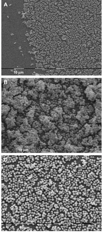
To examine the giant colony morphology of S. xylosus, op-tical and electron microscopic analyses were performed on spreading strain S. xylosus S04002. Optical microscopic obser-vations of the edge of the giant colony on low-agar medium showed a monolayer of cells with spaces between the cells. In the median zone, cells seemed to be more packed (data not shown). This observation was confirmed by electron mi-croscopy (Fig. 2A,B). In the median growth area of the giant colony, cells were aggregated in multiple layers separated by intercellular spaces (Fig. 2B). When the giant colony was formed on medium supplemented with DNase I, the appear-ance of the median zone was clearly modified and showed few or no aggregated cells (Fig. 2C).
4. Discussion
The majority of S. xylosus strains, independently of their or-igin, spread rapidly on a soft agar medium surface, colonizing the entire surface available. The average size of the giant col-onies was dependent on the strain, agar concentration, temper-ature and time of incubation. Spreading growth of S. xylosus was optimally observed on 0.3e0.4% agar media between
Fig. 2. Electron microscopic observations of the edge (A) and median growth area of a giant colony of S. xylosus S04002 strain formed at 25!C on 0.4% agar BHI supplemented (C) or not (B) with DNase I.
and minimal medium MX with various sources of carbon sup-ported spreading of S. xylosus strains. Spreading behavior was thus independent of the growth medium and did not require amino acids, glucose or complex nutrients. Some strains of S. aureus can spread rapidly on low-agar medium[12]. Unlike S. xylosus, 40!C was the optimum temperature for surface
translocation of S. aureus. No spreading of S. aureus was ob-served below 30!C or when LB medium was used. Moreover,
microscopic analyses revealed that the micromorphology of a giant colony of S. xylosus was different from that of S. au-reus. The edge of the S. xylosus colony comprised a single layer of spacing cells, while that of S. aureus consisted of closely packed cells in several layers[12]. The surface trans-location of S. xylosus does not appear to correspond to any of the six known bacterial surface translocations described by Henrichsen[9].
The flagellum-independent spread could depend on the pro-duction of surfactants, which lower the surface tension of the water and enhance surface migration[13,19]. We were unable to detect a surfactant in S. xylosus giant colonies, but colony spreading of this species is probably facilitated by the wetta-bility of the media, as drying of the surface media decreased the phenomenon. But the addition of Tween 80, which is known to modify the surface tension of the medium and im-prove surface wettability[20], did not have a significant effect on S. xylosus colony spreading. Complementation of the me-dium with B. subtilis surfactin did not induce spreading in non-efficient strains and reduced the phenomenon in positive spreading strains.
Addition of trypsin, a serine protease, moderately im-proved the surface migration of S. xylosus strains, probably by limiting cellecell adhesion due to proteins. But DNase I strongly enhanced S. xylosus surface translocation. DNase I acts upon single-chain and double-stranded DNA. It can pre-vent cellecell aggregation, which might be induced by extra-cellular DNA (eDNA) [25]. In S. xylosus, the presence of DNase limited the formation of cell aggregates in the median area of the giant colony. Some bacteria, such as Pseudomonas aeruginosa and S. aureus, produce eDNA, which forms a ma-trix participating in cellecell contact in biofilms[26,28]. We presume that eDNA reduces the spreading of S. xylosus. We had previously reported the ability of S. xylosus to form bio-films[23]. In this study, we have shown that most S. xylosus strains isolated from various niches can form biofilm on poly-styrene surfaces. No clear correlation between spreading abil-ity and biofilm formation can be established. Biofilm-forming S. xylosus strains may or may not spread on semi-solid me-dium, and conversely, spreading strains may or may not form biofilm. The mechanisms implicated in spreading seem to be independent of those implicated in biofilm formation in S. xylosus species.
Colony spreading could be of great benefit for S. xylosus, which is ubiquitous and colonizes naturally fermented food. The spreading motility of S. xylosus strains may provide some advantages for colonization of surfaces. The biological
Acknowledgements
We wish to thank Young-Suk Won for providing the strain implicated in mouse dermatitis, Pascal Rainard for strains iso-lated from mastitis, Miche`le Bes, Emmanuel Coton and Jac-ques Schrenzel for clinical strains, Andrea Laukova` for strains isolated from rabbit and dog, Thomas Rinsoz for the strain isolated from poultry and Christine Vernozy for strains isolated from mice. We would like to thank Brigitte Duclos for secretarial assistance and David Marsh for revision of the English. Emilie Dordet-Frisoni is the recipient of a fellow-ship of the French Ministry of ‘‘Education Nationale et Recherche’’.
References
[1] Arciola, C.R., Campoccia, D., An, Y.H., Baldassarri, L., Pirini, V., Donati, M.E., et al. (2006) Prevalence and antibiotic resistance of 15 minor staphylococcal species colonizing orthopedic implants. Int. J. Artif. Organs. 29, 395e401.
[2] Bergman, S., Selig, M., Collins, M.D., Farrow, J.A., Baron, E.J., Dickersin, G.R., et al. (1995) ‘‘Streptococcus milleri’’ strains displaying a gliding type of motility. Int. J. Syst. Bacteriol. 45, 235e239. [3] Cocolin, L., Rantsiou, K., Iacumin, L., Urso, R., Cantoni, C., Comi, G.
(2004) Study of the ecology of fresh sausages and characterization of populations of lactic acid bacteria by molecular methods. Appl. Environ. Microbiol. 70, 1883e1894.
[4] Corbie`re Morot-Bizot, S., Leroy, S., Talon, R. (2006) Staphylococcal community of a small unit manufacturing traditional dry fermented sausages. Int. J. Food Microbiol. 108, 210e217.
[5] Cunha Mde, L., Lopes, C.A., Rugolo, L.M., Chalita, L.V. (2002) Clinical significance of coagulase-negative staphylococci isolated from neonates. J. Pediatr. (Rio J). 78, 279e288.
[6] Fiegler, H., Bruckner, R. (1997) Identification of the serine acetyltrans-ferase gene of Staphylococcus xylosus. FEMS Microbiol. Lett. 148, 181e187.
[7] Go¨tz, F. (2002) Staphylococcus and biofilms. Mol. Microbiol. 43, 1367e1378. [8] Harshey, R.M. (2003) Bacterial motility on a surface: many ways to
a common goal. Annu. Rev. Microbiol. 57, 249e273.
[9] Henrichsen, J. (1972) Bacterial surface translocation: a survey and a classification. Bacteriol. Rev. 36, 478e503.
[10] Irlinger, F., Morvan, A., Elsolh, N., Bergere, J.L. (1997) Taxonomic characterization of coagulase-negative staphylococci in ripening flora from traditional French cheeses. Syst. Appl. Microbiol. 20, 319e328. [11] Jain, D., Collins-Thompson, D., Lee, H., Trevors, J. (1991) A
drop-collapsing test for screening surfactant-producing. J. Microbiol. Methods 13, 271e279.
[12] Kaito, C., Sekimizu, K. (2007) Colony spreading in Staphylococcus aureus. J. Bacteriol. 189, 2553e2557.
[13] Kinsinger, R.F., Shirk, M.C., Fall, R. (2003) Rapid surface motility in Bacillus subtilis is dependent on extracellular surfactin and potassium ion. J. Bacteriol. 185, 5627e5631.
[14] Kinsinger, R.F., Kearns, D.B., Hale, M., Fall, R. (2005) Genetic require-ments for potassium ion-dependent colony spreading in Bacillus subtilis. J. Bacteriol. 187, 8462e8469.
[15] Kloos, W.E., Schadewaldt, P., Schleifer, K.H. (1981). In: M.P. Starr, H. Stolp, H.G. Tru¨per, A. Balows, & H.G. Schlegel (Eds.), The prokar-iotes. A handbook on habitats, isolation, and identification of bacteria (pp. 1548e1569). Berlin, Heidelberg: Springer Verlag.
[16] Kloos, W.E., Schleifer, K.H. (1986) Bergey’s manual of systematic bacteriology. Baltimore, MD: Williams & Wilkins. 1013e1035.
[17] Martin, B., Garriga, M., Hugas, M., Bover-Cid, S., Veciana-Nogues, M.T., Aymerich, T. (2006) Molecular, technological and safety characterization of Gram-positive catalase-positive cocci from slightly fermented sausages. Int. J. Food Microbiol. 107, 148e158.
[18] Martinez, A., Torello, S., Kolter, R. (1999) Sliding motility in mycobacteria. J. Bacteriol. 181, 7331e7338.
[19] Matsuyama, T., Kaneda, K., Nakagawa, Y., Isa, K., Hara-Hotta, H., Yano, I. (1992) A novel extracellular cyclic lipopeptide which promotes flagellum-dependent and -independent spreading growth of Serratia marcescens. J. Bacteriol. 174, 1769e1776.
[20] Matsuyama, T., Bhasin, A., Harshey, R.M. (1995) Mutational analysis of flagellum-independent surface spreading of Serratia marcescens 274 on a low-agar medium. J. Bacteriol. 177, 987e991.
[21] Montel, M.C., Talon, R., Cantonnet, M., Fournaud, J. (1992) Identification of Staphylococcus from French dry sausage. Lett. Appl. Microbiol. 15, 73e77.
[22] Musk, D.J., Banko, D.A., Hergenrother, P.J. (2005) Iron salts perturb biofilm formation and disrupt existing biofilms of Pseudomonas aeruginosa. Chemical Biol. 12, 789e796.
[23] Planchon, S., Gaillard-Martinie, B., Dordet-Frisoni, E., Bellon-Fontaine, M.N., Leroy, S., Labadie, J., et al. (2006) Formation of biofilm by Staphylococcus xylosus. Int. J. Food Microbiol. 109, 88e96. [24] Rashid, M.H., Kornberg, A. (2000) Inorganic polyphosphate is needed
for swimming, swarming, and twitching motilities of Pseudomonas aeruginosa. Proc. Natl. Acad. Sci. U.S.A. 97, 4885e4890.
[25] Renner, W.A., Jordan, M., Eppenberger, H.M., Leist, C. (2004) Cellecell adhesion and aggregation: influence on the growth behavior of CHO cells. Biotechnol. Bioeng. 41, 188e193.
[26] Rice, K.C., Mann, E.E., Endres, J.L., Weiss, E.C., Cassat, J.E., Smeltzer, M.S., et al. (2007) The cidA murein hydrolase regulator contributes to DNA release and biofilm development in Staphylococcus aureus. Proc. Natl. Acad. Sci. U.S.A. 104, 8113e8118.
[27] Talon, R., Leroy-Se´trin, S., Fadda, S. (2002). In: F. Toldra´ (Ed.), Research advances in quality of meat and meat products e Chapter 10, Research Signpost (pp. 175e191).
[28] Whitchurch, C.B., Tolker-Nielsen, T., Ragas, P.C., Mattick, J.S. (2002) Extracellular DNA required for bacterial biofilm formation. Science 295, 1487.