Publisher’s version / Version de l'éditeur:
Photonic Therapeutics and Diagnostics VI, 7548, 2010-01-23
READ THESE TERMS AND CONDITIONS CAREFULLY BEFORE USING THIS WEBSITE. https://nrc-publications.canada.ca/eng/copyright
Vous avez des questions? Nous pouvons vous aider. Pour communiquer directement avec un auteur, consultez la
première page de la revue dans laquelle son article a été publié afin de trouver ses coordonnées. Si vous n’arrivez pas à les repérer, communiquez avec nous à PublicationsArchive-ArchivesPublications@nrc-cnrc.gc.ca.
Questions? Contact the NRC Publications Archive team at
PublicationsArchive-ArchivesPublications@nrc-cnrc.gc.ca. If you wish to email the authors directly, please see the first page of the publication for their contact information.
NRC Publications Archive
Archives des publications du CNRC
This publication could be one of several versions: author’s original, accepted manuscript or the publisher’s version. / La version de cette publication peut être l’une des suivantes : la version prépublication de l’auteur, la version acceptée du manuscrit ou la version de l’éditeur.
For the publisher’s version, please access the DOI link below./ Pour consulter la version de l’éditeur, utilisez le lien DOI ci-dessous.
https://doi.org/10.1117/12.842403
Access and use of this website and the material on it are subject to the Terms and Conditions set forth at
Durable phantoms of atherosclerotic arteries for optical coherence
tomography
Bisaillon, Charles-Étienne; Dufour, Marc L.; Lamouche, Guy
https://publications-cnrc.canada.ca/fra/droits
L’accès à ce site Web et l’utilisation de son contenu sont assujettis aux conditions présentées dans le site LISEZ CES CONDITIONS ATTENTIVEMENT AVANT D’UTILISER CE SITE WEB.
NRC Publications Record / Notice d'Archives des publications de CNRC:
https://nrc-publications.canada.ca/eng/view/object/?id=9ad1415e-ce45-4476-a46e-2e80b8922af9 https://publications-cnrc.canada.ca/fra/voir/objet/?id=9ad1415e-ce45-4476-a46e-2e80b8922af9Durable phantoms of atherosclerotic arteries for optical
coherence tomography
Charles-Etienne Bisaillon, Marc L. Dufour, and Guy Lamouche
Industrial Materials Institute, National Research Council, 75 bd. de Mortagne, Boucherville,
Quebec, J4B 6Y4, Canada
ABSTRACT
We previously presented a method to fabricate phantoms of normal coronary arteries. This method allows the deposition of multiple layers on a tubular structure, each layer replicating optical and mechanical properties of coronary artery layers. We now present an improved method to produce phantoms of arteries affected by atherosclerosis. The method now includes techniques to introduce structures that mimics the OCT signature of a calcification and of a lipid pool.
Keywords: Optical Coherence Tomography, phantoms, atherosclerosis, arteries
1. INTRODUCTION
Intravascular optical coherence tomography (IV-OCT) has now reached a stage close to widespread clinical use. A review of the recent technology and applications of OCT in cardiology can be found in Ref. [1]. Tremendous progress has been made on the technical side and assessment of IV-OCT as a diagnostic tool in cardiology has been convincingly demonstrated. A few companies are now working towards the commercialization of the technology. Acceptance in the clinical world is under way since IV-OCT is already approved in a few countries. The field is nevertheless still missing diseased artery phantoms. These are essential to ease further development of the technology, to insure training of personnel, and to serve as calibration and validation targets. In the current paper, we present a method to produce artery phantoms containing diseased structures. We first recall our previously described method to fabricate phantoms of normal arteries. We then describe how the fabrication process is modified to introduce structures that provide the OCT signatures of a calcification and of a lipid pool. We finally present OCT images of diseased artery phantoms that were fabricated with the proposed method.
2. PHANTOMS OF NORMAL ARTERIES
In a previous communication,2we described a method to fabricate phantoms of normal arteries. These phantoms are fabricated by the successive deposition of three different layers: intima, media, and adventitia. The material for each layer is deposited on a rotating shaft while being continuously wiped to control the thickness. The setup is illustrated in Fig. 1. The material used for each layer mimics the OCT signature of a corresponding artery layer in terms of backscattering amplitude and attenuation. The material is a silicone matrix containing aluminum oxyde to provide the backscattered OCT amplitude. Aluminum oxyde also provides a certain level of attenuation that is further increased when needed by the addition of carbon black. Figure 2(a) shows a normal artery phantom. The intima and adventitia layers are white since only aluminum oxyde is used for these layers. Carbon black is introduced in the media material resulting in a black layer. Figure 2(b) shows an OCT image of the phantom with its three well defined layers. The optical properties of the layers were chosen to mimic those measured on a porcine coronary artery. Theses phantoms also mimic the mechanical properties of arteries in the low deformation regime.
Further author information: (Send correspondence to G.L.)
Figure 1. Setup used to fabricate multilayer tubular phantoms. The material is deposited on a rotating shaft and continuously wiped by a blade to control the thickness. The blade is inclined in the figure to show that tapered layers can be fabricated.
Figure 2. (a) Photography and (b) OCT image of a normal artery phantom.
3. PHANTOMS OF DISEASED ARTERIES
Calcifications and lipid pools provide very clear signatures in IV-OCT images.3A calcification is characterized by low backscattering and low attenuation and provides a signal-poor region with defined boundaries. A lipid pool is characterized by high backscattering and large attenuation and provides a signal-poor region with diffused boundaries. The OCT signatures of such structures can be mimicked with inclusions made of the same material used to fabricate the normal artery layers, but with different concentrations of aluminum oxyde and/or carbon black. As a first step towards phantoms of diseased arteries, we modify our fabrication method to introduce such structures in a thickened intimal layer.
The various steps of the fabrication process are illustrated in Fig. 3. We begin in Fig. 3(a) with a grooved shaft that serves as a mold for an asymmetric D-shaped lumen over a delimited length of the phantom. In Fig. 3(b), a thin layer of intima material is deposited, it covers the inclusion from a lumen point of view. In Fig. 3(c),
an inclusion is fabricated and then deposited over the intima material. Additional intima material is added in Fig. 3(d) to cover the inclusion and to fill the groove. The remaining of the fabrication process is similar to that of a normal artery. The intima layer is completed in Fig. 3(e), the media layer is deposited in Fig. 3(f), followed by the adventitia layer in Fig. 3(g). Since the phantom is made of elastic material, it is stretched to be removed from the shaft. The completed phantom is illustrated in Fig. 3(h).
Figure 3. Steps in the fabrication process of a diseased artery phantom. See text for the detailed description.
4. OCT IMAGING
Measurements were performed with a custom-built Mach-Zehnder SS-OCT interferometer using a Santec swept source with a repetition rate of 30 kHz, a bandwidth of 110 nm, and a center wavelength of 1310 nm. Images are extracted from pullbacks made with custom-built pullback unit and intravascular probe.
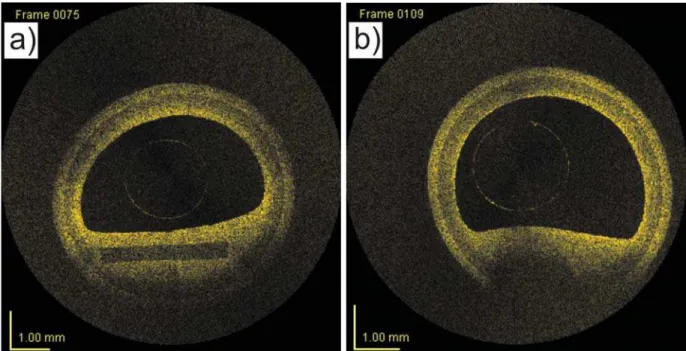
The OCT image of an artery phantom with a calcification is presented in Fig. 4(a). The lumen is asymmetric due to the D-shaped thickened intimal layer. The three layers are visible with the transition from the media to the adventitia being very well defined. The inclusion is made of silicone with a small content of aluminum oxyde leading to small backscattering with low attenuation. Thereby, the clear OCT signature of a calcification is obtained: a signal-poor region with defined boundaries. The inclusion was made with a regular rectangular shape to enhance the clear definition of the boundaries.
Figure 4(b) presents a phantom with a lipid pool. The inclusion is made of silicone with high concentrations of aluminum oxyde and of carbon black leading to high backscattering and high attenuation. Thereby, the clear OCT signature of a lipid pool is obtained: a signal-poor region with diffused boundaries.
5. CONCLUSION
We have presented a method to include diseased structures in artery phantoms. The resulting phantoms do provide OCT signatures similar to what is observed in clinical measurements. Since the material used to fabricate these phantoms is silicone, they are durable. Future work will aim at obtaining inclusions with optical properties that match more closely those of the true diseased structures. This will be possible when the needed data
Figure 4. OCT images of an artery with (a) a calcification and (b) a lipid pool.
will be more widely available in the literature. Up to now, only a few papers have addressed the quantitative measurement of the optical properties of atherosclerotic structures. Future work will also consider inclusions that are different from silicone. This will provide mechanical properties more representative of atherosclerotic structures. Future work also includes the fabrication of more complex structures, like the thin-cap fibroatheroma.
ACKNOWLEDGMENTS
The authors acknowledge the financial support of the Genomics and Health Initiative from National Research Council Canada. The authors also thank Christian deGrandpr´e for its contribution to the phantom fabrication and Fr´ed´eric D’Amours for performing the OCT measurements.
REFERENCES
[1] Regar, E., Serruys, P.W., and Van Leeuwen, T.G. (Eds), [Optical Coherence Tomography In Cardiovascular Research], InformaHealthcare, London(UK)(2007).
[2] Bisaillon, C.-´E. , Lanthier, M.-M., Dufour, M.L., and Lamouche, G., “Durable coronary artery phantoms for optical coherence tomography,” Proc. SPIE 7161, 71612E (2009).
[3] Xu, C., Schmitt, J. M., Carlier, S. G., and Virmani, R.,”Characterization of atherosclerosis plaques by measuring both backscattering and attenuation coefficients in optical coherence tomography,” Journal of Biomedical Optics 13(3), 034003 (2008).