HAL Id: hal-02323104
https://hal.archives-ouvertes.fr/hal-02323104
Submitted on 1 Dec 2020
HAL is a multi-disciplinary open access
archive for the deposit and dissemination of
sci-entific research documents, whether they are
pub-lished or not. The documents may come from
teaching and research institutions in France or
abroad, or from public or private research centers.
L’archive ouverte pluridisciplinaire HAL, est
destinée au dépôt et à la diffusion de documents
scientifiques de niveau recherche, publiés ou non,
émanant des établissements d’enseignement et de
recherche français ou étrangers, des laboratoires
publics ou privés.
Impact of Hemoglobin S Trait on Cell Surface Antibody
Recognition of Plasmodium falciparum-Infected
Erythrocytes in Pregnancy-Associated Malaria
Margaux Chauvet, Marilou Tetard, Gilles Cottrell, Florentin Aussenac,
Emeline Brossier, Luc Denoyel, Marion Hanny, Murielle Lohezic, Jacqueline
Milet, Nicaise Tuikue Ndam, et al.
To cite this version:
Margaux Chauvet, Marilou Tetard, Gilles Cottrell, Florentin Aussenac, Emeline Brossier, et al..
Im-pact of Hemoglobin S Trait on Cell Surface Antibody Recognition of Plasmodium falciparum-Infected
Erythrocytes in Pregnancy-Associated Malaria. Open Forum Infectious Diseases, Oxford University
Press, 2019, 6 (4), �10.1093/ofid/ofz156�. �hal-02323104�
M A J O R A R T I C L E
Open Forum Infectious Diseases
Received 26 November 2018; editorial decision 18 March 2019; accepted 21 March 2019. Presented in part: 29th Molecular Parasitology Meeting, September 2018, Woods Hole, MA.
aM. C. and M. T. ; F. M.-N. and A.M. contributed equally to this work.
Correspondence: A. Merckx, PhD, UMR261 MERIT, IRD - Université Paris Descartes (anais. merckx@parisdescartes.fr).
Open Forum Infectious Diseases®
© The Author(s) 2019. Published by Oxford University Press on behalf of Infectious Diseases Society of America. This is an Open Access article distributed under the terms of the Creative Commons Attribution-NonCommercial-NoDerivs licence (http://creativecommons.org/licenses/ by-nc-nd/4.0/), which permits non-commercial reproduction and distribution of the work, in any medium, provided the original work is not altered or transformed in any way, and that the work is properly cited. For commercial re-use, please contact journals.permissions@oup.com DOI: 10.1093/ofid/ofz156
Impact of Hemoglobin S Trait on Cell Surface Antibody
Recognition of Plasmodium falciparum-Infected
Erythrocytes in Pregnancy-Associated Malaria
Margaux Chauvet,a,1,2 Marilou Tétard,a,3 Gilles Cottrell,1,4 Florentin Aussenac,4 Emeline Brossier,1 Luc Denoyel,1 Marion Hanny,1 Murielle Lohezic,1,2
Jacqueline Milet,1 Nicaise Tuikue Ndam,1,2,5 Damien Pineau,1 Jocelyne Roman,1 Adrian J. F. Luty,1,2,4 Benoît Gamain,3 Florence Migot-Nabias,1,2,a and
Anaïs Merckx1,2,a
1MERIT, Institut de Recherche pour le Développement (IRD), Université Sorbonne Paris Cité, Université Paris Descartes, France; 2Laboratoire d’Excellence GR-Ex, Paris, France; 3UMR_S1134,
Université Sorbonne Paris Cité, Université Paris Diderot, Inserm, INTS, Unité Biologie Intégrée du Globule Rouge, Laboratoire d’Excellence GR-Ex, Paris, France; 4Centre d’Etude et de Recherche
pour le Paludisme Associé à la Grossesse et à l’Enfance, Cotonou, Benin; 5Department of Parasitology, Noguchi Memorial Institute for Medical Research, College of Health Sciences, University of
Ghana, Legon
Background. Sickle cell trait (HbAS) confers partial protection against malaria by reducing the adhesion of Plasmodium
falciparum-infected erythrocytes to host receptors, but little is known about its potential protection against placental malaria.
Methods. Using flow cytometry, we assessed the recognition of HbAA and HbAS VAR2CSA-expressing infected erythrocytes,
by plasma from 159 Beninese pregnant women with either HbAA (normal) or HbAS. Using multivariate linear models adjusted for gravidity, parasite infection at delivery, glucose-6-phosphate dehydrogenase deficiency, and α-thalassemia carriage, we observed significantly reduced cell surface antibody binding of HbAS-infected erythrocytes by plasma from HbAS compared with HbAA women (P < 10–3).
Results. The difference in cell surface antibody binding was only observed when infected erythrocytes and plasma were
asso-ciated according to the same hemoglobin genotype. Similar levels of VAR2CSA-specific antibody were measured by enzyme-linked immunosorbent assay in the 2 groups, suggesting that the altered interaction between VAR2CSA and HbAS women’s antibodies could reflect abnormal display of VAR2CSA on HbAS erythrocytes.
Conclusions. Our data stress the need for assessments of erythrocyte disorders such as the sickle cell trait in a population group
when studying immunological responses to P falciparum.
Keywords. Cell surface antibody binding; hemoglobin S; pregnancy-associated malaria; VAR2CSA. Each year, in sub-Saharan Africa, more than 30 million women
are exposed to the risk of developing pregnancy-associated
ma-laria (PAM) [1]. Pregnancy-associated malaria is a major cause
of maternal and fetal anemia, fetal growth restriction, preterm
birth, and low birth weight (LBW) [2]. Pregnancy-associated
malaria is caused by the adhesion and the sequestration of
Plasmodium falciparum-infected red blood cells (iRBCs) in
pla-cental intervillous spaces [3]. Adhesion of iRBCs in the placenta
is mediated by a single var gene, VAR2CSA [4, 5], the PfEMP1
variant that interacts with chondroitin sulfate A (CSA) on
syncytiotrophoblasts [6–8].
Primigravid women are the most susceptible to PAM because they lack specific protective immunity. Indeed, previous studies have shown that the level of VAR2CSA-specific antibodies
increases with gravidity [9–11], and that the presence of
these antibodies in the placenta is related to better pregnancy
outcomes [12]. These antibodies inhibit the binding of iRBCs to
CSA [13]. Women with higher plasma binding inhibition ability
at delivery are less susceptible to PAM and to give birth to a
LBW baby [11]. The genetic background of mothers may have
an impact on the ability of surface-expressed VAR2CSA to me-diate interaction with endothelial receptors.
Hemoglobin (Hb) S, responsible for sickle cell disease, results from a genetic mutation (Glu6Val) (rs334) at the sixth position within the β-globin chain (HBB, chromosome 11) of normal
Hb (HbA) [14]. Hemoglobin S is highly prevalent in human
populations living in malaria-endemic areas. The highest frequencies of HbS are observed in sub-Saharan Africa, the
Middle East, and India, up to 18% [15], because of the
protec-tion afforded by HbS heterozygous carriage (HbAS, sickle cell trait) against severe P falciparum malaria-related symptoms and
death, without serious hematological disadvantage [15]. HbAS is
It has been shown that HbAS iRBCs display lower amounts of VAR2CSA, which are aberrantly presented because they are anchored in enlarged and dispersed knobs, correlating with
reduced cytoadherence capacity to CSA [17, 18]. However, little
is known about the potential protection of HbS against PAM, despite some studies having reported that there was no signifi-cant interaction between sickle cell trait and P falciparum
infec-tion during pregnancy [19, 20]. Therefore, the influence of the
sickle cell trait genotype needs to be investigated in the context of VAR2CSA in the PAM.
Other red blood cell (RBC) disorders, such as glucose-6-phosphate dehydrogenase (G6PD) deficiency and α-thalassemia, coexist in populations affected by P falciparum malaria and by abnormal Hb, and they also confer protection
against malaria [21, 22]. The influence of G6PD deficiency on
iRBCs cytoadherence has not yet been determined, but a pre-vious study highlighted the abnormal display of PfEMP1 at the
surface of α-thalassemia erythrocytes [23].
In addition, α-thalassemia results from 1 or several deletions of the alpha gene(s) (HBA1 and HBA2) located in the α-globin
cluster on chromosome 16 [24]. The most common deletion
encountered in sub-Saharan Africa is a deletion of 3.7 kb (α−3.7
deletion). The heterozygous carriage of this deletion (α−3.7
thalassemia trait) is not or only slightly symptomatic, but ho-mozygous deletion can cause hematological alterations or be lethal [25].
In order to analyze the influence of HbS heterozygous car-riage in the cell surface antibody binding during pregnancy, we assessed the ability of Beninese pregnant women’s plasma to recognize HbAS and HbAA VAR2CSA-expressing iRBCs. With HbAA iRBCs, the cell surface antibody binding did not change according to maternal HbS carriage, but when iRBCs and plasma were associated according to the same Hb geno-type, we observed significantly lower cell surface antibody binding by plasma from HbAS compared with HbAA women. We were able to show that the level of cell surface antibody binding was associated with the anti-VAR2CSA antibody levels in these plasmas, although we did not observe a significant dif-ference between these antibody levels in HbAA compared with HbAS women.
METHODS
Ethics Statement
The study was approved by the Comité Consultatif de Déontologie et d’Éthique of the Institut de Recherche pour le Développement ([IRD] Marseille, France) and the Comité d’Éthique de la Faculté des Sciences de la Santé (Université d’Abomey Calavi, Benin; FSS 026/2007/CE/FSS/UAC). All study procedures were performed in accordance with the in-stitutional policies, guidelines, and regulations pertaining to research involving human subjects. Written informed consent was obtained for all participants.
Study Site and Population
The STOPPAM study, conducted in Benin between 2008 and 2011, enrolled 1037 pregnant women early in pregnancy and
observed them every month until delivery [26]. Recent data
identified 15.8% of women in the cohort as carriers of the
HbAS genotype [19]. In the present work, for each woman, we
considered some of the data and samples collected during the STOPPAM study, such as peripheral venous blood collected at delivery (plasma was purified from these samples) and thick and thin blood smears prepared to detect P falciparum infec-tion. Thus, we performed a case-control study with 159 plasmas collected at delivery. The cases and controls were matched (1 case for 2 controls) according to the gravidity and P
falcip-arum placental infection, because both are known to increase
anti-P falciparum antibody levels [11] (34 primigravidae, 25
secundigravidae, and 100 multigravidae all distributed equiv-alently into 26 infected and 133 noninfected mothers). Cases comprised women with abnormal HbS (52 HbAS), and controls were women with normal erythrocytes (107 HbAA).
Blood Samples for Erythrocyte Use
Human HbAA erythrocytes were supplied from the “Etablissement Français du Sang”. After informed written consent was obtained in accordance with the Declaration of Helsinki, HbAS RBCs were obtained from voluntary donors. Red blood cells were separated from plasma and leucocytes and stored at 4°C before use. All HbAA and HbAS RBCs came from blood group O donors.
Plasmodium falciparum-Infected Erythrocytes Culture
Plasmodium falciparum FCR3 parasites expressing VAR2CSA
(selected for their CSA) (Sigma-Aldrich, Saint-Quentin
Fallavier, France) adhesion phenotype [27]) were grown in vitro in
human HbAA and HbAS RBCs according to adapted procedures
from “Methods in Malaria Research” (https://www.beiresources.
org/portals/2/MR4/Methods_In_Malaria_Research-6th_edition.
pdf) adapted from Trager and Jensen [28]. In brief, HbAA iRBCs
were cultured in RNase protection assay (Roswell Park Memorial Institute [RPMI] 1640 medium (Gibco, Fisher Scientific, Illkirch, France) supplemented with 25 mM HEPES (Gibco), 2 mM L-glutamine (Gibco), 0.05 mg/mL gentamicin (Gibco), 2% AB human serum, and 0.5% AlbuMAX (Gibco). Cultures were
maintained at 5% hematocrit in a gas mixture of 5% O2, 5.5% CO2,
and 89.5% N2 and incubated at 37°C. HbAS RBCs were infected
through coculture with late trophozoite and schizont-infected HbAA erythrocytes obtained after magnetic-activated cell sorting (MACS) (Miltenyi Biotec, Paris, France). HbAA and HbAS IRBs were cultured in the same medium described above.
Cell Surface Antibody Binding of Plasmodium falciparum-Infected Red Blood Cells by Plasmas Using Flow Cytometry
Cell surface antibody binding of iRBCs by plasma was conducted
red blood cells collected after MACS were resuspended in phosphate-buffered saline ([PBS] Gibco)-1% bovine serum albumin ([BSA] Roche Diagnostics, Meylan, France) and dis-tributed in 96-well plates (500 000 iRBCs per well). After centrif-ugation (300 ×g for 2 minutes at room temperature), iRBCs were resuspended in PBS-1% BSA containing plasma (diluted 1:50) and mixed at 1000 rpm for 30 seconds. After 1-hour incubation at room temperature, the cells were washed twice with PBS-1% BSA. Then, antihuman immunoglobulin (Ig)G antibody phycoerythrin (PE)-conjugated (Jackson ImmunoResearch [Interchim], Montluçon, France) (dilution 1:100 in PBS-1% BSA) was added and mixed with the cells at 1000 rpm for 30 seconds. After 1-hour incubation at room temperature and 3 washes with 1% BSA, the cells were resuspended in PBS-2% paraformaldehyde and kept in darkness at 4°C overnight. The following day, the iRBCs were washed 2 times with PBS-1% BSA and resuspended with TO-PRO-3 (Thermo Fisher Scientific) (dilution 1:10 000 in PBS) just before acquisition and analysis by flow cytometry (FACS Canto II BD). Using FlowJo software, the geomean values of PE fluorescence (gated ac-cording to TO-PRO-3 fluorescence) were used to characterize the level of cell surface antibody binding of iRBCs. To nor-malize data between plates, each value was divided by that of a pool of plasmas from pregnant Europeans who had never been infected by P. falciparum.
Molecular Determination of the Glucose-6-Phosphate Dehydrogenase Deficiency and the α-Thalassemia 3.7 Deletion
For each woman, deoxyribonucleic acid (DNA) was extracted primarily from the blood leucocyte fraction (QIAGEN, Courtaboeuf, France). When leucocytes were not available,
DNA was extracted by the Chelex method [30] from blood
spotted and dried onto Whatman 3 paper.
Glucose-6-phosphate dehydrogenase deficiency results
from different mutations in the G6PD gene [31]. Normal
G6PD activity is ensured by the G6PD B enzyme proteoform. An A376G mutation (rs1050829) is responsible for the G6PD A variant associated with a minimal loss in enzyme activity
[32]. An additional mutation (mainly G202A or T968C
in sub-Saharan populations) is responsible for the G6PD
A− variant associated with enzyme deficiency. The G6PD
A and A− variants were detected using polymerase chain
reaction-restriction fragments length polymorphisms
(PCR-RFLP), with a method adapted from Carter et al [33], and
according to the GoTaqFlexi DNA Polymerase (Promega, Charbonnières-les-Bains, France) requirements. For G6PD A determination, Fok I (New England Biolabs, Evry, France) digestion was realized after PCR amplification. When G6PD A variant was identified, the additional mutations G6PD-G202A and G6PD-T968C were sought by PCR-RFLP, using the restriction enzymes NlaIII and NciI (New England Biolabs), respectively.
For α-thalassemia, the most common deletion encountered
in sub-Saharan Africa is a deletion of 3.7 kb (α−3.7 deletion)
resulting in an α-globin synthesis defect [25]. The gene
dele-tion of 3.7 kb was detected by multiplex PCR with a method
adapted from Liu et al [34] and according to the GoTaqFlexi
DNA Polymerase (Promega) requirements.
Statistical Analysis
A χ2 test was used to compare the proportions of G6PD A−
var-iant and α−3.7 deletion carriers among HbAA and HbAS women.
Fisher’s exact test was performed to analyze the associations
between carriage of HbS, G6PD A− variant, and α−3.7 deletion.
Multivariate linear regression models were used to analyze the associations between the cell surface antibody binding of iRBCs (quantitative dependent variable) and different explanatory variables of interest: maternal genotypes (binary variable HbS vs
HbA), G6PD A− variant (binary variable), and α-thalassemia−3.7
deletion carriage (binary variable). Our analyses were adjusted for gravidity and placental P falciparum infection at delivery, and the robust variance method was used to derive the confidence interval and P values of the regression coefficients. The analyses were carried out with STATA software, version 13.1 (STATA Corporation). The significance of deviation from
Hardy-Weinberg equilibrium was tested using the χ2 goodness-of-fit test.
RESULTS
Characterization of Hemoglobin S, Glucose-6-Phosphate Dehydrogenase, and α-Thalassemia Maternal Genotypes
Of the 1037 women from the STOPPAM study [26], 635 were
already genotyped for HbS carriage [19]. From these 635, we
selected 159 individuals to constitute our subcohort for the case-control study.
We were then able to determine both G6PD and α-thalassemia
genotypes for 79 HbAA and 37 HbAS women (Supplementary
Table 1). Globally, 26.7% and 51.7% of women from our
pop-ulation group carried, respectively, the A− variant responsible
for G6PD deficiency or the α-thalassemia−3.7 deletion. For the
G6PD A− variant, only the additional mutation G6PD-G202A
was found. No statistically significant deviation from Hardy-Weinberg equilibrium was observed for any polymorphism:
G6PD A and A− variants ([χ2 test] χ2 = 2.98, P = .57, based on
10 000 permutations) and α-thalassemia−3.7 deletion ([χ2 test]
χ2 = 0.11, P = .66, df = 1). Hemoglobin S was not tested
be-cause of lethality and therefore absence of HbSS individuals.
The G6PD A− variant and α−3.7 deletion were equally
distrib-uted between HbAA and HbAS women (χ2 test, both P > .53,
n = 116). We defined 2 groups for G6PD according to the en-zyme deficiency resulting from the presence or absence of the
variant A−. The “G6PD A−variant” group includes the women
carrying the G6PD A− variant in either homozygous or
hetero-zygous forms (A−B, A−A and A− A− genotypes), the second one
also separated women into 2 groups according to the presence
of the α−3.7 deletion. Therefore, we considered an “α−3.7 deletion”
group, including women with the α−3.7 deletion in either
homo-zygous (α−3.7/α−3.7) or heterozygous forms (αα/α−3.7), and a “no
α−3.7 deletion” group, comprising women with normal α genes
(αα/αα). We checked whether certain combinations of genetic variants were more frequently observed than expected in our
sample with a χ2 test. Because G6PD, HBB, and HBA1/HBA2 are
located on 3 different chromosomes, we would expect these RBC disorders to be independent of each other. We first compared the proportions of women carrying none, 1, or both RBC disorders amongst the HbAA and HbAS groups, but we did not see any
significant differences between the different groups (Table 1).
We then tested the association between the inheritance of HbS, G6PD deficiency, and α-thalassemia genotypes in our cohort (data not shown). We only noticed a significant link between α-thalassemia and HbS genotypes (Fisher’s exact test, P = 0.007,
n = 116) consisting of more αα/α−3.7 genotypes and less α−3.7/α−3.7
genotypes in the HbAS compared with the HbAA group.
The Cell Surface Antibody Binding of HbAA- or HbAS-Infected Red Blood Cells Does Not Change According to Maternal Genotypes
We determined the level of cell surface antibody binding of normal HbAA and HbAS P falciparum-iRBCs and analyzed
the impact of Hb, G6PD deficiency, or α-thalassemia maternal genotypes. Using a multivariate analysis, we did not observe a statistically significant difference for the cell surface antibody binding of HbAA iRBCs according to the RBC genotypes of the mothers from whom plasmas were obtained (multivariable
linear regression, n = 111) (Table 2). We repeated the
experi-ment, using HbAS iRBCs. As was the case for HbAA iRBCs, we did not observe any significant difference in the cell sur-face antibody binding of HbAS iRBCs according to the RBC genotypes of the mothers from whom plasmas were obtained
(multivariable linear regression, n = 110) (Table 2).
Separately, we assessed the difference in cell surface anti-body binding to HbAA and to HbAS iRBCs for each individual plasma sample. Our results showed that binding to HbAA iRBC
was higher than to HbAS iRBC (Wilcoxon paired-test, P < 10–3,
n = 140; data not shown).
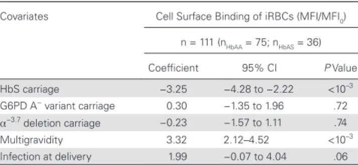
Cell Surface Antibody Binding of Infected Red Blood Cells Is Lower for HbAS Mothers When Matching Infected Red Blood Cells to Plasmas From Mothers With the Same Hemoglobin Genotype
To get closer to in vivo biological conditions, we matched iRBCs to plasma samples from mothers with the same Hb genotype. Thus, we compared the cell surface antibody binding to HbAA iRBCs of plasma from HbAA mothers and similarly the cell surface antibody binding to HbAS iRBCs of plasma from HbAS mothers. We observed significantly lower cell surface antibody binding by plasma from HbAS mothers (multivariable linear
re-gression, P < 10–3, n = 111, median value of HbAA group = 2.60;
median value of HbAS group = 1.13) (Figure 1). The G6PD A−
variant and α−3.7 deletion carriages were also included in the
multivariate analysis as covariates but did not impact this
ob-servation (Table 3). All subsequent tests were conducted
con-sidering cell surface antibody binding when iRBCs and plasmas were matched by Hb genotype.
The Cell Surface Antibody Binding of Infected Red Blood Cells Is Associated With the Anti-VAR2CSA Antibody Level
VAR2CSA is the antigen involved in the cytoadherence of iRBCs to CSA, causing their sequestration in the placenta, and
Table 1. Proportion of Women Carrying G6PD Deficiency and/or
α-Thalassemia Among HbAA and HbAS Groupsa
Combined RBC Genetic Defects HbAA HbAS
P Value
n (%) n = 79 n = 37
No G6PDA− variant and no α−3.7 deletion 28 (35.4%) 14 (37.8%) .96
Only G6PD A− variant 10 (12.7%) 4 (10.8%) .99
Only α−3.7 deletion 28 (35.4%) 15 (40.6%) .66
Both G6PD A− variant and α−3.7 deletion 13 (16.5%) 4 (10.8%) .60
Abbreviations: G6PDA, glucose-6-phosphate dehydrogenase A; HbAA, ; HbAS, sickle cell trait; RBC, red blood cells.
aWomen were separated into 4 groups according to their carriage of the G6PD A− variant
and the α−3.7 deletion. P value was based on a χ2 test to analyze the distribution of women
according to their G6PD A− variant and α−3.7 deletion carriages among HbAA and HbAS
groups.
Table 2. Factors Tested for the Association With the Cell Surface Antibody Binding of HbAA iRBCs or HbAS iRBCs Using a Linear Multivariate Regressiona
Cell surface binding (MFI/MFI0) Covariates Coefficient 95% CI P Value
HbAA iRBCs n = 111 (nHbAA = 75; nHbAS = 36) HbS carriage G6PD A− variant carriage α−3.7 deletion carriage Multigravidity Infection at delivery 0.04 −0.08 −0.24 4.48 1.76 −1.68 to 1.76 −1.91 to 1.76 −1.84 to 1.36 3.04–5.92 −0.67 to 4.19 .96 .93 .77 <10–3 .15 HbAS iRBCs n = 110 (nHbAA = 74; nHbAS = 36) HbS carriage G6PD A− variant carriage α−3.7 deletion carriage Multigravidity Infection at delivery −0.05 −0.16 0.02 0.75 0.49 −0.36 to 0.26 −0.43 to 0.12 −0.26 to 0.30 0.48–1.03 0.03–0.94 .74 .27 .88 <10–3 .04
Abbreviations: CI, confidence interval; G6PD, glucose-6-phosphate dehydrogenase; HbS, hemoglobin S; iRBCs, infected red blood cells; MFI, mean fluorescent intensity.
aCell surface antibody binding fluorescent intensities (MFI) were normalized to the value (MFI
0) obtained by a pool of plasmas derived from European pregnant women. Covariates included in
linear multivariate model regression were maternal genotypes: HbS carriage, G6PD A− variant carriage, and α−3.7 deletion carriage, as well as multigravidity and infection at delivery. A positive
is a promising placental malaria vaccine candidate [35]. The cell surface antibody binding of VAR2CSA-expressing iRBCs that we assessed could plausibly reflect multiple antibody-antigen interactions. Previous work measured the anti-VAR2CSA an-tibody levels for all plasma samples of the STOPPAM cohort taken at delivery. In brief, levels of anti-VAR2CSA antibodies were determined in plasma samples (at dilution 1:100) using an enzyme-linked immunosorbent assay (ELISA) assay targeting
0.5 μg/mL full-length ectodomain of VAR2CSA [11, 36]. We
used these data to test the association of the iRBCs cell surface
antibody binding and the corresponding antibody levels, when
RBC and plasma Hb genotypes were matched (Table 4). We
observed a significant positive association between the anti-VAR2CSA antibody levels and the iRBCs cell surface antibody
binding (multivariable linear regression, P < 10–3, n = 140). We
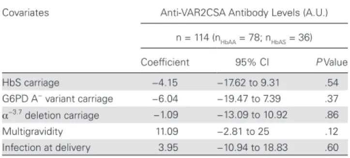
then assessed the association between this antibody level and maternal Hb genotype, but we found no significant difference in the level of anti-VAR2CSA antibodies between HbAA and
HbAS women (multivariable linear regression, n = 114) (Figure
2). In addition, these antibody levels did not vary according to
G6PD A− variant or α−3.7 deletion carriages (Table 5).
DISCUSSION
It is well known that HbS heterozygous carriage affords rela-tive protection against severe forms of malaria, but whether the HbAS genotype also confers a selective protective advan-tage against PAM remains to be determined. In this study, using plasma samples from a cohort of Beninese pregnant women, we assessed the impact of maternal HbAS genotype on the cell surface antibody binding of plasma antibodies to iRBCs during PAM. Our findings are consistent with previous studies, showing a significant increase in the level of surface recognition of iRBCs by plasma as a function of gravidity, independently of maternal
genotypes [11]. However, we observed significantly lower cell
sur-face antibody binding of iRBCs by plasma from HbAS mothers than by plasmas from HbAA mothers when iRBCs and plasmas were matched according to Hb genotype. This lower cell surface antibody binding suggests modifications in the interactions be-tween antigens and antibodies and could be explained by 2 main hypotheses: (1) HbAS women have a lower amount of antibodies recognizing the different antigens present on the iRBC surface, and (2) HbAS iRBCs display lower amounts of the recognized antigens and/or abnormally presented forms of these antigens, altering the capacity for antibody-mediated recognition. In pre-vious work, we measured the level of anti-VAR2CSA antibody
in women in the STOPPAM cohort [11], offering us a way to
test the first hypothesis. Because VAR2CSA is the parasite li-gand involved in the cytoadherence of iRBCs to CSA in PAM, and because women develop anti-VAR2CSA antibodies during successive pregnancies, it is plausible to imagine that the cell surface antibody binding of VAR2CSA is a major component of the overall cell surface antibody binding of iRBCs. To confirm this hypothesis, we first tested the association of the cell surface binding with the level of anti-VAR2CSA antibodies, and we re-vealed a positive association between these 2 factors. The levels of the antibodies measured by ELISA were nevertheless not signifi-cantly different between HbAA and HbAS mothers. However, cell surface antibody binding for each individual sample was shown by paired analyses to be significantly higher to HbAA compared with HbAS iRBC. All of these results suggest that the lower level of cell surface antibody binding of iRBCs by plasma from HbAS
20 P < 10–3 15 10 HbAA n = 75 HbASn = 36
Cell surface binding
(MFI/MFI
0
)
5 0
Figure 1. Association between cell surface antibody binding and maternal
hemo-globin (Hb) genotype when infected red blood cells and plasmas are matched by Hb genotype. Cell surface antibody binding fluorescent intensities (mean fluorescent intensity [MFI]) were normalized to the value (MFI0) obtained by a pool of plasmas
derived from European pregnant women. P value was based on a multivariate linear regression to determine the association between cell surface antibody binding and maternal Hb genotype (HbAA or HbAS), adjusted for gravidity, malaria infec-tion, glucose-6-phosphate dehydrogenase A− variant, and α−3.7 deletion carriages
(n = 111). Box and whisker plots illustrate medians with 75th and 25th percentiles (boxes) and 90th and 10th percentiles (whiskers).
Table 3. Factors Tested for the Association With the Cell Surface Antibody Binding of iRBCs (When iRBCs and Plasmas Are Matched by HbS Genotype) Using a Linear Multivariate Regressiona
Covariates Cell Surface Binding of iRBCs (MFI/MFI0)
n = 111 (nHbAA = 75; nHbAS = 36) Coefficient 95% CI P Value HbS carriage −3.25 −4.28 to −2.22 <10–3 G6PD A− variant carriage 0.30 −1.35 to 1.96 .72 α−3.7 deletion carriage −0.23 −1.57 to 1.11 .74 Multigravidity 3.32 2.12–4.52 <10–3 Infection at delivery 1.99 −0.07 to 4.04 .06
Abbreviations: CI, confidence interval; G6PD, glucose-6-phosphate dehydrogenase; HbS, hemoglobin S; iRBCs, infected red blood cells; MFI, mean fluorescent intensity.
aCell surface antibody binding fluorescent intensities (MFI) were normalized to the value
(MFI0) obtained by a pool of plasmas derived from European pregnant women. Covariates
included in the linear multivariate model regression were maternal genotypes: HbS car-riage, G6PD A− variant carriage, and α−3.7 deletion carriage, as well as multigravidity and
infection at delivery. A positive regression coefficient shows a positive association with the factor, and a negative regression coefficient shows a negative association.
women, when RBC and plasma Hb genotypes were matched, is not due to lower amounts of antibodies recognizing VAR2CSA. Thus, our findings suggest that the expression of VAR2CSA is decreased and/or abnormal on HbAS iRBCs. Previous studies
have documented lower levels of display of VAR2CSA [17]
or PfEMP1 [37] at the HbAS iRBC surface. These studies also
highlighted the abnormally shaped, heterogeneously distributed
knobs on HbAS RBCs [37]. In this study, we cannot exclude the
possibility that the cell surface antibody binding of other iRBC surface antigens may account for the lower total surface recog-nition of iRBCs by plasma of HbAS mothers, but the study by
Chan et al [38] suggests that it is indeed PfEMP1 on iRBCs that
is the principal target of anti-VSA antibodies. However, we could
not exclude the possibility that the IgG affinity for the recognized antigens at the iRBC surface may be stronger for HbAA women.
CONCLUSIONS
Immunity during pregnancy, notably antibody-mediated rec-ognition of VAR2CSA-expressing iRBCs, has been investigated
in previous studies [11, 39]. Nevertheless, this study is the
first to investigate the impact of abnormal HbS in the devel-opment of humoral immunity to malaria during pregnancy. It is also important to note that over half of our study group
carried the α−3.7 deletion, in line with what has been previously
found in neighboring Togo [40]. Likewise, over one quarter of
investigated women presented the A− variant responsible for
G6PD deficiency, consistent with already observed values in
locations from coastal West Africa [41]. The G6PD A− variant
and α−3.7 deletion were equally distributed between HbAA and
HbAS women. Only 35.4% and 37.8%, respectively, carried
nei-ther the G6PD A− variant nor the α−3.7 deletion. Furthermore,
we observed a significant link between α-thalassemia and
HbS genotypes, with more αα/α−3.7 genotypes and less α−3.7/
α−3.7 genotypes among the HbAS compared with the HbAA
women. Malaria-protective effects of the combination of both polymorphisms have been studied, and a negative epistasis between sickle cell trait and heterozygous α-thalassemia car-riage in relation to protection against malaria has already been
described [42, 43]. However, according to our findings, it is
im-portant to match plasma and erythrocyte genotypes. To char-acterize the influence of G6PD deficiency and α-thalassemia, which do not show here any impact on cell surface antibody binding of HbAS erythrocytes, it would be necessary to study first these genetic defects separately and to consider their cocarriage with HbS thereafter, matching ideally in each experi-ment the plasma and the erythrocyte genotypes to better repro-duce in vivo conditions, as we did here with HbAS iRBCs. Our results highlight the importance (1) of taking into account the
150 NS 100 Antibodies anti-V AR2CSA (A.U .) 50 0 HbAA n = 78 HbAS n = 36
Figure 2. Association between the level of anti-VAR2CSA antibody and
ma-ternal hemoglobin (Hb) genotype. The antibody levels data come from a previously published work. The antibody levels were measured by enzyme-linked immu-nosorbent assay [11], and values were converted to arbitrary units (A.U.) [36]. P value was based on a multivariate linear regression to determine the association between the level of anti-VAR2CSA antibody and maternal Hb genotype (HbAA or HbAS), adjusted for gravidity, malaria infection, glucose-6-phosphate dehydro-genase A− variant, and α−3.7 deletion carriages (n = 114). Box and whisker plots
illus-trate medians with 75th and 25th percentiles (boxes) and 90th and 10th percentiles (whiskers).
Table 5. Factors Tested for the Association With the Anti-VAR2CSA Antibody Level Using Linear Multivariate Regressiona
Covariates Anti-VAR2CSA Antibody Levels (A.U.) n = 114 (nHbAA = 78; nHbAS = 36) Coefficient 95% CI P Value HbS carriage −4.15 −17.62 to 9.31 .54 G6PD A− variant carriage −6.04 −19.47 to 7.39 .37 α−3.7 deletion carriage −1.09 −13.09 to 10.92 .86 Multigravidity 11.09 −2.81 to 25 .12 Infection at delivery 3.95 −10.94 to 18.83 .60
Abbreviations: A.U., arbitrary units; CI, confidence interval; G6PD, glucose-6-phosphate dehydrogenase; HbS, hemoglobin S.
aCovariates included in linear multivariate model regression were maternal genotypes:
HbS carriage, G6PD A− variant carriage, and α−3.7 deletion carriage, as well as multigravidity
and infection at delivery. A positive regression coefficient shows a positive association with the factor, and a negative regression coefficient shows a negative association.
Table 4. Association Between the iRBCs Surface Cell Surface Antibody Binding (When iRBCs and Plasmas Are Matched by Hb Genotype) and the Level of Anti-VAR2CSA Antibody Using a Linear Multivariate Regressiona Covariates Cell Surface Binding of iRBCs
(MFI/MFI0)
Coefficient 95% CI P Value Anti-VAR2CSA antibody levels (A.U.) 0.05 0.03–0.7 <10–3
HbS carriage −3.67 −4.62 to −2.73 <10–3
Multigravidity 3.05 2.02–4.09 <10–3
Infection at delivery 1.19 −0.37 to 2.75 .13
Abbreviations: A.U., arbitrary units; CI, confidence interval; HbS, hemoglobin S; iRBCs, infected red blood cells; MFI, mean fluorescent intensity.
aCell surface antibody binding fluorescent intensities (MFI) were normalized to the value
(MFI0) obtained by a pool of plasmas derived from European pregnant women. Covariates
included in linear multivariate model regression were the anti-VAR2CSA antibody levels and maternal HbS carriage, as well as multigravidity and infection at delivery. A positive re-gression coefficient shows a positive association with the factor, and a negative rere-gression coefficient shows a negative association.
Hb genotype of the mothers from whom plasmas are derived and (2) of the erythrocytes used for the in vitro assay of cell sur-face antibody binding, because RBC disorders could influence the export and/or the presentation of antigens. Furthermore, this study reinforces the hypothesis of VAR2CSA abnormal display at the HbAS erythrocyte surface, illustrating the conse-quence on HbAS iRBC surface recognition by HbAS plasmas. Despite the fact that sickle cell trait is the most widespread erythrocyte disorder in Africa, on the basis of our biological data we could not draw definitive conclusions on the impact of the HbAS genotype on PAM. Nevertheless, the same scien-tific strategy needs to be applied with respect to all the different genetic disorders, such as HbAC that is known to have an
im-pact on LBW for example [19], as well as G6PD deficiency and
α-thalassemia.
Supplementary Data
Supplementary materials are available at Open Forum Infectious Diseases online. Consisting of data provided by the authors to benefit the reader, the posted materials are not copyedited and are the sole responsibility of the authors, so questions or comments should be addressed to the corre-sponding author.
Acknowledgments
We thank the voluntary blood donors for their participation to this project; and the Universities Paris Diderot and Paris Descartes for the doc-toral scholarships awarded to Marilou Tétard and Margaux Chauvet. This study was part of the STOPPAM collaborative project. We are grateful to the women who participated in the study.
Author contributions. B. G., A. J. F .L., A. M., and F. M.-N. conceived
and designed the project. M. C., M. L., A. M., M. T., and N. T. N. performed the experiments. M. C., G. C., A. M., F. M.-N., and J. M. performed the anal-ysis. F. A., E. B., M. C., L. D., M. H., A. M., F. M.-N., and D. P. performed the genotyping. M. C., A. J. F. L., A. M., F. M. N., and M. T. wrote the paper. All authors reviewed the manuscript before submission.
Financial support. This work was funded by the Laboratoire d’Excellence
GR-Ex, Paris, France, reference ANR-11-LABX-0051, that is funded by the program Investissements d’Avenir of the French National Research Agency, reference ANR-11-IDEX-0005-02.
Potential conflicts of interest. All authors: No reported conflicts of
in-terest. All authors have submitted the ICMJE Form for Disclosure of Potential Conflicts of Interest.
References
1. World Health Organization. World Malaria Report. Geneva; World Health Organization; 2018.
2. Rogerson SJ, Desai M, Mayor A, et al. Burden, pathology, and costs of malaria in pregnancy: new developments for an old problem. Lancet Infect Dis 2018; 18:e107–18.
3. Andrews KT, Lanzer M. Maternal malaria: Plasmodium falciparum sequestration in the placenta. Parasitol Res 2002; 88:715–23.
4. Salanti A, Dahlbäck M, Turner L, et al. Evidence for the involvement of VAR2CSA in pregnancy-associated malaria. J Exp Med 2004; 200:1197–203.
5. Viebig NK, Gamain B, Scheidig C, et al. A single member of the Plasmodium fal-ciparum var multigene family determines cytoadhesion to the placental receptor chondroitin sulphate A. EMBO Rep 2005; 6:775–81.
6. Viebig NK, Nunes MC, Scherf A, Gamain B. The human placental derived BeWo cell line: a useful model for selecting Plasmodium falciparum CSA-binding parasites. Exp Parasitol 2006; 112:121–5.
7. Fried M, Duffy PE. Adherence of Plasmodium falciparum to chondroitin sulfate A in the human placenta. Science 1996; 272:1502–4.
8. Srivastava A, Gangnard S, Round A, et al. Full-length extracellular region of the var2CSA variant of PfEMP1 is required for specific, high-affinity binding to CSA. Proc Natl Acad Sci U S A 2010; 107:4884–9.
9. Duffy PE. Plasmodium in the placenta: parasites, parity, protection, prevention and possibly preeclampsia. Parasitology 2007; 134:1877–81.
10. O’Neil-Dunne I, Achur RN, Agbor-Enoh ST, et al. Gravidity-dependent pro-duction of antibodies that inhibit binding of Plasmodium falciparum-infected erythrocytes to placental chondroitin sulfate proteoglycan during pregnancy. Infect Immun 2001; 69:7487–92.
11. Ndam NT, Denoeud-Ndam L, Doritchamou J, et al. Protective antibodies against placental malaria and poor outcomes during pregnancy, Benin. Emerg Infect Dis
2015; 21:813–23.
12. Tuikue Ndam NG, Salanti A, Le-Hesran JY, et al. Dynamics of anti-VAR2CSA immunoglobulin G response in a cohort of Senegalese pregnant women. J Infect Dis 2006; 193:713–20.
13. Fried M, Nosten F, Brockman A, et al. Maternal antibodies block malaria. Nature
1998; 395:851–2.
14. Flint J, Harding RM, Boyce AJ, Clegg JB. The population genetics of the haemoglobinopathies. Baillieres Clin Haematol 1998; 11:1–51.
15. Piel FB, Patil AP, Howes RE, et al. Global distribution of the sickle cell gene and geographical confirmation of the malaria hypothesis. Nat Commun 2010; 1:104. 16. Williams TN, Mwangi TW, Wambua S, et al. Sickle cell trait and the risk of
Plasmodium falciparum malaria and other childhood diseases. J Infect Dis 2005; 192:178–86.
17. Cyrklaff M, Srismith S, Nyboer B, et al. Oxidative insult can induce malaria-protective trait of sickle and fetal erythrocytes. Nat Commun 2016; 7:13401. 18. Kilian N, Srismith S, Dittmer M, et al. Hemoglobin S and C affect protein export
in Plasmodium falciparum-infected erythrocytes. Biol Open 2015; 4:400–10. 19. Tétard M, Milet J, Dechavanne S, et al. Heterozygous HbAC but not HbAS is
associated with higher newborn birthweight among women with pregnancy-associated malaria. Sci Rep 2017; 7:1414.
20. Patel JC, Mwapasa V, Kalilani L, et al. Absence of association between sickle trait hemoglobin and placental malaria outcomes. Am J Trop Med Hyg 2016; 94:1002–7.
21. Mbanefo EC, Ahmed AM, Titouna A, et al. Association of glucose-6-phosphate dehydrogenase deficiency and malaria: a systematic review and meta-analysis. Sci Rep 2017; 7:45963.
22. Taylor SM, Parobek CM, Fairhurst RM. Haemoglobinopathies and the clinical epidemiology of malaria: a systematic review and meta-analysis. Lancet Infect Dis
2012; 12:457–68.
23. Krause MA, Diakite SA, Lopera-Mesa TM, et al. α-Thalassemia impairs the cytoadherence of Plasmodium falciparum-infected erythrocytes. PLoS One 2012; 7:e37214.
24. Piel FB, Weatherall DJ. The α-thalassemias. N Engl J Med 2014; 371:1908–16. 25. Medeiros Alcoforado GH de, Bezerra CM, Araújo Moura Lemos TM, et al.
Prevalence of α-thalassemia 3.7 kb deletion in the adult population of Rio Grande do Norte, Brazil. Genet Mol Biol. 2012; 35:594–598.
26. Huynh BT, Fievet N, Gbaguidi G, et al. Influence of the timing of malaria infec-tion during pregnancy on birth weight and on maternal anemia in Benin. Am J Trop Med Hyg 2011; 85:214–20.
27. Pouvelle B, Meyer P, Robert C, et al. Chondroitin-4-sulfate impairs in vitro and in vivo cytoadherence of Plasmodium falciparum infected erythrocytes. Mol Med
1997; 3:508–18.
28. Trager W, Jensen JB. Human malaria parasites in continuous culture. Science
1976; 193:673–5.
29. Tuikue Ndam NG, Fievet N, Bertin G, et al. Variable adhesion abilities and overlapping antigenic properties in placental Plasmodium falciparum isolates. J Infect Dis 2004; 190:2001–9.
30. Wooden J, Kyes S, Sibley CH. PCR and strain identification in Plasmodium falcip-arum. Parasitol Today Pers Ed 1993; 9:303–5.
31. Cappellini MD, Fiorelli G. Glucose-6-phosphate dehydrogenase deficiency. Lancet 2008; 371:64–74.
32. Howes RE, Dewi M, Piel FB, et al. Spatial distribution of G6PD deficiency variants across malaria-endemic regions. Malar J 2013; 12:418.
33. Carter N, Pamba A, Duparc S, Waitumbi JN. Frequency of glucose-6-phosphate dehydrogenase deficiency in malaria patients from six African countries enrolled in two randomized anti-malarial clinical trials. Malar J 2011; 10:241.
34. Liu YT, Old JM, Miles K, et al. Rapid detection of alpha-thalassaemia deletions and alpha-globin gene triplication by multiplex polymerase chain reactions. Br J Haematol 2000; 108:295–9.
35. Chêne A, Houard S, Nielsen MA, et al. Clinical development of placental malaria vaccines and immunoassays harmonization: a workshop report. Malar J 2016; 15:476.
36. Guitard J, Cottrell G, Magnouha NM, et al. Differential evolution of anti-VAR2CSA- IgG3 in primigravidae and multigravidae pregnant women infected by Plasmodium falciparum. Malar J 2008; 7:10.
37. Cholera R, Brittain NJ, Gillrie MR, et al. Impaired cytoadherence of Plasmodium falciparum-infected erythrocytes containing sickle hemoglobin. Proc Natl Acad Sci U S A 2008; 105:991–6.
38. Chan JA, Howell KB, Reiling L, et al. Targets of antibodies against Plasmodium falciparum-infected erythrocytes in malaria immunity. J Clin Invest 2012; 122:3227–38. 39. Chandrasiri UP, Randall LM, Saad AA, et al. Low antibody levels to pregnancy-specific malaria antigens and heightened cytokine responses associated with se-vere malaria in pregnancy. J Infect Dis 2014; 209:1408–17.
40. Segbena AY, Kueviakoe I, Messie AK, et al. Hemoglobin anomalies at the univer-sity hospital center in Lome, Togo. Med Trop (Mars) 2002; 62:51–4.
41. Howes RE, Piel FB, Patil AP, et al. G6PD deficiency prevalence and estimates of affected populations in malaria endemic countries: a geostatistical model-based map. PLoS Med 2012; 9:e1001339.
42. Williams TN, Mwangi TW, Wambua S, et al. Negative epistasis between the malaria-protective effects of alpha+-thalassemia and the sickle cell trait. Nat Genet 2005; 37:1253–7.
43. Mpimbaza A, Walakira A, Ndeezi G, et al. Associations between erythrocyte polymorphisms and risks of uncomplicated and severe malaria in Ugandan children: A case control study. PLoS One 2018; 13:e0203229.