HAL Id: tel-01685142
https://tel.archives-ouvertes.fr/tel-01685142
Submitted on 16 Jan 2018HAL is a multi-disciplinary open access archive for the deposit and dissemination of sci-entific research documents, whether they are pub-lished or not. The documents may come from teaching and research institutions in France or abroad, or from public or private research centers.
L’archive ouverte pluridisciplinaire HAL, est destinée au dépôt et à la diffusion de documents scientifiques de niveau recherche, publiés ou non, émanant des établissements d’enseignement et de recherche français ou étrangers, des laboratoires publics ou privés.
Protéine-kinases et cancer du rein : Découverte et
validation d’une nouvelle combinaison d’inhibiteurs
ciblant les protéine-kinases ATM et CK2
Sofia Giacosa
To cite this version:
Sofia Giacosa. Protéine-kinases et cancer du rein : Découverte et validation d’une nouvelle combinaison d’inhibiteurs ciblant les protéine-kinases ATM et CK2. Médecine humaine et pathologie. Université Grenoble Alpes, 2016. Français. �NNT : 2016GREAV035�. �tel-01685142�
THÈSE
Pour obtenir le grade de
DOCTEUR DE LA COMMUNAUTE UNIVERSITE
GRENOBLE ALPES
Spécialité : Doctorat CSV/Biologie Cellulaire
Arrêté ministériel : 7 août 2006
Présentée par
Sofía Inés Giacosa
Préparée au sein du Laboratoire Biologie du Cancer et l’Infection dans l'École Doctorale Chimie et Sciences du Vivant
Protéine-kinases et cancer du
rein : Découverte et validation
d’une combinaison d’inhibiteurs
ciblant les protéine-kinases ATM
et CK2.
Protein kinases and renal carcinoma: discovery and validation of a novel combinational target therapy through co-inhibition of CK2 and ATM kinases
Thèse soutenue publiquement le « 14 Octobre 2016 », devant le jury composé de :
Prof. Bernard DUCOMMUN
CNRS-USR3505 ITAV - Centre Pierre Potier-Toulouse Rapporteur Dr. Gilles PAGES
CNRS UMR 7284 INSERM U 1081 IRCAN-Nice Rapporteur Dr. Yannick ARLOT-BONNEMAINS
CNRS -UMR 6290 IGDR- Rennes Examinatrice Prof. Sylvie NEGRIER
Université Lyon I Centre Léon Bérard- Lyon Examinatrice Prof. Pierre HAINAUT
U1209 Institute Albert Bonniot-Grenoble Président du Jury Dr. Claude COCHET
INSERM, U1036 BCI CEA- Grenoble co-Directeur Dr. Odile FILHOL-COCHET
Résumé:
L’incidence du cancer du rein et sa mortalité associée se sont accrues au cours des dernières années. Le type de cancer rénal le plus fréquent est celui nommé Cancer Rénal à Cellules Claires (CRCC) où le plus souvent, le gène suppresseur de tumeur Von Hippel Lindau (VHL) est inactivé. Malgré une détection plus précoce, l’évolution de la pathologie demeure incertaine, en particulier quand les patients développent des métastases ou acquièrent une résistance au traitement (25-30% des patients). De nouvelles thérapies ciblant des kinases (Sunitinib, Sorafenib ou Temsirolimus) bien que très prometteuses conduisent très souvent à l’acquisition de résistance. Dans ce contexte, il est urgent de développer de nouveaux modèles prédictifs de la réponse des patients aux traitements et d'identifier de nouvelles cibles thérapeutiques.
Ma thèse de science visait trois objectifs complémentaires : 1) Identifier par criblage chimio-génomique des kinases comme cibles thérapeutiques combinées. 2) Etablir deux modèles de culture 3D de cancer du rein qui intègrent le microenvironnement d’une tumeur: les sphéroïdes et la culture organotypique de coupe de tissus. 3) Etudier la chimio-sensibilité de ces modèles à une combinaison de molécules identifiées dans le criblage.
Un criblage cellulaire a été réalisé sur la plateforme de Criblage de Molécules BioActives (CEA- Grenoble). Il a consisté à tester 80 molécules inhibitrices de protéine-kinases en combinaison avec l’extinction génique par interférence ARN (shRNAs lentiviraux) de 36 cibles potentielles connues pour leur implication dans divers cancers. La lignée cellulaire choisie (786-O) est dérivée d’une tumeur rénale à cellules claires radio et chimio-résistante et dépourvue de VHL. Parmi les touches qui compromettent la viabilité des cellules 786-O, la combinaison choisie pour son efficacité cible deux kinases importantes dans le contrôle de la survie cellulaire et de la réparation de l’ADN : CK2 et ATM. Le statut VHL des cellules module de façon dramatique leur sensibilité à cette combinaison, l’association de ces deux inhibiteurs étant plus efficace sur les cellules 786-O (VHL-) que sur les mêmes cellules dans lesquelles VHL a été réintroduit (VHL+). Au sein d’une tumeur, les différents niveaux d’oxygénation constituent une variation environnementale supplémentaire créant des susceptibilités ou des résistances aux traitements thérapeutiques. Pour déterminer l’impact de nos molécules dans ce contexte, nous avons testé la viabilité des cellules 786-O VHL+ et VHL- dans des conditions normoxiques (21% O2) ou hypoxiques (1,5% O2), en présence des molécules seules ou en combinaison. En normoxie, une diminution synergique de la viabilité des cellules 786-O VHL- est observée en présence de la combinaison, alors que cet effet n’a pas lieu sur les cellules 786-O VHL+. Cette synergie est potentialisée en condition hypoxique. Au niveau mécanistique, les voies de signalisation de stress cellulaires sont d’avantage activées dans les cellules VHL- en présence de la combinaison de molécules comparé au traitement avec chacune des molécules seules. Dans les sphéroïdes tumoraux multicellulaires reproduisant l’organisation d’une micro-tumeur, nos résultats montrent que notre combinaison de molécules induit d’avantage l’apoptose des cellules VHL- que les molécules seules, alors que les cellules VHL+ ne sont sensibles à aucun des traitements.
Ces résultats montrent que l’action de nos molécules combinées est clairement plus efficace dans un modèle 3-D. Ils démontrent également qu’il est possible d’objectiver une pharmaco-modulation de la viabilité de cultures organotypiques de tumeur du rein par des combinaisons d’inhibiteurs chimiques de protéine-kinases. Les perspectives de ce travail sont la validation de cette combinaison sur des tumeurs humaines et l’exploitation des cultures organotypiques comme test personnalisé de réponse aux traitements.
Abstract :
Renal cell carcinoma accounts for 3% of all malignant diseases in adults making it the 10th most common cancer in France. The most frequent type of Kidney cancer is Clear Cell Renal Cell Carcinoma (CCRCC). Almost all CCRCC show an inactivation of the Von Hippel Lindau tumour suppressor gene (VHL). Between 25-30% of the patients will develop metastatic renal cell carcinoma (mRCC) by the time they are diagnose or become unresponsive to all treatments and in these cases, the disease has a rapid progression. Over the past years, kinase-targeted therapies (Sunitinib, Sorafenib, Temsirolimus) have become the mainstay of treatment for mRCC, however, most, if not all, patients acquire resistance to these approaches over time.
In this context my PhD had 3 goals: a) to find a new combinatory targeted therapy through a High Throughput Screening; b) to establish 3D models mimicking the real environment of the tumours (spheroids, Tissue Slice Culture); c) to validate the Hits through different molecular and cellular biology studies.
We conducted a synthetic lethal screen on the CMBA platform (CEA-Grenoble), choosing 36 potential genes targets and 80 kinases inhibitors drugs. Each of the target gene was silenced by a transduction with shRNA Lentivirus into the 786-O cell line derived from ccRCC that lacks the tumour suppressor VHL, is radio- and chemo-therapy resistant, has increased mobility and is highly metastatic. Among the hit combinations that affect cell viability, one of them was chosen because it targets two important kinases involved in cell survival and DNA repair: CK2 and ATM. Moreover, this combination is specifically more active in the 786-O VHL- cells than in 786-O VHL+ cell line. We evaluated the effect of our drugs on the viability of our 786-O VHL+ and VHL- cells in normoxic (21% O2) or hypoxic (1.5% O2) conditions that reflect different environments that are present in a tumour. Surprisingly, in normoxia, we found a synergetic effect of the drug mix only on the 786-O VHL- cells but not on 786-O VHL+ cells. Furthermore, this effect was even stronger in conditions of Hypoxia (up to 20% of synergism).
Mechanistically, an up-regulation of the stress pathways was much stronger in the VHL- cells in the presence of the combination than with the drugs alone. No apoptosis was detected in this 2D models. In Multi-Cellular Tumour Spheroid (MCTS) where the organization of a micro-tumour is reproduced, our drugs are even more effective in inducing cell apoptosis than in 2D monolayers of 786-O VHL- cells. These results also demonstrate that pharmaco-modulation of viability of renal tumour organotypic culture by chemical combination targeting protein kinases can be studied. Perspectives of this work are the validation of this drug combination on human renal tumours and the use of organotypic culture as a test for personalized treatment response.
1 Je voudrais remercier les rapporteurs de ce jury de thèse Dr. Bernard Ducommun et Dr. Gilles Pages ainsi que les autres membres du jury Dr. Yannick Arlot-Bonnemais, Pr. Sylvie Negrier et Dr. Pierre Hainaut d’avoir accepté d’évaluer mon travail.
Aussi je voudrais remercier ma directrice de thèse Dr. Odile Filhol. Odile, merci pour avoir cru en moi, pour avoir tenté ta chance avec une petite argentine devant un poster dans un couloir presque noir au CEA Saclay. Pour ta patience et ton écoute pendant ces 4 ans de thèse, pour les heures et heures à l’animalerie en train de peser les souris en espagnol, pour partager ta connaissance sans restriction et pour m’aider à devenir une vraie chercheuse. C’était un honneur de travailler avec toi, et un plaisir de te connaître personnellement. Au Dr. Claude Cochet, mon co-directeur de thèse mais aussi un collègue avec qui j’ai partagé tant de discussions scientifiques. De la même manière que les argentins se disputent pour le football, nous avons mis notre passion pour la science dans nos discussions et parfois il fallait un arbitre pour nos arrêter de parler haha. Merci de ton soutien dans chaque nouvelle « hypothèse » mais aussi pour me transmettre ta connaissance avec une humilité digne des grands. Avec Odile vous m’avez formé pour qu’un jour je sois aussi qualifiée que vous et je vous serai toujours reconnaissant de cette générosité. Et oui, tu pourras continuer à m’envoyer des publis et je continuerai à te dire que je ne les lirai pas (juste pour continuer à t’embêter lol).
A Cathy, or half or moitié. Une personne qui ne transmet pas seulement son savoir mais aussi sa passion pour le travail bien fait. Nous avons tellement rigolé pendant cette thèse. Mais aussi, sans ton soutien dans les moments difficiles je ne serai jamais arrivé à la fin. Tu es une personne exceptionnelle qui en plus m’a appris beaucoup de choses avec patience, joie et humilité. Ton courage pour te battre dans la vie était une de mes inspirations pendant cette thèse.
A mes collègues Caroline, Magali, Megghane, le premier groupe comme on dirait. Merci pour m’avoir accueilli et si vite intégrer à la vraie culture française. Merci pour les heures partagées dans et en dehors du labo. Cette époque tous ensembles m’a manqué énormément durant les dernières années de thèse.
2 A Jean-Jacques Feige, merci pour ton soutien depuis le début dans le laboratoire et pour les suggestions scientifiques au cours de ces 4 ans.
Nadia Cherradi, Nadia Alfaidy, Sabine, Daniel, Emmanuelle, Aude, Agnés, Mariela, Olivier, Wael, Sarah, Deborah, Marie, Christine Cogne et Christine Mallet. Chacun d’entre vous a tellement apporté à cette expérience d’une manière ou d’une autre. Merci pour les conseils, les sourires, les balades et d’avoir toujours bien voulu m’aider.
A Nicolas, mon mari, mon ami, le pilier de notre famille. Cette expérience nous a rendus encore plus forts. Merci de toujours m’encourager à continuer à donner le meilleur de moi, de me dire que ça va aller et de me soutenir chaque jour. Depuis cette aventure, j’ai hâte de continuer à partager une infinité d’expériences ensembles.
A vos mamá, que siempre me diste alas para volar. Que difícil debe haber sido dejar partir a tus hijos, ahora lo entiendo mejor. Pero siempre me enseñaste que una mujer tiene que hacerse el futuro sola y compartirlo con un hombre que la valore. Te dedico esta tesis por ser la persona que más creyó en mí desde que me fui. Créeme que te extraño todos los días y que me gustaría poder salir a tomar un café juntas de vez en cuando.
A mi papá, que me enseño a dar todo por un objetivo. A buscarle la vuelta y la re vuelta. A pensar que siempre existe una solución para todo. Gracias chochan!
A mis hermanos y al resto de mi familia que son un pilar de apoyo enorme.
No quiero olvidar a mis amigas de aquí, Guillermina y Natalia, por ese constante apoyo en todas las áreas de mi vida. Por escucharme una y otra vez decir lo mismo o quejarme de lo mismo jaja.
Y agradezco también a mis amigas y amigos de la Argentina, que aunque sea con un mensajito estuvieron y están presentes para darme ánimos, hacerme sonreír en los días difíciles y darme un abrazo en la distancia cuando lo necesito.
Et à Helena, ma fille, que est venue comment une broche d’or s’épingler à toutes ces merveilleuses années. Cette petite thèse est pour te dire que rien n’est impossible dans la vie, qu’il ne faut jamais baisser les bras et que tu dois toujours essayer de faire ce que tu aimes.
3
TABLE OF CONTENTS
INTRODUCTION ... 11
CHAPTER 1: Kidney and Cancer ... 12
1. WHY IS YOUR KIDNEY SO IMPORTANT?. ... 12
Kidney Physiology. ... 12
1.2 Kidney Functions ... 14
2. KIDNEY CANCER ... 15
Frequency and mortality ... 15
2.2 Causes of kidney cancer ... 17
A) Non genetic associated factors ... 17
B) Genetic associated risk factors ... 18
3. CLEAR CELL RENAL CELL CARCINOMA (ccRCC) ... 20
Histopathology and frequency. ... 20
3.2 Somatic Genetics ... 21
3.3 Kidney cancer stages, treatment and prognosis ... 22
4. TREATMENT OF METASTATIC RCC ... 25 Surgery ... 25 4.2 Adjuvants therapies ... 25 • Immunotherapies ... 25 A) Interferon α (IFN α) ... 25 B) Interleukin-2 (IL-2) ... 26 • Targeted Therapies ... 27
A) VEGF/VEGF Receptor (VEGFR) inhibitors ... 30
B) mTOR inhibitors ... 33
C) MET inhibitors ... 34
Sequential and Combinational therapy ... 35
5.MECHANISM OF ACQUIRED RESISTANCE IN THE TREATMENT OF ccRCC ... 39
Resistance to Tyrosine Kinase Inhibitors ... 39
5.2 Resistance to mTOR inhibitors ... 44
CHAPTER 2: THE ROLE OF HYPOXIA IN ccRCC ... 49
1.VON HIPPEL LINDAU TUMOR SUPPRESSOR GENE (VHL) ... 49
VHL gene products ... 49
4
2. HYPOXIA INDUCIBLE FACTORS ... 55
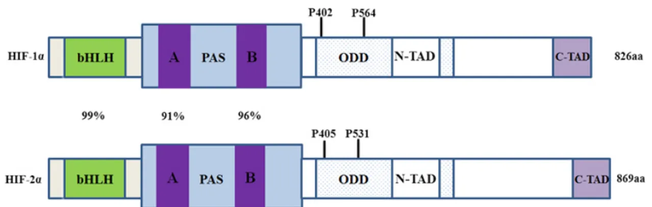
Structures and tissue expression level. ... 55
2.2 Implications of HIF-1α and HIF-2α in the ccRCC. ... 59
2.3 HIF isoforms as prognosis factors ... 61
CHAPTER 3: NOVELS TARGETS AGAINST ccRCC ... 62
1. ATAXIA TELANGESTASIA MUTATED KINASE (ATM) ... 62
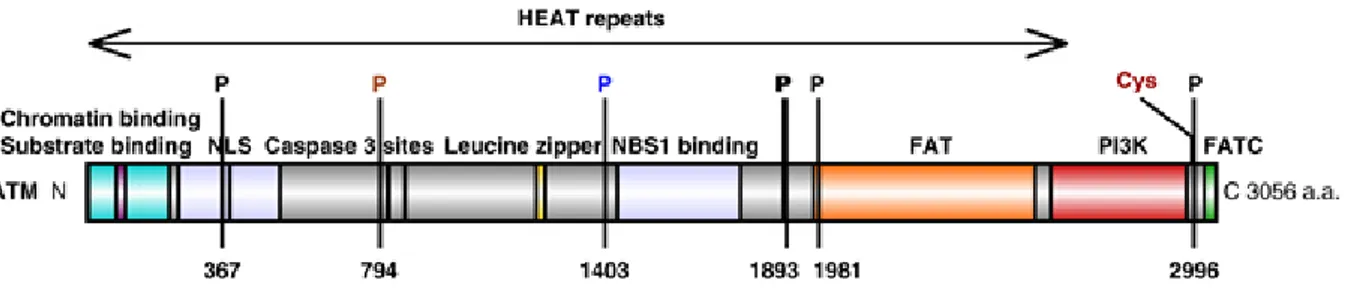
ATM structure ... 62
1.2 ATM cell function ... 63
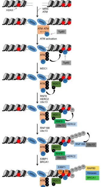
A) ATM role in DNA damage response activity. ... …63
B) ATM signaling in cancer cells. ... 66
C) ATM Dependent Modulation of Signalling Pathways Outside DDR Implicated in Cancer. ... 68
1.3 Modulation of ATM Activity in Cancer Therapy ... 71
2.PROTEIN KINASE CK2 ... 74
CK2 Structure ... 74
2.2 Cellular functions in Cancer Disease... 75
2.3 CK2 Protein expression in Renal Cancer ... 78
2.4 CK2 and the HIF axis ... 79
2.5 CK2 is a druggable target. ... 79
A) CK2 inhibitors ... 80
2.6 The CX-4945 inhibitor ... 82
A) Specificity and cell biology function. ... 82
B) Clinical Phase trials ... 83
C) CX-4945 in combinational therapy ... 84
RESULTS (i) ... 85
Search for novel therapeutic targets against ccRCC by High-Throughput screening. ... 86
Set up of conditions for High-throughput Screening (HTS) ... 86
A) 786-O as cell line model ... 86
B) Kinases as targets ... 87
C) Cells seeding and Cell Viability Markers ... 92
D) Determination of Z’Score Factor ... 92
1.2 The Screening ... 93
1.3 Validation of hits through molecular biology studies ... 97
5
Conclusions ... 103
RESULTS (ii) ... 105
PATENT: “Novel therapeutic drug combination against Kidney Cancer ». ... 106
Introduction ... 106
Materials and Methods ... 107
Results ... 113
Claims…. ... 117
Tables and Figures ... 119
Figures legends ... 128
Discussion ... 132
SUPPLEMENTARY RESULTS (i) ... 139
Study of the phenotype given by drug inhibitors ... 140
Intracellular vacuolar structures ... 140
Origin of vacuoles ... 146
A) Endocytosis ... 146
B) Early and Late endosomes ... 153
Mechanism of resistance ... 156
D) Lysosomal sequestration ... 156
E) Autophagy ... 158
Discussion ... 161
Conclusion ... 163
SUPPLEMENTARY RESULTS (ii) ... 165
Preliminary results on the mechanistic of CK2/ATM drug combination ... 166
ATM/CK2 dysregulation in ccRCC human samples ... 166
HIF-2α: The Redemption ... 167
HIF2α is a substrate for CK2. ... 172
Discussion ... 175
FINAL CONCLUSION AND PERSPECTIVES ... 177
REFERENCES ... 183
APPENDIX ... 243
6
LIST OF FIGURES
Figure 1: Internal Anatomy of a human kidney. ... 13
Figure 2: Scheme of a kidney nephron. ... 14
Figure 3: Incidence and mortality if kidney cancer worldwide ... 16
Figure 4: Picture of a Clear cell renal cell carcinoma ... 21
Figure 5: Kidney cancer stages and 5 years survival rates. ... 23
Figure 6: Dysregulated pathways of ccRCC and target therapies mechanism of action. ... 29
Figure 7: Mechanisms of resistance to anti-angiogenic targeted therapies. ... 43
Figure 8: Scheme of the mTOR pathway ... 45
Figure 9: HIFα Oxygen-dependent HIF regulation. ... 51
Figure 10: The structural domains of HIF-1α and HIF-2α. ... 55
Figure 11: Domain structure of human ATM. ... 62
Figure 12: ATM activation in response to DSBs ... 65
Figure 13: The ATM signalling pathway. ... 67
Figure 14: Chemical structures of ATM inhibitors. ... 71
Figure 15: Tetrameric representation of CK2. ... 75
Figure 16:CK2 contributes to the “Hallmarks of Cancer”. ... 76
Figure 17: Chemical structures of CK2 inhibitors ... 80
7
LIST OF TABLES
Table 1: Summary of life-style related RCC risk factors. ... 18
Table 2: Retrieved phase 2 and 3 studies from systematic research in the cytokine- refractory and in the post-vascular endothelial growth factor inhibition setting ... 37
Table 3: Retrieved studies from systematic research of cytokine-based combination and targeted therapy–based combination ... 38
Table 4: Mechanism of resistance to Tyrosine kinases inhibitors in ccRCC. ... 43
Table 5: pVHL functions in HIFα-dependent and independent fashions ... 54
Table 6: List of genes in the ccRCC context that are regulated either by both HIF-1α and HIF-2α or only one of them. ... 57
Table 7: Role of ATM in independent-DNA damage pathways. ... 70
Table 8: Dysregulated CK2 mRNA expression levels in 6 types of cancer.. ... 77
Table 9: Most representative ATP-competitve inhibitors of CK2.. ... 82
LIST OF CHARTS
Chart 1:Percent of cases & 5-year relative survival by stage at diagnosis in kidney and renal pelvis cancer. ... 248
LIST OF ACRONYMS
(AKT) protein kinase B
(ATM) Ataxia telangestasia mutated kinase (BEV) Bevacizumab
(ccRCC) Clear cell renal cell carcinoma (CK2) CK2 previously named Casein kinase 2 (COPD) chronic obstructive pulmonary disease (CTDA) C- terminal activation domain
(CUL2) Cullin 2
(DDR) DNA damage response (DSBs) Double Strands Breaks
(Elongin C) transcription elongation factors C (Elongin B) transcription elongation factors B (EMT) Epithelial to Mesenchymal transition (FDA) Food and drug administration
(FIH1) asparaginyl hydroxylase (GBM) glioblastoma
(HIF) Hypoxia inducible factor (HR) Hazard ratio
(HRE) Hypoxia Response Elements (IFN α) Interferon α
(IL-2) Interleukin-2 (IR) Ionizing radiation
(MAPK) mitogen activated protein kinases (MPPs) Matrix Metalloproteinases
(mRCC) Metastatic renal carcinoma (mTOR) Mammalian target of rapamycin (NK) natural killer cells
9 (NNKOAc) 4-[(acetoxymethyl) nitrosamino]-1-(3-pyridyl)-1-butanone
(NTAD) N-terminal transactivation domain (ORR) Objective response rate
(ORR) objective response rate (OS) Overall survival
(PDGF) Platelet-derived growth factor
(PDK1) phosphoinositide –dependent-kinase (PFS) Progression Free survival
(PI3K) phosphatidylinositol 3-kinase (RET) Receptor Tyrosine Kinase (ROS) Reactive oxygen species (RXB1) RING finger protein (siRNA) small interfering RNAs (shRNA) short hairpin RNA (SSBs) single strand breaks
(TAM) Tumor associated macrophages
(TCEB1) Transcription elongation factor polypeptide 1 (TRAIL) TNF-related apoptosis-inducing ligand
(TSC2) tuberin
(VEGF) Vascular endothelial growth factor (VHL) von Hippel-Lindau tumor suppressor (AKT) protein kinase B
(ATM) Ataxia telangestasia mutated kinase (BEV) Bevacizumab
11
12
CHAPTER 1
1. WHY IS YOUR KIDNEY SO IMPORTANT?
Kidney Physiology.
Kidneys are pair of bean-shape organs that are localized on the upper level at the back of the abdominal cavity, side by side the vertebral column. They show a dissymmetry in their position due to that right kidney is “pushed down” by the liver compared to the left kidney that is free in peritoneal area, nevertheless both kidneys are protected by the lower ribs. The kidney itself is enclosed in the fibrous capsule and surrounded by the perinephric and the paranephric fat. As well, there are renal glands that are on the outside part of the kidney and have extreme important functions as hormones production.
In a transversal cut section (figure 1) we can observe the different areas of a kidney. The white area is the cortex, just below the renal capsule, where the nephron bodies are located. Lower we have a pink section called medulla, where we find the tubules forming a renal triangle shape structure called renal pyramid. Between the cortex and the medulla, there are the renal columns that will collect the filters coming through the cortex section and will take them to the renal papilla. Renal papilla is formed by minor calix that together form the major calix and address the filter to the renal pelvis and finally to the ureter.
The renal artery enters the kidney in the hilum, crosses the renal lobule (formed by the renal pyramid and the cortex) and archers around the kidney changing its name interlobular renal and collecting the filter through branches. The renal vein has the same structure as the renal artery but it will “leave” the kidney at the hilum.
13 Figure 1: Internal Anatomy of a human kidney.
Inside the kidney it can be distinguished 4 main sections: The renal capsule (light pink), the cortex that is just below the capsule where the nephron bodies are inserted, the medulla where the nephron is located and the renal hilum containing the renal artery and renal vein. Picture taken from (Openstack College 2016)
Nephrons are part of the microstructures of the kidney and are the main responsible elements for the blood filtration (Figure 2). Nephrons consist in two main structures, the renal corpuscle and the renal tubule. In the renal corpuscle the blood will enter and will be filtrated by the glomerulus, leaving through the proximal renal convoluted tubules. Part of this filter will be absorbed (as glucose, electrolytes, etc.) at this level and the other part will continue towards the distal tubule, then to the renal papilla until the ureter. (Bard 2003) (Openstack College 2016)
14 Figure 2: Scheme of a kidney nephron.
The renal corpuscle is at the beginning of the filtering process by separating the solutes from the blood to then send the small solutes to the renal corpuscle for reabsorption and secretion (Picture taken from (Openstack College 2016)).
1.2 Kidney Functions
Kidneys have several functions (Greger 1996) (Tanner 2009) (Owen 1969)
I. Regulation of extracellular flux volume and blood pressure by adjusting sodium excretion and producing various substances, for example renin, that can affect blood pressure.
II. Regulation of osmotic pressure (osmolarity) of the body fluids by excreting osmotically dilute or concentrated urine.
III. Production of hormones including erythropoietin and 1, 25-dihydroxy vitamin D3.
IV. Homeostatic regulation of PH: Regulation of acidosis is done by reabsorbing bicarbonate through the tubular cells and generating ammoniagenesis (formation of
15 NH3 buffer) by the collecting duct cells. Lowering rates of glutamine metabolism and ammonium secretion makes regulation of alkalosis.
V.
Gluconeogenesis when starvation: In short term starvation, kidney’s main substrate for gluconeogenesis is glutamine together with other amino acids being converted into it by transamination. As a result, ammonia product will be excreted by the urine and partly reutilized for protein production.
VI. Excretion of waste: Kidneys eliminate the products of metabolism, including urea (the main nitrogen-containing end product of protein metabolism in humans), uric acid (an end product of purine metabolism), and creatinine (an end product of muscle metabolism).
2. KIDNEY CANCER
Frequency and mortality
Renal Cell Carcinoma represents the 12th most frequent type of cancer and the 2% of all
adult malignancies (Ferlay 2015). In 2012, worldwide, there were 338.000 new cases diagnosed, with a mortality reaching 20-40% of the patients. As well, the predictions for 2020 for the medium and more developed countries are 23.000 new patients per year, compared with 2.200 new patients in the less developed countries, counting both sex (GLOBOCAN PROYECT).
The frequency of kidney cancer is still 2.1 in men versus women; one of the reason could be that men tends to smoke more and to be exposed to cancer-causing chemicals at work. Although due to the increase stress factors added to the everyday life, this ratio is getting more equal (Znaor 2015).
It is encouraging that, due to the use of imaging techniques that allows better and earlier detection of small renal masses, RCC shows decrease mortality and the 5 years survival after RCC treatment has increased (Pierorazio 2012) (Cairns 2011).
16 Figure 3: Incidence and mortality of kidney cancer worldwide
Although RCC incidence is still increasing in most countries, there is a marked trend in increase of RCC mortality in countries with lower human development. Picture taken from GLOBOCAN.
17
2.2 Causes of kidney cancer
A) Non genetic associated factors
Smoking
It is considered as a risk factor in the RCC. (International Agency for Research Cancer IARC) In non-smokers and smokers populations, the possibility of getting RCC increases by 50% in men and 20% in women that smoke regularly. The reasons why smoking increases the risk of having RCC could be tissue chronic hypoxia as a result of the exposure to carbon monoxide, as well as the smoking-related diseases such as chronic obstructive pulmonary disease (COPD) (Sharifi 2006). In addition, several studies suggest cigarette smoke is a major risk factor for RCC (Hunt 2004) (IARC 2004). 4-(methylnitrosamino)-1-(3-pyridyl)-1-butanone (NNK) is one of the most abundant carcinogenic N-nitrosamines present in cigarette smoke. As show by (Clague 2009) lymphocytes derived from patients with RCC showed higher levels of DNA damage compare to the samples coming from control that were treated with a NNK precursor 4-[(acetoxymethyl) nitrosamino]-1-(3-pyridyl)-1-butanone (NNKOAc). These studies suggest that patients that have higher sensitivity to NNKOAc-induced DNA damage exhibited a higher risk of developing RCC.
Obesity
It is now well accepted that overweight is implicated in more than 40% of RCC cases in USA and in 30% of the cases in Europe (Calle 2004). Several studies world wide, have shown that patients that had overweight or obesity at the beginning of a prospective study, had an increase risk to develop RCC (24% for men and 34% for women for 5 kg/m2 in their body
mass index (Reeves 2007), (Pischon 2006), (Adams 2008). The global rise in obesity likely has contributed to the upward RCC incidence trends, but
does not explain the recent levelling of RCC in some countries. Several mechanisms could explain the increased risk for an obese person to acquire RCC, among them it has been described chronic tissue hypoxia, insulin resistance and a compensatory hyperinsulinemia, altered endocrine milieu and production of adipokines, obesity-induced inflammatory
response, and lipid peroxidation and oxidative stress (Drabkin 2010).
18 In the next table you will find a summary of the life-style related RCC risk factors.
Table 1: Summary of life-style related RCC risk factors. Taken from (Chow 2010)
B) Genetic associated risk factors
Inherited renal cancer is known to be part of a certain number of familial cancers, mostly in the case of the von Hippel-Lindau (VHL) syndrome disease. The disease is named after the German ophthalmologist Eugen von Hippel, who identified and described characteristic retinal manifestations and the Swedish pathologist Arvid Lindau, who discovered the frequent co-occurrence of retinal and cerebellar hemagioblastoma with tumors and cysts in visceral organs (Gläsker 2015).
This syndrome is characterized for alteration of the VHL gene and a higher susceptibility to develop several types of tumors as pheochromocytomas, hemangioblastomas of the
19 cerebellum, and spinal cord, retinal angiomas, pancreatic cysts, and bilateral renal carcinoma including the RCC subtype Clear cell renal cell carcinoma (ccRCC). Only a small portion of renal cancer is known to occur in patients with this disease (estimated birth incidence of one in 36000 people (Maher 1991) (Latif 1993), indicating that in most cases RCC have a sporadic origin (Glasker 2015).
Nevertheless, (Claque 2009) showed that there is up to 2.2 fold increase on the risk to acquire sporadic RCC between the persons that have close relative family diagnosed with RCC than those who doesn’t. In conclusion, it is believed that the different exposures to environmental risk factors and genetic susceptibility of exposed individuals can influence the risk of developing sporadic renal cell cancer.
20
3. CLEAR CELL RENAL CELL CARCINOMA (ccRCC)
Histopathology and frequency.
Renal carcinoma are separated in two groups: 1) “Non- papillary renal carcinoma” have a loss of heterozygosity in the chromosome 3 containing the VHL gene, due to somatic mutation (in 50% of the cases). In 10 % of the cases there is a inactivation of the VHL gene by epigenetic changes as hypermethylation in its promoter); 2) those named “Papillary Renal Carcinoma” that are characterized with chromosomal abnormalities at loci other than chromosome 3. Genetically, these tumors are characterized by trisomies (chromosomes 3q, 7, 12, 16, 17, and 20) and loss of the Y chromosome (Störkel 2000) (Zar 1994). According to the World Health Organization , from all subtypes of RCC, non-papillary ccRCC is diagnosed in the 70-85% of cases versus only 10-15% papillary RCC and 4-5% chromophobe RCC.
Non-papillary clear cell of Renal carcinoma is considered to belong to the malignant group of kidney neoplasm. In histology this renal cortical tumor is typically characterized by epithelial cells with clear cytoplasm, because it is filled with lipids and glycogen that are dissolved during the histologic processing, creating the distinct clear cytoplasm (Haddad 1993). It is frequent as well, to find foci in which cells have an eosinophilic staining, often in the case of high grade tumors and adjacent to areas with necrosis or haemorrhage. These tumors cells have an acinar or very compact –alveolar growth (Storkel 2000).
21
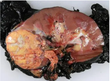
Figure 4: Picture of a Clear cell renal cell carcinoma
The clear cell renal cell carcinoma is typically golden yellow due to the rich lipid content of its cells; cholesterol, neutral lipids, and phospholipids are abundant. Tumor extension can be observed along the renal vein. Picture taken from Pathology of Clear Cell Carcinoma-emedicine.
3.2 Somatic Genetics
As previously described, the majority of ccRCC does not have an origin in the von Hippel Lindau disease. Nonetheless, deletions in the short arm of the chromosome 3 leading to partial or total inactivation of the VHL gene has shown to be a constant in the sporadic clear cell Renal Carcinoma (Brouch 2010) (Foster 1994) (Gnarra 1994). In the von Hippel Lindau disease, mutation in the tumor suppressor gene is inherited and the malignant kidney cancer arises due to a second sporadic mutation in the wild type allele gene. In the case of sporadic ccRCC, the mutations occur on the same cell type after somatic inactivation of both alleles of the VHL gene by allelic deletion, mutation, or epigenetic silencing in 70% or more of the cases. (Gnarra 1994) (Knudson 1985) (Herman 1994) (Latif 1993) (Schram 2002). These data suggest that the VHL gene is most likely a tumor suppressor gene in sporadic clear cell RCC.
22
3.3 Kidney cancer stages, treatment and prognosis
At first basis, the type of treatment that will be applied to a patient with RCC is determined by the probability of cure, that in most cases is directly related on how early the patient is diagnosed, the state of the tumor and the degree of tumor dissemination (American Cancer Facts 2015; Sachdeva 2016; RCC is mostly a silenced disease, probably because kidneys are localized deep in the body and there may not be any symptoms until the tumor size is large, only 10% of patients will approach their doctor due to classic triad of flank pain, hematuria, and flank mass.
Chart N°1 reveals the different sizes and stages of RCC. Chart N°2 describes the 5 years survival prognosis. More than 50% of patients with early stage renal cell carcinoma are cured, but according to the Cancer Center treatments of America and the Surveillance, Epidemiology and End Results (SEER) database of the National Cancer Institute in USA, the 5 years survival outcome for stage IV disease is poor (around 10% of patients).
23 Figure 5: Kidney cancer stages and 5 years survival rates.
RCC is often an asymptomatic disease in early stages. Often, patients are diagnosed in an advanced stage of the disease, greatly decreasing their chances
of survival. Picture taken from http://www.aboutcancer.com/renal_survival_section_0808.htm
24 Chart 1: Percent of cases & 5-year relative survival by stage at diagnosis in kidney and renal pelvis cancer. Taken from the Surveillance, Epidemiology and End Results (SEER) database of the National Cancer Institute (USA).
Chart 2: Patients survival rate in patients with distant metastatic cancer according to Cancer Center treatments of America and the Surveillance, Epidemiology and End Results (SEER) database of the National Cancer Institute (USA).
25
4. TREATMENT OF METASTATIC RCC
Surgery
Partial nephrectomy has been so far the first line of treatment for organ-confined ccRCC (tumors smaller than 4 cm), and in some cases a total nephrectomy can be applied in larger tumors. In the last years there have been an increased interest in including studies that will evaluate not only the appearance of local recurrence after surgery but also the way metastasis respond to drug therapies in presence or absence of the primary tumor (Coppin 2011; Lams 2006).
4.2 Adjuvants therapies
Adjuvants therapies are the treatments that are given in addition to the primary or initial therapy to maximize their effectiveness. Usually, in different types of cancer, the adjuvant is the treatment given after surgery where the detectable disease has been removed but when there is still risk of relapse due to non-detectable residual disease (Pal 2014; National Cancer Institute: Dictionary of cancer terms).
In this section I will discuss the therapies that are used in a monotherapy or as an adjuvant in the systemic treatment of mRCC.
• Immunotherapies
A) Interferon α (IFN α)
Interferon α was the first cytokine therapy approved for the systemic treatment of mRCC. This natural glycoprotein expressed by leukocytes, stimulates natural killer cells (NK), decreases cell proliferation through the inhibition of cyclin kinases, increases immunogenicity of tumor cells and inhibits angiogenesis (Fossa 2001; Goldstein 1988; Coppin 2000; Oliver 1989).
26 • IFN-α as monotherapy
As reviewed by (Canil 2010) although IFN-α had a promising start in the treatment of advanced or metastatic clear cell carcinoma, comparison analyses of 8 randomized trials in patients with inoperable renal cell carcinoma, have proven that there is limit in the response rate (up to 20%) in patients treated with IFN-α versus control (placebo).
Moreover, there is a high toxicity of this treatment including lack of appetite (51%), tiredness (68%), nausea/vomiting (26%/9%), lack of energy (65%), dry mouth (41%), shivering (23%) and depressed mood (25%) after 4 weeks of treatment with IFN-α. (Medical Research Council Renal Cancer Collaborators1999) has led to limit the use of this treatment or to try several combinations with others agents as first or second-line therapy for metastatic renal cell carcinoma. This last subject will be later discussed.
• IFN-α as adjuvant treatment
Surgery in the case of mRCC has been subject of several studies. As showed by Flanigan 2001 the median overall survival of patients that were treated with IFN-α after cytoreductive nephrectomy went up to 11.1 months survival compared to the 8.1 months when treated with IFN-α only. This was also proved in another study by (Mickisch 2001) where they observed and increase of median overall survival up to 17 months when there was a treatment with IFN-α compared to the 7 months of patients that were treated with α-interferon only. This suggest that IFN-α should be considered for the treatment of patients with high risk of relapse after surgery, although this is not possible in all the cases due to the high toxicity associated with this agent.
B) Interleukin-2 (IL-2)
IL-2 is a T-cell growth factor and activator of T cells, monocytes, macrophages and natural killer (NK) cells. IL-2 affects tumor growth by activating lymphoid cells in vivo without affecting tumor proliferation directly.
27 • Il-2 as monotherapy
Several studies performed by (Law 1995 , Rosemberg 1993) have shown that high-dose of IL-2 can induce durable long term remission but in a very small portion of patients (1 of 36 and 4 of 48 patients respectively). In the study performed by (Fisher 1997) and colleagues a median of response duration for all partial responses was 20 months. As well, 20% of patients were estimated to be alive, 5 to 10 years following treatment.
The main limitation in this treatment was that serious adverse effects occur. Toxic effects associated with high-dose of IL-2 are related to increased vascular permeability and secondary cytokine secretion (eg, IL-1, interferon gamma, tumor necrosis factor, nitric oxide). The management of high-dose toxicities requires in patient monitoring, often in an intensive care unit. (Rosemberg 1993; Law 1995; Fisher 1997). All these studies had mix number of patients that had gone under nephrectomy so they cannot be evaluated as either monotherapy or surgery adjuvant therapies.
• Il-2 as adjuvant treatment
The role of IL-2 after nephrectomy, remains controversial (Belledgrun 2000 and Fligin 1997). Studies showed that the combination of nephrectomy with high-doses of IL-2 significantly improves survival outcomes in patients with metastatic renal cell carcinoma.
In contrast, the results obtained by Clark 2013 showed no difference in the overall survival (OS) after one cycle of IL-2 high-dose when added as adjuvant in patients with locally advanced and metastatic disease that had been completely resected.
• Targeted Therapies
Overall, the systemic therapy of advanced RCC has not been satisfactory since, as discussed before, cytokine treatments have shown limited efficacy as well as high toxicity.
Since then, the better understanding of the signaling pathways involved in the development of ccRCC has led to the development of novel therapies that target specifically the dysregulated pathways inside the tumor cell and/or the cells that are in the tumor environment.
28 As described before, the inactivation of the VHL gene leads to the accumulation of the transcription factor HIF, over-expression of vascular endothelial growth factor (VEGF) and platelet-derived growth factor (PDGF) (Wiessener 2001; Cookman 2000; lliopoulos 1996). These proteins are known to promote angiogenesis, a key process in tumor development and progression (Takahashi 1994).
As well, the mammalian target of rapamycin (mTOR), a protein kinase, is in the upstream pathway regulating not only HIF but also other factors as angiogenesis, cell proliferation, metabolism that are critical for the pathogenesis of mRCC (Hudson 2002).
29 Figure 6: Dysregulated pathways of ccRCC and targeted therapies mechanism of action.
The inactive form of VHL in the ccRCC leads to stabilization and accumulation of the hypoxia inducible factor HIFα. This accumulation can also result as a consequence of the activation of mTOR and the PI3-K/AKT pathway due to cell stimuli. Increased levels of HIF induce activation of hypoxia inducible genes as for example vascular growth factor (VEGF), platelet-derived growth factor (PDGF), GLUT1, that are implicated in several processes as neovascularization, glucose uptake, and others cell growth survival processes. Temsirolimus and Everolimus inhibit the kinase activity of the mTOR complex 1 (mTORC1); Bevacizumab is a VEGF ligand-binding antibody; sunitinib, sorafenib, axitinib, and pazopanib are small molecule inhibitors of multiple tyrosine kinase receptors including VEGF-R and PDGFR (Picture taken from Shuch 2015).
30 During the last 8 years, targeted therapies against these different dysregulated pathways have been developed and tested in different trials either alone or in combination (figure N°6).
Note: Definitions to understand trials outcomes (According to National Cancer institute dictionary)
Progression Free survival (PFS): The length of time during and after the treatment of a disease, such as cancer, that a patient lives with the disease but he does not get worse. In a clinical trial, measuring the PFS is one way to evaluate how a new treatment works.
Overall Survival (OS): The length of time from either the date of diagnosis or the start of treatment for a disease, that patients diagnosed with the disease are still alive. In a clinical trial, measuring the overall survival is one way to see how well a new treatment works. Hazard ratio (HR): A measure of how often a particular event happens in one group compared to another group, over time. In cancer research, hazard ratios are often used in clinical trials to measure survival at any point in time in a group of patients who have been given a specific treatment compared to a control group with another treatment or a placebo. A hazard ratio of one means that there is no difference in survival between the two groups. A hazard ratio of greater than one or less than one means that survival was better in one of the groups.
Objective response rate (ORR): Objective response means either a partial or complete response. Rate is expressed in percentage of each case.
A) VEGF/VEGF Receptor (VEGFR) inhibitors
• Bevacizumab
Bevacizumab (BEV) is a humanized monoclonal antibody that interacts with circulating VEGF and blocks it’s binding to its receptor (VEGFR) thus inhibiting the angiogenesis pathways implicated in ccRCC (Presta 1997).
This novel targeted agent was tested in ccRCC in a randomized phase II trial with intravenous treatment every 2 week with 10 mg/kg of BEV. Results showed that, compared to the
31 control (placebo), the PFS was extended in median 4.8 vs 2.5 months. However the response rate was low (10%) (Yang 2003).
More recently, the AVOREN trial (Escudier 2007) and the CALGB 90206 trial (Rini 2008) compared the BEV + IFNα versus IFNα alone. The objective response rate (28.4 vs 12.9% for BEV + IFNα vs IFNα or IFNα + placebo), and the progression free survival (10.2 months vs 5.4 months for BEV + IFNα vs IFNα or IFNα + placebo) were substantially improved when there was a combination of BEV+ INFα versus the INFα alone or in combination with placebo.
In 2009, the FDA approved the use of BEV+ INFα for the treatment of advanced RCC. (FDA) • Sunitinib
Sunitinib is an orally administered multi-kinase tyrosine kinase that blocks VEGFR-1, -2, platelet derived growth factor receptor-α and β (PDGFR-α/β), and related tyrosine kinases (RTKs) (Roskoski 2007).
The pivotal phase 3 clinical trial NCT83889 (Motzer 2007) including patients with previously untreated mRCC and mainly favourable or intermediate prognostic risk, showed that median progression free survival (PFS) was significantly longer with Sunitinib than IFNα (11 months versus 5 months). A higher objective response rate (ORR) was also reported with Sunitinib versus IFNa (independent review, 39% vs 8%; p < 0.000001). The OS gave a very small statistical significance (P= 0.051). Further analysis revealed that the Sunitinib’s group of patients that did not received any treatment after the trial, extended their overall survival compared to the INFα group (28.1 months versus 14 months) (Motzer 2009) . Sunitinib is today, a standard of care in the first line treatment of mRCC.
• Sorafenib
Sorafenib is another multi-kinase inhibitor targeting VEGFR-1, VEGFR-2, VEGFR-3, PDGFR-α and –β and Receptor Tyrosine Kinase (RET) although it was found originally as a Ras-Raf-MEK-ERK pathway inhibitor (Wilhelm 2004 ; Carlomagno 2006).
32 As the first studies to determine its efficacy, showed little difference between the PFS reported for Sorafenib alone (5.7 months) versus IFNα alone (5.6 months), Sorafenib was not approved as a first line of treatment.
In another study that compared groups of patients treated with INF-α or Sorafenib alone, where after disease progression both were switch to sorafenib (dose escalation in the second case), authors observed tumor size reduction, better quality of life, and improved tolerability (Escudier 2007). Results showed better outcome in these patients that were cytokine refractory with an improved PFS as compared with that of placebo (5.5 versus 2.8 months) (Escudier 2009).
In conclusion, Sorafenib is recommended to be use in cytokine-refractory patients with advanced or metastatic renal cell carcinoma, although there are very high toxicities associated. Some of them are diarrhea (48%), rash and desquamation (41%), alopecia (31%), hand-foot syndrome (33%), and fatigue (29%) (Escudier 2009).
• Pazopanib
Pazopanib is a multi-kinase inhibitor of VEGFR-1, -2, and -3, PDGFR-α and -β, and c-Kit (Sonpavde 2007).
Its activity over mRCC was demonstrated in a phase III randomized trial (Sopavde 2008). In this study, two arms were examined: a population of patients that were naïve for any other treatment versus placebo and another arm with a group who had pre-cytokine-based treatment versus the naïve group treated with Pazopanib. In the treatment-naïve subpopulation, the PFS for Pazopanib was 11.1 months versus the 2.8 months with placebo. In the case of the cytokine-pre-treated subpopulation, the PFS was of 7.4 months compared to the 4.2 months of placebo.
The toxicity of Pazopanib is similar to, if not less severe than, that of similar molecules such as Sunitinib and Sorafenib, becoming a promising alternative treatment against mRCC.
• Axitinib
Axinitib emerged as an alternative for patients who had failed in the response to prior systemic therapy. This VEGFR-1, VEGFR-2, and VEGFR-3 tyrosine kinase inhibitor, has been
33 shown to be 450 times more potent to target these receptors than the first generation of VEGF inhibitors.
The AXIS trial corresponding to therapies in patients with mRCC refractory to Sunitinib plus Sorafenib cytokines and Sorafenib (group 2; n = 29), or Sorafenib alone (group 3; n = 15), showed a benefit of 2 months in the PFS compared to Sorafenib (6.7 months versus 4.7 months), as well, the ORR survival was increased. For groups with mRCC refractory to Sunitinib and Sorafenib, the ORRs were 7% with PFS of 7.1 months, whereas, in patients with mRCC refractory to cytokines, the ORR was 28% with PFS of 9 months and in those refractory to Sorafenib the ORRs were 28% with PFS of 7.7 months. Toxicity and adverse events included fatigue (13%), hypertension (11%), and hand–foot syndrome (11%) (Cella 2013; Motzer 2013; Escudier 2014; Rini 2011; Ueda 2013). These results establish Axitinib as a second-line treatment option for patients with metastatic renal cell carcinoma.
B) mTOR inhibitors
• Temsilorimus
Temsilorimus is an inhibitor of the mammalian target rapamycin mTOR. Treatment of cancer cells with this agent induces cell cycle arrest and inhibition of angiogenesis through regulation of VEGF that is under the regulation of the HIF pathway (Hudson 2002; Faivre 2006).
Its efficacy was investigated in a randomized phase-III study (Hudes 2007) that included patients that had poor prognosis factors according to the Memorial Sloan-Kettering Cancer Center (MSKCC) prognostic criteria (Motzer 2002). The comparison was done between three arms: The first one, treatment with only IFN α; the second one, treatment with Temsilorimus and the third one, the combination of both. The results of ORR did not differ significantly between arms: INFα (4.8%), Temsirolimus (8.6%), and combination therapy (8.1%). PFS showed a benefit of 2 months for the second group (Temsilorimus alone, compared to the first group of INF-α alone). OS was higher as well in the second group (10.9 versus 7.3 months). However, when patients on the combination regimen were compared with the interferon-α group, PFS and OS were similar, being 4.7 and 8.4 months, respectively.
34 Nevertheless, there are high toxicities associated to this treatment. In 11% of the patients the most common grade 3 and 4 side effects are asthenia and hyperglycemia.
Based on positive survival and PFS results, Temsirolimus is recognised in recent guidelines as a first-line treatment option for patients with mRCC who have poor MSKCC prognostic factors.
• Everolimus
Compared to Temsilorimus, this mTOR inhibitor is administrated orally and has a different active form. In RECORD-1 trial (Motzer 2008), patients with metastatic renal cell carcinoma, which had progressed on Sunitinib, Sorafenib, or both, were randomly assigned into a group treated with Everolimus versus a group treated with placebo. The outcome of this study showed no difference in the median OS between groups at the end of double-blind analysis. PFS was evaluated in an unblinded analysis, since at disease progression patients with placebo were offered to turn into Everolimus. Median PFS was significantly improved on this late group compared to the placebo group (4.9 versus 1.9 months). Lack of increased OS was likely the result of the crossover design of the study (Motzer 2010).
Toxicities associated to this treatment are mostly of grade 3 and are observed in 15% of patients. This toxicities, stomatitis (44%), infections (37%), asthenia (33%), fatigue (31%), diarrhea (30%), rash (29%), nausea (26%), anorexia (25%), peripheral edema (25%), vomiting (24%), thrombocytopenia (23%), hypercholesterolemia (77%), hypertriglyceridemia (73%), and hyperglycemia (57%) are comparable to those induced by the Temsilorimus treatment (Hudson 2007). In addition, Everolimus-induced pneumonitis is a side effect in 4% of patients (Motzer 2010).
Based on the data from this trial, Everolimus is now the recommended therapy in patients who have progressed on prior VEGF-targeted therapy.
C) MET inhibitors
• Cabozantinib
Cabozantinib has arise as a novel alternative and second line treatment for patients that have failed to VEGFR therapy. This inhibitor of tyrosine kinases including MET, VEGFR, and
35 AXL has shown to improve the median overall survival (21,4 months) compared to everolimus (16,5 months).
Cabozantinib treatment also resulted in improved progression-free survival (7.4 months in patients treated with cabozantinib compared to 3,8 months in patients treated with everolimus ) . The most frequent type of adverse effects were grade 3 or 4. hypertension (49 [15%] in the cabozantinib group vs 12 [4%] in the everolimus group), diarrhoea (43 [13%] vs 7 [2%]), fatigue (36 [11%] vs 24 [7%]), palmar-plantar erythrodysaesthesia syndrome (27 [8%] vs 3 [1%]), anaemia (19 [6%] vs 53 [17%]) , between others. (Choueiri 2016, Grassi 2016)
Based on these results, cabozantinib has been approved for the treatment of mRCC as a second line therapy.
4.3 Sequential and Combinational therapy
Often, mRCC become recurrent due to the acquisition of resistance to the first line treatment. Sequential therapy, allows the treatment of this cancer as a chronic disease. Table N°2 summarises some of the several efforts that have been done to establish the best strategy of sequencing targeted therapies after cytokine pre-treated disease progression. The main treatments for mRCC are molecular target therapies focused in two axes: The VEGF based therapies and the mTOR inhibitor therapies.
The current paradigm in mRCC treatment is sequential monotherapy where patients are treated with first line therapy in naïve patients until the disease progress and then a second line therapy is applied. As discussed before, VEGF-based therapies are the most standard first treatment for mRCC, although complete response and durable treatments are not frequent. Resistance to VEGF-TKIs appears around 6 to 11 months after treatment (Motzer 2007; Escudier 2007 ).
Combinational therapies
Numerous combinations of cytokines, with or without other systemic therapies, have been tried, but no regimen has been found to be consistently superior. In addition, it appears that
36 only a relatively small proportion of highly selected patients with mRCC benefit from cytokine therapy.
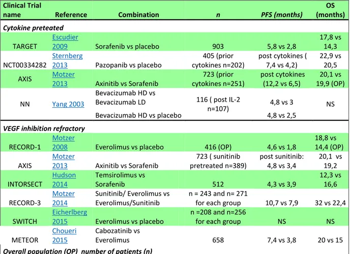
In the next two tables (Table N°2 and Table N°3), I will resume the several trials that have been performed to obtain the best systematic and combinational therapies in cytokine and VEGF inhibitor refractory patients.
37
Clinical Trial
name Reference Combination n PFS (months) (months) OS
Cytokine preteated
TARGET Escudier 2009 Sorafenib vs placebo 903 5,8 vs 2,8 17,8 vs 14,3 NCT00334282 Sternberg 2013 Pazopanib vs placebo cytokines n=202) 405 (prior post cytokines ( 7,4 vs 4,2) 22,9 vs 20,5 AXIS Motzer 2013 Axinitib vs Sorafenib cytokines n=251) 723 (prior post cytokines (12,2 vs 6,5) 19,9 (OP) 20,1 vs
NN Yang 2003
Bevacizumab HD vs
Bevacizumab LD 116 ( post IL-2
n=107) 4,8 vs 3 NS
Bevacizumab HD vs placebo 4,8 vs 2,5
VEGF inhibition refractory
RECORD-1 Motzer 2008 Everolimus vs placebo 416 (OP) 4,6 vs 1,8 18,8 vs 14,4 (OP) AXIS Motzer 2013 Axinitib vs Sorafenib pretreated n=389) 723 ( sunitinib post sunitinib: 4,8 vs 3,4 20,1 vs 19,2 INTORSECT Hudson 2014 Temsirolimus vs Sorafenib 512 4,3 vs 3,9 12,3 vs 16,6 RECORD-3 Motzer 2014 Sunitinib/ Everolimus vs Everolimus/Sunitinib n = 243 and n= 271 for each group 10,7 vs 7,9 32 vs 22,4
SWITCH Eicherlberg 2015 Everolimus vs placebo n =208 and n=256 for each group NS NS METEOR Choueri 2015 Cabozatinib vs Everolimus 658 7,4 vs 3,8 20 vs 15
Overall population (OP) number of patients (n)
Table 2: Retrieved phase 2 and 3 studies from systematic research in the cytokine- refractory and in the post-vascular endothelial growth factor inhibition setting. PFS: Progression Free survival, OS: Overall survival, n: number of patients. Table adapted from (Albiges 2015)
38
Clinical Trial
name Reference Combination n RR% months) PFS (
INF based
AVOREN Escudier 2007 IFN and Bevacizumab vs IFN placebo 306/289 31/13 10,2/5,4
Escudier 2010 IFN and Bevacizumab vs IFN 366/349 26/13 8,5/5,2
ARCC Hudes 2007 IFN vs Temsilorimus alone 207/209 4,8/8,6 3,1/5,5
IFN vs IFN and Temsilorimus 207/210 4,8/8,1 3,1/4,7
NN Jonasch 2010 Sorafenib and IFN vs Sorafenib alone 40/40 25/30 7,6/7,4 IL-2 based
ROSORC Procopio 2011 Sorafenib and IL-2 vs Sorafenib 66/62 27/15 7,6/6,9 Bevacizumab based
TORAVA
Bevacizumab and Erlotinib vs Bevacizumab and placebo 53/50 14/13 9,9/8,5
Négrier 2011 Bevacizumab and Temsilorimus vs Sunitinib 88/42 27/29 8,2/8,2
Bevacizumab and Temsilorimus vs Bevacizumab and INF 88/41 27/46 8,2/16,8
INTORAC Rini 2013 Bevacizumab and Temsilorimus vs Bevacizumab and INF 400/391 27/27,4 9,1/9,3
RECORD-2 Ravaud 2012 Bevacizumab and Everolimus vs Bevacizumab and INF 182/183 N/A 9,3/10
BEST
Bevacizumab vs Bevacizumab and Temsilorimus 89/91 12/28 8,7/7,3
McDermonTT
2013 Bevacizumab vs Bevacizumab and Sorafenib 89/90 12/30 8,7/11,3
Bevacizumab vs Sorafenib and Temsilorimus 89/91 12/27 8,7/7,7 Sorafenib based
NCT00467025 Rini 2012
Sorafenib and AMG 386 ( 3mg/kg) vs or
(10mg/kg) 51/50 37/38 8,5/9
Sorafenib and AMG386 (10 mg/kg) vs
placebo 51/51 37/25 8,5/9
Table 3: Retrieved studies from systematic research of cytokine-based combination and targeted therapy–based combination PFS: Progression Free survival, OS: Overall survival, n: number of patients, RR: response rate Albiges 2015
.
39
5. MECHANISM OF ACQUIRED RESISTANCE IN THE
TREATMENT OF ccRCC
5
Resistance to Tyrosine Kinase Inhibitors
According to the Response Evaluation Criteria in Solid Tumors or RECIST criteria, resistance is defined as a progression of the disease despite treatment application. The progression itself corresponds to at least 20% increase in the sum of the diameters of target lesions, taking as reference the smallest sum on study. As well the appearance of new lesions is considered as progression (Rini 2009; Eisenhuaer 2009).
The inhibitory effect of a certain targeted agent relies on how much the tumor is dependent on the particular pathway that is inhibited, to survive. So, resistance comes when the tumor becomes independent from the activity of drug-targeted pathway.
This independence can have two origins:
1. An intrinsic resistance, due to genetic alterations that pre-exist inside the tumor and that don’t allow proper drug binding as a consequence of the lack of expression of crucial proteins.
2. An acquired resistance, defined as progression of the disease after the patients had have a benefit from a first treatment. This progression is due to the activation of alternative pathways or increased expression of specific molecules as an answer to the inhibition, this could be defined as a compensation (Rosa 2013).
Key players in signaling pathways are tyrosine protein kinases that can be divided in two major groups: Receptor Tyrosine kinases that have a crucial role in the transduction of extracellular signals into the cell, and the non-receptor tyrosine kinases that are implicated in intracellular communications. Tyrosine kinases targeted antiangiogenic inhibitors as sunitinib, sorafenib, pazopanib and axitinib, were actually designed as multi-target inhibitors (targeting both VEGF and PDGF pathways) since their effects could be broader than a single-targeted inhibitor (Gotink 2010). Unfortunatly during the last years, there has been several reports of acquired resistance to these anti-angiogenic tyrosine kinase inhibitors (Motzer
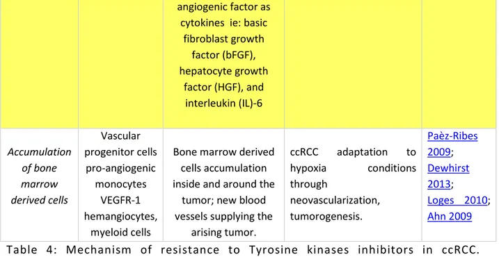
40 2007; Escudier 2007; Karashima 2013). The response to anti-angiogenic targeted therapy can activate several mechanisms to induce resistance as described in the next Table N°4 and represented in Figure N°7 and reviewed in Giuliano 2013).
41
Process name Cell type involved Mechanism of resistance Results References EMT (Epithelial to Mesenchymal transition) Epithelial cells Protein accumulation take place after long
exposure of cells to extra-cellular stimuli.
Polarized cells convert into motile mesenchymal cells, allowing escape from their biological structure. Β- catenin translocates to the nucleus activating the Wnt canonical pathway. Matrix
Metalloproteinases (MPPs) are upregulated
and decrease cell-cell interaction. Harada2012; Hugo2007; Klymkoswky 2009; Chaffer 2011; Chuang 2008 EMT induced CSCs (Cancer Stem cells) Separated population
Inducers of EMT such as TGF-β, Wnt or Notch
cause cells to acquire cellular markers of
CSCs.
EMT induces cell to transform into CSCs associated with reversible
epigenetic changes. Then these cells will form a new
tumor population (metastasis) with chemo
refractory features. Huang 2013; Hollier 2009; Singh 2010 TAM ( Tumor associated macrohages) Tumor associated macrophages
M2 are highly present in infiltrate ccRCC. TAMs triggers EMT through regulation of
TLR4/IL-10, Versican V1, TGF-β1, Gas6/Ax1,
miR488, FoxQ1 and NFκBIL-10. Increases
overproduction of PDGF, TGFβ , VEGF.
Through increase of proangiogenic factors, there is increase vessel formation and tumor adaption to hypoxia. EMT is increase. Patients having TAMs’high levels in their serum have poor prognosis outcome Ikemoto 2003; Zhang 2015; Santoni 2013; Dannennman 2013 Process name Cell type involved Mechanism of resistance Results References
42
Lysosomal Sequestration
ccRCC cells sunitinib is hydrophobic, weak base drug and thus is sequestered in acidic
lysosomes via ion trapping mechanism.
Antiangiogenic inhibitors affect not only endothelial cells but also tumor cells, although this is dependent of the drug concentration. Inside the human or experimental tumors the
drug concentration correspond to the one that
can inhibit tumor cell proliferation. Drug concentration in serum are 10 times lower than inside the tumor. Continous exposure to sunitinib induces lysosomal sequestration in ccRCC cells, so, therapeutic
concentrations are not achieved. Gotink 2011; Logan 2013 Giuliano2015 Santoni 2013 Increase Pericyte coverage of tumor cells Perivascular cells/ vascular smooth muscle cells In tumoral conditions, endothelial cells activate pericytes that
induce newly highly differentiated vessels. Stabilizing process of abnormally complicated vascular system formation. Pericytes depletion leads to EMT associated with increase hypoxia conditions. Excessive angiogenesis of the ccRCC, more aggressive tumor type with increase number of
metastasis, poor prognosis. Ribatti 2011; Armulik 2011; Hall 2006; Cao 2013; Angiogenic switch Vascular cells Elevated expression of IL-8 (potent proangiogenic factor). Expression of independent VEGF ccRCC progression, neo-angiogenesis after tyrosine
kinase treatment.
Huang 2010; Mizukami 2005; Porta 2013
43
angiogenic factor as cytokines ie: basic
fibroblast growth factor (bFGF), hepatocyte growth factor (HGF), and interleukin (IL)-6 Accumulation of bone marrow derived cells Vascular progenitor cells pro-angiogenic monocytes VEGFR-1 hemangiocytes, myeloid cells
Bone marrow derived cells accumulation inside and around the
tumor; new blood vessels supplying the
arising tumor. ccRCC adaptation to hypoxia conditions through neovascularization, tumorogenesis. Paèz-Ribes 2009; Dewhirst 2013; Loges 2010; Ahn 2009
Table 4: Mechanism of resistance to Tyrosine kinases inhibitors in ccRCC. (Adapted from Bielecka 2014)
Figure 7: Mechanisms of resistance to anti-angiogenic targeted therapies.Several processes are activated after long exposure to inhibitors that induces hypoxia in the tumor. These responses can have a tumor intrinsic origin as for example
44 augmentation of pro-angiogenic factors, increased number or cells that have become resistant to chemo- and radiotherapy, increased selection of hypoxia-tolerant and pro-angiogenic cancer stem cells (CSCs), and enhanced invasion and migration properties of tumor cells. Host-related mechanisms of resistance: hypoxia increases recruitment of endothelial progenitor cells (EPCs) and of bone marrow–derived circulating cells (RBCCs) toward the tumor vasculature, where they induces endothelial differentiation by secretion of angiogenic cytokines. Tumor -associated macrophages (TAMs) can infiltrate the tumor and mediate resistance by secreting tumor-promoting, angiogenic, and lymphangiogenic cytokines. Cancer-associated fibroblasts (CAFs) support tumor growth and angiogenesis by secreting fibroblast growth factors (FGFs). Increased coverage with pericytes enhances resistance by stabilization of the new formed vessels. Picture taken from Sonjas 2010.
5.2 Resistance to mTOR inhibitors
The discovery of the molecular basis in the ccRCC has led to design more targeted therapies against it. As discussed before, the Hipoxia Inducing Factor HIF has a determinant role in the tumorigenesis as mediator of the angiogenesis due the mutation of the VHL tumor suppressor. Angiogenesis inhibitors prevent the formation of blood vessels rather than inhibiting proliferation of tumor cells (Dordevic 2009; Nicol 1997).
In the case of the mTOR pathway, its targeting become crucial since its activation leads to constitutive HIF -1α expression, indicating then that inhibiting this pathway could not only affect tumor angiogenesis but also cell proliferation as this pathway is hyperactivated both in epithelial and cancer cells. ( Salvadori 2012)
There are two main mTOR inhibitors that are now frequently used, Temsilorimus and Everolimus alone or in combination (see Treatment of RCC section), however they do not offer long lasting response and almost all patients show disease progression during the treatment. (Batelli 2012)
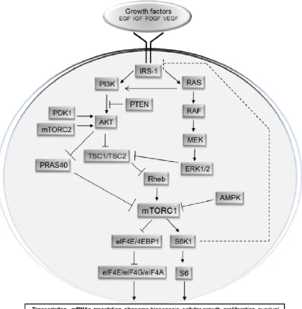
45 To better understand the mechanism of resistance to the mTOR, first we will take a look to Figure N°8, that represents the complexity of the mTOR pathway.
Figure 8: Scheme of the mTOR pathway
mTOR is involved in a complicated network of interactions linking growth factors receptors and intracellular regulatory factors in the process of angiogenesis, tumorigenesis and cellular metabolism. Picture taken from (Populo 2012)
46 It is useful to differentiate two basic signalling cascades leading to mTOR activation. In the first pathway, insulin binds to its Insulin receptor substrate (IRS1) inducing its activation. The signal is then transduced by the phosphatidylinositol 3-kinase (PI3K), which subsequently will activate phosphoinositide –dependent-kinase (PDK1) and then AKT (protein kinase B) (Laplante 2012; Taniguchi 2006; Wang 2006 ).
In a second pathway, after Ras activation, there will be a cascade of activation through Raf and the MEK 1 /2 to mitogen activated protein kinases (MAPK) and ribosomal s6 kinases (RSKs). Together these two signal cascades will increase phosphorylation of tuberin (TSC2) inactivating the harmatin-tuberin complex formed with TSC1 (Carrière 2008; Chang 2003). Inactivation of TSC1/TSC2 leads to mTORC1 activation, although another mechanism can also activate mTORC1 independently of TSC1/TSC2 (Laplante 2009, Wang 2007).
There are crosstalks between elements of PI3K/AKT, leading to activation of mTORC1 through activation of not only PDK1 but also itmTORC2 that phosphorylates and fully activates AKT. Indicating that interaction between mTOR complexes are bidirectional (Sarbassov 2005).
There are two main downstream targets of mTORC1 activity, the eukaryotic initiation factor 4E (IeF4E)-binding protein 1 (4E-BP1) and the ribosomal p70 S6 kinase 1 (S6K1). (Khaleghpour 1999) Stimulation of this last one, can lead to activation of HIF-1α and cell cycle regulators as c-myc and cyclin D1 (Choo 2008).
In ccRCC, VHL inactivation impedes HIF-1α and/or HIF-2α degradation. Upregulated levels of HIF’s subunits should inhibit mTORC1 through the upregulation of REDD1. However, this is not the case, since it was reported that mTORC1 is often activated in ccRCC (Rini 2009). There are several genes that are mutated in the PI3K signalling pathway regulating mTORC1 and its inhibition. Table N°6, represents some of the mutated genes and their implications in drug resistance.


![[18F]UCB-H RADIOTRACER AS A TOOL TO UNDERSTAND THE ROLE OF THE SV2A PROTEIN](data:image/gif;base64,R0lGODlhAQABAIAAAP///wAAACH5BAEAAAAALAAAAAABAAEAAAICRAEAOw==)