Disorientation in amnesia
A confusion of memory traces
Armin Schnider, Christine von Daniken and Klemens Gutbrod
Division of Neuropsychological Rehabilitation, UniversityDepartment of Neurology, Inselspital, Bern, Switzerland
Correspondence to: Dr med. Armin Schnider,
Neurologische Universitdtsklinik, Inselspital, CH-3010 Bern, Switzerland
Summary
Disorientation is a common phenomenon in delirium and amnesia. It is thought to have an obvious explanation, i.e. disoriented patients fail to store the information crucial for the maintenance of orientation. In this study, we explored whether disorientation was indeed associated with a failure to learn new information or rather with a confusion of information within memory. Twenty-one patients with severe amnesia were examined. Orientation was tested with a 20-item questionnaire. Two runs of a continuous recognition task were used to test the ability to acquire information (first run of the task) and the tendency to confuse the temporal context of information acquisition (comparison of the second with the first run). We found that orientation was much
better predicted by the measure of temporal context confusion (r = 0.90) than by the ability to simply acquire information (r = 0.54). Superimposition of neuroradiological scans demonstrated that increased temporal context confusion was associated with medial orbitofrontal or basal forebrain damage; patients with normal levels of temporal context confusion did not have damage to these areas. We conclude that disorientation more often indicates a confusion of memory traces from different events, i.e. increased temporal context confusion, than an inability to learn new information. Disorientation appears to reflect primarily a failure of the orbitofrontal contribution to memory.
Keywords: temporal order amnesia; disorientation; confusion; frontal lobes; orbitofrontal cortex; memory
Abbreviations: CVLT = California Verbal Learning test; IR = item recognition; TCC = temporal context confusion
Introduction
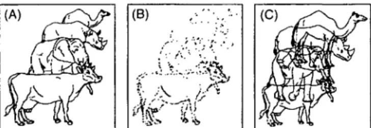
Disorientation to time, place and situation, rarely also to person, is a common finding in clinical practice. Disorientation is a regular component of acute confusional states (delirium) (Horenstein et at., 1967; Chedru and Geschwind, 1972; Mesulam et al., 1976; Devinsky et al., 1988) and is sometimes present in dementia (Cummings and Benson, 1992) and amnesia. The mechanism of disorientation appears to be obvious: clinical wisdom holds that disoriented subjects cannot store new information and therefore fail to continuously update their knowledge about time and the environment (Benton et al., 1964; High et al., 1990). However, there is an alternative possibility as schematized in Fig. 1: normal memory function demands not only that information has been stored but also that the temporal order among pieces of information is maintained (Fig. 1A). Disorientation might not only ensue from a failure to simply store information (Fig. IB) but also, and possibly more so, if a subject did store information, but confused the temporal sequence of information within memory (Fig. 1C) (Von © Oxford University Press 1996
Cramon and Saring, 1982; Baddeley and Hitch, 1993). This would make it difficult to realize what piece of stored information pertained to the present situation. In a recent study we found that.this type of memory failure sets spontaneously confabulating patients apart from other amnestic patients (Schnider et al., 1996ft).
(A) (B)
Fig. 1 Types of memory failure: schema illustrating (A) normal storage of both item and temporal sequence information in memory, (B) failure to retain new information in memory; and (C) confusion of the temporal sequence of information acquisition within memory despite storage of the information itself.
Patients and methods
Twenty-one amnestic patients hospitalized for neuro-psychological rehabilitation participated in the study. Aeti-ologies of amnesia were as follows: traumatic brain injury (n = 8); haemorrhage and surgery of an aneurysm of the anterior communicating artery (n = 5) or right posterior communicating artery (n = 2); herpes simplex encephalitis (n = 2); surgery of an invasive left olfactory meningioma (n = 1); Wernicke-Korsakoff syndrome (n = 1); right frontal haemorrhage (n = 1); left thalamic infarction (n = 1). All patients' amnesia was evident in everyday behaviour and was confirmed with several memory tests as documented in our previous study (Schnider et al., 1996i>). However, patient selection was based on performance in the California Verbal Learning test (CVLT; Delis et al, 1987). Because all patients finally judged as disoriented had a long delay free recall (of =£4 in the CVLT, only patients with similarly deficient recall were included. All patients were mobile on the ward throughout the day. Only patients who had been in our unit for at least 2 weeks were included to ensure that all patients had been living in a similar setting and had thus received a similar amount of information to help orientation. Patients were excluded if they had insufficient attention (digit span <5) or another cognitive deficit precluding participation in the experiment (e.g. aphasia, visual agnosia). The tests reported here were performed 75±55 (17-270) days after the occurrence of brain damage. Fifteen age- and education-matched controls with no history of neurological or psychi-atric illness (mostly family members of patients) were also tested. All subjects gave their informed consent to being tested.
Orientation
The orientation test described by Von Cramon and Saring (1982) was used. This is in the form of a questionnaire designed for German speaking subjects. It contains questions that are particularly appropriate for hospitalized patients and comprises five questions for each of four domains of orientation: (i) orientation to person: name, age, profession, citizenship, eye colour; (ii) orientation to place: city, name of building, unit or floor, approximate direction of home town, county; (iii) orientation to situation: reason for being here, type of treatment, sources of support, name of a person on the ward, party covering the costs of the sojourn; (iv) orientation to time: day of the week, date, month, year, time. A correctly oriented subject will give at least four correct answers for domains (i) to (iii) and at least three
Free recall ('Do you remember the words that I told you before?') requires both that the demanded information has been stored and that it can be retrieved from memory. All patients in this study failed to recall previously learned words. Pure information storage is better reflected by the ability to recognize previously learned information (Lezak, 1995). Since this study aimed to juxtapose the impact of failed storage of information and of increased confusion of the temporal context of information acquisition on disorientation, it was desirable to test temporal context confusion with a recognition task too, so that the two processes could be directly compared. The following experiment, which was more extensively discussed in a previous article (Schnider et al., 1996fo), tested both processes with two components of the same recognition task:
Run 1: item recognition (IR)
To test pure information storage, a continuous recognition task of design similar to the recognition tests of Sturm and Willmes (1995) for nonsense stimuli was composed with 120 meaningful, concrete drawings from Snodgrass and Vanderwart (1980). The picture series consisted of six series with 20 pictures each. Each series contained eight items that appeared in all six series (thus, they were repeated five times after initial presentation) and 12 distracter items that were not repeated in other series. Each picture was presented on a computer screen for 2 s. For each picture the subjects were requested to answer the question: 'Have you already seen precisely this picture in this run?'. Answers were recorded by the examiner pressing the appropriate response key and immediately followed by presentation of the next picture. According to Sturm and Willmes (1995), the item recognition score was calculated as: IR = hits - false positives. The maximum score was therefore 40 (40 hits, no false positives).
Run 2: temporal context confusion (TCC)
One hour after the recognition task (run 1), a second run was made with precisely the same design. For this run, target items were replaced so that eight distracter items from the first run now served as the target items, while the target items from the first run now ranked among the distracters. Subjects were instructed to 'forget that [they] had already taken a similar test before' and were requested to answer the question: 'Have you already seen precisely this picture in this run?' for each picture. The central idea behind the experiment was that false familiarity with a distracter item
40 30 20 10 0
Item recognition (IR)
•0.2 0 0.2 0.4 0.6 0.8 1 1.2 1.4
Temporal context confusion (TCC)
Fig. 2 Amnestic patients' association of total orientation score (Z-ORI) with (A) item recognition (IR)
and (B) the temporal context confusion (TCC). Open circles indicate patients with IR in the chance range; in these patients, TCC was not determined. The dashed horizontal lines separate patients with disorientation in at least one domain from normally oriented amnesties. The bars in the lower left corners indicate the controls' range of performance (maximal to minimal values) and the controls' mean.
Table 1 Association of orientation scores with item
recognition (IR) and temporal context confusion (TCC)
Variable IR TCC I-ORI TIME-ORI PLACE-ORI SIT-ORI 0.54* 0.38 0.57* 0.55* 0.90*** 0.78** 0.79** 0.84*** Association of orientation scores with IR and TCC. Numbers indicate second order polynomial regression coefficients. Orientation measures: I-ORI = total orientation score; TIME-ORI = time orientation score; PLACE-TIME-ORI = orientation to place score; SIT-ORI = orientation to situation score. Significance levels: *0.05 >P> 0.01; **0.01 **P> 0.0001; ***P =s 0.0001.
(i.e. a false positive response) was based on an inability to distinguish between the item's previous occurrence in the first rather than the second run (irrespective of whether it has been a target or a distracter in the first run), i.e. on temporal context confusion (TCC). Thus, TCC was defined as the relatiev increase of false positives in the second over the first run, i.e. TCC = (FP2/ Hits2) - (FP, / Hits,), where
FPj and FP2 = false positives in run 1 and 2, respectively;
HitS] and Hit2 = hits in run 1 and 2, respectively. Since this
experiment could measure temporal context confusion only if a subject was able to store information at all, run 2 was made only with subjects who had performed significantly above chance in run 1 (a" > 1.64, Brophy, 1986). Two patients with herpes simplex encephalitis and one patient with traumatic brain injury did not meet this criterion.
Lesion analysis
Most patients had several CT scans. An MRI was available for four patients. An attempt was made to account for the different lesion types: in patients with traumatic brain injury, all haemorrhagic lesions visible in the early scans (after removal of subdural or epidural haematomas in two patients)
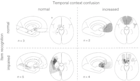
were taken into account because each parenchymal haemorrhage is likely to indicate an area of axonal damage (Eisenberg and Levin, 1989). Lesions were also taken into account if they were subsequently invisible in later scans. With all other aetiologies, the scan performed closest in time to our experiment was analysed to prevent overestimation of the lesion area due to perifocal oedema. Lesions were reconstructed with the templates of Damasio and Damasio (1989) and referred to a composite axial slice containing the hippocampus, amygdala and basal forebrain and to the midsagittal plane (see Fig. 3). The lesion areas were superimposed in a commercial drawing program. Four patients had CT and MRI scans with no visible focal brain lesion (clipping of an anterior communicating artery aneurysm, n = 1; Wernicke-Korsakoff syndrome, n = 1; traumatic brain injury, n = 2).
Results
Amnesties versus controls
Figure 2 shows the performances of the patients and controls. Item recognition discriminated much better between amnesties and controls (f(34) = 4.2, P = 0.0002; Fig. 2A) than TCC (r(31) = 1.9, P = 0.06; Fig. 2B). The IR and TCC scores were not significantly correlated (P > 0.05) either in the controls (r = -0.48) nor in the amnesties (r = -0.28).
Determinants of disorientation
Eleven patients were disoriented to time, place and situation, three patients were disoriented only to time, and seven patients were normally oriented. The following analysis was limited to the amnesties to determine the contribution of IR and TCC to disorientation. Because all patients were oriented to person, this domain of orientation was omitted from further analysis. Highest regression coefficients with orientation scores were obtained using second order polynomial
o
I
o CD 2 "§ — ^» CD n=2Fig. 3 Lesion reconstruction showing the projection of lesions to the midsagittal plane and a combined axial plane encompassing the hippocampus (H), the amygdala (A), and the orbitofrontal area (including basal forebrain, F) as indicated in the upper left design. Patients are separated according to whether their item recognition and temporal context confusion were within ('normal') or outside the controls' range ('impaired' IR, 'increased' TCC). In the sagittal plane, shaded areas indicate lesions close to the midline, empty polygons with dashed lines indicate lateral hemispheric lesions, 'n' indicates the number of patients in the respective group.
regression rather than simple regression. All domains of orientation were much better predicted by TCC than by IR (see Fig. 2 and Table 1).
The total number of correct answers (Z-ORI in Table 1) did not significantly correlate with the following parameters: days after brain injury (r = 0.23), age (r = -0.06), number of years at school (r = -0.08); or with several measures of frontal lobe function: verbal fluency (Thurstone and Thurstone, 1963), r = 0.29; figural fluency (Regard et al., 1982), r = -0.20; colour-word interference (Stroop, 1935), r = 0.14.
Lesion analysis
Patients were separated according to whether their IR and TCC were within or outside the range of the controls. By accepting this broad range of 'normality', classification in the 'impaired' group has a high specificity for true impairment while there is a risk that some patients with a true impairment would be classified as 'normal'. This was the case with three amnestic patients who scored in the 'normal' range on both IR and TCC. The three patients with chance IR scores were excluded from this analysis because their TCC was not determined. Patients with increased TCC and normal IR (n = 2) had medial orbitofrontal lesions sparing the basal forebrain (Fig. 3). Patients with impaired IR but normal TCC (n = 5) had diverse lesions sparing both the medial orbitofrontal cortex and the basal forebrain. Patients with
both impaired IR and increased TCC (n = 4) had lesions that mostly involved the basal forebrain.
Discussion
Our results indicate that disorientation in amnesia is based primarily on a confusion of information within memory rather than a lack of stored information. Although severe failure to store new information was associated with disorientation, increased temporal context confusion predicted disorientation much better (Table 1 and Fig. 2). Although correlations do not prove a causal link, our study strongly suggests such a link: first, this study prospectively tested the possibility that the two examined mechanisms of memory contributed to orientation, a possibility which was a priori reasonable; secondly, other measures of frontal lobe function did not correlate with orientation, indicating a specificity of temporal context confusion.
It has been surmised that disorientation after brain damage reflects anterograde and retrograde amnesia (Benton et al., 1964; High et al., 1990), i.e. an insufficient amount of information in memory to maintain orientation (Fig. IB). This explanation cannot account for the observation that disoriented patients' responses to questions of orientation may vary from one interview to another; the answers may be correct at one time and wrong at other times (Daniel et al, 1987). Our results suggest that the main problem of disoriented patients is a confusion of memory traces from
diverse events rather than a lack of information in memory. While a healthy person normally has a feeling for the recent flow of information and does not have any difficulty in realizing what acquired knowledge refers to the present (Fig. 1A), a disoriented patient may be unable to distinguish intuitively between knowledge acquired some minutes ago and knowledge acquired some days, months, or even years ago (Fig. 1C). The patient may therefore confuse the date, place and the reason of his being in a particular place and his responses may vary from one occasion to the other. Incorrect responses to questions of orientation may reflect a subject's problem in selecting the currently correct answer from memory rather than a lack of this knowledge.
This study was designed to seek a common mechanism of disorientation in amnesia and therefore included patients with diverse types of brain damage. Certain aetiologies of amnesia were not represented in our study group. Notwithstanding these caveats, our data suggest that increased temporal context confusion emanates from prefrontal, especially medial orbitofrontal, damage or disconnection. Conversely, a decreased capacity to store and subsequently recognize new information appears to have less anatomical specificity as it may result from lesions in diverse locations (Fig. 3). Basal forebrain lesions appear typically to produce a combination of these two types of memory failure. Our finding of a functional and anatomical dissociation between item recognition failure and temporal context confusion is in agreement with earlier studies on temporal order recognition (Squire, 1982; Milner et al., 1985; Hunkin and Parkin, 1993). Our study does not definitively determine whether increased temporal context confusion reflects a failure of the process of information storage or information retrieval. In our opinion, defective retrieval is unlikely because it would similarly affect recollection of recent and remote information. However, many disoriented patients and many patients with spontaneous confabulations—which are also accounted for by increased temporal context confusion (Schnider et al., 1996b)—readily give precise accounts of remote events (Schnider et al., 1996a). We suggest that increased temporal context confusion is mainly due to a specific defect of information storage, i.e. a failure to co-encode temporal order information. Temporal order information should not be conceived of as separate from item information but rather in terms of the saliency that recent information may attain in memory, as schematized in Fig. 1. This saliency, which distinguishes recent from remote information, may be determined by the behavioural relevance attributed to new information (Schnider et al., 1996a). Neurons in the orbitofrontal cortex have been shown to react specifically to stimuli of behavioural significance (Rosenkilde et al., 1981). We have previously suggested that defective temporal labelling of information, resulting in increased temporal context confusion, may result from damage of the circuit connecting the amygdala, the dorsomedial thalamic nucleus, and the orbitofrontal cortex, whereas failed retention of information in memory, resulting in defective item
recognition, may result from an interruption of the classic Papez circuit, i.e. the circuit connecting the hippocampus with the anterior thalamic nucleus (Schnider et al., 1996a). Both circuits have multiple, spatially close connections in the anteromedial thalamus (e.g. mamillo-thalamic tract, ventral amygdalo-fugal pathways) and basal forebrain (e.g. septum verum, ventral striatum); a lesion in this area may thus interrupt either limbic circuit and produce either type of memory failure.
Ackowledgement
We wish to thank Dr E. Markus for her support. This study was supported financially by the Swiss National Science Foundation (Grant number 32-40 432.94).
References
Baddeley AD, Hitch G. The recency effect: implicit learning with explicit retrieval? [Review]. Mem Cognit 1993; 21: 146-55. Benton AL, Van Allen MW, Fogel ML. Temporal orientation in cerebral disease. J Nerv Ment Dis 1964; 139: 110-9.
Brophy AL. Alternatives to a table of criterion values in signal detection theory. Behav Res Methods Instrum Computers 1986; 18: 285-6.
Chedru F, Geschwind N. Disorders of higher cortical functions in acute confusional states. Cortex 1972; 8: 395-411.
Cummings JL, Benson DF. Dementia. A clinical approach. 2nd ed. London: Butterworth-Heinemann, 1992.
Damasio H, Damasio AR. Lesion analysis in neuropsychology. New York: Oxford University Press, 1989.
Daniel WF, Crovitz HF, Weiner RD. Neuropsychological aspects of disorientation. Cortex 1987; 23: 169-87.
Delis DC, Kramer JH, Kaplan E, Ober BA. The California Verbal Learning Test. New York: Psychological Corporation, 1987. Devinsky O, Bear D, Volpe BT. Confusional states following posterior cerebral artery infarction. Arch Neural 1988; 45: 160-3. Eisenberg HM, Levin HS. Computed tomography and magnetic resonance imaging in mild to moderate head injury. In: Levin HS, Eisenberg HM, Benton AL, editors. Mild head injury. New York: Oxford University Press, 1989: 133-41.
High WM Jr, Levin HS, Gary HE Jr. Recovery of orientation following closed-head injury. J Clin Exp Neuropsychol 1990; 12: 703-14.
Horenstein S, Chamberlain W, Conomy J. Infarctions of the fusiform and calcarine regions: agitated delirium and hemianopia. Trans Am Neurol Assoc 1967; 92: 85-9.
Hunkin NM, Parkin AJ. Recency judgements in Wemicke-Korsakoff and post-encephalitic amnesia: influences of proactive interference and retention interval. Cortex 1993; 29: 485-99.
Lezak MD. Neuropsychological assessment. 3rd ed. New York: Oxford University Press, 1995.
Rosenkilde CE, Bauer RH, Fuster JM. Single cell activity in ventral prefrontal cortex of behaving monkeys. Brain Res 1981; 209: 375-94.
Schnider A, Gutbrod K, Hess CW, Schroth G. Memory without context. Amnesia with confabulations following infarction of the right capsular genu. J Neurol Neurosurg and Psychiatry 1996a; 61:
186-93.
Schnider A, Gutbrod K, von Daniken C. The mechanisms of spontaneous and provoked confabulations. Brain 1996b; 119: 1365-75.
Snodgrass JG, Vanderwart M. A standardized set of 260 pictures:
Sturm W, Willmes K. NVLT bzw. VLT - Nonverbaler und Verbaler Lerntest. Modling: Dr. G. Schuhfried GmbH, 1995.
Thurstone LL, Thurstone TG. Chicago Test of Primary Mental Abilities. Chicago: Research Associates, 1963.
Von Cramon D, Saring W. Storung der Orientierung beim hirnorganischen Psychosyndrom. In: Bente D, Coper H, Kanowski S, editors. Hirnorganische Psychosyndrome im Alter. Berlin: Springer,
1982: 38^19.
Received March 7, 1996. Revised May 3, 1996. Accepted May 13, 1996


