HAL Id: hal-02555961
https://hal.archives-ouvertes.fr/hal-02555961
Submitted on 27 Apr 2020
HAL is a multi-disciplinary open access
archive for the deposit and dissemination of sci-entific research documents, whether they are pub-lished or not. The documents may come from teaching and research institutions in France or abroad, or from public or private research centers.
L’archive ouverte pluridisciplinaire HAL, est destinée au dépôt et à la diffusion de documents scientifiques de niveau recherche, publiés ou non, émanant des établissements d’enseignement et de recherche français ou étrangers, des laboratoires publics ou privés.
on connectional architecture atlas.
Haixiang Wang, Aileen Mcgonigal, Kai Zhang, Qiang Guo, Bingqing Zhang,
Xiu Wang, Xiao Wang, Jiuluan Lin, Xiancheng Song, Qian Feng, et al.
To cite this version:
Haixiang Wang, Aileen Mcgonigal, Kai Zhang, Qiang Guo, Bingqing Zhang, et al.. Semiologic sub-groups of insulo-opercular seizures based on connectional architecture atlas.. Epilepsia, Wiley, 2020, Epub ahead of print. �10.1111/epi.16501�. �hal-02555961�
1
DOI: 10.1111/epi.16501
Semiologic subgroups of insulo-opercular seizures based on
connectional architecture atlas
Haixiang Wang
1, Aileen McGonigal
2,3, Kai Zhang
4, Qiang Guo
5, Bingqing Zhang
1,
Xiu Wang
5, Xiao Wang
6, Jiuluan Lin
1, Xiancheng Song
1, Qian Feng
1, Siyu Wang
1,
Mengyang Wang
6, Xiaoqiu Shao
7, Xiaoyan Liu
8, Liang Wang
9,10, Wenjing Zhou
11Epilepsy Center, Tsinghua University Yuquan Hospital, Beijing, China 2Aix Marseille University, INSERM, INS, Inst Neurosci Syst, Marseille, France 3Clinical Neurophysiology and Epileptology Department, Timone Hospital, APHM, Marseille, France
4Department of Neurosurgery, Beijing Tiantan Hospital, Capital Medical University, Beijing, China
5Epilepsy Center, Guangdong Sanjiu Brain Hospital, Guangzhou, China
6Department of Neurology, Beijing Sanbo Brain Hospital, Capital Medical University, Beijing, China
7Department of Neurology, Beijing Tiantan Hospital, Capital Medical University, Beijing, China
8Department of Pediatrics, Peking University First Hospital, Beijing, China 9CAS Key Laboratory of Mental Health, Institute of Psychology, Beijing, China 10Department of Psychology, University of Chinese Academy of Sciences, Beijing, China
Correspondence
Wenjing Zhou, Epilepsy Center, Tsinghua University Yuquan Hospital, No.5, Shijingshan Road, Beijing, China. Email: [email protected] Liang Wang, CAS Key Laboratory of Mental Health, Institute of Psychology, No.16, Lincui Road, Beijing, China. Email: [email protected]
Abstract
Objective: Insulo-opercular seizures are characterized by diverse semiology, related
to the insula's multiple functional roles and extensive connectivity. We aimed to iden-tify semiologic subgroups and correlate these with insulo-opercular subregions based on connectional architecture.
Methods: We retrospectively collected a large series of 37 patients with
insulo-opercular seizures explored by stereoelectroencephalography (SEEG) from three epilepsy centers. A new human brain atlas (Brainnetome Atlas, BNA) based on both anatomic and functional connections was employed to segment insulo-opercular cor-tex. Semiology and SEEG changes were carefully reviewed and quantified. Principal component analysis and cluster analysis were used to correlate semiologic character-istics with insulo-opercular subregions.
Results: Four main semiologic subgroups were identified, organized along an
anter-oventral to posterodorsal axis based on BNA. Group 1 was characterized by epigastric sensation and/or integrated gestural motor behaviors with or without feelings of fear or rage, involving the anteroventral insular regions and mesial temporal lobes. Group 2 was characterized by auditory sensations and symmetric proximal/axial tonic signs involving the posteroventral temporal operculum. The characteristics of group 3 were orofacial and laryngeal signs, involving the intermediate insulo-opercular regions. The features of group 4 were somatosensory signs followed by nonintegrated gestural motor behaviors and/or asymmetric tonic signs involving the posterodorsal insulo-opercular regions with propagation to the mesial frontal lobes. Thus anteroventral seizure organizations predominantly showed limbic system semiology, whereas more posterodorsal regions were associated with semiology involving mainly the sensori-motor system. Subjective symptoms proved to be particularly discriminating factors.
Significance: Insulo-opercular seizures can be categorized in terms of clinical
semiol-ogy and correlate with connectional architecture subregions along an anteroventral-pos-terodorsal axis in line with the cytoarchitectonic gradient rather than the gyral anatomy
1
|
INTRODUCTION
The insular lobe is a deeply located structure covered by dense vasculature and the frontal-parietotemporal opercula. Stereoelectroencephalography (SEEG) is a well-suited ap-proach that provides direct access by combining lateral orthogonal trajectories through opercula, with an oblique ap-proach through frontal and/or parietal cortices.1A variety of subjective and objective signs have been recorded through SEEG insular explorations, including viscerosensory, soma-tosensory, and auditory sensations; autonomic symptoms; gestural motor behaviors; and elementary motor signs.2-6 The diversity of insular seizures reflects distinctive propagation pathways including perisylvian, temporal, and frontal pat-terns7,8; as such, insular seizures are great mimickers of sei-zures originating from elsewhere in the brain.9 On the other hand, seizures arising from other lobes also often involve insulo-opercular cortex, such as temporal-plus epilepsy.10 Consequently, the diversity of clinical semiology and epilep-togenic networks often leads to failure of diagnosis and surgi-cal treatment in insulo-opercular seizures.
Insulo-opercular seizures became a subject of study and clinical interest through pioneering works using SEEG, no-tably from the Lyon group.4,5 However, since their first de-scriptions, a main difficulty in achieving an overview of the spectrum of clinical semiology has been lack of large published series, previous studies having included as few 6 patients5 and the largest series to date being based on 22 patients.11Thus, although several previous studies involv-ing insulo-opercular seizures exist, these either have fewer cases,5,12 do not investigate semiologic features,11,13,14 or do not focus on spontaneous seizures.15,16 In addition, previous studies have not addressed the potential role of specific in-sulo-opercular functional subregions in different semiologic and propagation patterns. The insula can be divided into seven cytoarchitectonic subdivisions with a posterodorsal to anteroventral gradient of hypergranular to granular, dys-granular, and agranular fields.17 In line with this cytoarchi-tectonic gradient, a structural anteroposterior connectivity gradient without any distinct borders was shown using dif-fusion-weighted imaging (DWI).7,18 Functional connec-tivity studies also reported two19 or four20 distinct regions within the insula along a ventral-anterior to dorsal-posterior gradient. Hence, we postulated that semiologic features of
insulo-opercular seizures might correlate with connectional architecture subregions.
The human Brainnetome Atlas (BNA), with 210 corti-cal and 36 subcorticorti-cal subregions, provides a fine-grained, cross-validated atlas and contains information on both an-atomic and functional connections.21 In this atlas, insular cortex is subdivided into six subregions that closely relate to previous functional and histologic maps of the insular cor-tex.17,20,22 Thus, it provides a very appropriate atlas for in-sulo-opercular subregion localization based on connectional architecture rather than gyral anatomy.
The present study collected a large series of patients with seizures arising from the insulo-opercular cortex determined by SEEG. Quantified semiologic characteristics and SEEG changes were examined using principal component analysis and cluster analysis to correlate the distinctive semiologies with insulo-opercular subregions.
2
|
PATIENTS AND METHODS
2.1
|
Patient selection and SEEG recording
We collected patients with drug-resistant focal epilepsy who underwent SEEG in three epilepsy centers (Tsinghua
Funding information
Natural Science Foundation of China, Grant/Award Number: 31771255, Z171100000117014 and XDB32010300; Beijing Municipal Science and Technology Commission; Strategic Priority Research Program of Chinese Academy of Science
of the insula cortex. This provides new insights into facilitating differential diagnosis and presurgical localization but also highlights the importance of considering connec-tional architecture in determining neural correlates of complex semiologic patterns.
K E Y W O R D S
Brainnetome Atlas, cluster analysis, insular seizure, semiology, stereoelectroencephalography
Key Points
• Four main semiologic subgroups of insulo-oper-cular seizures were identified by using principal component analysis and cluster analysis.
• These groups organized along an anteroventral to posterodorsal axis based on a novel parcellation method, the Brainnetome Atlas.
• Semiology, particularly subjective symptoms, sig-nificantly correlated with insulo-opercular subre-gion localization.
• Connectional architecture segmentation rather than gyral anatomy may be important in deter-mining neural correlates of complex semiologic patterns.
University Yuquan Hospital, Beijing Tiantan Hospital, and Guangdong Sanjiu Brain Hospital) from July 2014 to June 2018. We included 37 patients with seizures arising from in-sula ± opercular cortex. The seizure-onset zone was defined from SEEG recordings based on the first clear ictal electri-cal change, providing: (a) that this change occurred prior to the clinical onset of the seizure, and (b) that it manifested by a fast synchronizing discharge (low-voltage fast activ-ity or recruiting fast discharge of spikes).23We excluded pa-tients whose SEEG recording was inconclusive or in whom seizures arose from adjacent cortex and secondarily spread to insulo-opercular cortex. This study was approved by the Tsinghua University Yuquan Hospital Human Ethics Review Committee (No. 2016005) and informed consent was signed by the patients or their guardians.
All patients underwent comprehensive evaluation, includ-ing 3T magnetic resonance imaginclud-ing (MRI), and long-term scalp video EEG, and SEEG recordings. The SEEG explora-tions were performed by using intracerebral multiple contact electrodes (HKHS Healthcare Co., Ltd, Beijing, China; 8-16 contacts, length: 2 mm, diameter: 0.8 mm, 1.5 mm apart) that were placed following the guide of Sinovation Robot (Sino Precision Medical Technology Co., Ltd, Beijing, China) or the Leksell Stereotactic System (Elekta, Stockholm, Sweden). For detailed methods, please see Wang et al.24 Video-SEEG (256 channels Nihon Kohden digital EEG system, Nihon Kohden, China) was recorded for 1-3 weeks in order to record patients’ habitual seizures. Signals were recorded with a bandpass filter between 0.08 and 600 Hz and with a sampling rate of 2000 Hz. Interictal and ictal SEEG changes were analyzed using a bipo-lar montage between adjacent contacts of the electrode, with the reference electrode chosen in the white matter.
2.2
|
Automatic localization and cortical
labeling of depth intracranial electrodes in
Brainnetome Atlas
We have developed an integrative toolbox "FIELD" based on the MATLAB software platform to efficiently and accu-rately reconstructed and labeled the intracranial electrodes implanted in patients with drug-resistant epilepsy.25 In this toolbox, the electrode coordinates were normalized to a standard Montreal Neurological Institute (MNI) space and the probabilistic value of each contact belonging to a speci-fied area or functional network was also provided. The MNI coordinates were projected onto the BNA to obtain accurate localization of all electrode contacts for each patient.
The human Brainnetome Atlas was used for insulo-oper-cular parcellation. Thus, insular cortex was divided into six functional subregions rather than anatomic gyral parcellation. Subregions of insular cortex according to the BNA included INS1 (G, hypergranular insula), INS2 (vIa, ventral agranular
insula), INS3 (dIa, dorsal agranular insula), INS4 (vId/vIg, ventral dysgranular and granular insula), INS5 (dIg, dorsal granular insula), and INS6 (dId, dorsal dysgranular insula). The opercular cortex was defined as consisting of four subre-gions created from this atlas: OpF (A44v, A44op and A45c); OpC (A4tl); OpP (A1/2/3tonla, A40rv); and OpT (A41/42, TE1.0/TE2.0). In addition, mesial frontal lobe (supplemen-tary motor area, SMA; pre-SMA; anterior/middle cingulate gyrus; and orbitofrontal cortex) and mesial temporal lobe closely connected to insulo-opercular cortex were included.
2.3
|
Analysis of
anatomic-electroclinical features
Ictal clinical and electrical modifications were analyzed in each patient and each seizure following the method devel-oped by a previous study of frontal lobe seizures.26 Video SEEG–recorded clinical semiology and electrical data were independently analyzed by three epileptologists (H. Wang, B. Zhang, and Q. Feng).
All recorded seizures were reviewed, and the presence or absence of 14 ictal signs observed in this series was noted. An overall semiologic score was established for each pa-tient with values ranging from 0 (=absence of that sign), to a maximum of 2 for major features (=constant and early sign present in each seizure), with minor features being scored as 1 (=sign not always present). With regard to semiologic de-scription, the terms that were best able to categorize seizures according to different anatomic involvement were selected, and nondiscriminating signs were removed. Semiologic cat-egories included those motor semiologies described in the previous frontal seizures study.26In addition, multiple sen-sory symptoms were listed because of the known repertoire of subjective clinical responses observed during insular and opercular stimulation.16,27In contrast, consciousness was pre-served during the majority of insulo-opercular seizures, and as such the presence or absence of this sign did not corre-spond to differences in anatomic localisation.5 Therefore, the term “impairment of consciousness” was excluded. Orofacial signs included elementary motor and/or sensory signs, such as facial tonic or clonic signs, contraction of mouth corner, and/or tongue or facial anesthesia. We did not count within this group oroalimentary automatisms such as lip smacking or swallowing.
We also estimated the level of participation of each re-gion in initial seizure organization. The rere-gions not involved by SEEG ictal activity were scored as 0, whereas those im-mediately involved at electrical onset were scored as 2. The intermediate score 1 was used when SEEG ictal activity was seen later. For further detail of this method, the reader is re-ferred to Bonini et al.26 Notably, not only regions involved at seizure onset but also regions involved by early spread during
the period of appearance of clinical signs were evaluated and scored.
Two matrices encompassing 14 (clinical signs) and 12 (brain areas) variables, scored 0-1-2 for each of the 37 pa-tients, were thus obtained.
2.4
|
Statistical analysis
The process of cluster analysis and principal component analysis was similar to the previous study.26 We summarized the process steps as below. Two dissimilarity matrices were computed based on the two matrices (signs × patients and areas × patients), which were entered into hierarchical clus-ter analysis in MATLAB. Clusclus-ter analysis was performed to allow ordering of variables. The proximity of variables within the dendrogram represented their degree of similar-ity. Ordered sequences of ictal signs and of brain areas were thus obtained, arranged according to the frequency of their co-occurrence. A Kendall correlation matrix was then per-formed to demonstrate the relationship between ictal signs and involved areas, and to measure the strength of this as-sociation. The correlation test was finally used to assess the significance at P < .05 for each correlation.
Then, principal component analysis (PCA) was per-formed in order to convert all possibly correlated variables (signs) into a smaller number of linearly uncorrelated vari-ables (principal components). Consequently, the size of the transformed data was reduced, and the first three compo-nents could explain the variance in the data. Finally, patient position within the new lower-dimensional space supplied by PCA was computed, as defined by ictal signs used as coor-dinates. Using these new coordinates, hierarchical clustering
was performed, aiming to distinguish clinically homoge-neous groups of patients.
3
|
RESULTS
3.1
|
General characteristics
We included 37 patients (17 male, 20 female) with seizures arising from insula ± opercular cortex. Mean age at record-ing was 18 ± 7.9 years and mean epilepsy duration was 9.7 ± 7 years. Cerebral magnetic resonance imaging (MRI) was negative for 22 patients (59.5%). Subsequent to SEEG exploration, all patients underwent tailored resections that included the insulo-opercular cortex and parts of anterior temporal lobe and mesial temporal structures in four patients. The main postsurgical complication was transient hemiple-gia (12/37, 32.4%), especially for middle-posterior insular resection. However, occurrence of persistent hemiplegia was low (2/37, 5.4%). Of patients with at least 1-year follow-up (n = 31), 24 were in International League Against Epilepsy (ILAE)28 class 1(77.4%), 4 patients were in class 2, and 3 patients were in class 3. In terms of histopathology, 27 (73%) had focal cortical dysplasia, 5 (13.5%) had gliosis, and 1 (2.7%) had tuberous sclerosis. The other 4 patients showed nonspecific histopathological change.
A combination of lateral orthogonal SEEG trajectories through the frontoparietal and temporal opercula with an oblique approach through the frontal or parietal cortices was performed to allow larger insular sampling.1,4 A total of 372 electrodes was implanted, with an average number (mean ±SD) per patient of 10.3 ± 2.6 and an average of 5.2 ± 1.5 elec-trodes sampling insulo-opercular cortex. Electrode placement
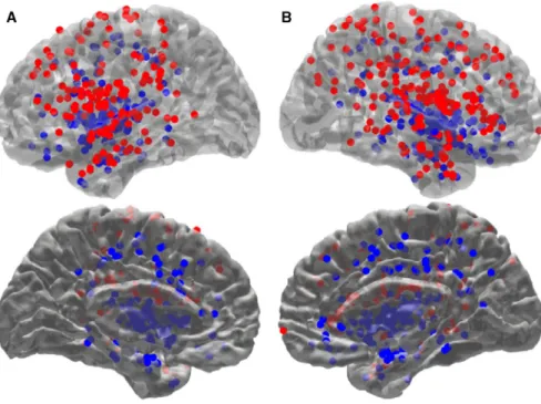
FIGURE 1 Electrode position in standard MNI space. Entry (red dots) and target (blue dots) points obtained from each electrode for each patient (n = 37) are displayed in the left (A) and right (B) hemisphere. Top and bottom rows show lateral and medial views with different transparencies for visual illustration, respectively
including numbers of electrodes sampling insula and opercu-lum subregions for each patient is listed in Table 1. Electrode position in standard MNI space is shown in Figure 1.
Seizures were frequently nocturnal (23/37, 62.2%) and often occurred daily (29/37, 78.4%). Almost all patients
(34/37, 91.9%) had seizures with preserved awareness at the beginning. Hence, 32 patients (86.5%) reported clear subjec-tive signs including somatosensory (10/32), auditory sensation (10/32), laryngeal sensation (8/32), and autonomic sensa-tion (6/32). However, no patient had vestibular or olfactory
TABLE 1 Regions sampled by SEEG implantation for each patient
Pts SEEG explored Opercular electrodes/number Insular electrodes/number SOZ
1 L:INS, Op, T OpF/2; OpC/1; OpP/2; OpT/1 INS1/2; INS5/1; INS6/1 INS5 2 R:INS, Op, P, C, CG OpP/2 INS1/1 OpP 3 R:INS, Op, C, T OpF/2; OpC/1; OpP/1; OpT/2 INS1/1; INS5/1; INS4/1; INS2/1 INS1 + OpP 4 R:INS, Op, F, CG OpF/1; OpC/1; OpP/2; OpT/1 INS1/2; INS5/1; INS6/1; INS4/2; INS3/1 INS1 + OpP 5 R:INS, Op, F, CG, C, T OpF/1; OpC/1; OpP/3; OpT/1 INS1/1; INS5/1; INS6/1; INS4/1; INS3/1 INS5 6 R:INS, Op, F, CG OpF/2; OpC/1; OpP/2 INS1/1; INS5/1; INS3/1 INS1 + OpP 7 L:INS, Op, F, C, T OpF/1; OpC/1; OpP/2 INS1/2; INS5/2; INS6/3; INS2/1; INS3/1 INS5 + OpC 8 L:INS, Op, F, C, CG OpF/1; OpC/1; OpP/2 INS1/2; INS5/1; INS6/2; INS3/1; INS4/2 INS1 + OpP 9 Bilateral: INS, Op; L: T OpC/1; OpP/3; OpT/1 INS1/3; INS5/3; INS6/1; INS4/1 INS5 10 L:INS, Op, F, CG; R: F OpF/1; OpC/1; OpP/1; OpT/1 INS1/1; INS5/1; INS6/2 OpT 11 R:INS, Op, T, CG, P OpF/1; OpC/1; OpP/1 INS1/1; INS5/2; INS4/1 INS5 + OpC 12 Bilateral: INS, Op; L: F, CG OpF/2; OpC/2; OpP/3 INS5/3; INS6/2; INS2/1; INS3/1 OpC 13 Bilateral: INS, Op; L: T OpC/1; OpP/3; OpT/3 INS1/4; INS5/1; INS4/3; INS2/1; INS6/3 OpT 14 R:INS, Op, T, P OpF/2; OpT/2; OpP/2 INS1/2; INS6/1; INS4/1; INS3/1 OpT + INS1 15 R:INS, Op, T, F OpC/1; OpP/2; OpT/3 INS4/1; INS5/1 OpT 16 L:INS, Op, T OpF/1; OpC/1; OpT/2; OpP/2 INS1/2; INS5/1; INS6/1; INS2/1; INS3/1 INS5 + INS6 17 R:INS, Op, F, C, CG OpF/1; OpC/2; OpP/2 INS5/1; INS6/1 OpC 18 R:INS, Op, F OpF/2; OpC/2; INS5/1; INS6/2; INS3/1 INS5 + INS6 19 R:INS, Op, C, F OpF/1; OpC/2; OpP/1 INS5/2; INS6/1; INS3/1 INS5 + INS6 20 L:INS, Op, T OpF/1; OpC/1; OpP/2; OpT/2 INS1/1; INS5/2; INS4/1; INS2/1; INS3/1 INS2 21 R:INS, Op, T, F, CG OpF/2; OpT/1 INS2/1; INS3/1; INS4/1 INS2 + INS3 22 R:INS, Op, F, T OpF/1; OpC/1; OpP/1 INS5/2; INS6/1; INS3/1; INS4/1; INS2/1 INS5 + OpC 23 R:INS, Op, F, T OpF/1; OpC/1 INS2/1; INS3/1; INS4/1; INS6/1 INS2 + INS3 24 L:INS, Op, C, P OpF/1; OpC/1; OpP/1; OpT/2 INS1/2; INS5/1; INS6/1; INS4/1 INS1 25 R:INS, Op, T, C OpC/1; OpT/1 INS1/1; INS5/1; INS2/1; INS4/1 OpT 26 L:INS, Op, T OpT/2 INS1/1; INS2/1; INS4/2 OpT + INS1 27 Bilateral: INS, Op, T; R:CG OpF/1; OpC/1; OpP/1; OpT/1 INS5/2; INS6/1; INS4/2; INS3/2 INS5 + INS4 28 L:INS, Op, F, CG OpF/1; OpC/1 INS2/1; INS3/1; INS6/1 INS2 + INS3 29 L:INS, Op, C, F, P, CG OpF/1; OpC/1; OpP/1; OpT/1 INS1/1; INS5/1; INS3/1; INS6/1; INS4/1 INS5 30 R:INS, Op, F OpF/2; OpC/1; OpT/1 INS1/1; INS5/1; INS3/1; INS6/1; INS4/1 INS5 + OpC 31 Bilateral: INS, Op, F, T OpF/2; OpC/1; OpT/1 INS2/1; INS3/2; INS6/2; INS4/1 INS2 + INS4 32 L:INS, Op, F, T OpF/1; OpC/1 INS5/1; INS6/1 INS5 + INS6 33 L:INS, Op, F, T OpF/2; OpC/1; OpT/1 INS2/1; INS3/1; INS6/1; INS5/1 OpF 34 R:INS, Op, F, C OpF/1; OpC/1 INS5/1; INS6/1 INS5 35 R:INS, Op, T OpF/1; OpC/1; OpP/1; OpT/1 INS1/1; INS5/1; INS6/1; INS4/1 INS5 + OpC 36 R:INS, Op, C, CG, F, P OpC/1; OpP/1; OpT/1 INS1/2; INS5/1; INS6/1 OpP 37 R:INS, Op, T, P, CG OpC/1; OpT/1 INS1/1; INS5/2; INS6/1 INS5 Abbreviations: C, central lobe; CG, cingulate gyrus; F, frontal lobe; INS, insula; INS1-6, subregion of insular cortex; L, left; Op, operculum; OpF/C/P/T, frontal/ central/parietal temporal operculum; P, parietal lobe; R, right; SOZ, seizure-onset zone; T, temporal lobe.
symptoms. Only six patients (16.2%) manifested focal to bi-lateral tonic-clonic seizures. Four patients had reflex seizures, one being eating-evoked and three auditory-evoked. Main se-miologic features for each patient are described in Table S1.
3.2
|
Cluster analysis and
anatomic-electroclinical correlations
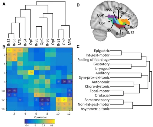
Ictal signs and brain regions classified with hierarchical cluster analysis are shown in Figure 2. These clusters indicate that ictal signs in one group are most frequently present together during seizures. Cluster analysis of the brain regions allows grouping together of anterior ventral regions (INS2, INS3, INS4), poste-rior ventral regions(OpT), intermediate regions (INS5, INS6, OpC, OpF) and posterior dorsal regions (INS1, OpP). Smaller clusters of insulo-opercular subregions grouped together at lower cluster levels also show strong associations: INS2 and INS3; INS5 and OpC; and INS1 and OpP (Figure 2A).
Matrix correlation between clinical signs and involved regions shows a diagonal pattern of correlation (Figure 2B).
Yellow-orange squares indicate positive correlation and blue squares indicate negative correlation. At the top-left corner, epigastric sensations and integrated motor behaviors are correlated with subregions located in the more anteroven-tral regions (INS2 and INS3). At the bottom-right corner, somatosensory, non-integrated gestural motor behaviors and asymmetric tonic signs are correlated with the more postero-dorsal subregions. Therefore, the matrix forms an antero-ventral-posterodorsal gradient according to the occurrence of clinical signs. Such a diagonal structure is conserved for significant correlation (P < .05; Figure 2B, starred squares). Matrix correlation also demonstrates that some clinical signs seem to be more specific to certain subregions: Auditory sensations are correlated only with OpT and somatosensory aura is correlated only with posterodorsal regions. However, some clinical signs such as autonomic signs and focal motor signs are more widely distributed and have weaker localiza-tion value.
Hierarchical classification of patients results in clusters of clinically homogeneous patients. A cut was chosen that resulted in four groups (Figures S1 and S2). Clustered groups
FIGURE 2 Correlation matrix between cortical areas and clinical features, as ordered by hierarchical clustering basing on their reciprocal distance. A, Clustering of brain regions is shown on the horizontal axis. B, Correlation between areas and signs as a function of the color (orange, positive correlation; blue, negative correlation; starred squares, P < .05) follows a diagonal pattern of association running from anteroventral (top left corner) to more posterodorsal regions (bottom right corner). C, Clustering of clinical signs is shown on the vertical axis. In these two ordered sequences, neighboring regions as well as neighboring signs occur more frequently together than distant ones. The gray star shown in the squares denotes a significant Kendall correlation (P < .05). D, Subdivision of insulo-opercular areas according to the Brainnetome Atlas.21 Each area is color-coded for visual illumination. INS1-6, subregion of insular cortex; MTL, mesial temporal lobe;OpT, temporal operculum; OpC, central operculum; OpF, frontal operculum; OpP, parietal operculum; MFL, mesial frontal lobe; Sym-pro-axi-tonic, Symmetric proximal/axial tonic posture; Int-gest-motor, Integrated gestural motor behaviors; Non-int-gest-motor, Non-integrated gestural motor behaviors
for clinical signs and regions for the four groups of patients obtained with hierarchical clustering performed on new PCA coordinates were shown (Figure 3). The involved brain areas were analyzed as a function of the resulting homogeneous groups of patients and the characteristics of the early spread networks obtained for each group (Figure 4).
Group 1 (four patients) is characterized by epigastric
sen-sation and integrated gestural motor behaviors and/or feel-ings of fear/rage. Gustatory sensations could also occur in these patients. Moreover, this group is characterized by the absence of focal motor and orofacial signs (Figure 3, group 1). Significant involvement of INS2, INS3, and INS4, as well as mesial temporal lobes occurred in this group.
Group 2 (nine patients), is characterized by auditory
sensations and symmetric proximal/axial tonic seizures. In a similar way to group 1, this group is characterized by the ab-sence of focal motor and orofacial signs (Figure 3, group 2). Significant involvement (value-test >1) of OpT occurred in this group. INS4 could be involved in this group because of adjacent location, but the value-test was <0.5.
Group 3, the largest group, was composed of 17 patients.
They manifested elementary orofacial signs and laryngeal signs (Figure 5). Autonomic signs and focal motor signs often appeared in this group compared with the other three groups (Figure 3, group 3). Involved regions included INS5, INS6, OpC, and OpF.
Characteristic semiologic features of Group 4 (7 patients) were somatosensory auras followed by nonintegrated gestural motor behaviors and/or asymmetric tonic signs (Figure 3, group 4). Autonomic signs never occurred and involved re-gions included INS1 and parietal operculum, with propaga-tion to mesial frontal lobe (SMA, pre-SMA, and anterior/ middle cingulate gyrus).
4
|
DISCUSSION
Here, we report semiologic features of 37 patients with insulo-opercular seizures, correlating electroclinical sub-groups with insulo-opercular subregions. To our knowl-edge, this represents the largest reported series to date. Despite the diverse symptoms and complex anatomy of insulo-opercular cortex, our results allowed identification of electroclinical subgroups according to similarity of se-miology by means of PCA and cluster analysis. Moreover, using a novel approach based on the Brainnetome Atlas, which takes into account both structural and functional connectivity, we demonstrate that these subgroups were organized along an anteroventral-posterodorsal axis, rather than according to anatomic gyri of insula cortex as found in previous studies.12,29
FIGURE 3 Clustered groups for clinical signs (A) and regions (B) for the four groups of patients obtained with hierarchical clustering performed on new PCA coordinates. Full names for clinical signs and regions refer to Figure 2
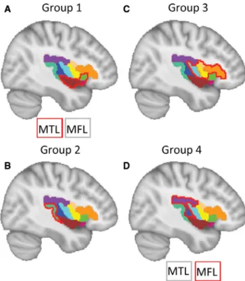
FIGURE 4 Cortical regions that characterize the four groups of patients. Brain areas significantly involved in one group than the others are outlined in red. The MTL and MFL that cannot be shown in the brain due to mesial regions are listed beside. The color-coded areas refer to Figure 1D. MTL, mesial temporal lobe; MFL, mesial frontal lobe
4.1
|
Electroclinical subgroups of
insulo-opercular seizures
Cluster analysis was selected as a well-suited method to identify similarities across the heterogeneous subjects. The clustered groups were organized along an anteroventral to posterodorsal axis. Anteroventral seizure organizations pre-dominantly showed limbic system semiology, whereas more posterodorsal regions were associated with semiology mainly involving the sensorimotor system.
In group 1, seizures were organized within anteroventral insular regions including INS2, INS3, and INS4, character-ized by epigastric sensations, integrated gestural motor behav-iors, as well as feelings of fear and rage. These seizures shared features also seen in mesial temporal lobe seizures (MTLS) and prefrontal seizures. The mesial temporal lobes showed significant involvement in the present study, but on the other hand, orbitofrontal and anterior cingulate regions were not necessarily involved, even in fear-related behaviors. The next more posterior ventral group, which significantly involved temporal operculum, was characterized by auditory sensations and symmetric proximal/axial tonic signs. The auditory fea-tures can be readily understood by the fact that Heschl gyrus is buried within the lateral sulcus. Auditory sensations are also obtained in response to stimulation of the posterior insula, and
especially in the midposterior long gyrus of the insula.16 INS4 corresponding to ventral posterior insula could be involved in this group. However, according to the lower value-test statis-tic, INS4 was not classified within group 2. Group 3 was a large and intermediate one in the vicinity of insular central sulcus characterized by elementary orofacial signs and laryn-geal signs. Orofacial signs included asymmetric facial tonic or tonic-clonic signs and unilateral eyelid blinking. These signs seem to reflect frontocentral operculum involvement. Laryngeal signs included uncomfortable subjective sensations ranging from constriction to strangulation, often associated with vocalization. In accordance with results from electrical stimulations of the human insula, throat constrictive sensa-tions were obtained from contacts located around, or rostral to, the central insular sulcus.16 In previous reports of visceral sensations elicited by cortical electrical stimulations, oropha-ryngeal sensations were most related to the opercular part of the precentral and postcentral gyri and the insula, whereas other digestive sensations including epigastric sensations and retrosternal sensations, were preferentially related to the temporal lobe and the anterior cingulate cortex, respec-tively.30 Therefore, the laryngeal symptoms strongly pointed to the involvement of dorsal central insula and frontocentral operculum categorized here as group 3. In group 4, seizures arising from posterodorsal insulo-opercular regions were
FIGURE 5 Anatomic-electroclinical features of a patient from group 3. A, Semiology is characterized by laryngeal discomfort followed by laryngeal constriction with open mouth, and right facial and arm tonic signs. B, Ictal stereoelectroencephalography (SEEG) shows rhythmic spikes evolving to a low-voltage rapid discharge at electrical onset in the intermediate insular region and central operculum. C, SEEG implantation scheme with electrode labels on a three-dimensional (3D) reconstruction of the brain; electrodes A, B, C are oblique, and the others are orthogonal. D, Sagittal view of patient's T1 MRI with implanted electrodes. Regions showing major involvement in the generation of the ictal discharge are colored in orange
typified by somatosensory auras followed by nonintegrated gestural motor behaviors and/or asymmetric tonic signs. Somatosensory auras were described as electrical paresthesia, anesthesia, pain, or burning sensations, which reflects the role of the parietal operculum in networks subserving basic and higher-order somatosensory processing.31
Nonintegrated gestural motor behaviors including hy-perkinetic movements without emotional content have been reported in seizures arising from premotor and lateral prefrontal regions26 and the temporal lobe,32 and also from operculo-insular and parietal lobes.33 This is proposed to reflect seizure spread to mesial frontal lobes including SMAs and cingulate gyrus.24,34 The pattern of hyperkinetic movements seen here in posterodorsal regions was in con-trast to the anteroventral group 1 in which gestural motor behaviors like grasping and groping were integrated and naturalistic, in keeping with the anteroposterior gradient seen in frontal lobe seizures.26 Nonintegrated behaviors in group 4 cases were frequently associated with tonic pos-ture, as also reported in frontal seizures.26 In the present series, these were asymmetric tonic signs, as has been de-scribed previously in insulo-opercular cases,12 particularly those arising from posterior insula.6
4.2
|
Anteroventral-posterodorsal axis based
on connectional architecture
Mesulam and Mufson proposed a cytoarchitectonic organiza-tion in the insula with three major domains: agranular, dys-granular, and dys-granular, concentrically arranged around the piriform olfactory cortex at the limen insula.35 Morel et al di-vided insular cortex into seven architectonic subdivisions (G, Ig, Id1-3, Ia1-2) with a posterodorsal to anteroventral gradient of hypergranular to granular. All the cytoarchitectonic and con-nectivity data reflect a functional and structural gradient within the insula from the anteroventral to the posterodorsal parts.
In the present study, we introduce the human Brainnetome Atlas, which contains information on both anatomic and func-tional connections.21 This atlas compares favorably to other parcellation methods.36 The insular cortex has been subdi-vided into six subregions in this atlas, which relate closely to previous functional and histological maps of the insular cor-tex. In line with the cytoarchitectonic and functional gradient, the axis from group 1 to group 4 corresponded to the gradient from agranular cortex in the anteroventral social-emotional region to dysgranular and granular cortex in the intermediate orofacial sensorimotor region to hypergranular cortex in the posterodorsal somatosensory and pain region.
Mazzola et al proposed a bipolar organization of major insular functions through direct cortical stimulations: a pos-terior part assigned to somatosensory functions, and notably to pain perception; and an anterior part assigned to visceral
sensitivity and visceromotor functions.37 This hypothesis is congruent with semiologic features seen in the present study. Furthermore, they proposed that the mid-dorsal insula (an in-termediate region in the present study) may be an integrated oral sensory region evoking gustatory, olfactory, and somato-sensory sensation.
4.3
|
Subjective symptoms of
insulo-opercular seizures
The insula is considered as a highly eloquent cortical struc-ture, since 80% of electrical stimulations elicited a clinical response in previous studies.37 Moreover, the insula is the only cortical region where stimulations can evoke such a multi-modal representation, perhaps supporting its integra-tive functions of polymodal inputs.37 Insular cortex stimu-lations through stereotactic electrodes can often reproduce ictal semiology, especially subjective symptoms. In the pre-sent study, several subjective symptoms such as epigastric, auditory and somatosensory sensations were indeed strongly discriminating clinical indicators for the identified groups. This localizing importance of subjective semiology in insulo-opercular seizures is in contrast to a previous study of frontal lobe seizures in which motor semiology was the key factor allowing clinical categorization.26 On the other hand, specific patterns of motor signs were also associated with certain lo-calizations in the present series.
4.4
|
Limitations
Our study has some limitations, the main one being that, as for any intracranial EEG method, SEEG sampling was nec-essarily limited to those sites deemed necessary for surgical pre-evaluation. As such, we cannot exclude the possibility that some nonexplored regions, especially those connected to insulo-opercular regions, had some involvement in initial seizure organization. On the other hand, the clear anatomic electroclinical correlations shown here and the favorable surgical results obtained in the vast majority of cases tend to reinforce the interest of these data. Although the current series represents the largest to date, as with any group-level analysis a larger data set would allow even more fine-grained anatomic electroclinical correlations.
5
|
CONCLUSIONS
Insulo-opercular seizures can be categorized in terms of clin-ical semiology and correlated with connectional architecture subregions along an anteroventral -posterodorsal axis, in line with the cytoarchitectonic gradient rather than gyral anatomy
of insula cortex. Review of a large series allows the full spectrum of semiologic features to be assessed. Subjective symptoms were the key indicators for clinical categoriza-tion, although certain motor patterns were also discriminat-ing. While epilepsy is a network disorder38-40 and frontal, prefrontal, temporal, or parietal seizures can share common semiologic features with insulo-opercular seizures, such semiologic categorization of insulo-opercular seizures pro-vides new insights in facilitating differential diagnosis and presurgical localization. Through use of a novel parcellation method, the Brainnetome Atlas, the present results also high-light the importance of considering connectional architecture when investigating neural correlates of complex semiologic patterns. Future work should take into account not only ana-tomic relations of structures involved in epileptic networks, but also dynamic aspects such as discharge frequency and coherence that contribute to semiologic expression.41-43
ACKNOWLEDGMENTS
The authors would like to appreciate Professor Lingzhong Fan(Institute of Automation, Chinese Academy of Sciences)for his helpful advice on use of the Brainnetome Atlas. Liang Wang was supported by the Natural Science Foundation of China (31771255), the Beijing Municipal Science and Technology Commission (Z171100000117014), and the Strategic Priority Research Program of Chinese Academy of Science (XDB32010300).
CONFLICTS OF INTEREST
None of the author has any conflict of interest to disclose. We confirm that we have read the Journal's position on issues involved in ethical publication and affirm that this report is consistent with those guidelines.
ORCID
Haixiang Wang https://orcid.org/0000-0001-8901-801X
Aileen McGonigal https://orcid.org/0000-0001-6775-5318
Qian Feng https://orcid.org/0000-0002-5050-3445
Wenjing Zhou https://orcid.org/0000-0002-7272-6236
REFERENCES
1. Afif A, Chabardes S, Minotti L, Kahane P, Hoffmann D. Safety and usefulness of insular depth electrodes implanted via an oblique ap-proach in patients with epilepsy. Neurosurgery. 2008;62: ONS471– 80; discussion 479–480.
2. Isnard J, Hagiwara K, Montavont A, Catenoix H, Mazzola L, Ostrowsky-Coste K, et al. Semiology of insular lobe seizures. Rev Neurol (Paris). 2019;175:144–9.
3. Obaid S, Zerouali Y, Nguyen DK. Insular Epilepsy: Semiology and Noninvasive Investigations. J Clin Neurophysiol. 2017;34:315–23. 4. Isnard J, Guenot M, Ostrowsky K, Sindou M, Mauguiere F. The role of the insular cortex in temporal lobe epilepsy. Ann Neurol. 2000;48:614–23.
5. Isnard J, Guenot M, Sindou M, Mauguiere F. Clinical Manifestations of Insular Lobe Seizures: A Stereo-electroencephalographic Study. Epilepsia. 2004;45:1079–90.
6. Peltola ME, Trebuchon A, Lagarde S, Scavarda D, Carron R, Metsahonkala L, et al. Anatomoelectroclinical features of SEEG-confirmed pure insular-onset epilepsy. Epilepsy Behav. 2020;105:106964.
7. Cerliani L, Thomas RM, Jbabdi S, Siero JC, Nanetti L, Crippa A, et al. Probabilistic tractography recovers a rostrocaudal trajectory of connectivity variability in the human insular cortex. Hum Brain Mapp. 2012;33:2005–34.
8. Jobst BC, Gonzalez-Martinez J, Isnard J, Kahane P, Lacuey N, Lahtoo SD, et al. The insula and its epilepsies. Epilepsy Curr. 2019;19:11–21.
9. Nguyen DK, Nguyen DB, Malak R, Leroux JM, Carmant L, Saint-Hilaire JM, et al. Revisiting the role of the insula in refractory par-tial epilepsy. Epilepsia. 2009;50:510–20.
10. Barba C, Minotti L, Job AS, Kahane P. The insula in temporal plus epilepsy. J Clin Neurophysiol. 2017;34:324–7.
11. Wang X, Hu W, McGonigal A, Zhang C, Sang L, Zhao B, et al. Electroclinical features of insulo-opercular epilepsy: an SEEG and PET study. Ann Clin Transl Neurol. 2019;6:1165–77.
12. Proserpio P, Cossu M, Francione S, Tassi L, Mai R, Didato G, et al. Insular-opercular seizures manifesting with sleep-related paroxysmal motor behaviors: a stereo-EEG study. Epilepsia. 2011;52:1781–91. 13. Mullatti N, Landre E, Mellerio C, Oliveira AJ, Laurent A, Turak B,
et al. Stereotactic thermocoagulation for insular epilepsy: Lessons from successes and failures. Epilepsia. 2019;60:1565–79. 14. Freri E, Matricardi S, Gozzo F, Cossu M, Granata T, Tassi L.
Perisylvian, including insular, childhood epilepsy: Presurgical workup and surgical outcome. Epilepsia. 2017;58:1360–9. 15. Ostrowsky K, Magnin M, Ryvlin P, Isnard J, Guenot M, Mauguiere
F. Representation of pain and somatic sensation in the human in-sula: a study of responses to direct electrical cortical stimulation. Cereb Cortex. 2002;12:376–85.
16. Mazzola L, Mauguiere F, Isnard J. Electrical stimulations of the human insula: their contribution to the Ictal Semiology of Insular Seizures. J Clin Neurophysiol. 2017;34:307–14.
17. Morel A, Gallay MN, Baechler A, Wyss M, Gallay DS. The human insula: Architectonic organization and postmortem MRI registra-tion. Neuroscience. 2013;236:117–35.
18. Ghaziri J, Tucholka A, Girard G, Houde JC, Boucher O, Gilbert G, et al. The corticocortical structural connectivity of the human insula. Cereb Cortex. 2017;27:1216–28.
19. Cauda F, D'Agata F, Sacco K, Duca S, Geminiani G, Vercelli A. Functional connectivity of the insula in the resting brain. NeuroImage. 2011;55:8–23.
20. Kurth F, Zilles K, Fox PT, Laird AR, Eickhoff SB. A link between the systems: functional differentiation and integration within the human insula revealed by meta-analysis. Brain Struct Funct. 2010;214:519–34.
21. Fan L, Li H, Zhuo J, Zhang Y, Wang J, Chen L, et al. The Human brainnetome atlas: a new brain atlas based on connectional archi-tecture. Cereb Cortex. 2016;26:3508–26.
22. Kelly C, Toro R, Di Martino A, Cox CL, Bellec P, Castellanos FX, et al. A convergent functional architecture of the insula emerges across imaging modalities. NeuroImage. 2012;61:1129–42. 23. Bartolomei F, Chauvel P, Wendling F. Epileptogenicity of brain
structures in human temporal lobe epilepsy: a quantified study from intracerebral EEG. Brain. 2008;131:1818–30.
24. Wang H, David O, Zhou W, Wang L, Zhang B, Song X, et al. Distinctive epileptogenic networks for parietal operculum seizures. Epilepsy Behav. 2019;91:59–67.
25. Qin C, Tan Z, Pan Y, Li Y, Wang L, Ren L, et al. Automatic and precise localization and cortical labeling of subdural and Depth Intracranial Electrodes. Front Neuroinform. 2017;11:10.
26. Bonini F, McGonigal A, Trebuchon A, Gavaret M, Bartolomei F, Giusiano B, et al. Frontal lobe seizures: from clinical semiology to localization. Epilepsia. 2014;55:264–77.
27. Maliia MD, Donos C, Barborica A, Popa I, Ciurea J, Cinatti S, et al. Functional mapping and effective connectivity of the human operculum. Cortex. 2018;109:303–21.
28. Wieser HG, Blume WT, Fish D, Goldensohn E, Hufnagel A, King D, et al. Commission Report. Proposal for a new classification of outcome with respect to epileptic seizures following epilepsy sur-gery. Epilepsia. 2001;42:282–6.
29. Dylgjeri S, Taussig D, Chipaux M, Lebas A, Fohlen M, Bulteau C, et al. Insular and insulo-opercular epilepsy in childhood: An SEEG study. Seizure. 2014;23:300–8.
30. Mulak A, Kahane P, Hoffmann D, Minotti L, Bonaz B. Brain map-ping of digestive sensations elicited by cortical electrical stimula-tions. Neurogastroenterol Motil. 2008;20:588–96.
31. Eickhoff SB, Jbabdi S, Caspers S, Laird AR, Fox PT, Zilles K, et al. Anatomical and functional connectivity of cytoarchitectonic areas within the human parietal operculum. J Neurosci. 2010;30:6409–21. 32. Vaugier L, Aubert S, McGonigal A, Trebuchon A, Guye M, Gavaret M, et al. Neural networks underlying hyperkinetic seizures of "temporal lobe" origin. Epilepsy Res. 2009;86:200–8.
33. Gibbs SA, Proserpio P, Francione S, Mai R, Cardinale F, Sartori I, et al. Clinical features of sleep-related hypermotor epilepsy in re-lation to the seizure-onset zone: A review of 135 surgically treated cases. Epilepsia. 2019;60:707–17.
34. Hagiwara K, Jung J, Bouet R, Abdallah C, Guenot M, Garcia-Larrea L, et al. How can we explain the frontal presentation of insular lobe epilepsy? The impact of non-linear analysis of insular seizures. Clin Neurophysiol. 2017;128:780–91.
35. Mesulam MM, Mufson EJ. Insula of the old world monkey. I. Architectonics in the insulo-orbito-temporal component of the paralimbic brain. J Comp Neurol. 1982;212:1–22.
36. Arslan S, Ktena SI, Makropoulos A, Robinson EC, Rueckert D, Parisot S. Human brain mapping: a systematic comparison of parcellation methods for the human cerebral cortex. NeuroImage. 2018;170:5–30.
37. Mazzola L, Mauguiere F, Isnard J. Functional mapping of the human insula: data from electrical stimulations. Rev Neurol (Paris). 2019;175:150–6.
38. Bartolomei F, Lagarde S, Wendling F, McGonigal A, Jirsa V, Guye M, et al. Defining epileptogenic networks: contribution of SEEG and signal analysis. Epilepsia. 2017;58:1131–47.
39. Kramer MA, Cash SS. Epilepsy as a disorder of cortical network organization. Neuroscientist. 2012;18:360–72.
40. Richardson MP. Large scale brain models of epilepsy: dy-namics meets connectomics. J Neurol Neurosurg Psychiatry. 2012;83:1238–48.
41. Chauvel P, McGonigal A. Emergence of semiology in epileptic sei-zures. Epilepsy Behav. 2014;38:94–103.
42. Aupy J, Noviawaty I, Krishnan B, Suwankpakdee P, Bulacio J, Gonzalez-Martinez J, et al. Insulo-opercular cortex gener-ates oroalimentary automatisms in temporal seizures. Epilepsia. 2018;59:583–94.
43. McGonigal A. Semiology and epilepsy networks. Neurosurg Clin N Am. 2020. In press.
SUPPORTING INFORMATION
Additional supporting information may be found online in the Supporting Information section.
How to cite this article: Wang H, McGonigal A,
Zhang K, et al. Semiologic subgroups of insulo-opercular seizures based on connectional architecture atlas. Epilepsia. 2020;00:1–11.