HAL Id: tel-01838213
https://tel.archives-ouvertes.fr/tel-01838213
Submitted on 13 Jul 2018HAL is a multi-disciplinary open access
archive for the deposit and dissemination of sci-entific research documents, whether they are pub-lished or not. The documents may come from teaching and research institutions in France or abroad, or from public or private research centers.
L’archive ouverte pluridisciplinaire HAL, est destinée au dépôt et à la diffusion de documents scientifiques de niveau recherche, publiés ou non, émanant des établissements d’enseignement et de recherche français ou étrangers, des laboratoires publics ou privés.
treatment of sleep apnea syndromes
Diego Oswaldo Pérez Trenard
To cite this version:
Diego Oswaldo Pérez Trenard. Optimal control of non-invasive neuromodulation for the treatment of sleep apnea syndromes. Signal and Image processing. Université Rennes 1, 2018. English. �NNT : 2018REN1S014�. �tel-01838213�
ANNÉE 2018
THÈSE / UNIVERSITÉ DE RENNES 1
sous le sceau de l’Université Bretagne Loire pour le grade de
DOCTEUR DE L’UNIVERSITÉ DE RENNES 1
Mention : Signal, Image, Vision
Ecole doctorale MATHSTIC
présentée par
Diego Oswaldo Pérez Trenard
préparée à l’unité de recherche INSERM, U1099 Laboratoire Traitement du signal et de l’image
UFR ISTIC: Informatique et Électronique
Optimal Control of
Non-Invasive
Neuromodulation
for the Treatment of
Sleep Apnea Syndromes
Thèse soutenue à Rennes le 6 Avril 2018
devant le jury composé de :
Raimon JANÉ
PU, Universitat Politècnica de Catalunya /rapporteur
Jacques FELBLINGER
PH-PU, Université de Lorraine /rapporteur
Corinne MAILHES
PU, Université de Toulouse /examinateur
Philippe MABO
PH-PU, Université de Rennes 1 /examinateur
Lotfi SENHADJI
PU, Université de Rennes 1 /Co-directeur de thèse
Alfredo I HERNÁNDEZ
Acknowledgements
I would like to express my sincere gratitude to my supervisor, Alfredo Hernández, for the continuous support not only during my Ph.D study but from the first moment we started working together during my engineering internship. His guidance, motivation and immense knowledge helped me all the time during my research and while writing this thesis. More than my supervisor, I consider him a friend. I would also like to express my sincere appreciation to my co-supervisor, Lotfi Senhadji, for his constant availability and cheerfulness that motivates the entire laboratory team. My deepest sense of gratitude to all the members of the LTSI for making me feel part of the team and supporting me at every moment. To my office colleague, Thomas Janvier, for our extremely interesting conversations and brainstorming moments that always result in brilliant ideas. To Fabrice Tudoret who always, with smiles and jokes, helped me with anything I needed. To Patricia, Soizic and Muriel for their logistic support and pleasant disposition. To my colleagues Nadine, Matthieu and Daniel for becoming my friends and for all the moments of joy that we shared together.
I want to seize this moment to thank all my old venezuelan friends that despite the distance we support each other with the same friendship as always. Special thanks to a friend who has become my brother, Ragde, who has been my friend during all my life and who has supported me in every moment since I’ve been here. A big thanks to all the new friends I have made in this wonderful country for showing me a new culture and another sense of seeing life.
No tengo palabras para describir el agradecimiento que tengo a mi querida familia que sin ellos nada de esto hubiese sido posible. Gracias a mi padre, Oswaldo, que con sus consejos, experiencia, extrema inteligencia y comprensión me ha guiado en cada paso de mi vida. A mi madre, Carolina, que con su infinito amor y dedicación me convirtieron en la persona que soy, no puedo imaginar una mejor madre. A mi hermano, Andrés, con su contagiosa alegría y apoyo incondicional me motivan a seguir adelante, créeme hermano, no tengo palabras para expresarte el orgullo y admiración que siento hacia ti. A Mireia, que con su amor y comprensión inigualable se ha convertido en uno de los pilares más importantes de mi vida, grandes aventuras aún nos deparan. Por último, quiero agradecer a mis abuelos y tíos que siempre con sus palabras de aliento me han acompañado en cada momento. Un agradecimiento especial a mi nana, Pura, que siempre la tengo en mis pensamientos.
Abstract
Sleep apnea syndrome (SAS) is a multifactorial disease characterized by recurrent episodes of breathing pauses or significant reductions in respiratory amplitude during sleep. Typically, these episodes are followed by events of significant drops of oxygen levels in the blood (hypoxemia) that may provoke acute cardiorespiratory responses along with alterations of the sleep structure, which may be deleterious in the long term. Several therapies have been proposed for the treatment of SAS, being continuous positive airway pressure (CPAP) the gold standard treatment. Despite their excellent results in symptomatic patients, there is a 15% initial refusal rate and long term adherence is difficult to achieve in minimally symptomatic patients. Therefore, the development of non-invasive SAS treatment methods, with improved acceptability, is thus of major importance. The objective of this PhD thesis is to propose new signal processing and control methods of non-invasive neuromodulation for the treatment of SAS. The hypothesis underlying this work is that bursts of kinesthetic stimulation delivered during the early phase of apneas or hypopneas may elicit a controlled startle response that can activate sub-cortical centers controlling upper airways muscles and the autonomic nervous system, stopping respiratory events without generating a cortical arousal.
In this context, the first part of this manuscript is dedicated to the description of a novel real-time monitoring and therapeutic neuromodulation system (PASITHEA system), which functions as a multi-purpose device for SAS diagnosis and treatment through kinesthetic stimulation. This system has been developed in the framework of an ANR TecSan project (PASITHEA project), led by our laboratory, with the participation of Sorin CRM SAS. The main contributions in this thesis are focused on the signal processing and control aspects of this system, as well as the electronics associated with one of the main system components. Another contribution is related to the evaluation of these methods and devices through specific clinical protocols.
In the second part of this work, we propose a first control method for delivering triggered kinesthetic stimulation. An On/Off control method using as control variable the output of a real-time respiratory event detector is proposed. When respiratory event detection is confirmed, a command is sent to the kinesthetic stimulator to activate it and it is stopped when the respiration flow is resumed. A unique stimulation strategy where a constant stimulation amplitude is applied upon event detention was implemented in a first clinical protocol (HYPNOS study), dedicated to assessing the patient response to therapy
implementing the previously mentioned control method. Several signal processing methods were developed and applied to estimate the patient response. Results showed that 75% of the patients responded correctly to the proposed therapy in terms of respiratory event duration. Moreover, significant decreases in the SaO2 variability were also found in more
than half of the patients when implementing a novel method for the estimation of the variations in the SaO2 signal.
Since we hypothesized that inappropriate patient selection could be one of the factors that could explain the observed lack of response in 25% of patients. The next chapter proposes a method to differentiate patients who could benefit from kinesthetic stimulation therapy, based on the estimation of complexity-based indexes of heart rate variability (HRV). The results of these analyses showed that the effectiveness of this therapy seems correlated to a functional autonomic nervous system (ANS) and that future developments should be focused on the description of predictive models based on the combination of heart rate variability (HRV) and heart rate complexity (HRC) parameters, in order to identify appropriate candidates.
Finally, the last part of this thesis proposes an improved closed-loop control method, in-tegrating concurrent, coupled proportional-derivative (PD) controllers in order to adaptively change the kinesthetic stimulation amplitude delivered to the patient by the therapeutic system, using as control variables three physiological signals recorded in real-time: Nasal pressure (NP), oxygen saturation (SaO2) and the electrocardiogram signal (ECG). A second clinical protocol comprising 40 patients (EKINOX study) with the main objective of validating the control algorithm for patient-specific adaptive kinesthetic stimulation was launched. Several improvements to the first version of the system were developed to allow the integration of the proposed controller. The last chapter presents preliminary results from the first phase of this study, where 10 patients were analyzed in order to validate the correct functioning of the controller and determine the set of optimal control parameters to be used for the second phase. The obtained results validated the proposed controller operation and showed that the controller was able to provide adaptive kinesthetic stimulation in function of the patient-specific responses. In addition, qualitative results show that the control algorithm tends to minimize the delivered amplitude of stimulation while eliciting the desired physiological response. A second phase of this study implementing the proposed controller and the set of the selected control parameters from the first phase is currently ongoing with the inclusion of the remaining 30 patients.
This manuscript ends with a chapter presenting the general conclusions and perspectives of this work.
Résumé
Le syndrome d’apnée du sommeil (SAS) est une maladie sous-diagnostiquée qui affecte environ 5% des hommes et des femmes d’âge moyen. Elle se caractérise par des épisodes récurrents de collapsus des voies respiratoires supérieures (apnée) ou par des réductions importantes de l’amplitude du flux d’air respiratoire (hypopnée), suivis d’un réveil transitoire qui entraîne le rétablissement de la perméabilité des voies respiratoires supérieures. Ces épisodes sont répétés plusieurs fois pendant le sommeil, ce qui entraîne une fragmentation du sommeil ainsi que des altérations cardiorespiratoires aiguës associées au développement de l’hypertension, des maladies coronariennes, des accidents vasculaires cérébraux et d’autres complications cardiovasculaires à long terme. Les effets délétères du SAS sur les manifestations cardiovasculaires sont principalement déclenchés par la gravité de l’hypoxie intermittente (HI) causée par les pauses respiratoires, induisant l’activation subséquente du système nerveux autonome.
Parmi les thérapies proposées pour le traitement des SAS, la thérapie de référence dans la prise en charge des SAS modérés à sévères est la pression positive continue des voies aériennes (CPAP), suivie de l’utilisation de dispositifs d’avancement mandibulaire (MAD). La CPAP est associée à d’excellents résultats chez les patients symptomatiques.
Cependant, il y a un taux de refus initial de 15% et il est difficile d’atteindre une adhérence à long terme chez les patients présentant des symptômes minimaux. Des études récentes sur des patients traités par CPAP ont montré un taux relativement élevé d’abandon et de faible conformité avec des taux moyens d’adhérence entre 39% et 50%. La compliance à la MAD est plus élevée que la CPAP, mais les traitements ne sont pas aussi efficaces et les thérapies à base de MAD dépendent fortement de la morphologie du patient. Des améliorations à la CPAP classique, telles que la pression positive continue automatique des voies aériennes (Auto-CPAP), ont été proposées. Néanmoins, une méta-étude récente a montré que la méthode Auto-CPAP n’apporte pas de valeur ajoutée significative en ce qui concerne l’acceptation ou la conformité à la thérapie par rapport à la CPAP classique à titrage manuel. Les thérapies alternatives sont donc souhaitables dans le domaine des SAS.
En plus de la CPAP, de la MAD et de la chirurgie pour élargir les voies respiratoires supérieures, les thérapies basées par stimulation ont gagné de l’intérêt pour le traitement des SAS. Un mécanisme clé sous-jacent aux collapsus pharyngés répétés pendant le sommeil est la réduction de l’activité des muscles dilatateurs du pharynx à un niveau qui n’est pas en mesure de maintenir la perméabilité des voies respiratoires supérieures dans le contexte d’un
affaiblissement de l’anatomie des voies respiratoires supérieures. De nouvelles approches de stimulation dédiées à l’augmentation du débit neural vers les muscles dilatateurs des voies respiratoires supérieures comme la stimulation du nerf hypoglosse ou la stimulation électrique directe des muscles transcutanés sub-mentaux sont en cours d’évaluation. En outre, des dispositifs de stimulation directe du nerf phrénique ont été développés pour le traitement de l’apnée centrale. Ces thérapies font actuellement l’objet de recherches cliniques et, bien que prometteuses, elles nécessitent une intervention invasive qui peut entraîner des effets secondaires importants.
Les paragraphes ci-dessus soulignent le fait qu’une grande partie de la population des SAS reste encore insuffisamment traitée, voire non traitée. Le développement de méthodes de traitement SAS non invasives, avec une meilleure acceptabilité, est donc d’une importance majeure. Dans des études antérieures, il a été démontré que la stimulation kinesthésique déclenche un réflexe physiologique appelé réflexe de sursaut, capable d’entraîner des réponses motrices systémiques et une activation cardiaque autonome. L’hypothèse qui sous-tend ce travail de thèse est que des poussées de stimulation kinesthésique, délivrées au cours de la phase précoce de l’apnée ou de l’hypopnée, peuvent réduire la durée des événements respiratoires et, par la suite, limiter les désaturations d’oxygène associées, par une activation contrôlée du réflexe de sursaut.
Le premier chapitre de ce travail décrit le contexte clinique dans lequel s’inscrit cette thèse. Un aperçu général du SAS, comprenant la pathogenèse, l’épidémiologie, la strati-fication des risques, la gestion clinique, ainsi qu’un examen des thérapies actuelles, est présenté. De plus, ce chapitre inclut également une présentation de la physiologie du système cardiorespiratoire et de sa régulation par le système nerveux autonome ainsi que la structure du sommeil humain. Le deuxième chapitre présente le cadre théorique général des méthodes de traitement du signal et de théorie du contrôle, utilisées tout au long de ce travail. La terminologie et les définitions liées à ces domaines sont introduites, ainsi que leurs applications dans le domaine biomédical.
Dans ce contexte, la première partie de ce manuscrit est consacrée à la description d’un nouveau système de surveillance et de neuromodulation thérapeutique en temps réel (système PASITHEA), qui fonctionne comme un appareil multifonctionnel pour le diagnostic et le traitement du SAS par stimulation kinesthésique. Ce système a été développé dans le cadre d’un projet ANR TecSan (projet PASITHEA), piloté par notre laboratoire, avec la participation de Sorin CRM SAS. Les principales contributions de cette thèse se concentrent sur les aspects du traitement des signaux et contrôle de ce système, ainsi que sur l’électronique associée à l’un des principaux composants du système. Une autre contribution est liée à l’évaluation des dispositifs et des méthodes développées par de protocoles cliniques spécifiques.
Le troisième chapitre présente une description complète de la première version de ce nouveau système de surveillance et de neuromodulation thérapeutique pour le SAS, basé sur la stimulation kinesthésique adaptative non invasive proposée dans cette thèse. Dans ce chapitre, on décrit la première version du système (PASITHEA v1) qui a été conçu
Résumé vii
pour détecter, surveiller et traiter les désordres respiratoires. Le système est composé de trois éléments : i) un enregistreur ambulatoire cardiorespiratoire (Holter), ii) un système de stimulation kinesthésique et iii) une application de contrôle en temps réel pour la stimulation kinesthésique adaptative, fonctionnant sur un ordinateur standard. Ces éléments communiquent entre eux par un protocole de communication sans fil basé sur la technologie Bluetooth (BT). Dans cette première version, nous proposons une première méthode de contrôle de la stimulation kinesthésique. Une méthode de contrôle On/Off utilisant comme variable de contrôle la sortie d’un détecteur d’événements respiratoires en temps réel est proposée. Lorsque la détection d’événements respiratoires est confirmée, un ordre est envoyé au stimulateur kinesthésique pour l’activer et il est arrêté lorsque le flux respiratoire reprend. Une stratégie de stimulation où une amplitude de stimulation constante est appliquée lors de la détention d’un événement, a été mise en œuvre dans le cadre d’un premier protocole clinique (étude HYPNOS), dédié à l’évaluation de la réponse du patient à la thérapie, mettant en œuvre la méthode de contrôle précédemment mentionnée.
Dans le cadre de ce premier étude clinique, le système a été testée sur 46 patients qui ont effectué une nuit complète d’enregistrement avec une polysomnographie (PSG). Ce test était constitué de périodes de stimulation intermittentes de 30 minutes suivies de périodes de 30 minutes pendant lesquelles le stimulateur était inactif. Une analyse qualitative, patient par patient, des réponses cardiorespiratoires obtenues lors de la thérapie kinesthésique a été réalisée, dans le but d’évaluer la réponse de chaque patient à la stimulation. Une réponse correcte a été définie comme un retour à une respiration normale, pendant ou immédiatement après la stimulation kinesthésique. L’absence de retour à une respiration normale après la fin de la troisième poussée de stimulation a été considérée comme une non-réponse. Les résultats préliminaires ont montré que le système a fonctionné de façon satisfaisante lors de cette première évaluation clinique. Dans le groupe de patients répondeurs, les conséquences cardiorespiratoires aiguës aux événements respiratoires observés pendant les périodes non stimulées, pouvant être délétères pour le patient à long terme, sont interrompues ou significativement atténuées pendant les périodes où la stimulation a été délivrée.
Les résultats quantitatifs obtenus à partir de cette première phase d’étude sont exposés dans le quatrième chapitre, afin d’analyser l’une des hypothèses principales de notre travail. Les poussées de stimulation kinesthésique délivrées au cours de la phase précoce de l’apnée ou de l’hypopnée peuvent provoquer une activation contrôlée du réflexe de sursaut. Les centres sous-corticaux, contrôlant les muscles des voies respiratoires supérieures et le système nerveux autonome, sont alors activés stoppant ainsi les événements respiratoires sans générer un réveil cortical. Plusieurs méthodes de traitement des signaux ont été appliquées pour estimer la réponse du patient. Les résultats montrent que le traitement proposé diminue significativement la durée des apnées ou des hypopnées (environ 5 secondes) chez 75% des patients inclus dans l’analyse. De plus, ils montrent que lors de la implémentation d’une nouvelle méthode d’estimation des variations du signal de SaO2, le traitement diminue
également l’amplitude des chutes de SaO2chez 54.2% des patients étudiés avec une réduction
d’hypopnée. La plupart des patients n’ont montré aucune différence significative entre les périodes de stimulation (stimulées et non stimulées) ni pour le stade de sommeil, ni en ce qui concerne l’indice micro-arousal. Dans ce chapitre, nous avons émis l’hypothèse que l’un des facteurs expliquant l’absence de réponse observée chez 25% des patients pourrait être attribuable à une sélection inappropriée de patients. Les individus, susceptibles de répondre de manière optimale à la thérapie, pourraient être sélectionnés en analysant certains indices de la fonction autonomique lors d’un test préliminaire.
Le cinquième chapitre présente les résultats obtenus à partir d’une nouvelle méthode permettant de différencier les patients qui pourraient bénéficier d’une thérapie de stimulation kinesthésique. Nous pensons que le mécanisme principal qui sous-tend l’efficacité de ce traitement est le bon état de fonctionnement du système nerveux autonome (SNA) des patients admissibles. Par conséquent, une analyse comparative de la fonction du SNA, basée sur des paramètres de variabilité (HRV) et de complexité (HRC) de la fréquence cardiaque, a été réalisée sur des données de patients souffrant de SAS avec des réponses différentes à la thérapie de stimulation kinesthésique. Les résultats de ces analyses ont montré que l’efficacité de cette thérapie dépend d’un SNA fonctionnant correctement et que les développements futurs devraient être centrés sur la description de modèles prédictifs basés sur la combinaison des paramètres HRV et HRC, afin d’identifier les candidats pouvant bénéficier de la thérapie de stimulation kinesthésique.
Le dernier chapitre propose une méthode améliorée de contrôle en boucle fermée, intégrant des contrôleurs proportionnels-dérivés (PD) couplés et simultanés, afin de modifier de façon adaptative l’amplitude de stimulation kinesthésique délivrée au patient par le système thérapeutique, en utilisant comme variables de contrôle trois signaux physiologiques enregistrés en temps réel : Pression nasale (NP), saturation en oxygène (SaO2) et signal
électrocardiographique (ECG). Un deuxième protocole clinique comprenant 40 patients (étude EKINOX) avec l’objectif principal de valider l’algorithme de contrôle de la stimulation kinesthésique adaptative spécifique au patient a été initié. Plusieurs améliorations de la première version du système ont été réalisées. Ce chapitre présente aussi les résultats préliminaires de la première phase de cette étude, où les données de 10 patients ont été analysées afin de valider le bon fonctionnement du contrôleur et de déterminer l’ensemble des paramètres de contrôle à utiliser pour la deuxième phase. Les résultats ont permis de valider le fonctionnement du contrôleur en boucle fermée proposé et sa capacité à fournir une stimulation kinesthésique adaptative en fonction des réponses spécifiques au patient. De plus, les résultats qualitatifs démontrent que l’algorithme de contrôle tend à minimiser l’amplitude de stimulation délivrée, tout en induisant la réponse physiologique désirée. Certaines préoccupations d’ordre technique ainsi que les travaux futurs prévus sont également exposés dans ce chapitre. Une deuxième phase de cette étude mettant en œuvre le contrôleur proposé et l’ensemble des paramètres de contrôle sélectionnés pour la première phase est actuellement en cours avec l’inclusion des 30 patients restants.
Enfin, un chapitre de conclusion est présenté dans lequel les principales conclusions et contributions de ce travail de thèse sont exposées.
Contents
Abstract III
Résumé V
Contents IX
List of acronyms XIII
1 Introduction 1
References . . . 4
2 General context: Sleep apnea syndrome (SAS) 7 2.1. Sleep apnea syndrome (SAS) . . . 7
2.1.1. Pathogenesis of Sleep Apnea . . . 8
2.1.2. Consequences of sleep apnea . . . 9
2.2. The cardio-respiratory system response to SAS . . . 11
2.2.1. The respiratory system . . . 12
2.2.2. Autonomic regulation of the cardiovascular system . . . 14
2.2.2.1. Mechanoreceptors . . . 16 2.2.2.2. Human reflexes . . . 16 2.2.2.3. Baroreflex . . . 16 2.2.2.4. Chemoreflex . . . 17 2.3. Sleep structure . . . 17 2.4. Conclusion . . . 19 References . . . 19
3 Problem statement and general methodological approach 23 3.1. Problem statement: patient-specific treatment of SAS . . . 23
3.1.1. Current therapies for sleep apnea syndrome . . . 24
3.1.2. Proposed approach, objectives and underlying hypotheses . . . 25
3.2. Brief presentation of classical control algorithms implemented in biomedical applications . . . 27
3.2.1. Feedback strategies . . . 27 ix
3.2.1.1. On-Off control . . . 29
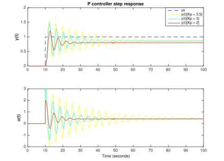
3.2.1.2. Proportional control . . . 31
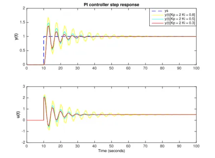
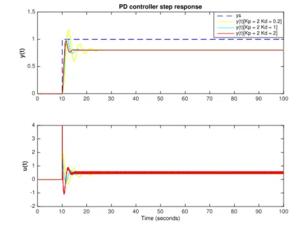
3.2.1.3. Proportional-integrative-derivative (PID) control . . . 32
3.2.2. Performance measurement of control methods . . . 36
3.2.3. Optimal control . . . 38
3.3. Conclusion . . . 39
References . . . 40
4 Novel kinesthetic stimulation system for the treatment of sleep apnea syndromes (PASITHEA system) 45 4.1. General description of the PASITHEA system . . . 45
4.1.1. Cardiorespiratory Holter . . . 46
4.1.2. Kinesthetic stimulation system . . . 47
4.1.3. Real time processing and control application . . . 48
4.2. On-Off kinesthetic stimulation for the treatment of SAS . . . 50
4.2.1. Real-time respiratory event detector . . . 50
4.2.2. On-off control of the kinesthetic stimulation . . . 52
4.3. Evaluation methodology . . . 52
4.3.1. The HYPNOS study . . . 52
4.3.2. Data acquisition and patient population . . . 53
4.3.3. Evaluation of the respiratory event detector . . . 54
4.3.4. Preliminary, qualitative evaluation of the therapy . . . 55
4.4. Results . . . 55
4.4.1. Evaluation of the apnea/hypopnea detector . . . 55
4.4.2. Qualitative preliminary responses of the kinesthetic stimulation . . . 56
4.4.2.1. Responder patient . . . 57
4.4.2.2. Partially responder patient . . . 59
4.4.2.3. Non-responder patient . . . 60
4.5. Discussion . . . 60
4.6. Conclusion . . . 62
References . . . 63
5 On-off Kinesthetic Stimulation Therapy for Sleep Apnea Syndrome: Effects in event duration and oxygen saturation levels 65 5.1. The proposed On-Off controller . . . 65
5.2. Quantitative evaluation of the on-off controller . . . 66
5.2.1. Recall of study design and participants . . . 66
5.2.2. Data processing and statistical analysis . . . 67
5.3. Results . . . 71
5.3.1. Event duration . . . 71
Contents xi
5.3.3. Local, acute SaO2 analysis . . . 71
5.3.4. Sleep analysis . . . 73
5.3.5. Other outcomes . . . 73
5.4. Discussion . . . 73
5.5. Conclusion . . . 76
References . . . 77
6 Autonomic differences based on the response to kinesthetic stimulation therapy in sleep apnea patients 81 6.1. Introduction and hypothesis . . . 81
6.2. Methodology . . . 82
6.2.1. Study design . . . 82
6.2.2. General data analysis approach . . . 82
6.2.2.1. Sleep onset period detection . . . 82
6.2.2.2. RR series extraction . . . 83
6.2.2.3. ECG-Derived Respiration signal processing . . . 83
6.2.2.4. Spectral heart rate variability analysis . . . 85
6.2.2.5. Detrended fluctuation analysis . . . 86
6.2.2.6. Sample entropy . . . 86
6.2.3. Statistical analysis . . . 87
6.3. Results . . . 87
6.3.1. Heart rate variability . . . 87
6.3.2. Heart rate complexity . . . 88
6.3.3. Leave-one-out cross-validation . . . 88
6.4. Discussion . . . 88
6.5. Conclusion . . . 91
References . . . 91
7 Closed-loop Kinesthetic Stimulation for the Treatment of Sleep Apnea Syndromes 95 7.1. Introduction and hypothesis . . . 95
7.2. Methodology . . . 96
7.2.1. Methodological and technical improvements of the PASITHEA system 96 7.2.1.1. Apnea/hypopnea detector optimization . . . 97
7.2.1.2. Kinesthetic stimulation device . . . 100
7.2.1.3. Real-time control application . . . 100
7.2.2. Coupled PD controller for adaptive kinesthetic stimulation . . . 100
7.2.3. Closed-loop algorithm . . . 102
7.2.4. Acceleration signal inclusion . . . 104
7.2.5. Study design (EKINOX study) . . . 105
7.3.1. Detector optimization . . . 106 7.3.2. Controller evaluation . . . 106 7.3.3. Acceleration evaluation . . . 109 7.4. Discussion . . . 110 7.5. Conclusion . . . 112 References . . . 113 8 Conclusion 115 References . . . 118
A List of associated publications 121 International journals . . . 121
International conferences . . . 121
International abstracts . . . 121
Software registration . . . 122
B Kinesthetic stimulation therapy of sleep apnea syndrome: duration, acute SaO2 and sleep analyses results 123 B.1. Event durations . . . 124
B.2. Local, acute SaO2 results . . . 126
B.3. Sleep stage . . . 128
B.4. Micro-arousal . . . 133
B.5. Whole night sleep overview . . . 134
List of Figures 135
List of acronyms
AHR Apnea or hypopnea responder ANS Autonomic nervous system AUC Area under the ROC curve AR Apnea responder
Auto-CPAP Automatic continuous positive airway pressure BT Bluetooth technology
BTLE Bluetooth Low Energy CNS Central nervous system
CPAP Continuous positive airway pressure CVS Cardiovascular system EA Evolutionary algorithms ECG Electrocardiogram HF High frequency HR Heart rate HR Hypopnea responder HRC Heart rate complexity HRV Heart rate variability IH Intermittent hypoxia LF Low frequency
MAD mandibular advancement device NP Nasal pressure
NR No responder
NREM Non rapid eye movement OSA Obstructive sleep apnea
PASITHEA Personalized and Adaptive kinesthetic StImulation Therapy, based on cardiorespiratory Holter monitoring, for slEep Apnea syndromes PCO2 Partial carbon dioxide pressure
PVC Premature ventricular contractions PO2 Partial oxygen pressure
PNS Peripheral nervous system REM Rapid eye movement RS Respiratory system SaO2 Oxygen saturation
SAS Sleep apnea syndrome SNS Somatic nervous system SOP Sleep-onset period
CHAPTER
1
Introduction
Sleep apnea syndromes (SAS) is an under-diagnosed disease affecting up to 5% of middle-aged men and women (Heinzer et al., 2015; Jennum et al., 2009; Peppard et al., 2013). It is characterized by recurrent episodes of upper airway collapse (apnea) or significant reductions in respiratory airflow amplitude (hypopnea), followed by transient awakening that leads to the restoration of upper airway permeability. These episodes are repeated several times every hour, leading to sleep fragmentation along with acute cardio-respiratory alterations which are associated with the development of hypertension, coronary heart diseases, stroke and other cardiovascular complications in the long term (Lévy et al., 2014; Somers et al., 2008; Stansbury et al., 2015). The deleterious effects of SAS on cardiovascular outcomes are mainly triggered by the intermittent hypoxia (IH) severity caused by the respiratory pauses, inducing the subsequent activation of the autonomic nervous system (Dick et al., 2007; Drager et al., 2010; Kasai et al., 2011).
Patients with SAS have varying degrees of symptomatology and SAS-related co-morbid conditions (Bailly et al., 2016; Deacon et al., 2016). Several therapies have been proposed for the treatment of SAS, being the gold standard therapy in the management of moderate to severe SAS, the continuous positive airway pressure (CPAP) (Berkani et al., 2015; Marin et al., 2005), followed by the use of mandibular advancement devices (MAD) (Bratton et al., 2015; Hoffstein, 2007). CPAP, the first line therapy, is associated with excellent results in highly symptomatic patients. However, there is a 15% initial refusal rate and long term adherence is difficult to achieve in mildly symptomatic patients (Craig et al., 2012). Recent studies on CPAP-treated patients have shown a relatively high rate of abandonment and poor compliance with average adherence rates between 39% and 50% (Kushida et al., 2012). Compliance with MAD is higher than CPAP, yet treatments are not as effective (Phillips et al., 2013). In addition, the efficacy of MAD-based therapies strongly depends on the morphology of the patient (Fritsch et al., 2001) and more than 30% of SAS patients are contraindicated to MAD owing to dental or joint problems (Petit et al., 2002). Improvements to the classical CPAP, such as the automatic continuous
positive airway pressure (Auto-CPAP), have been proposed. However, a recent meta-study has shown that the Auto-CPAP method does not provide significant added value concerning the acceptance or compliance to the therapy, with respect to classical, manually titrated CPAP (Gao et al., 2012). Thus, alternative therapies are desirable for the appropriate treatment of SAS.
In addition to CPAP, MAD and surgery to enlarge the upper airways, stimulation therapies have recently gained interest in the treatment of SAS (Strollo et al., 2016). A key mechanism underlying repeated pharyngeal collapse during sleep is the reduction of pharyngeal dilator muscle activity to a level that is not able to maintain upper airway patency in the context of impaired upper airway anatomy. New stimulation approaches dedicated to increase the neural output to upper airway dilator muscles as hypoglossal nerve stimulation (Strollo Jr et al., 2014) or direct electrical stimulation of submental transcutaneous muscles are currently under evaluation (Pengo et al., 2016). Also, devices for the direct stimulation of the phrenic nerve have been developed, which may be useful for the treatment of central apnea (Ponikowski et al., 2011). These therapies are currently under clinical investigation and, although promising, they require an invasive procedure that may cause significant side effects (Eastwood et al., 2011). MRI incompatibility may also be a limitation to the widespread use of these systems.
The above paragraphs underline the fact that there is still a large portion of the SAS population that remains inadequately treated or even untreated. The development of non-invasive SAS treatment methods, with improved acceptability, is thus of major importance. In this context, the objective of this PhD thesis is to propose new signal processing and control methods of non-invasive neuromodulation for the treatment of SAS, based on kinesthetic stimulation. Additional objectives are the development of an integrated system prototype, embedding the proposed methods, and the evaluation of this prototype system in a clinical setup. Indeed, previous studies have shown that kinesthetic stimulation may trigger a physiological reflex, called the "startle reflex", which is capable of eliciting systemic motor responses and cardiac autonomic activation (Taylor et al., 1991; Yeomans et al., 2002). The hypothesis underlying this thesis is that bursts of kinesthetic stimulation, delivered during the early phase of apneas or hypopneas, may reduce respiratory event duration and subsequently limit related oxygen desaturations, through a controlled activation of the startle reflex.
This manuscript is divided into eight chapters. The first chapter provides the clinical context in which this thesis is framed. A general overview of the SAS, including pathogenesis, epidemiology, risk stratification, clinical management, as well as a state-of-the-art review of the current therapies, is presented. Also, a brief review about the cardiorespiratory system physiology and its regulation through the autonomic nervous system along with the human sleep nature is also presented. Chapter 3 introduces the general signal processing and control theory framework that is consistently employed throughout this work. It details the terminology and formalized definitions related to the context of control and signal processing, and presents some applications of the state of the art in the biomedical domain.
3
After these introductory chapters, chapter 4, provides a complete description of a novel real-time monitoring and therapeutic neuromodulation system (PASITHEA system), which serves as a multi-purpose device for delivering kinesthetic stimulation (Hernandez et al., 2016). The different control methods that are proposed throughout this work were embedded into this device, which was then validated in clinical protocols. In the first stage of this thesis, we proposed a first control algorithm for delivering kinesthetic stimulation. An On/Off control method using as control variable the output of a real-time respiratory event detector was integrated. Briefly, when respiratory event detection is confirmed, a command is sent to the kinesthetic stimulator to activate it and it is stopped when the respiration flow is resumed. A unique stimulation strategy with a constant stimulation amplitude was applied upon event detention. The complete system was tested in a first clinical protocol (HYPNOS study), dedicated to assessing the patient response to therapy implementing the previously mentioned control method. Several signal processing methods were developed to estimate the patient response. Chapter 5 presents the implemented methods and the main results obtained from this first clinical protocol. In addition, a novel method developed in the context of this thesis for the analysis of the variations in the SaO2 signal is also presented in that chapter.
In chapter 6, we propose a new method to estimate patients who could benefit from kinesthetic stimulation therapy. Several analyses in the time and frequency domains were implemented in order to extract heart rate variability (HRV) and heart rate complexity (HRC) markers from heart rate time series. Beat detection was obtained from the recorded electrocardiogram (ECG), through a wavelet-based algorithm previously proposed in our laboratory and adapted to this signals. Results from the retrospective application of this method for the estimation of patient response on the data obtained from the previously mentioned clinical protocol are also presented.
Finally, the last part of this thesis proposes a novel closed-loop control method, inte-grating concurrent, coupled proportional-derivative (PD) controllers in order to manage the kinesthetic stimulation amplitude delivered to the patient by the therapeutic system, using as control variables three physiological signals recorded in real-time: Nasal pressure (NP), oxygen saturation (SaO2) and the ECG signal. A complete description of a second clinical
protocol comprising 40 patients (EKINOX study) with the main objective of validating the control algorithm for patient-specific adaptive kinesthetic stimulation is presented in chapter 7. This chapter also describes the different improvements to the first version of the system that were developed to allow the integration of the proposed controller. Preliminary results from the first phase of this study, where 10 patients were analyzed in order to validate the correct functioning of the controller and determine the set of optimal control parameters to be used for the second phase with the inclusion of the remaining 30 patients are also presented.
References
Bailly, S., M. Destors, Y. Grillet, P. Richard, B. Stach, I. Vivodtzev, J.-F. Timsit, P. Lévy, R. Tamisier, J.-L. Pépin, et al. (2016). “Obstructive sleep apnea: a cluster analysis at time of diagnosis”. In: PLoS One 11.6, e0157318.
Berkani, K. and J. Dimet (2015). “[Acceptability and compliance to long-term continuous positive pressure treatment]”. In: Revue des maladies respiratoires 32.3, pp. 249–255. Bratton, D. J., T. Gaisl, A. M. Wons, and M. Kohler (2015). “CPAP vs mandibular
advancement devices and blood pressure in patients with obstructive sleep apnea: a systematic review and meta-analysis”. In: Jama 314.21, pp. 2280–2293.
Craig, S. E., M. Kohler, D. Nicoll, D. J. Bratton, A. Nunn, R. Davies, and J. Stradling (2012). “Continuous positive airway pressure improves sleepiness but not calculated vascular risk in patients with minimally symptomatic obstructive sleep apnoea: the MOSAIC randomised controlled trial”. In: Thorax, thoraxjnl–2012. Deacon, N. L., R. Jen, Y. Li, and A. Malhotra (2016). “Treatment of obstructive
sleep apnea. Prospects for personalized combined modality therapy”. In: Annals of the American Thoracic Society 13.1, pp. 101–108.
Dick, T. E., Y.-H. Hsieh, N. Wang, and N. Prabhakar (2007). “Acute intermit-tent hypoxia increases both phrenic and sympathetic nerve activities in the rat”. In: Experimental physiology 92.1, pp. 87–97.
Drager, L. F., J. C. Jun, and V. Y. Polotsky (2010). “Metabolic consequences of intermittent hypoxia: relevance to obstructive sleep apnea”. In: Best practice & research Clinical endocrinology & metabolism 24.5, pp. 843–851.
Eastwood, P. R., M. Barnes, J. H. Walsh, K. J. Maddison, G. Hee, A. R. Schwartz, P. L. Smith, A. Malhotra, R. D. McEvoy, J. R. Wheatley, et al. (2011). “Treating obstructive sleep apnea with hypoglossal nerve stimulation”. In: Sleep 34.11, pp. 1479–1486.
Fritsch, K. M., A. Iseli, E. W. Russi, and K. E. Bloch (2001). “Side effects of mandibular advancement devices for sleep apnea treatment”. In: American journal of respiratory and critical care medicine 164.5, pp. 813–818.
Gao, W., Y. Jin, Y. Wang, M. Sun, B. Chen, N. Zhou, and Y. Deng (2012). “Is automatic CPAP titration as effective as manual CPAP titration in OSAHS patients? A meta-analysis”. In: Sleep and Breathing 16.2, pp. 329–340.
Heinzer, R., S. Vat, P. Marques-Vidal, H. Marti-Soler, D. Andries, N. Tobback, V. Mooser, M. Preisig, A. Malhotra, G. Waeber, et al. (2015). “Prevalence of sleep-disordered breathing in the general population: the HypnoLaus study”. In: The Lancet Respiratory Medicine 3.4, pp. 310–318.
Hernandez, A., G. Guerrero, D. Feuerstein, L. Graindorge, D. Perez, A. Am-blard, P. Mabo, J.-L. Pépin, and L. Senhadji (2016). “Pasithea: An integrated monitoring and therapeutic system for sleep apnea syndromes based on adaptive kinesthetic stimulation”. In: IRBM 37.2, pp. 81–89.
References 5
Hoffstein, V. (2007). “Review of oral appliances for treatment of sleep-disordered breathing”. In: Sleep and Breathing 11.1, pp. 1–22.
Jennum, P. and R. L. Riha (2009). “Epidemiology of sleep apnoea/hypopnoea syndrome and sleep-disordered breathing”. In: European Respiratory Journal 33.4, pp. 907–914. Kasai, T. and T. D. Bradley (2011). “Obstructive sleep apnea and heart failure:
pathophysiologic and therapeutic implications”. In: Journal of the American College of Cardiology 57.2, pp. 119–127.
Kushida, C. A., D. A. Nichols, T. H. Holmes, S. F. Quan, J. K. Walsh, D. J. Gottlieb, R. D. Simon Jr, C. Guilleminault, D. P. White, J. L. Goodwin, et al. (2012). “Effects of continuous positive airway pressure on neurocognitive function in obstructive sleep apnea patients: the Apnea Positive Pressure Long-term Efficacy Study (APPLES)”. In: Sleep 35.12, pp. 1593–1602.
Lévy, P., M. Kohler, W. T. McNicholas, F. Barbé, R. D. McEvoy, V. K. Somers, L. Lavie, and J.-L. Pepin (2014). “Obstructive sleep apnoea syndrome.” In: Nature reviews. Disease primers 1, pp. 15015–15015.
Marin, J. M., S. J. Carrizo, E. Vicente, and A. G. Agusti (2005). “Long-term cardiovascular outcomes in men with obstructive sleep apnoea-hypopnoea with or without treatment with continuous positive airway pressure: an observational study”. In: The Lancet 365.9464, pp. 1046–1053.
Pengo, M. F., S. Xiao, C. Ratneswaran, K. Reed, N. Shah, T. Chen, A. Douiri, N. Hart, Y. Luo, G. F. Rafferty, et al. (2016). “Randomised sham-controlled trial of transcutaneous electrical stimulation in obstructive sleep apnoea”. In: Thorax, thoraxjnl–2016.
Peppard, P. E., T. Young, J. H. Barnet, M. Palta, E. W. Hagen, and K. M. Hla (2013). “Increased prevalence of sleep-disordered breathing in adults”. In: American
journal of epidemiology 177.9, pp. 1006–1014.
Petit, F.-X., J.-L. Pépin, G. Bettega, H. Sadek, B. Raphaël, and P. Lévy (2002). “Mandibular advancement devices: rate of contraindications in 100 consecutive obstruc-tive sleep apnea patients”. In: American journal of respiratory and critical care medicine 166.3, pp. 274–278.
Phillips, C. L., R. R. Grunstein, M. A. Darendeliler, A. S. Mihailidou, V. K. Srinivasan, B. J. Yee, G. B. Marks, and P. A. Cistulli (2013). “Health outcomes of continuous positive airway pressure versus oral appliance treatment for obstructive sleep apnea: a randomized controlled trial”. In: American journal of respiratory and critical care medicine 187.8, pp. 879–887.
Ponikowski, P., S. Javaheri, D. Michalkiewicz, B. A. Bart, D. Czarnecka, M. Jastrzebski, A. Kusiak, R. Augostini, D. Jagielski, T. Witkowski, et al. (2011). “Transvenous phrenic nerve stimulation for the treatment of central sleep apnoea in
heart failure”. In: European heart journal 33.7, pp. 889–894.
Somers, V. K., D. P. White, R. Amin, W. T. Abraham, F. Costa, A. Culebras, S. Daniels, J. S. Floras, C. E. Hunt, L. J. Olson, et al. (2008). “Sleep apnea and
cardiovascular disease: An American heart association/American college of cardiology foundation scientific statement from the American heart association council for high blood pressure research professional education committee, council on clinical cardiology, stroke council, and council on cardiovascular nursing in collaboration with the national heart, lung, and blood institute national center on sleep disorders research (national institutes of health)”. In: Journal of the American College of Cardiology 52.8, pp. 686– 717.
Stansbury, R. C. and P. J. Strollo (2015). “Clinical manifestations of sleep apnea”. In: Journal of thoracic disease 7.9, E298.
Strollo Jr, P. J., R. J. Soose, J. T. Maurer, N. De Vries, J. Cornelius, O. Froymovich, R. D. Hanson, T. A. Padhya, D. L. Steward, M. B. Gillespie, et al. (2014). “Upper-airway stimulation for obstructive sleep apnea”. In: New England Journal of Medicine 370.2, pp. 139–149.
Strollo, P. J. and A. Malhotra (2016). Stimulating therapy for obstructive sleep apnoea.
Taylor, B. K., R. Casto, and M. P. Printz (1991). “Dissociation of tactile and acoustic components in air puff startle”. In: Physiology & behavior 49.3, pp. 527–532.
Yeomans, J. S., L. Li, B. W. Scott, and P. W. Frankland (2002). “Tactile, acoustic and vestibular systems sum to elicit the startle reflex”. In: Neuroscience & Biobehavioral Reviews 26.1, pp. 1–11.
CHAPTER
2
General context: Sleep apnea
syndrome (SAS)
This chapter describes the clinical context in which this thesis is framed. First, a general overview of the disease under study, the sleep apnea syndrome (SAS), is presented. This frequent syndrome is characterized by repeated episodes of breathing pauses, associated with acute cardiorespiratory responses, that may be deleterious in the long term. Since the upper airway anatomy plays a significant role in the pathophysiology, a description of this structure is presented in section 2.2.1. An explanation of the physiological sub-systems that are involved in the pathogenesis of SAS is also resented in section 2.2. Then, a brief review on the physiological basis of the cardiorespiratory system and its modulation through the autonomic nervous system is introduced. Finally, the last sections are dedicated to the description of the different physiological concepts that will be evoked in this work.
2.1. Sleep apnea syndrome (SAS)
Sleep apnea syndrome (SAS) is characterized by repeated episodes of breathing pauses (apnea) or reductions in respiratory amplitude (hypopnea) during sleep. These episodes often induce significant arterial hypoxemia and hypercapnia that usually lead to transient sleep arousals, sleep fragmentation and acute over compensatory responses of the autonomic nervous system, that may be deleterious in the long term, being associated with higher cardiovascular and metabolic morbidities (Peppard et al., 2013; Somers et al., 2008; Stansbury et al., 2015). SAS represent a major healthcare issue, affecting more than 5% of the middle-aged population (Heinzer et al., 2015; Jennum et al., 2009; Peppard et al., 2013). Commonly, SAS is divided into two event categories:
Central events: Central apnea (CA) or hypopnea (CH) are denoted by an absence or marked reduction of brain stem respiratory output, and thus, neuromuscular respiratory drive to respiratory pump muscles.
Obstructive events: Obstructive apnea (OA) or hypopnea (OH) are characterized by an extra-thoracic upper airway obstruction along with respiratory efforts. Mixed events: Mixed apnea or hypopnea result from a combination of central and obstructive events.
Population-based studies have shown that most SAS events are driven by anatomical and neurochemical anomalies in the control of the upper airway and chest wall respiratory musculature. The dominant risk factors are the body weight, followed by male gender and craniofacial structure and aging (Dempsey et al., 2002; Partinen et al., 1988a; Peppard et al., 2000; Young et al., 2003). Figure 2.2 presents an example of the typical physiological signals recorded during the night from patients suffering from severe SAS. In this example, the consequences of the repeated respiratory events in each signal can be observed.
Three biological systems are primarily involved in SAS: i) the respiratory system, ii) the cardiovascular system and iii) the autonomic nervous system (ANS). The next sections will present a brief description of these systems and their relation to SAS, in order to fix the clinical framework of this work.
2.1.1. Pathogenesis of Sleep Apnea
In humans, one of the major structures that comprise the respiratory system are the upper airways (description in section 2.2.1). This structure performs complex motor behaviors required to generate speech and are possible because the hyoid bone, which is a key anchoring site for pharyngeal dilator muscles which is not rigidly attached to skeletal structures, leaving the human pharynx with no rigid support unlike in other animals (Morgan et al., 2007). This attribute gives to the upper airway the chance to narrow or collapse when the compensatory neural activation of dilator muscles is lost at sleep onset. However, these compensatory processes are the product of multiple factors and vary markedly among individuals (Dempsey et al., 2010). A diagram of the pathogenesis of SAS is shown in figure 2.1. First, the diagram shows the principal structural and functional determinants of an anatomical predisposition for airway closure. Generally, a patient having any of them is more prone to suffer from SAS. Then, after sleep, two paths to develop either an obstructive or central apnea are presented. The diagram emphasizes the mechanisms underlying both types of respiratory events, and integrates anatomical deficits with mechanisms for central neurochemical control of breathing stability and compensatory neuromuscular control of upper airway muscles in order to explain the cyclical nature of SAS.
An illustration of these different mechanisms can also be appreciated in figure 2.2. Figure 2.2-A shows the nasal pressure (NP) signal with the presence of several respiratory events. These events are provoked, as previously described, by a decrease of the tonic activity of the upper airway dilator muscles causing the upper airway to narrow/collapse (obstructive apnea) or by an unstable central respiratory motor output leading to a stoppage
2.1. Sleep apnea syndrome (SAS) 9
Pathogenesis of sleep apnea syndrome (SAS)
Anatomical predisposition to airway closure
Adipose soft tissue deposition Compromised craniofacial structures Muscle injury Anomalies on upper airway structures (size, length, position) Age Cyclical SAS event Sleep Upper airway narrowing / closure Ventilatory overshoot Arousal and/or airway open
Obstructive apnea / hypopnea
Hypocapnia
Apnea / hypopnea Motor output to airway
and chest wall Tonic activity to upper airway dilatator muscles
Chemoreceptor feedback to airway Critical dependance of the
ventilatory control system on chemical stimuli (PCO2)
Unstable central respiratory motor output
Central apnea / hypopnea
Passive airway narrowing / closure Motor output to airway
and chest wall
Chemoreceptor stimuli Arousal / ventilatory overshoot
Figure 2.1: Scheme of the sleep apnea syndrome cyclical pathogenesis. Modified figure from (Dempsey et al., 2010).
of the respiration (central apnea). After these alterations in the respiratory system, the lack of uptake of oxygen produces a decrease of oxygen concentration in the blood (hypoxemia) which induces intermittent hemodynamic variations in the cardiovascular system, this behavior can be observed in figure 2.2-B, where the oxygen saturation (SaO2) signal
is presented. Moreover, acute autonomic responses can also be seen in the heart rate (HR) signal (figure 2.2-C), where regulating mechanisms in the cardiovascular system are induced by recurrent activations of the baroreflex and chemoreflex as a response to hypoxia. Additionally, these respiratory events may provoke sleep micro-arousals (figure 2.2-D) which are the main factor of the sleep fragmentation related to SAS. The next sections will describe the different physiological concepts previously mentioned.
2.1.2. Consequences of sleep apnea
As previously introduced in section 2.1, SAS is recognized as a risk factor for the development of hypertension and other cardiovascular diseases. Recurrent episodes of SAS produce arterial oxygen desaturation, hypercapnia, significant intrathoracic pressure oscillations, and sleep disruption. They are also characterized by a marked, transient increase in systemic arterial pressure caused by sympathetic nervous system activation
Apnea Hypopnea
A
B
C
D
Figure 2.2: Example of a typical recording of a patient suffering from sleep apnea syndrome (HYPNOS study). The first panel shows the nasal pressure (NP) signal, where the different apnea and hypopnea events are highlighted. The second panel represents the oxygen saturation (SaO2) signal with the intermittent hypoxia events associated with SAS. The
third panel, presents the instantaneous heart rate (HR) signal, showing recurrent acute autonomic responses that are due to the respiratory events. Finally, the fourth panel presents a hypnogram signal (MA = micro-arousal, A = Awake, REM = Rapid eye movement, LS = Light sleep and DS = Deep sleep) representing the sleep structure of the patient during the recording. The hypnogram shows the presence of sleep arousals and sleep fragmentation produced by respiratory events.
(Katragadda et al., 1997; O’Donnell et al., 1996). Such responses can occur a significant number of times for severe SAS patients, as shown in figure 2.2. These acute over compensatory responses of the autonomic nervous system, due to the effects of SAS, are deleterious in the long-term and they may induce the following consequences:
Cardiovascular and metabolic defects: One of the most well-known risk factors for hypertension is SAS. The causal link between SAS and hypertension has been firmly established via animal models (Brooks et al., 1997; Fletcher et al., 1992) and studies that have shown that blood pressure decreases in hypertensive patients when SAS is treated (Becker et al., 2003; Pepperell et al., 2002). Other diseases, such as left ventricular dysfunction, cardiac arrhythmias and stroke have been also associated with SAS (Arzt et al., 2005; Guilleminault et al., 1983; Shahar et al., 2001). In addition, recent studies have associated SAS with metabolic defects and to the predisposition to the eventual development of Type 2 diabetes (McArdle et al., 2007; Punjabi et al., 2005).
Sleep fragmentation: Sleep deprivation, or sleep restriction, may not reflect the disturbances in sleep that occur in SAS. Typically, total sleep time is not significantly restricted in SAS, but rather sleep is fragmented by repetitive arousals resulting
2.2. The cardio-respiratory system response to SAS 11
from the impaired breathing during sleep. Common symptoms associated with sleep fragmentation include decreased psychomotor performance on task involving short term memory, reaction time, vigilance and degraded mood (Bonnet et al., 2003; Martin et al., 1996). These symptoms lead to daytime sleepiness which is one of the main risk factors to suffer everyday accidents (NODA et al., 1998; Stoohs et al., 1994).
2.2. The cardio-respiratory system response to SAS
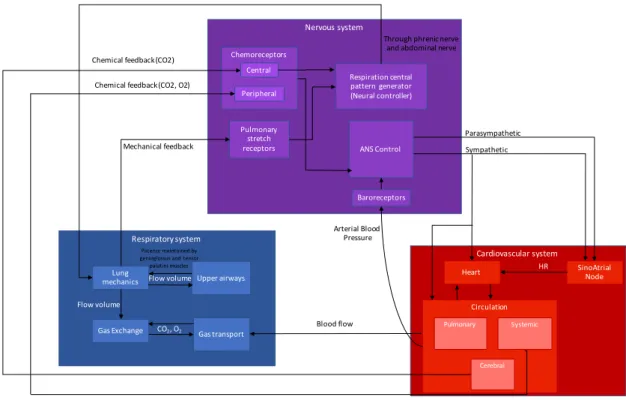
This section presents a brief description of the main components of the cardio-respiratory system and its control, in order to introduce the main physiological concepts needed for the understanding of the rest of the manuscript. Figure 2.3 presents a modular diagram of the three biological systems that are involved in response to SAS and the general interactions among them.
The blue module represents the respiratory system, where the airflow coming from the upper airways interacts with the lungs in order to allow for gas exchange and transport of oxygen (O2) and carbon dioxide (CO2) to the rest of the body. The red module represents
the cardiovascular system, comprising the heart and the circulatory system. Moreover, the sinoatrial node in charge of the heart rhythm control has been represented separately. The heart acts upon the circulatory system and its different components: pulmonary, systemic and cerebral. Finally, the purple module represents the nervous system, comprised by central respiratory and ANS controls and three sensory receptors (chemoreceptors, baroreceptors and pulmonary stretch receptors).
The respiratory system is affected by the cardiovascular system due to alterations in blood flow and, more importantly, by the nervous system. The central nervous system induces the ventilatory activity by sending electrical impulses through the phrenic and abdominal nerves, which will generate a negative intra-thoracic pressure, allowing for airflow intake. In turn, the nervous system receives mechanical feedback from the lungs captured by pulmonary stretch receptors. On the other hand, sympathetic and parasympathetic branches of the autonomic nervous system regulate the cardiovascular system function. Baroreceptors and chemoreceptors measure arterial blood pressure and gases in circulation respectively in order to close the loop between these two systems.
In the case of SAS, the balance among systems is lost by a narrow/collapse of the upper airways (obstructive apnea) or by unstable central respiratory motor drive (central apnea) as described in section 2.1.1. These alterations limit the gas exchange and activate peripheral chemoreceptors and pulmonary stretch receptors due to the lack of air and therefore oxygen. In turn, these activations produce an increase of the sympathetic tone which leads to an increase in HR and thus in arterial blood pressure. They also induce an activation of the neural controller that forces respiration to resume by controlling lung mechanics and in consequence generating a cortical arousal. A general physiological
description of each of the previously mentioned modules are described in the following sections. Respiratory system Lung mechanics Gas Exchange Gas transport Upper airways Cardiovascular system Circulation SinoAtrial Node Heart Nervous system Respiration central pattern generator (Neural controller) Pulmonary stretch receptors ANS Control Chemoreceptors Baroreceptors Central Peripheral Parasympathetic Sympathetic HR Arterial Blood Pressure Blood flow CO2, O2 Mechanical feedback Flow volume Flow volume Chemical feedback (CO2, O2) Pulmonary Systemic Chemical feedback (CO2) Cerebral Through phrenic nerve and abdominal nerve Patency maintained by genioglossus and tensor palatini muscles
Figure 2.3: Diagram of the three biological systems that are involved in response to SAS. Figure adapted from the ongoing PhD thesis work of Gustavo Guerrero, SEPIA team -LTSI.
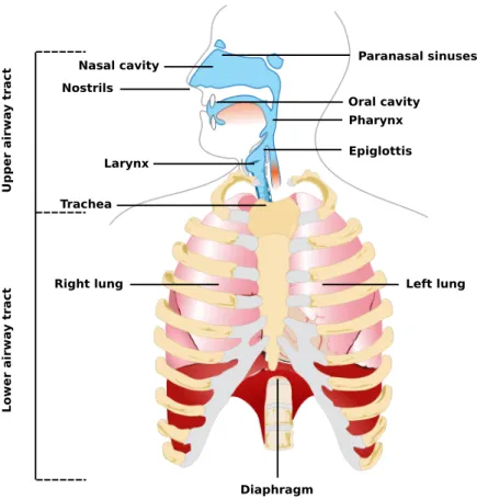
2.2.1. The respiratory system
The respiratory system (RS) (see figure 2.4) is a biological system composed by three major structures: the airways, the lungs, and different respiratory muscles. Its main function is to perform the gas exchange that is required by the human body to maintain a constant stream of oxygen, in order to sustain the cellular lifecycle. Gas exchange is particularly important for respiration, which involves the uptake of oxygen (O2) and release
of carbon dioxide (CO2).
As illustrated in figure 2.4, the RS is divided in two main parts: the upper and the lower airway tracts. The latter is comprised by the lungs and the diaphragm. The lungs are the largest organs in the respiratory system and their function is to extract O2 from the outside
and to transfer it into the bloodstream to then release CO2 from the bloodstream to the
outside in a process know as gas exchange. On the other hand, the diaphragm is a muscle capable of voluntarily contracting in order to pull or push the membrane surrounding the lungs facilitating air to flow inside or outside them.
2.2. The cardio-respiratory system response to SAS 13 Paranasal sinuses Oral cavity Pharynx Epiglottis Nasal cavity Nostrils Larynx Trachea
Right lung Left lung
Diaphragm Up p er a irw a y tra c t Low er a irw a y tra c t
Figure 2.4: The respiratory system. Adapted from "Human respiratory system pedagogical fr" by Michka B licensed under CC BY 4.0.
cavities (Pohunek, 2004). However, some authors also include the larynx, trachea and oral cavity because of their role in the RS. These structures are involved in performing functional tasks such as air warming and humidification, pathways for olfaction, speech, swallowing and allowing the passage of air for breathing. It is composed of numerous muscles and soft tissue which allows the upper airway to change shape in order to grant to humans the ability to speak and swallow. Although these features are widely used during wakefulness, the lack of rigid or bony support creates a collapsible portion that extends from the hard palate to the larynx, that may collapse during sleep due to a significantly reduced muscle tone (Pierce et al., 1999).
The upper airway patency is maintained by activation of dilator muscles driven by chemoreceptors responding to chemical stimuli (hypercapnia and hypoxia) and by mechanoreceptor feedback responding to negative pharyngeal pressure (Loewen et al., 2011). In the case of SAS, during airways obstruction an increase in negative pharyngeal pressure is due to an elevation of the respiratory effort against the obstructed airway, which in turn is provoked due to chemical stimulation. The most well studied muscles are the genioglossus and tensor palatini (MALHOTRA et al., 2000). Anatomic airway narrowing is compensated by increased activation of upper airway muscles in awake patients.
However, an important mechanism in the pathogenesis of OSA relates to the interaction between pharyngeal anatomy and a diminished ability of the upper airway dilator muscles to maintain a patent airway during sleep (Mezzanotte et al., 1992). Consequently, several studies have shown that whereas healthy individuals experience a loss of upper airway muscle tone at sleep onset, an individual reliant on muscle tone due to an anatomic vulnerability will be particularly susceptible to developing OSA (Mezzanotte et al., 1996; Worsnop et al., 1998).
In addition, as previously mentioned, the genioglossus is importantly modulated by locally mediated mechanoreceptive reflex mechanisms that respond to negative pharyn-geal pressure (Pillar et al., 2001). One such mechanism is the genioglossus negative pressure reflex, whereby the muscle is activated in response to rapid changes in negative intrapharyngeal pressure (Horner et al., 1991). Nevertheless, although genioglossus muscle responsiveness may be impaired during sleep compared with wakefulness, studies have shown that the muscle does respond to sustained negative pressure and potentially hypercapnia (Lo et al., 2006; Stanchina et al., 2002). However, there is a substantial interindividual variability in the effectiveness of these compensatory responses to restore airflow during sleep (Jordan et al., 2007).
2.2.2. Autonomic regulation of the cardiovascular system
The cardiovascular system (CVS) is a closed circulatory system mainly composed of the heart, blood vessels and blood (figure 2.5). Its function is to maintain an adequate supply of oxygen to all tissues of the body and to transport nutrients, carbon dioxide, hormones and blood cells throughout the organism. However, in order to maintain this function, the autonomic system must process visceral information and coordinate neural elements that innervate the heart, blood vessels, and the respiratory system. Regarding SAS, this system is closely involved in the responses to the breathing pauses as figure 2.3 shows. During a SAS event, activations of the nervous system are primarily elicited by activations on chemoreceptors due to the lack of oxygen. These activations trigger different over compensatory responses in the cardiovascular system such as: increased HR, changes in cardiac contractility and changes in the intrathoracic pressure due to the airway occlusion. As mentioned in section 2.1, these responses may be deleterious in the long term, being associated with cardiovascular diseases.
On the other hand, the nervous system is a network of nerve cells and fibres which, along the brain and spinal cord, receives and interprets information from the entire organism and transmits impulses to the effector organs. It is subdivided in four different subsystems:
Central Nervous System (CNS): It is composed of the brain and spinal cord and it behaves as the processing center of the nervous system. It integrates and coordinates the information that it receives and also influences the activity of the organism.
2.2. The cardio-respiratory system response to SAS 15
Figure 2.5: Cardiovascular system, composed of the heart and the pulmonary and systemic circulations. Adapted from "CardiovascularSystem" by Dcoetzee licensed under CC BY 4.0.
Peripheral Nervous System (PNS): It consists on nerves and glands outside of the brain and spinal cord, and it is responsible of the connection and information transport between CNS and the rest of the body. This subsystem is also subdivided in two parts:
• The somatic nervous system (SNS): Comprising the nerves associated with the voluntary control of body movements, and innervating skeletal muscles and external sensory organs.
• The autonomic nervous system (ANS): Innervating smooth muscles and glands in order to influence the function of internal organs, and thus regulate involuntary body functions. It is subdivided in three branches: the sympa-thetic, parasympathetic and enteric nervous systems. The sympathetic and parasympathetic nervous systems have opposite actions, where the former is often considered as the “fight or flight” system and is generally activated in cases of stress whereas the latter is mainly activated when organisms are in a relaxed state. Finally, the enteric nervous system is responsible of the intestinal motility in digestion and is capable of acting independent of the sympathetic and parasympathetic nervous systems.
2.2.2.1. Mechanoreceptors
Mechanoreceptors are sensory receptors that respond to mechanical forces applied to the body (Biswas et al., 2015; Catton, 1970). These receptors allow animals to perceive the different mechanical stimuli coming from outside, by initiating nerve impulses in sensory neurons when they are physically deformed by an outside force such as: touch, pressure, motion and stretching. There are four main types of mechanoreceptors in glabrous mammalian skin, depending of their function (Parvizi, 2010):
Pacinian corpuscles: They are egg-shaped structures embedding free nerve endings responsible for sensitive to vibration and pressure.
Meissner’s corpuscles: This type of nerve endings are located near the surface of the skin and they are responsible for sensitivity to light touch and vibration.
Merkel nerve endings: They consist of large cells with lobulated nuclei and dense-core granules in the cytoplasm facing the associated afferent nerve terminal. They are slowly adapting mechanoreceptors responsible of adapting to maintained stimuli. Ruffini corpuscle: These nerve endings are located in the dermis, subcutaneous tissues, and connective tissues. They are slowly adapting endings that respond when skin is stretched.
2.2.2.2. Human reflexes
A reflex is one of the simplest kind of neural activity in which a stimulus leads to an immediate automatic response that does not receive or need conscious thought. The anatomical pathway of a reflex is called the reflex arc and it consists of an afferent (or sensory) nerve, usually one or more interneurons within the central nervous system, and an efferent (motor, secretory, or secreto-motor) nerve.
The cardiorespiratory system is regulated by sets of neurons that form two major types of reflex circuit. The first type is called baroreflex or baroreceptor reflex, which causes a decrease in the discharge of sympathetic vasomotor and cardiac outflows whenever an increase in blood pressure occurs. The second major type is the chemoreflex or chemoreceptor reflex which regulates respiration, cardiac output, and regional blood flow, ensuring that proper amounts of oxygen are delivered to the brain and heart.
2.2.2.3. Baroreflex
The baroreflex is a mechanism that regulates acute blood pressure changes via controlling heart rate, contractility, and peripheral resistance. These blood pressure changes are sensed by baroreceptors, which are mechanoreceptors located in the carotid sinus and in the aortic arch. Impulses sent from the baroreceptors are relayed to the nucleus of the tractus solitarius and ultimately to the vasomotor center of the brain. A sudden increase in blood
2.3. Sleep structure 17
pressure stretches the baroreceptors and the increased firing results in the vasomotor center inhibiting sympathetic drive and increasing vagal tone. On the other hand, in a sudden drop in blood pressure, baroreceptors will decrease the firing of impulses provoking the vasomotor center to uninhibit sympathetic activity in the heart and blood vessels and decrease vagal tone causing an increase in heart rate.
2.2.2.4. Chemoreflex
The chemoreflex is a mechanism that regulates respiratory activity in order to maintain arterial blood partial oxygen pressure (PO2), partial carbon dioxide pressure (PCO2), and
pH within appropriate physiological ranges. Falls in arterial PO2 are called hypoxemia
whereas increases in arterial PCO2 are called hypercapnia. Both phenomenon are sensed by
chemoreceptors which are sensory extensions located in carotid and aortic bodies (Peripheral chemoreceptors) and in medullary neurons (central chemoreceptors). They send impulses in order to increase the rate and depth of respiration, however, they also affect cardiovascular function either directly (by interacting with medullary vasomotor centers) or indirectly (via altered pulmonary stretch receptor activity).
2.3. Sleep structure
Sleep is a complex process in which the body rests and where the consciousness is in partial or complete abeyance along with a partial suspension of body functions, including sensory activity. This process is required in humans in order to maintain proper function and health, and it is influenced by two interacting biological mechanisms: the circadian rhythm and the sleep-wake homeostasis. The former, which is the main mechanism that coordinates the day-night / light-dark cycle and regulates the body’s sleep patterns and brainwave activity among other biological processes, interacts together with the latter which is an internal biochemical system that operates as a timer, generating a homeostatic sleep drive or pressure to sleep, in order to determine the timing of the transitions from wakefulness to sleep and vice-versa (typically 8 hours of sleep).
Usually, the sleep process is divided into four stages: rapid eye movement (REM), stage 1, stage 2 and stage 3. However, some studies separate these stages into two categories: REM and non-REM (NREM) sleep. These stages progress cyclically and represent brain activity during sleep, which is estimated among other factors, by synchronized electrical pulses from masses of neurons communicating with each other called brainwaves. Figure 2.6 shows the different brainwaves and their associated sleep stages.
Stage 1: is a light transition sleep stage where brainwave activity gradually slows down, drifting form sleep to awake. During this stage, eye movement decreases as well as muscle activity. However, it is an easily disrupted stage where sudden changes in brainwave activity pattern accompanied by shifts from deep sleep to light sleep