HAL Id: hal-00272473
https://hal.archives-ouvertes.fr/hal-00272473
Submitted on 11 Apr 2008
HAL is a multi-disciplinary open access archive for the deposit and dissemination of sci-entific research documents, whether they are
pub-L’archive ouverte pluridisciplinaire HAL, est destinée au dépôt et à la diffusion de documents scientifiques de niveau recherche, publiés ou non,
Cell Wall
Elisabeth Jamet, Hervé Canut, Georges Boudart, Cécile Albenne, Rafael F.
Pont-Lezica
To cite this version:
Elisabeth Jamet, Hervé Canut, Georges Boudart, Cécile Albenne, Rafael F. Pont-Lezica. Cell Wall. G. K. Agrawal & R. Rakwal. Plant Proteomics: Technologies, Strategies, and Applications, John Wiley & Sons, Inc., pp.293-307, 2008. �hal-00272473�
Published in: Plant Proteomics: Technologies, Strategies, and
Applications. (G.K. Agrawal & R Rakwal, eds.) John Wiley & Sons,
Inc. pp. 293-307 (2008)
Chapter 20
CELL WALL
Elisabeth JAMET, Hervé CANUT, Cécile ALBENNE, Georges BOUDART, and Rafael PONT-LEZICA
Surfaces Cellulaires et Signalisation chez les Végétaux, UMR 5546 CNRS-Université Paul Sabatier-Toulouse III, Pôle de Biotechnologie végétale, 24 Chemin de Borde Rouge BP 42617 Auzeville, 31326 Castanet-Tolosan, France.
1. Introduction:
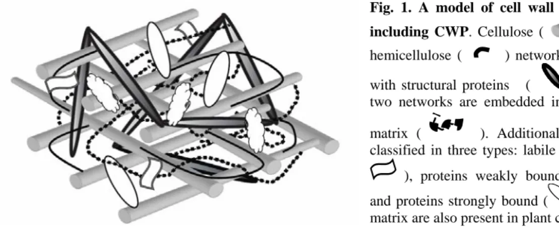
Plant cell walls are dynamic structures essential not only for cell division, enlargement and differentiation, but also for response to environmental constraints (1-3). They are also sources of signals for cell recognition within the same or between different organisms (4, 5). Each cell type is surrounded by a specific cell wall, leading to a great diversity of cell wall structures and compositions (1, 6). Cell walls are natural composite structures, mostly made up of high molecular mass polysaccharides, proteins, and lignins, the latter found only in specific cell types. Polysaccharides represent up to 95% of cell wall mass whereas cell wall proteins (CWP) only account for 5 to 10% of that mass. Models of cell wall structure describe the arrangement of their components into two structurally independent but interacting networks, embedded in a pectin matrix (7, 8). Cellulose microfibrils and hemicelluloses constitute the first network; the second one is formed by structural proteins among which extensins (Fig. 1).
CWP contribute to all cell wall functions and are essential actors in plants. Several studies exemplify unexpected roles for CWP during development or during interactions with pathogens. For instance, the roles of extensins have been restricted to that of structural proteins for many years. However, the phenotype of the rsh (At1g21310,
root-shoot-hypocotyl-defective) mutant showed that a particular extensin is essential for
correct positioning of the cell plate during cytokinesis in embryo cells (9). Due to incorrect position of planes of division, rsh embryos have irregular cell shape and size giving rise to seedlings incapable of normal development. The A. thaliana COBRA (At5g60920) cell wall protein was shown to be essential for cellulose microfibril orientation: the cob mutant has severe defects in anisotropic expansion associated with
disorganization of cellulose microfibrils and reduction of crystalline cellulose (10).
PMR6 (At3g54920, powdery mildew resistant) encodes a protein showing homology to
pectate lyases and is assumed to play a key role during the infection of A. thaliana by
Erysiphe cichoracearum (3). Indeed, the pmr6 mutant becomes resistant to infection
without induction of well-known defense mechanisms. The cell wall was found to be enriched in pectins with a low degree of esterification and modified at the level of cellulose microfibril H-bonding environment. This new type of disease resistance seems to be based on the loss of a gene required during a compatible interaction.
Fig. 1. A model of cell wall structure including CWP. Cellulose ( ) and hemicellulose ( ) network interacts with structural proteins ( ). These two networks are embedded in a pectin matrix ( ). Additional proteins, classified in three types: labile proteins ( ), proteins weakly bound ( ) and proteins strongly bound ( ) to the matrix are also present in plant cell wall.
Extraction and analysis of CWP present specific constraints in addition to the difficulties usually encountered in proteome analysis, such as protein separation and detection of scarce proteins. Protocols should be adapted to take into account the following specificities. (i) The lack of a delimiting membrane may result in the loss of CWP during the isolation procedure. Thus, ionic strength of grinding buffer should be very low. (ii) Polysaccharide networks of cellulose, hemicelluloses and pectins form potential traps for intracellular proteins that contaminate CWP. A significant decrease in the level of CWP contamination can be obtained by working on living cells without altering their plasma membrane or by purifying uncontaminated cell walls. (iii) CWP are embedded in a polysaccharide matrix and interact in different ways with other cell wall components, making the extraction of some of them challenging. The composition of solutions for extraction of CWP must be adapted to improve their elution from cell walls. (iv) Separation of CWP by 2-dimensional electrophoresis (2-DE) is not very efficient since most CWP are basic glycoproteins. Efficient alternatives to 2-DE depend on a pre-fractionation of CWP by ion-exchange chromatography followed by separation by 1-dimensional electrophoresis (1-DE) or in replacing the electrophoresis step by a direct identification of CWP by peptide sequencing using mass spectrometry coupled to ionic and/or reverse-phase liquid chromatography (LC-MS/MS). (v) Identification of heavily
O-glycosylated CWP is very difficult using peptide mass mapping by MS. It requires
Three types of CWP can be distinguished on the basis of their interactions with cell wall components (11). CWP can have little or no interactions with cell wall polysaccharides or other CWP and thus move in the extracellular space. Such proteins can be found in liquid culture media of cell suspensions and seedlings or can be extracted with low ionic strength buffers. We call this fraction “labile proteins”. Most of them have acidic pI ranging from 2 to 6. Alternatively, CWP can be weakly bound to the matrix by Van der Waals interactions, hydrogen bonds, hydrophobic or ionic interactions. Such proteins can be extracted by salts. Most of them have basic pI ranging from 8 to 11, so that they are positively charged at the acidic pH of cell walls. Even though most of the cell wall polysaccharides are neutral, pectins contain polygalacturonic acid residues that provide negative charges for interactions with basic proteins. Such interactions would be modulated by pH, degree of pectin esterification, Ca2+ concentration, as well as mobility and diffusion coefficients of these macromolecules (8). Two protein domains involved in interactions with pectins have been described. A four Arg residues-domain of a peroxidase was shown to have a high affinity for Ca2+-pectate in vitro (12). A domain of two polygalacturonase-inhibiting proteins comprising four residues of Arg and Lys interacts in vitro with pectin (13). Finally, CWP can be strongly bound to cell wall components so that they are resistant to salt-extraction. As examples, extensins are cross-linked by covalent links (14).
This chapter will provide (i) an overview of plant cell wall proteomic studies, (ii) several strategies to analyze cell wall proteomes, (iii) a description of experimental results and (iv) some proposals for future research.
2. Brief bibliographic review:
Despite the difficulties specific to CWP extraction and analysis, cell wall proteomics has become an active field during the last years. Main results have been obtained with the model plant Arabidopsis thaliana. However, some studies have been performed on
Medicago sativa (15) and Zea mays (16).
Many studies have been performed on cell suspension cultures since it is an abundant material that has been widely used for analysis of composition and structure of cell walls. CWP have been extracted in two different ways: (i) without grinding, by harvesting proteins present in culture medium (17), or by extracting proteins from the surface of living cells with salt solutions (17-19); (ii) starting with cell wall purification, by extracting proteins with solutions containing salts, detergents or denaturing agents (20, 21).
Cell wall proteomic studies of various plant organs have been performed: roots (16), stems (15, 22), rosette leaves (23), etiolated hypocotyls (24), and seedlings cultured in liquid medium (25). Such studies are complementary to those performed on cell suspension cultures confirming that cell wall structure and composition are regulated
during development (1, 6). They also allow comparisons between cell wall proteomes of different organs in relation to their functions.
Due to technical limitations, proteomic studies cannot yet give an exhaustive inventory of CWP. This is the reason why certain studies have been focused on specific sub-proteomes that are not well-represented using global approaches. A Yariv-binding proteome was analyzed to describe arabinogalactan proteins (AGP) that are proteoglycans comprising up to 90% of polysaccharides and specifically recognized by the Yariv reagent through an interaction of the antigen-antibody type (25). Since CWP go through the secretory pathway, most of them are supposed to be glycosylated. Concanavalin A (ConA) was used to select N-glycosylated proteins (22). A sub-proteome was actually obtained: it was enriched in glycoside hydrolases and in multicopper oxidases, but it was missing expansins. Glycosylphosphatidylinositol (GPI)-anchored proteins (GAP) are located at the cell surface and can be involved in signaling or in cell adhesion. Several families of GAP were specifically isolated from lipid rafts using Triton X-114 phase partitioning and sensitivity to phosphatidylinositol-specific phospholipase C (26). CWP secreted in culture medium have been searched in culture medium of seedlings (27). Interestingly, this sub-proteome was missing proteins having interacting domains with proteins such as leucine-rich repeats (LRR) or with polysaccharides such as lectins.
Finally, the characterization of the Z. mays xylem sap proteome should be mentioned since it can reveal proteins related to different steps of xylem differentiation including transition from primary to secondary cell wall, programmed cell death and long-distance signaling between roots and aerial organs. Actually, 97% of the identified proteins were predicted to be secreted (28).
3. Specific methodology and strategies:
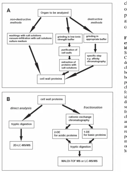
The choice of a protocol to extract CWP for proteomic analysis is dependent on the plant material and on the type of proteins to be released from cell walls. Working on living cells is probably the best solution to avoid intracellular contamination. Three types of non-destructive methods are available (Fig. 2A): (i) analysis of liquid medium of cell suspension cultures or seedlings (17, 27); (ii) washings of cells cultured in liquid medium with salt solutions (17, 18); (iii) vacuum infiltration of organs such as leaves or roots with appropriate extraction buffers (16, 23). Both labile and weakly-bound CWP can be released. When this is not possible or when a specific sub-proteome has to be studied, it is necessary to use a destructive method, i.e. to purify cell walls or any other fraction that can contain CWP such as xylem sap or lipid rafts (Fig. 2B). The main problem is to prevent contamination of CWP by intracellular proteins that will stick non-specifically to cell walls. Mostly weakly- or strongly-bound CWP can be extracted from purified cell walls since labile CWP are probably lost during cell wall preparation. Extracted proteins can be submitted to affinity chromatography to study a sub-proteome: ConA-affinity
chromatography can sort out N-glycosylated
proteins (22); Yariv-affinity chromatography can select AGP (25).
Fig. 2. Strategies for extraction, fractionation and identification of CWP. A. Strategies for extraction of CWP. Non-destructive or destructive methods can be chosen. A global approach can be used or a sub-proteome can be sorted by a specific step of purification, e.g. by affinity chromatography. B. Strategies for protein fractionation and identification. CWP can be directly identified using peptide sequencing by MS or fractionated by cation exchange chromatography into an acidic and a basic fraction prior to 2- and 1-DE electrophoresis respectively. Identification is performed through peptide mass mapping by MALDI-TOF MS or peptide sequencing by LC-MS/MS.
The composition of the extraction solution is critical and determines the type of CWP that can be released from cell walls. A solution of 0.3 M mannitol infiltrated in living tissues such as leaves can solubilize only a few acidic CWP. Such proteins are expected to have no interaction with negatively charged pectins and to be located only in intercellular spaces (23). NaCl is usually used for extraction of proteins retained by ionic interactions in cell walls. LiCl can extract hydroxyproline-rich glycoproteins from intact cells of Chlamydomonas reinhardii (cited in 11). Calcium chloride is probably the most efficient salt for extraction of CWP (23). The ability of acidic and neutral carbohydrates to strongly chelate calcium might explain, through a competition mechanism, that CWP weakly bound to cell wall polysaccharides can be selectively solubilized by CaCl2.
CDTA, a chelating agent, solubilizes Ca2+-pectate. It releases a small number of proteins having domains of interaction with polysaccharides, notably proteins showing homology to lectins (23).
Once extracted, CWP can be analyzed in different ways (Fig. 2B). A direct analysis can be performed by a tryptic digestion, followed by 2D-LC-MS/MS (2 dimensional-liquid chromatography-mass spectrometry) (21). This is a very powerful technique allowing the identification of many proteins by peptide sequencing. It does not require protein separation by electrophoresis thus avoiding the loss of certain types of CWP. An alternative is the fractionation of CWP by cationic exchange chromatography followed by 2-DE or 1-DE for acidic and basic proteins respectively. Tryptic digestion is performed in-gel prior to peptide mass mapping using MALDI-TOF MS or peptide sequencing by LC-MS/MS (23).
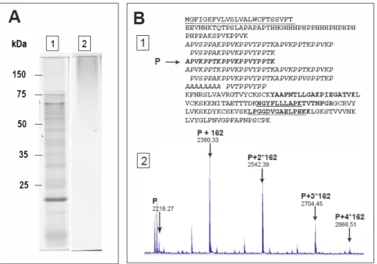
Fig. 3. Analysis of cell wall heavily-glycosylated proteins. A. In-gel staining of AGP with the Yariv reagent. Proteins present in culture medium of 14 day-old etiolated seedlings (27) were concentrated and separated by 1-DE. They were stained either with colloidal blue (A1) or with the Yariv reagent (A2). Molecular masses of markers are indicated on the left. B. Identification of a glycosylated PRP (At1g28290) through mass mapping using MALDI-TOF MS. This protein was found in CWP extract from 11 day-old hypocotyls (24). B1. Amino-acid sequence of the PRP encoded by At1g28290. The predicted signal peptide is underlined. Five non-glycosylated peptides (in boldface/boldface underlined) located in the non repetitive C-terminal domain of the protein allowed protein identification. A specific hydroxyproline-rich peptide (P: APVKPPTKPPVKPPVYPPTK) from the repetitive Pro-rich domain (bold italics) was identified with five Hyp (m/z=2218.27). B2. Close-up of the MALDI-TOF MS spectrum showing modified P peptides: unmodified P (m/z 2218.27), P modified by 162.06 Da mass increments (m/z=2380.30 [P+162]; 2542.39 [P+2*162]; 2704.45 [P+3*162]; 2866.51 [P+4*162)] that can correspond to the addition of one to four hexose residues respectively.
Specific difficulties are encountered for hydroxyproline-rich glycoproteins (HRGP) or proline-rich proteins (PRP) that can be heavily glycosylated CWP. Up to now, only a few of these proteins have been identified in proteomic studies. Among HRGP, extensins are structural proteins forming cross-linked networks whereas AGP can be released in cell walls after cleavage of their GPI anchor. Such proteins are poorly separated using classical techniques of electrophoresis, their biased composition in amino acids and their high level of glycosylation prevent appropriate tryptic digestion for peptide mass mapping. However, the presence of HRGP among CWP can be revealed by hydroxyproline measurement. Although AGP cannot be identified using mass spectrometry without deglycosylation (26), they can be revealed after 1-DE by staining with the Yariv reagent (Fig. 3A). Some PRP could be identified using MALDI-TOF MS through peptide mass mapping of their non-glycosylated parts (Fig. 3B1). In that case, it was also possible to identify modified peptides carrying hydroxyproline residues and sugars on MALDI-TOF MS spectra (Fig. 3B2). A more powerful method for protein identification consists in a preliminary step of deglycosylation with anhydrous hydrogen fluoride to remove O-linked sugars (25).
The reliability of protein profiling for a compartment like the cell wall strongly depends on the quality of the extraction protocol. The classical methods to check the purity of a particular fraction are not conclusive for proteomic studies, since the sensibility of the analysis by mass spectrometry is 10 to 1000 times higher than enzymatic or immunological tests using specific markers. There have been many discussions about the possibility to find non canonical proteins in cell walls, i.e. proteins known or predicted to be intracellular (11, 29). The most efficient way to evaluate the quality of a protocol of CWP extraction is (i) to identify all the proteins by mass spectrometry, and (ii) to perform extensive bioinformatic analysis to determine if the identified proteins contain a signal peptide, and no retention signals for other cell compartments. Several programmes should be used to ensure a reliable prediction: PSORT allows predicting any sub-cellular localization (http://psort.ims.u-tokyo.ac.jp/form.html); TargetP looks for the presence of signal peptides for protein secretion or transit peptides for mitochondrion or chloroplast targeting (http://www.cbs.dtu.dk/services/TargetP/). It is then possible to conclude about the quality of the extraction protocol by calculating the ratio of predicted secreted proteins to intracellular ones.
4. Experimental results and applications:
Results of proteomic studies are usually shown as tables of raw data making their interpretation very difficult. Prediction of protein function or functional domains by bioinformatics is a very powerful tool to classify CWP, to infer biological or biochemical functions performed by CWP and to point out proteins yet unknown that can perform new functions. Several programs have to be used to get reliable information: BLAST allows to find homologous proteins and to describe multigene families (http://www.ncbi.nlm.nih.gov/BLAST/A); ScanProsite looks for patterns or motifs stored
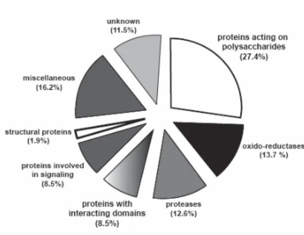
Fig. 4. Distribution of A. thaliana CWP identified through proteomic studies in functional classes. The database comprises 365 proteins from several proteomic studies (17-27). Protein sequences were analyzed with several bioinformatics programmes to look for functional domains and homologies to proteins of known functions. They were sorted into eight functional classes: proteins acting on polysaccharides, oxido-reductases, proteases, proteins with interacting domains, proteins involved in signaling, structural proteins, proteins of unknown function, and miscellaneous. The percentage of proteins in each functional class is indicated between brackets.
in the PROSITE database among which motifs with high probability of occurrence that can be useful to look for putative sites of post-translational modifications (http://www.expasy.ch/tools/scanprosite/); InterProScan searches for motifs or profiles stored in several databases (http://www.ebi.ac.uk/InterProScan/). Using such tools, an arrangement of proteins in functional classes was proposed to get an overview of the A.
thaliana cell wall proteomes (11). Of course this classification has to evolve to take into
account new experimental results obtained by biochemical or genetic approaches. However, the assumption is that proteins sharing conserved domains have the same activity. It should be noted that the biochemical function of only a small portion of the identified proteins was experimentally demonstrated. A CWP database was constructed (11) and now includes 365 proteins coming from several proteomic studies performed on
A. thaliana (17-27). About 88.5 % of these CWP could be distributed in seven categories
on the basis of predicted biochemical or biological functions (Fig. 4). The remaining 11.5% consist in proteins of yet unknown function, some of them being only present in plants. Proteins acting on polysaccharides are the most abundant proteins (27.4%). They are glycoside hydrolases, glycoside transferases, carbohydrate esterases, carbohydrate lyases and expansins. Two functional classes of CWP are of equal importance: oxido-reductases (13.7%) include peroxidases, multicopper oxidases, berberine-bridge enzyme (S)-reticulin:oxygen oxido-reductases and germins; proteases (12.6%) are of several types, i.e. subtilisins, aspartic proteases, cysteine proteases and serine carboxypeptidases. Proteins having interacting domains (8.5%) include proteins interacting with proteins through LRR domains, lectins interacting with sugars, enzyme inhibitors such as polygalacturonase inhibitor proteins (PGIP), pectin methylesterases inhibitors (PMEI) and protease inhibitors. Proteins involved in signaling (8.2%) are mainly AGP and LRR-receptor protein kinases that have been identified through their extracellular LRR domains. Other proteins of various functions (16.2%) were put together in a functional class called “miscellaneous” among which are proteins homolog to acid phosphatases, blue copper binding proteins and proteins having lipase/acyl hydrolase domains. These proteins are awaiting additional experimental data to be more precisely classified.
Are proteomes exhaustive descriptions of the protein content of cell walls? As mentioned above, global approaches cannot lead to the extraction of all proteins from cell walls due to the diversity of protein structures, abundance, and interactions with cell wall components. Additional limitations are separation and analysis techniques. Each proteome is thus missing CWP. A first example is provided by the rosette proteome that is lacking peroxidases whereas peroxidase activities have been found in rosette leaves (23). The use of several salt solutions to release CWP by vacuum-infiltration was probably not sufficient to elute peroxidases that can be bound to Ca2+-pectates (13). As a second example, structural proteins are under-represented in all cell wall proteomes. Either those proteins were not extracted from cell walls due to their cross-linking in networks through covalent links (12) or they were not identified using MS due to their high level of glycosylation.
Do proteomes take into account post-translational modifications? Although trains of spots separated by 2-DE are mentioned in many studies, only a few experimental data are available to show that identified proteins can be isoforms differing by post-translational modifications. Glycosylation of several CWP was visualized by fluorescent staining with ProQ Emerald® 300 (Molecular Probes, USA): as many as 12 and 17 isoforms were stained for a subtilisin serine protease and a PGIP respectively (28). The presence of N-glycosylations was shown on the COBRA protein by a shift in electrophoretic mobility following a treatment with a peptide N-glycosidase (10). The presence of a GPI anchor on COBRA was shown by treatment of membranes with a phosphatidylinositol specific phospholipase C that led to a release of the protein in the aqueous phase (10). Phosphorylation of a lectin (At1g78850) and a chitinase (At3g12500) was shown after separation by 2-DE and positive reaction to antibodies against phosphotyrosine residues (30).
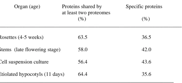
Are the same proteins found in the cell walls of different plant organs? It is yet difficult to answer this question for several reasons. Since quantitative data are missing, comparisons should rely on qualitative data, i.e. presence/absence of a protein. Proteomes should be obtained using comparable protocols for extraction, separation and identification of proteins by MS. Differences in polysaccharide composition, cell wall structure, a lower abundance of the protein, or post-translational modifications might be responsible for the absence of a protein in a proteome. However, taking into account all these restrictions, a comparison between several proteomes of A. thaliana is proposed in Table I: 4-5 week-old rosettes (23), stems at the late flowering stage (22), cell suspension cultures (17, 19-21) and 11 day-old hypocotyls (24). It is noteworthy that despite the presence of proteins of all functional classes in each proteome, a great part of each proteome (between 36.5 and 43.6% of identified proteins) is specific. These differences are mainly due to the presence of different members of the same protein families which might be differently regulated during development. The case of oxido-reductases is shown in Table II. As many as 23 peroxidases were identified as well as 8 multicopper oxidases and 6 berberine-bridge (S)-reticulin:oxygen oxido-reductases. Some of them
were common to several proteomes; others were specific to a single one. Only 6 proteins were found in the four proteomes compared: the XYL1 alpha-xylosidase (At1g68560), the AtXTH4 xyloglucan endotransferase (At2g06850), the SKS5 multicopper oxidase homolog to SKU5 (At1g76160), two lectins with curculin-like domains (At1g78850, At1g78860) and a protein homolog to the tomato xyloglucan-specific endoglucanase inhibitor protein (XEGIP, At1g03220).
5. Concluding remarks:
Proteomics greatly contributed to a better knowledge of CWP that are believed to play crucial roles in cell wall structure, cell wall polysaccharide modifications, plant defense, signaling and lignification. The organ-specificity of cell wall proteomes can be related to the diversity of cell walls and to the complexity of their composition and structures. The great diversity of CWP, and especially of glycoside hydrolases, found in rosette and stem proteomes was not expected. It suggests a great plasticity of cell walls, even in well-differentiated tissues. New proteins of yet unknown functions were identified among which major cell wall proteins. The characterization of post-translational modifications in relation to protein function has already started thanks to progress in MS technologies. The presence of phosphatases, proteases and glycoside hydrolases suggests a complex regulation of CWP involving various types of post-translational processing events such as de-phosphorylation and hydrolytic processing by proteases or glycosidases. Finally, the identification of different types of proteins contributing to the same physiological process should help understanding cell wall functions.
6. Five year viewpoint:
Although a lot of information on cell wall proteomes is now available, many questions concerning CWP remain unanswered. They concern the exhaustive description of proteomes, the regulation of gene expression through post-translational modifications and protein degradation, the role of the cell wall in signaling via oligosaccharides or oligopeptides generated by glycoside hydrolases or proteases, the precise biological function of proteins for which a biochemical function is predicted, and the role of proteins of yet unknown function.
Present cell wall proteomes are lacking strongly-bound proteins such as structural proteins. Big efforts should be made to extract and to analyze those CWP that are physically-linked to cell wall components. On the one hand, enzymes or chemicals could be used to degrade cell wall polysaccharides while maintaining protein integrity. Either new types of CWP should be released or the same types of proteins as with salt solutions but linked in other ways to cell wall components. On the other hand, separation of CWP should be improved prior to identification by MS. Steps of chromatography could be introduced prior to 1-DE for a better fractionation. MS analysis could also be done on peptide mixtures directly obtained from CWP to skip the electrophoresis step that appears to be very limiting.
A comprehensive understanding of gene regulation requires considering all steps from gene transcription to protein degradation. More and more, comparisons of data of transcriptomics and proteomics show that the amount of an mRNA is not strictly correlated to that of the translated protein (31). Protein degradation is rarely taken into account although it is certainly an essential step considering the high number of proteases among CWP. Development of reliable and easy-to-use techniques of protein quantification is required for accurate comparison of proteomes of different plant organs or from various physiological stages. Moreover, post-translational modifications can greatly affect protein function. Since CWP are frequently glycosylated, N- and O-deglycosylation protocols should be improved. The development of MS techniques should facilitate the analysis of post-translational modifications, thus increasing our knowledge of CWP structures in relation to protein activity (32).
In addition to its great contribution to the knowledge of CWP, proteomics can contribute understanding CWP functions. Cell walls are assumed to play key roles in signaling during development and in response to environmental constraints. First molecular basis for such roles were provided by the discovery of oligosaccharides named elicitors. Many glycoside hydrolases are present in all proteomes (21.1% of CWP) including those from “fully differentiated” organs. Looking for substrates of such enzymes would give a lot of information on oligosaccharides mobile in cell walls. Proteases are assumed to release oligopeptides, now considered as peptide hormones most of which are extracellular (33). As an example, the A. thaliana SDD1 (At1g04110,
stomatal density and distribution) cell wall protease was shown to be involved in
stomatal patterning (34). The sdd1-1 mutant exhibits stomata clustering and increased stomatal density. It is assumed that SDD1 generates an extracellular signal regulating the number of asymmetric divisions in satellite meristematoids. The challenge is now to identify the substrates of such proteases, to characterize released protein fragments as well as their targets. Indeed, oligopeptides below 10 kDa yet escape proteomic analysis.
The biological functions of the majority of the identified proteins have not yet been experimentally studied. Proteomics give information on the presence/absence of the protein in an organ or in response to environmental constraints and on post-translational modifications eventually essential for its functionality. Bioinformatic predictions provide useful clues to design relevant experiments for understanding its biochemical and biological functions. When proteins are encoded by orphan genes, no hint can be inferred. In all cases, a full description of CWP biological functions will require complementary approaches including genetics, biochemistry, study of pattern of expression and immunocytochemistry. Unexpected cell wall functions will probably arise from these studies.
The importance of protein/polysaccharide and protein/protein interactions has been neglected up to now to understand supra-molecular assembly of cell wall components. Actually, bioinformatic analysis of weakly-bound CWP identified by proteomic studies
showed that about 8.5% of them have domains of interaction with proteins or polysaccharides. Moreover, the importance of cell wall modifying enzymes in all proteomes (27.4%) was probably not anticipated. All these proteins contribute to cell wall structure and modifications during development or in response to environmental constraints. They are candidates to design new biotechnological tools to get cell walls with modified structures for industrial purposes.
Acknowledgements
The authors are grateful to the Université Paul Sabatier (Toulouse III, France) and the CNRS for support.
Bibliography
1. Roberts K. 2001. How the cell wall acquired a cellular context. Plant Physiol 125: 127-130.
2. Ellis C, Karafyllidis I, Wasternack C, Turner JG.The arabidopsis mutant cev1 links cell wall signaling to jasmonate and ethylene responses. 2002. Plant Cell 14: 1557-1566.
3. Vogel JP, Raab TK, Somerville CR, Somerville SC. Mutations in PMR5 result in powdery mildew resistance and altered cell wall composition. 2004. Plant J 40: 968-978.
4. Pennell R. 1998. Cell walls structures and signals. Curr Opin Plant Biol 1: 504-510. 5. Brownlee C. Role of the extracellular matrix in cell-cell signalling: paracrine
paradigms. 2002. Curr Opin Plant Biol 5: 396-401.
6. Freshour G, Clay RP, Fuller MS, Albersheim P, Darvill AG, Hahn MG. 1996. Developmental and tissue-specific structural alterations of the cell-wall polysaccharides of Arabidopsis thaliana roots. Plant Physiol 110: 1413-1429.
7. Carpita N, Gibeaut D. 1993. Structural models of primary cell walls in flowering plants: consistency of molecular structure with the physical properties of the walls during growth. Plant J 3: 1-30.
8. Cosgrove DJ. 2005. Growth of the plant cell wall. Nat Rev Mol Cell Biol 6: 850-861.
9. Hall Q, Cannon MC. 2002. The cell wall hydroxyproline-rich glycoprotein RSH is essential for normal embryo development in Arabidopsis. Plant Cell 14: 1161-1172. 10. Roudier F, Fernandez AG, Fujita M, Himmelspach R, Borner GHH, Schindelman G, Song S, Baskin TI, Dupree P, Wasteneys GO, Benfey PN. 2005. COBRA, an Arabidopsis extracellular glycosyl-phosphatidyl inositol-anchored protein, specifically controls highly anisotropic expansion through its involvement in cellulose microfibril orientation. Plant Cell 17: 1749-1763.
11. Jamet E, Canut H, Boudart G, Pont-Lezica RF. 2006. Cell wall proteins: a new insight through proteomics. Trends Plant Sci 11: 33-39.
12. Carpin S, Crevecoeur M, de Meyer M, Simon P, Greppin H, Penel C. 2001. Identification of a Ca(2+)-pectate binding site on an apoplastic peroxidase. Plant Cell13: 511-520.
13. Spadoni S, Zabotina O, Di Matteo A, Mikkelsen JD, Cervone F, De Lorenzo G, Mattei B, Bellincampi D. 2006. Polygalacturonase-inhibiting protein interacts with pectin through a binding site formed by four clustered residues of arginine and lysine. Plant Physiol 141: 557-64.
14. Brady JD, Sadler IH, Fry SC. 1996. Di-isodityrosine, a novel tetrameric derivative of tyrosine in plant cell wall proteins: a new potential cross-link. J Biochem 315: 323-327.
15. Watson BS, Lei Z, Dixon RA, Sumner LW. 2004. Proteomics of Medicago sativa cell walls. Phytochem 65: 1709-1720.
16. Zhu J, Chen S, Alvarez S, Asirvatham VS, Schachtman DP, Wu Y, Sharp RE. 2006. Cell wall proteome in the maize primary root elongation zone. I. Extraction and identification of water-soluble and lightly ionically bound proteins. Plant Physiol 140: 311-325.
17. Borderies G, Jamet E, Lafitte C, Rossignol M, Jauneau A, Boudart G, Monsarrat B, Esquerré-Tugayé MT, Boudet A, Pont-Lezica R. 2003. Proteomics of loosely bound cell wall proteins of Arabidopsis thaliana cell suspension cultures: a critical analysis. Electrophoresis 24: 3421-32.
18. Kwon HK, Yokoyama R, Nishitani K. 2005. A proteomic approach to apoplastic proteins involved in cell wall regeneration in protoplasts of Arabidopsis suspension-cultured cells. Plant Cell Physiol 46: 843-857.
19. Robertson D, Mitchell GP, Gilroy JS, Gerrish C, Bolwell GP, Slabas AR. 1997. Differential extraction and protein sequencing reveals major differences in patterns of primary cell wall proteins from plants. J Biol Chem 272: 15841-15848.
20. Chivasa S, Ndimba B, Simon W, Robertson D, Yu X-L, Knox J, Bolwell P, Slabas A. 2002. Proteomic analysis of the Arabidopsis thaliana cell wall. Electrophoresis 23: 1754-1765.
21. Bayer EM, Bottrill AR, Walshaw J, Vigouroux M, Naldrett MJ, Thomas CL, Maule AJ. 2006. Arabidopsis cell wall proteome defined using multidimensional protein identification technology. Proteomics 6: 301-311.
22. Minic, Z. 2006. Laboratoire de Biochimie des Signaux Régulateurs Cellulaires et Moléculaires, FRE2621 CNRS, Paris. Personal communication.
23. Boudart G, Jamet E, Rossignol M, Lafitte C, Borderies G, Jauneau A, Esquerré-Tugayé M-T, Pont-Lezica R. 2005. Cell wall proteins in apoplastic fluids of
Arabidopsis thaliana rosettes: Identification by mass spectrometry and
bioinformatics. Proteomics 5: 212-221.
24. Feiz L, Irshad M, Pont-Lezica RF, Canut H, Jamet E. 2006. Evaluation of cell wall preparations for cell wall proteomics. Plant Methods 2:10.
25. Schultz CJ, Ferguson KL, Lahnstein J, Bacic A. Post-translational modifications of arabinogalactan-peptides of Arabidopsis thaliana. 2004. J Biol Chem 279: 45503-44511.
26. Borner GH, Lilley KS, Stevens TJ, Dupree P. 2003. Identification of glycosylphosphatidylinositol-anchored proteins in Arabidopsis. A proteomic and genomic analysis. Plant Physiol 132: 568-577.
27. Charmont S, Jamet E, Pont-Lezica R, Canut H. 2005. Proteomic analysis of secreted proteins from Arabidopsis thaliana seedlings: improved recovery following removal of phenolic compounds. Phytochem 66: 453-461.
28. Alvarez S, Goodger JQD, Marsh EL, Chen EL, Chen S, Asirvatham VS, Schachtman DP. 2005. Characterization of the maize xylem sap proteome. J Proteome Res 5: 963-972.
29. Slabas AR, Ndimba B, Simon WJ, Chivasa S. 2004. Proteomic analysis of the Arabidopsis cell wall reveals unexpected proteins with new cellular locations. Biochem Soc Trans 32: 524-528.
30. Ndimba BK, Chivasa S, Hamilton JM, Simon WJ, Slabas AR. 2003. Proteomic analysis of changes in the extracellular matrix of Arabidopsis cell suspension cultures induced by fungal elicitors. Proteomics 3: 1047-1059.
31. Kolkman A, Daran-Lapujade P, Fullaondo A, Olsthoorn MM, Pronk JT, Slijper M, Heck AJ. 2006. Proteome analysis of yeast response to various nutrient limitations. Mol Syst Biol 2: 2006.0026.
32. Morelle W, Canis K, Chirat F, Faid V, Michalski JC. 2006. Characterization of N-glycans of recombinant human thyrotropin using mass spectrometry. Proteomics 6: 3993-4015.
33. Matsubayashi Y, Sakagami Y. 2006. Peptide hormones in plants. Annu Rev Plant Biol 57: 649-674.
34. von Groll U, Berger D, Altmann T. 2002. The subtilisin-like serine protease SDD1 mediates cell-to-cell signaling during Arabidopsis stomatal development. Plant Cell 14: 1527-1539.
Table I. Specificities of A. thaliana cell wall proteomes. Results are expressed as percentages of total number of proteins identified in each proteome: 4-5 week-old rosettes (23), stems at the late flowering stage (22), cell suspension cultures (17, 19-21, 26), 11 day-old etiolated hypocotyls (24).
Organ (age) Proteins shared by Specific proteins at least two proteomes
(%) (%)
____________________________________________________________________
Rosettes (4-5 weeks) 63.5 36.5
Stems (late flowering stage) 58.0 42.0
Cell suspension culture 56.4 43.6
Etiolated hypocotyls (11 days) 64.4 35.6
Table II. Origin of the specificity of cell wall proteomes: some examples taken from A. thaliana multigene families of oxido-reductases. Three families of oxido-reductases are analyzed: peroxidases, multicopper oxidases and homolog to berberine-bridge (S)-reticulin:oxygen oxido-reductases. Same proteomes as in Table I are compared: Ros, rosettes; St, stems; CSC, cell suspension culture; EH, etiolated hypocotyls; CM, culture medium of etiolated seedlings.
Gene Protein Ros St CSC EH CM
____________________________________________________________________ Peroxidases At1g05240 AtPrx01 + At1g49570 AtPrx10 + At1g71695 AtPrx12 + At2g18140 AtPrx14 + + At2g18150 AtPrx15 + + At2g38380 AtPrx22 + + At3g01190 AtPrx27 + At3g03670 AtPrx28 + At3g21770 AtPrx30 + + At3g28200 AtPrx31 + At3g32980 AtPrx32 + + At3g49110 AtPrx33 + At3g49120 AtPrx34 + + + + At4g08770 AtPrx37 + At4g30170 AtPrx45 + + At4g36430 AtPrx49 + At5g05340 AtPrx52 + At5g06720 AtPrx53 + + At5g17820 AtPrx57 + At5g42180 AtPrx6 4 + At5g64100 AtPrx69 + At5g64120 AtPrx71 + + At5g66390 AtPrx72 + Multicopper-oxidases At4g25220 SKS1 + At4g22010 SKS4 + + At1g76160 SKS5 + + + + At1g41830 SKS6 + + + At1g21860 SKS7 + At4g38420 SKS9 + At5g66920 SKS17 + At4g12420 SKU5 + +
Gene Protein Ros St. CSC EH CM
_______________________________________________________________ Homologs to berberine-bridge (S)-reticulin:oxygen oxido-reductase
At1g30730 + At2g34790 + + At4g20830 + + At5g44380 + + At5g44390 + At5g44400 +