HAL Id: hal-01275050
https://hal.archives-ouvertes.fr/hal-01275050
Submitted on 3 Feb 2017
HAL is a multi-disciplinary open access
archive for the deposit and dissemination of sci-entific research documents, whether they are pub-lished or not. The documents may come from teaching and research institutions in France or abroad, or from public or private research centers.
L’archive ouverte pluridisciplinaire HAL, est destinée au dépôt et à la diffusion de documents scientifiques de niveau recherche, publiés ou non, émanant des établissements d’enseignement et de recherche français ou étrangers, des laboratoires publics ou privés.
Biocidal Properties of a Glycosylated Surface:
Sophorolipids on Au(111)
Claire Valotteau, Christophe Calers, Sandra Casale, Jan Berton, Christian V.
Stevens, Florence Babonneau, Claire-Marie Pradier, Vincent Humblot, Niki
Baccile
To cite this version:
Claire Valotteau, Christophe Calers, Sandra Casale, Jan Berton, Christian V. Stevens, et al.. Biocidal Properties of a Glycosylated Surface: Sophorolipids on Au(111). ACS Applied Materials & Inter-faces, Washington, D.C. : American Chemical Society, 2015, 7 (32), pp.18086-18095. �10.1021/ac-sami.5b05090�. �hal-01275050�
IMPORTANT NOTE : Please be aware that slight modifications occurring after Proof
correction may occur between this version of the manuscript and the version on the
Publisher’s website---
Biocidal Properties of a Glycosylated Surface: Sophorolipids on Au(111)
Claire Valotteau,
1,2Christophe Calers,
2Sandra Casale,
2Jan Berton,
3Christian V. Stevens,
3Florence Babonneau,
1Claire-Marie Pradier,
2Vincent Humblot,
2,*Niki Baccile.
1,*
1 Sorbonne Universités, UPMC Univ Paris 06, CNRS, Collège de France, Laboratoire de Chimie de la
Matière Condensée de Paris, UMR 7574, 11 place Marcelin Berthelot, 75005 Paris, France.
2 Sorbonne Universités, UPMC Univ Paris 06, CNRS, Laboratoire de Réactivité de Surface, UMR 7197,
4 place Jussieu, 75005 Paris, France.
3 SynBioC Research Group, Departement of Sustainable Organic Chemistry and Technology, Faculty of
Bioscience Engineering, Ghent University, Coupure links 653, B-9000 Gent, Belgium
*Corresponding authors: [email protected] and [email protected]
Keywords: sophorolipid, glycolipid, surface functionalisation, self-assembled monolayer, antibacterial coating, killing-by-contact, biocidal mechanism
Abstract
Classical antibacterial surfaces usually involve antiadhesive and/or biocidal strategies. Glycosylated surfaces are usually used to prevent biofilm formation via antiadhesive mechanisms. We report here the first example of a glycosylated surface with biocidal properties created by the covalent grafting of sophorolipids (a sophorose unit linked by a glycosidic bond to an oleic acid) through a self-assembled monolayer (SAM) of short aminothiols on gold (111) surfaces. The biocidal effect of such surfaces on Gram+ bacteria was assessed by a wide combination of techniques including microscopy observations, fluorescent staining and bacterial growth tests. About 50% of the bacteria are killed via alteration of the cell envelope. In addition, the role of the sophorose unit and aliphatic chain configuration are highlighted by the lack of activity of substrates modified respectively with sophorose-free oleic acid and sophorolipid-derivative having a saturated aliphatic chain. This system
demonstrates thus the direct implication of a carbohydrate in the destabilization and disruption of the bacterial cell envelope.
Introduction
Biological contamination of surfaces is a major concern in medical and pharmaceutical industries 1,
food-processing installations, water pipes, naval ships as well as in monuments and cultural heritage materials. Bacteria, fungi or algae are inclined to adhere and grow onto solid surfaces, leading to the formation of biofilms 2, that may lead to undesired phenomena like loss of adhesion properties,
degradation or infection. Surface contamination has an important socio-economical impact. For instance, nosocomial infections in the U.S.A. were estimated to 1.7 million in 2002 and leading to slightly less than 100,000 death cases 3. Moreover, bacteria in biofilms are generally more resistant 4,
thus making the antibiotics ineffective in the long run. To prevent these problems, a large amount of research is carried out to develop preventive antimicrobial coatings on surfaces, including either antiadhesive or biocidal strategies. In the latter, one mainly finds either a killing-by-contact 5 or a
killing-by-release 6 approach, where the active compound can be antibiotic 7, quaternary ammonium
derivatives 5, nanoparticles 8, etc., either blended into or deposited onto a substrate 9, or chemically
grafted onto surfaces. Nevertheless, the ecotoxicity of most of these compounds, the increasing resistance of microorganisms, or the complexity of the grafting strategy restrict their practical use at large scales. Bio-derived compounds, e.g. enzymes, molecules able to interfere with quorum-sensing phenomena or antimicrobial peptides, appear as appealing alternatives for their broad range of action, their efficiency at very low concentration and the absence of bacterial resistance. However, their high cost of synthesis and purification, pH sensitivity, susceptibility to proteolysis, as well as potential local toxicity and allergy after repeated applications, may restrict their use 10.
In this context, carbohydrates, one of the most common compounds in living systems, are of increased interest for a large number of medical and pharmalogical-related applications, like vaccines and anticancer drugs 11, fabrication of glycoarrays for antibody recognition and cell adhesion 12, antibiofilm and antimicrobial formulations 13, just to cite some. Even if carbohydrates alone are a
source of nutrition to microorganisms rather than biocidal compounds, some reports relate the antimicrobial effects of sugar-containing molecules, such as bacterial exopolysaccharides, extracted from Escherichia coli, 14 and few glycoconjugates, carbohydrates covalently linked to an organic
moiety (from simple alkyl chain to more complex structures), of both synthetic and natural origins. For instance, amphotericin B, a complex glycolipid extracted from Streptomyces nodosus, is used to treat fungal infections 15 and other compounds display interesting antimicrobial properties 13. Until
now, only two forms of antimicrobial actions of carbohydrate derivatives have been described: 1) a biocidal action via cellular lysis promoted by glycolipids in solution 13; 2) an antiadhesive effect,
(glycoarray) or free in solution 13, 16-20. Even though the mechanisms of action are still largely debated,
the biocidal action is likely due to the amphiphilic nature of the glycolipids, which penetrate into the plasma membrane, causing lysis and possible leakage of cytoplasm material 21-23. Since neither pure
carbohydrates, nor most common lipids, like fatty acids 24, have specific biocidal properties, one
generally refers to as a “surfactant effect”, where the role in the membrane lysis cannot be attributed to a specific part of the molecule, but rather to its physico-chemical properties in solution
25, 26. In terms of the antiadhesive effect, this is believed to depend on competing protein-sugar
interactions between the pathogen and the substrate, where proteins refer to carbohydrate-binding proteins like lectins and adhesins present in the cell envelope. Playing with the nature of a carbohydrate coating (type, mobility, number of sugars), one can either increase or reduce the affinity of cells for a surface, thus promoting, or discouraging, cell adhesion phenomena 16-18.
In the context of carbohydrate-driven antimicrobial activity, we show here the first example of a glycosylated surface with biocidal properties. To do so, we use sophorolipids (SL), a family of bolaform glycolipids characterized by a sophorose unit (glucose β(1-2)), linked through a glycosidic bond to oleic acid (Figure 1), and having a COOH group free of access for chemical grafting at the opposite side of the carbohydrate. Sophorolipids are characterized by a low toxicity, high biodegradability, an environmental compatibility and possess both antifungal and/or antimicrobial properties in solution 13, 27, 28. They are produced in large amounts by the yeast Starmerella bombicola
29, they are already commercialized worldwide (e.g., Soliance, Ecover, IntoBio, Saraya) 30 and,
probably the most interesting feature, they are ready as-such, needing no specific chemical synthesis nor complex protection/deprotection modification. They constitute an ideal platform for large-scale antimicrobial surface applications, if compared to competitors like antimicrobial peptides or customed glyconjugates. In addition, our approach demonstrates the direct involvement in the cell envelope-destabilization/disruption of a carbohydrate, the role of which is largely debated in the biocidal properties of glycolipids in solution31 but also in the more general context of
carbohydrate/lipidic membrane interactions 32.
The open acidic form of sophorolipids (SL) will be grafted on model flat gold (111) surfaces using short thiolamines as primers (cysteamine, cys). The modified gold surfaces are characterized step by step by Polarization-Modulation Reflection Absorption Infrared Spectroscopy (PM-RAIRS) and X-ray Photoelectron Spectroscopy (XPS). Antimicrobial activity is tested against Listeria ivanovii, a Gram positive bacterium, surrounded by a thick peptidoglycan layer without outer membrane, since sophorolipids appears more effective in solution against bacteria exhibiting such cell envelope. Antimicrobial properties are qualitatively and quantitatively evaluated through a combination of Atomic Force Microscopy (AFM) and Scanning Electron Microscopy with Field Emission Gun
(SEM-FEG), fluorescent staining and bacterial growth tests and compared to blank samples constituted by the gold surface alone and the cysteamine-modified gold surface. In order to elucidate the role of the sophorose group in the biocidal action, we develop and graft on control gold surfaces three SL-related molecules, either bearing the same sugar but a different chain, or playing with the chain without sugar (Figure 1) : 1) oleic acid (OA), which mimics only the aliphatic chain of sophorolipids, 2) a sophorolipid-derivative having a fully-saturated aliphatic chain (SL0), important to observe the
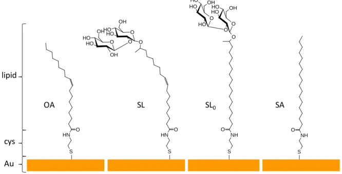
possible role of the molecular conformation at the gold surface due to the presence of a C=C double bond and 3) stearic acid (SA), which mimics the aliphatic chain of the fully-saturated sophorolipid-derivative. OA SL SL0 Au cys lipid SA N H O O O O H O H O OH O O H O H OH OH SH NH O S H N H O SH NH O O O O H O H O OH O O H O H O H OH S H
Figure 1. Functionalization of gold surfaces with oleic acid (OA), acidic monounsaturated sophorolipid (SL), acidic fully saturated sophorolipid (SL0) and stearic acid (SA). Cysteamine (cys) constitutes the primer for all compounds.
Materials and Methods
Cysteamine (cys), N-hydroxysuccinimide (NHS), 1-(3-dimethylaminopropyl)-N-ethylcarbodiimide hydrochloride (EDC), oleic acid (OA), stearic acid (SA), formaldehyde, dimethyl sulfoxide and sodium chloride (NaCl) were obtained from Sigma Aldrich (Saint Quentin Fallavier, France). Sophorolipids (SL) were derived from a commercial acidic and lactone mixture of sophorolipids purchased from Soliance (France) (Sopholiance S, batch number: 11103A, dry content: 60 ± 6 %). To obtain a high purity (> ~92 mol%, by 1H NMR) form of the non-acetylated acidic sophorolipids only, we employed a
classical hydrolysis route using 5 M NaOH followed by acidification by HCl and pentanol extraction (please refer to ref. 33 for more details) of the sophorolipid only. The fully-saturated sophorolipid
described in ref. 34. All solvents were reagent-grade and were used without any further purification.
Water was purified with a milliQ system (Millipore, resistivity > 18 MΩ.cm−1) from EMD Millipore
Corp. (Billerica, MA, USA). Glass substrates, coated successively with a 50 Å thick layer of chromium and a 200 nm thick layer of gold were purchased from Arrandee (Werther, Germany).
Surface preparation
The substrates have been prepared using a standard protocol 35. Prior to use, the gold-coated
substrates were annealed in a butane flame to ensure a good crystallinity of the top layer, after what they were exposed to UV-ozone during 15 minutes and rinsed successively in a bath of ultrapure water and in a bath of absolute ethanol during 10 minutes. First, the substrates were immersed in an ethanolic solution of Cys at 10 mM. After 3 hours, the substrates were sonicated in ultrapure ethanol to desorb the non-grafted molecules and thoroughly rinsed in ethanol and then in ultrapure water before being dried under a flow of dried air. Next, to promote the grafting of the SL on the thiol-amine, the carboxylic acid termination of SL (50 mg.L-1) was activated by succinimide ester using a
mixture of EDC (77 mg.L-1) and NHS (23 mg.L-1) in water. After 1 hour under stirring, the cys-modified
gold substrates were immersed for 3 hours in this solution. Successive rinsing in ultrapure water and ethanol were performed to remove non-covalently grafted reactants before drying under a flow of dried air. Quartz Crystal Microbalance was used to verify that the rinsing procedure efficiently eliminated the excess of unreacted SL. OA, SA and SL0 were grafted onto gold following a similar
protocol, using ethanol as solvent.The as-obtained surfaces are depicted in Figure 1. All samples were characterized by PM-RAIRS and XPS after each step of functionalization.
Characterization Techniques
Polarized Modulated Reflexion Absorption InfraRed Spectroscopy (PM-RAIRS)
PM-RAIRS measurements were performed using a Nicolet Nexus 5700 FT-IR spectrometer equipped with a nitrogen-cooled HgCdTe wide band detector. Infrared spectra were recorded at 8 cm-1
resolution, by co-addition of 128 scans. A ZnSe grid polarizer and a ZnSe photoelastic modulator were placed prior to the sample in order to modulate the incident beam between p and s polarizations (HINDS Instruments, PM90, modulation frequency = 36 kHz). The sum and difference interferograms were processed and underwent Fourier-transformation to yield the PM-RAIRS signal which is the differential reflectivity (ΔR/R°) = (Rp-Rs)/(Rp+Rs), where Rp is the signal parallel to the incident plane while Rs is the perpendicular contribution. The measurements were done at two different voltages applied to the modulator ZnSe crystal to optimize the sensitivity.
InfraRed Attenuated Total Reflexion (IR-ATR)
IR-ATR measurements were recorded using a Nicolet 5700 FT-IR spectrometer equipped with a nitrogen-cooled MCT detector. Infrared spectra were obtained by co-adding 256 scans at 8 cm-1
resolution and referencing against the background of air.
Atomic Force Microscopy Imaging (AFM)
AFM images of dried surfaces were recorded using a Caliber AFM microscope from Bruker Instruments Inc. Topographic images were taken in the tapping mode. Silicon nitride tips (resonance frequency 280-400 Hz, force constant 40-80 N/m) have been used. Images were obtained with a constant speed of 1 Hz with a resolution of 512 lines and 512 pixels each. The raw data were processed using the imaging processing software Nanoscope Analysis v.1.30. from Bruker (Nano Surfaces Corp. Santa Barbara, Ca, USA) and used mainly for slope corrections.
Scanning Electron Microscopy with Field Emission Gun (SEM-FEG)
SEM images were recorded with a Hitachi SU-70 Field Emission Gun Scanning Electron Microscope. The samples were fixed on an alumina SEM support with a carbon adhesive tape and were observed without metallization. In-Lens Secondary electron detector (SE-Upper) was used to obtain characterize our samples. The accelerating voltage was 1 kV and the working distance was around 5 mm. At least five different locations were analyzed on each surface, arising to the observation of a minimum of 100 single bacteria observed.
Water contact angle measurements
Static water contact angles were measured under ambient conditions (at 20°C and 40% relative humidity) analyzing the drop profile of sessile drops. 10 µL droplet of millQ water was deposited on the sample surface using a Krüss DSA100 apparatus (Germany) equipped with a CCD camera and an image analysis processor. 4 droplets were analyzed on different locations on each sample and the test was performed in triplicate on 3 different samples. The reported values are the averages of these 12 measurements for each kind of surface.
Molecular drawing of SL and SL0 on a flat surface
Sophorolipid SL and SL0 molecules covalently bonded on cysteamine via an amide bond had been
drawn respecting bond lengths, dihedral angles and sense of chirality for each atom. We referred to the IUPAC name for sophorolipids: 17-L-([2-O-β-D-glucopyranosyl-β-D-glucopyranosyl]-oxy)-octadecenoic acid. Drawing and structure relaxation was done with the Gaussview software, version 2.1 while the images have been done using the PyMOL molecular graphic system, version 1.3.
Antimicrobial activity
Strains and culture conditions
Non-human pathogenic bacteria Listeria ivanovii Li4pVS2 were used to investigate the modified surfaces. Unless otherwise indicated, bacterial suspensions were always prepared from stationary phase cultures incubated overnight in brain heart infusion (BHI) broth (BD Difco, France) at 37°C under agitation (250 rpm). The broth was then centrifuged at 10 000 g for 5 minutes, the supernatant was eliminated and the bacteria were re-dispersed in an isotonic sterile solution (NaCl 0.9%). The optical density of the broth was controlled at 620 nm and adjusted at 0.05, which corresponds to 5x10⁶ colony forming unity (CFU) per mL.
Deposition of bacteria on samples
A 100 µL drop of bacteria solution freshly prepared was deposited on each surface sample, which were previously washed in a 70% ethanol aqueous solution and dried in sterile environment. In all experiments, unless otherwise mentioned, the inoculated surfaces were incubated for 3 hours at room temperature under a wet atmosphere.
Evaluation of bacteria adhesion by infrared spectroscopy
After bacterial deposition and incubation during 3 hours, surfaces were washed five times with 100 µL drops of isotonic sterile solution (NaCl 0.9%) and dried under a laminar air flow. Ten infrared spectra were acquired by PM-RAIRS on each sample in order to scan their entire surface and collect the signal from all the adhered bacteria. All measurements (on bare gold and modified surfaces) had been done during the same experimental session, thus reducing at minimum intensity variations of the background, beam intensity and alignment. The relative amounts of bacteria adsorbed were evaluated by considering the amide bands area, the bacteria IR fingerprints. The areas were integrated from 1700 cm-1 to 1500 cm-1 to include the whole signal due to amides I and II
contributions, respectively at 1660 cm-1 and 1550 cm-1. The attachment of bacteria onto the different
surfaces is expressed as a percentage compared to gold substrates: Ad(%) = 100 *(area of amide
bands on sample) / (area of amide bands on gold). The uncertainty attached to this percentage
comes from the propagation of the uncertainties attached to the measurement of amide bands area on both the considered surface and the gold substrate. These results were confirmed by repeating the same procedure above on 3 different set of samples.
Observation of bacterial morphology by microscopy
Qualitative analysis of the bacterial morphology was done by mean of AFM and SEM-FEG microscopies, which enables to visualize the effect of antimicrobial products on bacteria and could help to identify general target sites 25. Since SEM-FEG observations are done under vacuum, samples
were previously treated as follows: after the incubation period, 12 µL of formaldehyde at 37% were added in order to fix the bacteria and avoid collapsing of bacteria upon drying. After 15 minutes, samples were washed six times with 100 µL drops of filtered ultrapure water to remove non-adhered bacteria and dried under a laminar air flow.
Evaluation of bacteria viability by fluorescent staining
Damaged bacteria on the surface were counted using the Live/Dead Bacterial Viability Kit (BacLight©). 1.5 µL of each fluorescent stains, Syto9 and propidium iodide (PI), were added to 1 mL of ultrapure water. After incubation, surfaces were rinsed five times using 100 µL drops of an isotonic sterile solution and 10 µL of the fluorochrome solution were deposited on the surfaces. Samples were then left during 10 minutes in dark and at room temperature prior to microscopy analysis. Surfaces were always kept under a humid environment during the whole time of the experiments to avoid water evaporation. Samples were then examined with an epifluorescence microscope (AXIO 100 Zeiss). Images were acquired with a 10 or 40 objective lens and recorded with a CCD camera (AxioCam MRm Zeiss). Fluorochromes were respectively excited and detected at 455-495 nm and 505-555 nm (for Syto9) and at 533-558 nm and 570-640 nm (for PI). About ten different locations of each surface were analyzed in order to have statistically relevant data; this experiment was conducted on 3 sets of samples and at least a thousand of bacteria were then enumerated. Bacterial counting (red: damaged membrane; green: intact membrane) was done using the software Image J and the viability is calculated as follows: Viability(%) = 100 * (number of green bacteria) / (number of
green bacteria + number of red bacteria). Bacteria growth capacity
The evaluation of bacterial growth capacity after being in contact with the samples completes the characterization data set and was done as follows: after bacterial deposition and incubation during 3 hours, the surfaces were washed five times with 100 µL drops of isotonic sterile solution. Samples were then transferred into a sterile tube containing 2 mL of isotonic sterile solution (NaCl at 0.9 %) and sonicated 5 minutes. The gold sample was removed and bacteria suspensions were diluted 100 and 1000 times. 50 µL of each dilution was deposited in duplicate on petri dishes filled with BHI agar (15 g/L). The plates were incubated at 37°C overnight before enumeration. Results are expressed in percentages of the number of attached and cultivable bacterial cells onto the different surfaces as compared to gold substrates: Growth(%) = 100* (number of colonies forming unity on sample) /
(number of colonies forming unity on bare gold substrate). These tests were done in triplicate on
uncertainty attached to these results follows from the statistical analysis of these repeated experiments.
Minimum Inhibitory Concentration (MIC) and Minimum Bactericidal Concentration (MBC) determination
The MIC values of sophorolipids and derivatives used for surface grafting were determined using a liquid growth inhibition assay. An exponential-phase bacterial culture of Listeria ivanovii was diluted in broth to an optical density of 0.01 (106 CFU/mL). 50 µL of this bacterial suspension were mixed
with 50 μl of two-fold serial aqueous dilutions of each product (final concentrations of sophorolipids and derivatives from 2 to 32 mg/mL). As OA, SA and SL0 are not soluble in water, these products
were first solubilized into 10% of DMSO. The bacterial growth was monitored by measuring the change in optical density. This test was performed in triplicate with positive (0.7% formaldehyde) and negative (isotonic solution) inhibition controls. MIC is expressed as the lowest concentration that completely inhibits bacterial growth after overnight incubation.
The bacterial suspension with a concentration of sophorolipid superior or equal to the MIC are diluted and deposited on agar plate in order to determine the Minimum Bactericidal Concentration (MBC). The MBC is expressed as the lowest concentration for which no colony forming unit are observed on agar plates after overnight incubation.
Results
SL grafting on Au(111)
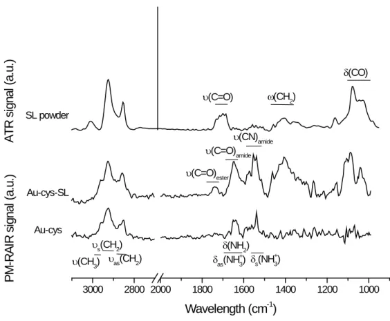
Figure 2 shows the PM-RAIRS spectra of gold surfaces after each functionalization step. After treatment in the cysteamine solution and rinsing, a typical IR spectrum of aminothiol is observed, with the expected absorption bands of NH and CH2 groups as described in literature 35 The grafting of
cysteamine is also confirmed by XPS analyses (bottom spectra in Figure S1 in the Supporting Information section). Then, the PM-RAIRS spectrum of the SL-modified sample (Au-cys-SL) shown in Figure 2 exhibits the typical vibrations expected for SL, as shown by the comparison with the corresponding IR-ATR powder spectrum, as well as the signature of the amide group at 1648 cm-1
(νC=O) and 1538 cm-1 (νCN & δNH). These results confirm that the SL molecules are covalently grafted
on the outer NH2 group of cysteamine via an amidation reaction with the COOH moiety of SL. They
are corroborated by XPS spectra recorded on the Au-cys-SL sample (top spectra in Figure S1 in the Supporting Information section). The additional weak band at 1738 cm-1 on the PM-RAIRS spectrum
of Au-cys-SL, ascribed to a νC=O vibration, may indicate the presence of spurious amounts of either
NHS or NHS-activated sophorolipid despite rinsing. The rinsing step enables yet to eliminate at best the poorly attached molecules and to obtain a stable layer, as shown by Quartz Crystal Microbalance experiments (cf. Figure S2 in Supporting Information section).
3000 2800 2000 1800 1600 1400 1200 1000 υas(CH2) δs(NH+3) δ(CO) υ(CN)amide υ(CH3) υ(C=O) ω(CH2) υs(CH2) δas(NH+3) δ(NH2) SL powder Au-cys-SL Au-cys
Wavelength (cm
-1)
υ(C=O)ester υ(C=O)amideA
T
R
s
ignal
(
a.
u.
)
P
M
-R
A
IR
s
ignal
(
a.
u.
)
Figure 2. IR-ATR spectrum of the monounsaturated acidic sophorolipid powder (SL powder) and PM-RAIRS spectra of the cystamine (Au-cys) and SL-grafted surfaces (Au-cys-SL) with identification of the main contributions
Antimicrobial activity
Surface antimicrobial properties
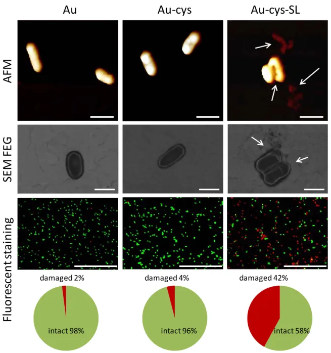
In order to evaluate the antimicrobial activity of the SL-functionalized gold surfaces, we have combined AFM, SEM-FEG with epifluorescence microscopy and microbial viability tests, on both SL-modified (Au-cys-SL) and SL-free gold surfaces (Au and Au-cys). SEM-FEG is a fast screening technique to evaluate eventual morphological changes of the microorganisms. To avoid possible artifacts due to vacuum conditions, bacteria have been fixed on the wet surfaces with formaldehyde, known to cross-link the membrane proteins in their current state. Moreover, we always deposit bacteria on bare gold substrate as a control sample. On the contrary, imaging by AFM, a non-invasive microscopy
technique commonly used to explore mechanical stress on biointerfaces, was performed on as deposited, non-cross linked, bacteria under ambient temperature and pressure conditions.
AFM and SEM-FEG images in Figure 3 show L. ivanovii deposited on SL-free samples (Au and Au-cys). Bacteria are of about 1 µm in length and exhibit the expected elongated rod shape 36. On the
SEM-FEG image, the plasma membrane of the Gram+ bacteria appears as a white line surrounding the ovoidal shape of the microorganism indicating that contact with these surfaces does not alter the cell envelope. On the contrary, on the SL-modified surfaces (Au-cys-SL), bacteria do not have their native oval shape and the cell envelopes are damaged and appear pierced, causing the leakage of the cytoplasmic content and the collapse of the bacteria, as indicated by the arrows in Figure 3 (Au-cys-SL). These observations show direct evidence that the gold surface-grafted with SL damages the plasma membrane of L. ivanovii in agreement with the antibacterial effect reported for sophorolipids in solution 23, 28, 37.
Au
Au-cys
Au-cys-SL
AFM
SE
M
FE
G
Fl
uo
res
cen
t s
tain
in
g
intact 98% damaged 2% damaged 4% intact 96% intact 58% damaged 42%Figure 3. Bacteria (L. ivanovii) deposited on Au (left column), Au-cys (middle column) and Au-cys-SL (right column) surfaces observed by AFM (top range), SEM-FEG (middle range) and epifluorescence microscopy (bottom range). The charts below each image represent the percentage of adhering intact and damaged bacteria according to fluorescent staining. The scale bare represents 1 µm on AFM and SEM-FEG images and 50 µm on images from epifluorescence microscopy.
To confirm these findings and quantify the number of damaged bacteria, bacterial staining with fluorescent markers, Syto9 and PI, was conducted in such way that green-stained bacteria indicate an intact plasma membrane, while red-stained bacteria show damaged cell membrane. Our results show that less than 5% of bacteria are damaged on both Au and Au-cys surfaces while almost half of the bacteria population has a damaged membrane on the Au-cys-SL sample (Figure 3).
However, epifluorescence microscopy does not constitute a direct proof of bacteria viability or death38. Conversely, bacteria were also cultivated on agar plate after having been in contact with the
modified surfaces in order to count the actual number of bacterial colonies. A drastic decrease of
ca 45% of bacterial growth was systematically observed after contact with SL-containing surfaces
compared to the Au and Au-cys samples (Growth (%): Au= 100%, Au-cys= 95%, Au-cys-SL= 54%). These numbers are in very good agreement with the fluorescent staining (Intact bacteria (%): Au= 98%, Au-cys= 96%, Au-cys-SL= 58%) and confirm the antibacterial effect of SL-modified surfaces via membrane lysis. Note in addition that the rate of damaged bacteria stays constant over time from 1 hour up to at least 6 hours in contact with the surface. Similarly, we have found the same biocidal efficiency at different growth stages (exponential phase or stationary phase) of L. ivanovii.
Understanding the origin of biocidal effect of SL
The data presented so far put in evidence several striking facts: 1) surface-grafted SL keep their biocidal action against Gram+ Listeria ivanovii; 2) the killing efficiency is in the 40 % - 45 % range; 3) the apparent mechanism of action occurs through membrane lysis, as described for the same compounds (and related glycolipids) in aqueous solutions 21-23, 28. The persistence of the antimicrobial
properties of SL after their grafting onto a surface demonstrates that the carboxylic acid group may not play a key role in their antibacterial property, since the COOH is neutralized by the anchoring strategy. This is in line with the comparisons of lactone-type and acid-type sophorolipids activity in solution, which show that the former has a higher antimicrobial activity 28.
To put in evidence the role of the sophorose unit, we have controlled the possible biocidal action of OA-grafted gold surface. In addition, to understand the potential role of the conformation of sophorose at the outer surface we have tested the biocidal action of the fully saturated form of acidic sophorolipids (SL0) and the corresponding fatty acid (SA), as shown in Figure 1. It is, in fact,
known that the anomer conformations of a synthetic glycolipids strongly influence their antibacterial activity in solution; for instance, the alpha anomer of the lauric ether derivative of methyl glucopyranoside has a MIC of 0.04 M, which is roughly 100 times smaller (hence, more efficient) than the corresponding beta anomer 31. For these reasons, even if OA and SA are not supposed to have
antibacterial effects in solution 24, and since we are not aware of such properties for SL
0, we have
tested the MIC in solutions of these compounds. Using DMSO to improve solubility, and then diluting the solution in water, SL0, OA and SA do not exhibit any inhibitory effect, even at high concentrations
(32 mg/mL, i.e. eight-fold above the MIC value found in this work for SL, 4 mg/mL) and bacteria show their oval native shape and intact plasma membranes when observed by SEM-FEG (images not shown). Nevertheless, since SL0 forms supramolecular water-insoluble fibers 34, it is difficult to obtain
reliable antibacterial data in solution and therefore the corresponding grafted surfaces appear as an interesting tool to explore the interactions between these molecules and bacteria.
OA, SA and SL0-grafted surfaces have been elaborated and characterized following the same
procedure as the one used for SL grafting (see description above): the corresponding infrared and XPS data (shown in Figure S3 and Figure S4 in the Supporting Information) confirm the grafting of SL0
on gold in equivalent amounts that SL (both layers have equivalent thickness, 30.0±2.0 Å for SL and 31±5 Å for SL0, according to estimation based on XPS analyses – see Supporting information), as well
as OA and SA, making the comparison of antibacterial properties of these surfaces possible. Figure 4 displays the observations of bacteria deposit on these modified surfaces.
Au-cys-OA
Au-cys-SL
0Au-cys-SA
SE
M
FE
G
Fl
uo
res
cen
t s
tain
in
g
intact 98% damaged 2% intact 98% damaged 2% intact 97% damaged 3%Figure 4. Bacteria (L. ivanovii) deposited on Au-cys-OA (left column), Au-cys-SL0 (middle column) and Au-cys-AS (right column) surfaces observed by SEM-FEG (top range) and epifluorescence microscopy (bottom range). The charts below each image represent the percentage of adhering intact and damaged bacteria according to fluorescent staining. The scale bares represent 1µm on AFM images and 50 µm on images from epifluorescence microscopy.
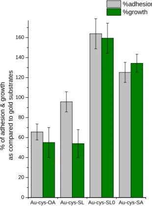
Bacteria exhibit their native oval shape on SEM images and look intact on all samples. The harmless features of these surfaces is confirmed by fluorescent staining: more than 97% of the adhered bacteria appear green after fluorescent staining, meaning that their membrane was not damaged by any of the tested compounds. The corresponding colony counting data after exposure are reported in Figure 5 (green bars); however, these data must be weighted by the adhesion (% with respect to bare gold) of the bacteria onto the surfaces (grey bars in Figure 5). In fact, the adhesion capacity of L.
ivanovii is not constant on all modified surfaces. When in contact with SL-modified surfaces, the
bacterial adhesion is very similar (95 %-100 %, within the experimental error bars) to what it is observed onto gold, but their growth is reduced by approximately 50 %. This difference nicely confirms, from a quantitative point of view, the biocidal action of grafted SL, previously highlighted by fluorescent staining. Interestingly, on OA-modified surfaces, both adhesion and growth are reduced by ~40 % with respect to the gold surface, thus meaning that this surface has a slight anti-adhesive effect, but it is not biocidal, as also highlighted by the fluorescence staining experiments, cf. Figure 4. Finally, the adhesive nature of SL0 and, at a lower extent, the one of SA are higher than bare
Au or SL surfaces (the adhesion on those surface being respectively estimated to 160 and 130 % as compared to gold); the growth counting, which is found to be almost 60 % and 30 % higher with respect to gold for SL0 and SA, respectively, is then solely attributed to the adhesive properties of
these surfaces.
Au-cys-OA Au-cys-SL Au-cys-SL0 Au-cys-SA 0 20 40 60 80 100 120 140 160 % of adhes ion & grow th as c ompared t o gol d s ubs tr at es %adhesion %growth
Figure 5. Adhesion (grey bars) and bacterial growth (green bars) of Listeria ivanovii after contact with OA, Au-cys-SL, Au-cys-SL0 and Au-cys-SA surfaces as compared to Au substrates.
In order to better understand the interactions between bacteria and surfaces, water contact angles on the modified surfaces have been determined (Figure 6) as it may influence the contact between bacteria and surfaces, and hence the biocidal effect.
Au cys OA SL SA SL0 45 50 55 60 65 70 75 W at er c ont ac t angl e ( °)
Figure 6. Water contact angle on bare gold surface and on functionalized surfaces.
The gold surface becomes more hydrophobic after functionalization with OA and SA, for which the water contact angles are respectively 55° ± 2° and 62° ± 2°. The sophorose units, hydroxyl-rich water-soluble disaccharides, of SL lead to an expected decrease of the contact angle with respect to OA from 55° ± 2° to 48° ± 2°, whereas the contact angle increases from 62° ± 2° to 71° ± 2° when passing from SA to SL0. The antinomic effect in terms of surface wetability between SL (48° ± 2°) and SL0 (71°
± 2°) is unexpected, if one looks at the chemical nature of both quasi identical molecules. Nevertheless, this is not surprising; examples of conformational changes strongly affecting the wetability of coatings exist for peptides, where the chirality sense of the peptide backbone governs the hydrophilic/hydrophobic character of the coated surface 39. It is also known that glucose-based
surfaces/compounds can display different wetability properties. Cyclodextrins, for instance, are water-soluble compounds with a hydrophobic cavity 40, as the orientation of the C-OH groups in
glucose identifies regions of different water-loving properties 41. A striking example is constituted by
two allomorphs of cellulose, Iα and Iβ, the (100) surfaces of which are, respectively, hydrophilic and
hydrophobic 41. The stronger wetability in Iα was demonstrated to come from both the larger
moieties of the Iβ are parallel to the surface (low roughness) and more C-OH groups are involved in
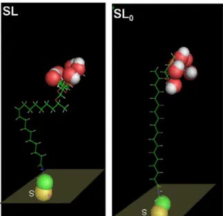
hydrogen bonding with their close neighbors. In the case of SL-modified surfaces, we then believe that the double bond present in the SL molecule favors the preferential exposure of C-OH groups compared to the SL0 coatings. Interestingly, if one lays SL and SL0 molecules on a flat surface
respecting the theorethical bond lengths, dihedral angles and chirality for both the sugar and C17 atom in the aliphatic chain, it is then possible to see that the actual orientation of the C-OH groups is orthogonal to the surface for SL, while it is parallel to it for SL0, as shown in Figure 7, and thus
explaining the hydrophilicity of the SL-containing substrate if compared to the SL0 substrate. In
addition, the bending of the SL molecule may increase local disorder and a loss of molecular packing, similarly to differences of molecular packing and water-solubility encountered in solution: at room temperature and under acidic conditions, SL forms micelles 33 (water-soluble) while SL0 forms
water-insoluble crystalline fibers with nanoscale chirality 34.
Figure 7. SL (left) and SL0 (right) lying on top of a model surface shown with highlighted C-OH group of sophorose using the sphere representation (Oxygen: red; Hydrogen: white; Sulfur: yellow; Nitrogen: green).
Discussion.
By comparing the bacteria morphology and viability on OA, SA, SL0 and SL grafted samples, one can
safely assert that only surfaces with the unsaturated form of sophorolipids have a biocidal activity (to this regard, please also see the comment in the PM-RAIRS section, page 4, of the SI). The simultaneous presence of the hydrophilic sophorose group and the double bonds appears to be necessary to induce bacterial killing. These two structural features have also a crucial influence on several physico-chemical properties. Sophorose is a bulky disaccharide and its steric hindrance influences the conformation of SL: the surface exhibits a loose packing as observed for antimicrobial peptides chemically grafted on thiolated self assembled monolayers, for instance 35. The presence of
the double bond induces curvature in the alkyl chain layer, thus contributing to the loose, probably defect-rich, packing of the SL compound. The orientation of sophorose is also directly related to the constraints imposed by the C=C double bond (120° angle), and so is influencing the intermolecular hydrogen bonding network. The use of a fully saturated (stearic) alkyl chain in SL0 has several
important consequences; it is in fact known that molecular packing is tighter for stearic acid based compound 42. The so different values of the water contact angles of Au-cys-SL and Au-cys-SL
0 suggest
that the orientation of the sophorose is different on gold, maybe correlated to the so different biocidal properties (40% to 50% growth inhibition on SL modified-surfaces compared to almost none on SL0 modified-surfaces). Consequently, a variability of the killing-by-contact efficiency could be due
to two combined reasons: the disaccharide orientation and local disorder.
To understand the importance of these findings, one must recall the role of carbohydrates in biocidal compounds, often mentioned but never clarified. In the first place, the biocidal properties of glycolipids have always been found for free molecules in solution, where it is impossible to dissociate the action of the sugar from its covalently bonded backbone. Nevertheless, some works show that the efficiency against a given organism depends on the nature of the sugar. For instance, sucrose monolaurate is less effective towards Enterococcus faecalis than lactose monolaurate, but their activities are comparable towards Listeria monocytogenes 43, 44. Besides, a stronger piece of evidence
that the nature of the carbohydrate has an important role was reported by Nobmann et al. on the effect of alpha or beta anomer on the MIC, as discussed previously 31.
When polysaccharides or sugars are grafted on a surface, the main reported effect concerns the enhancement, or reduction, of microorganisms adhesion, but no biocidal effect 19. The decrease of
bacterial viability observed on gold samples modified with monounsaturated sophorolipids suggests a killing-by-contact action related to the presence of covalently grafted molecules, whereas the hypothetical role of micelles is excluded, since the initial sophorolipid concentration is of 50 µg.mL-1,
by Quartz Crystal Microbalance experiments, the grafted layer remains stable after the rinsing procedure performed at the end of the grafting (cf. Figure S2 in Supporting Information). In any case, even if all sophorolipids present at the gold surface after rinsing (~180 ng according to QCM experiments, as exposed in the Supporting Information section) are released into the 100 µL drop of bacterial suspension used in the antibacterial tests, the concentration would be less than 2 µg.mL-1,
thus assuring to be below the typical MIC and the MBC for free sophorolipids (respectively 4 and 32 mg.mL-1, as measured in this study). Excluding the hypothesis according to which bacteria may be
killed by residual free sophorolipid molecules, it also automatically excludes the fact that grafted sophorolipids act via the classical “surfactant effect”, as described in solution for many other glycolipids, and more in general surfactant molecules, or even peptides 21-23, 28, 46. Here SL can only
interfere with the bacterial cell envelope through the sophorose moiety, which is not able to diffuse neither through the cell wall nor the plasma membrane. We also make the assumption that sophorolipids, characterized by an ether covalent bond between sophorose and oleic acid (ether-based glycolipids are known to be stable during their antibacterial activity 31), are stable at the gold
surface towards potential hydrolysis by bacterial esterase. Another favorable argument consists in the fact that if sophorolipids would be hydrolyzed by L. ivanovii, we would not expect an antibacterial activity at all, as the residual oleic acid moiety has none. The killing process is then directly related to the contact between the cell envelope and the sophorose headgroup. Moreover, there is a general consensus on the fact that the most effective biocidal chemical groups (e.g. antimicrobial peptides, alkylammonium derivatives) are able to approach and pierce the cell wall thanks to their rigid and electrically-charged nature. The action of cationic polymers for instance is usually described as disrupting the lipidic membranes of bacteria through electrostactic and hydrophobic interactions 47.
Similar mechanisms are also suggested for antimicrobial peptides 25. In comparison, because of their
electro-neutrality, monounsaturated sophorolipids have little chance to cause membrane lysis after grafting. We believe that the origin of their biocidal effect is rather related to specific sophorose/cell envelope interactions. However, these have never been described in the literature.
To understand the origin of cell envelope damage in our system, we thus make two hypotheses, biochemical and/or biophysical (or both simultaneously). The biochemical pathway refers to a possible interaction between sophorose and the microorganism which triggers a (series of) biochemical response(s) responsible for the cell envelope rupture. This is the classical way penicillin antibiotics work by inhibiting the transpeptidase, enzymes responsible for the cross-linking of the peptidoglycan layer.48 Similar arguments are also used to explain the mechanism of action of the
moenomycin antibiotic, a well-known glycolipid, and its derivatives. They inhibit the in vitro transglycosylation reaction in E. coli, thus blocking the synthesis of the cell wall and its eventual
death, with or without lysis 49-51. In addition, a potential role as signaling molecules that regulate
gene expression has been discussed to explain the antiadhesive properties of some bacterial exopolysaccharides 16. If these specific mechanisms do not seem to be applicable to the case of
surface-grafted SL against Listeria (since it seems difficult for anchoring SL to interfere with the inner metabolism of the bacteria), similar sophorose-induced biochemical pathways may not be completely excluded, even though unknown at the moment. The biophysical pathway refers to a mechanical, destabilizing, interaction between the carbohydrate and the Listeria cell envelope, which is mainly constituted by a peptidoglycan and phospholipid layers 52, 53. If the action of sophorose on
the cell envelope is out of doubt, the way such interaction occurs is still unknown. Specific literature data neither exist on this specific system nor, to the best of our knowledge, on the more general destabilization of the peptidoglycan layer by mono- and disaccharides. As far as an action on the plasma membrane, the lysis of which is put in evidence here by the epifluorescence observations (if PI reaches the nucleic acid of red bacteria, it means their plasma membrane is damaged), one could formulate two different mechanistic hypotheses, admitting the previous interpenetration of the cell wall: 1) intra-membrane sugar intercalation 54, 55, 56, perturbing the local liquid crystalline order, as
observed in the case of other non-reducing disaccharides (e.g., trehalose and sucrose) 32 .
2) Membrane destabilization mediated by recent fundamental studies by Abbott et al. go in this sense, showing the destabilizing effects the macromolecular adsorbents of biological relevance (e.g., proteins) may have on model phospholipid liquid crystal membranes 57. They have shown that not
only weak binding of proteins drive the reorganization of the phospholipids and trigger orientational transitions in the liquid crystals, but also that similar events can also occur by ligand (biotin)-receptor (streptavidin) recognition phenomena, where streptavidin is contained in the liquid crystal layer and biotin in an external phospholipid vesicles approaching it 58. It is not known at the moment if any of
these hypotheses, generally formulated in the case of interactions between free molecules and a cell envelope, can be applied to the present biocidal effect of surfaces-grafted sophorolipids and it deserves more fundamental studies. Besides, whatever the mechanism, the main reason for which the saturated SL0 sophorolipid does not seem to have any biocidal property could be multiple: poor
accessibility of individual surface sophorose groups, enhanced bacterial adhesion, both limiting the accessibility of sophorose and consequent interaction with membrane penetration.
Conclusion
This paper reports the conception of an innovative biocidal coatings using carbohydrates immobilized on a surface. Sophorolipids, yeast-derived bolaform biosurfactants, were successfully attached to a model flat gold surface modified by a thiolamine primer through amidation of their COOH group. The
resulting glycosylated surface, where sophorose (glucose β−1,2), a non-reducing disaccharide, lays on top of the surface, shows a biocidal activity towards Listeria ivanovii bacteria. So far, glycosylated surfaces have been known for their anti-adhesive properties. A combination of microscopy tools shows that bacteria are killed through membrane lysis. Bacterial growth and adhesive tests show that between 40 % and 50 % of exposed bacteria are damaged; these values are coherent with the quantitative data obtained from fluorescent staining. To better understand the mechanism of action, the effect of sophorolipids was compared to rational molecular variations of this compound: oleic acid, constituting the aliphatic backbone of sophorolipids, fully saturated sophorolipids and stearic acid, the aliphatic component of the latter, have been grafted on gold and no biocidal activity could be detected. This comparative study then underlines the crucial role played by sophorose but it also highlights the importance of the aliphatic chain configuration, influencing the substrate wettability, bacterial adhesion and biocidal effects. If this first example of a biocidal effect from a glycan array demonstrates the membrane disrupting properties of a non-reducing disaccharide, the actual mechanism of interaction between sophorose and the bacterial membrane is still unknown, and it may not still be confined to this particular sugar alone.
Financial Support
This work was supported by French state funds managed by the ANR within the Investissements d'Avenir programme under reference ANR-11-IDEX-0004-02, and more specifically within the framework of the Cluster of Excellence MATISSE.
Acknowledgements
The authors acknowledge IMPC (Institut des Matériaux de Paris Centre, FR2482) and the C'Nano projects of the Region Ile-de-France, for SEM-FEG and Omicron XPS apparatus funding. Christophe Méthivier is deeply acknowledged for his important help in XPS calculations. Frederik Tielens (LCMCP, Université Pierre et Marie Curie, Paris, France) is kindly acknowledged for his help in molecular drawing in the SI. Bastien Wild (LHYGES, Université de Strasbourg, France) is kindly acknowledged for his initial help in the experimental part.
Supporting Information showing detailed XPS, QCM and PM-RAIRS additional data and analyses of SL, SL0, OA and SA on Au-cys substrates. The material is available free of charge via Internet at
References
(1) Hall-Stoodley, L.; Costerton, J. W.; Stoodley, P. Bacterial Biofilms: From the Natural Environment to Infectious Diseases. Nat. Rev. Microbiol. 2004, 2, 95-108.
(2) Costerton, J. W.; Lewandowski, Z.; Caldwell, D. E.; Korber, D. R.; Lappin-Scott, H. M. Microbial Biofilms. Annu. Rev. Microbiol. 1995, 49, 711-745.
(3) Klevens, R. M.; Edwards, J. R.; Richards, C. L.; Horan, T. C.; Gaynes, R. P.; Pollock, D. A.; Cardo, D. M. Estimating Health Care-Associated Infections and Deaths in U.S. Hospitals, 2002. Public Health
Rep. 2007, 122, 160-166.
(4) Lewis, K. Multidrug Tolerance of Biofilms and Persister Cells. Curr. Top. Microbiol. 2008, 322, 107-131.
(5) Tiller, J. C.; Liao, C. J.; Lewis, K.; Klibanov, A. Designing Surfaces That Kill Bacteria on Contact.
Proc. Natl. Acad. Sci. U. S. A. 2001, 98, 5981-5985.
(6) Cado, G.; Aslam, R.; Séon, L.; Garnier, T.; Fabre, R.; Parat, A.; Chassepot, A.; Voegel, J. C.; Senger, B.; Schneider, F.; Frère, Y.; Jierry, L.; Schaaf, P.; Kerdjoudj, H.; Metz-Boutigue, M. H.; Boulmedais, F. Self-Defensive Biomaterial Coating against Bacteria and Yeasts: Polysaccharide Multilayer Film with Embedded Antimicrobial Peptide. Adv. Funct. Mater. 2013, 23, 4801-4809. (7) Aumsuwan, N.; Heinhorst, S.; Urban, M. W. The Effectiveness of Antibiotic Activity of Penicillin Attached to Expanded Poly(Tetrafluoroethylene) (Eptfe) Surfaces: A Quantitative Assessment. Biomacromolecules 2007, 8, 3525-3530.
(8) Lee, D.; Cohen, R. E.; Rubner, M. F. Antibacterial Properties of Ag Nanoparticle Loaded Multilayers and Formation of Magnetically Directed Antibacterial Microparticles. Langmuir 2005, 21, 9651-9.
(9) Rocha, M.; Ferreira, F. A.; Souza, M. M.; Prentice, C., Antimicrobial Films: A Review. In
Microbial Pathogens and Strategies for Combating Them: Science, Technology and Education, A.
Méndez-Vilas, E., Ed. Formatex: 2013; Vol. 1, pp 23-31.
(10) Glinel, K.; Thébault, P.; Humblot, V.; Pradier, C.-M.; Jouenne, T. Antibacterial Surfaces Developed from Bio-Inspired Approaches. Acta Biomater. 2012, 8, 1670-1684.
(11) Osborn, H. M.; Evans, P. G.; Gemmell, N.; Osborne, S. D. Carbohydrate-Based Therapeutics. J.
Pharm. Pharmacol. 2004, 56, 691-702.
(12) Laurent, N.; Voglmeir, J.; Flitsch, S. L. Glycoarrays--Tools for Determining Protein-Carbohydrate Interactions and Glycoenzyme Specificity. Chem. Commun. 2008, 4400-12.
(13) Cortes-Sanchez, A. d. J.; Hernandez-Sanchez, H.; Jaramillo-Flores, M. E. Biological Activity of Glycolipids Produced by Microorganisms: New Trends and Possible Therapeutic Alternatives.
Microbiol. Res. 2013, 168, 22-32.
(14) Valle, J.; Da Re, S.; Henry, N.; Fontaine, T.; Balestrino, D.; Latour-Lambert, P.; Ghigo, J. M. Broad-Spectrum Biofilm Inhibition by a Secreted Bacterial Polysaccharide. Proc. Natl. Acad. Sci. U. S.
A. 2006, 103, 12558-63.
(15) Brajtburg, J.; Powderly, W. G.; Kobayashi, G. S.; Medoff, G. Amphotericin B: Current Understanding of Mechanisms of Action. Antimicrob. Agents Chemother. 1990, 34, 183-188.
(16) Bernal, P.; Llamas, M. A. Promising Biotechnological Applications of Antibiofilm Exopolysaccharides. Microb. Biotechnol. 2012, 5, 670-673.
(17) Fessele, C.; Lindhorst, T. K. Effect of Aminophenyl and Aminothiahexyl Α-D-Glycosides of the Manno-, Gluco-, and Galacto-Series on Type 1 Fimbriae-Mediated Adhesion of Escherichia Coli.
(18) Hartmann, M.; Horst, A. K.; Klemm, P.; Lindhorst, T. K.; Hartmann, M.; Horst, A. K.; Klemm, P.; Lindhorst, T. K. A Kit for the Investigation of Live Escherichia Coli Cell Adhesion to Glycosylated Surfaces. Chem. Commun. 2010, 46, 330-332.
(19) Kesel, S.; Mader, A.; Seeberger, P. H.; Lieleg, O.; Opitz, M. Carbohydrate Coating Reduces Adhesion of Biofilm-Forming Bacillus Subtilis to Gold Surfaces. Appl. Environ. Microbiol. 2014, 80, 5911-5917.
(20) Wehner, J. W.; Hartmann, M.; Lindhorst, T. K. Are Multivalent Cluster Glycosides a Means of Controlling Ligand Density of Glycoarrays? Carbohydr. Res. 2013, 371, 22-31.
(21) Chapman, J.; Diehl, M.; Lyman, R. Biocide Susceptibility and Intracellular Glutathione in
Escherichia Coli. J. Ind. Microbiol. 1993, 12, 403-407.
(22) Glover, R. E.; Smith, R. R.; Jones, M. V.; Jackson, S. K.; Rowlands, C. C. An Epr Investigation of Surfactant Action on Bacterial Membranes. FEMS Microbiology Letters 1999, 177, 57-62.
(23) Lang, S.; Katsiwela, E.; Wagner, F. Antimicrobial Effects of Biosurfactants. Fett/Lipid 1989, 91, 363-366.
(24) Kabara, J. J.; Swieczkowski, D. M.; Conley, A. J.; Truant, J. P. Fatty Acids and Derivatives as Antimicrobial Agents. Antimicrob. Agents Chemother. 1972, 2, 23-28.
(25) Brogden, K. A. Antimicrobial Peptides: Pore Formers or Metabolic Inhibitors in Bacteria? Nat.
Rev. Microbiol. 2005, 3, 238-250.
(26) Hurdle, J. G.; O'Neill, A. J.; Chopra, I.; Lee, R. E. Targeting Bacterial Membrane Function: An Underexploited Mechanism for Treating Persistent Infections. Nat. Rev. Microbiol. 2011, 9, 62-75. (27) Baek, S. H.; Sun, X. X.; Lee, Y. J.; Wang, S. Y.; Han, K. N.; Choi, J. K.; Noh, J. H.; Kim, E. K. Mitigation of Harmful Algal Blooms by Sophorolipid. J. Microbiol. Biotechnol. 2003, 13, 651-659. (28) Kim, K.; Dalsoo, Y.; Youngbum, K.; Baekseok, L.; Doonhoon, S.; Eun-Ki, K. Characteristics of Sophorolipid as an Antimicrobial Agent. J. Microbiol. Biotechnol. 2002, 12, 235-241.
(29) Van Bogaert, I. N. A.; Zhang, J.; Soetaert, W. Microbial Synthesis of Sophorolipids. Process
Biochem. 2011, 46, 821-833.
(30) Mukherjee, S.; Das, P.; Sen, R. Towards Commercial Production of Microbial Surfactants.
Trends Biotechnol. 2006, 24, 509-515.
(31) Nobmann, P.; Smith, A.; Dunne, J.; Henehan, G.; Bourke, P. The Antimicrobial Efficacy and Structure Activity Relationship of Novel Carbohydrate Fatty Acid Derivatives against Listeria Spp. And Food Spoilage Microorganisms. Int. J. Food Microbiol. 2009, 128, 440-5.
(32) Moiset, G.; Lopez, C. A.; Bartelds, R.; Syga, L.; Rijpkema, E.; Cukkemane, A.; Baldus, M.; Poolman, B.; Marrink, S. J. Disaccharides Impact the Lateral Organization of Lipid Membranes. J. Am.
Chem. Soc. 2014, 136, 16167-75.
(33) Baccile, N.; Cuvier, A.-S.; Valotteau, C.; Van Bogaert, I. N. A. Practical Methods to Reduce Impurities for Gram-Scale Amounts of Acidic Sophorolipid Biosurfactants. Eur. J. Lipid Sci. Technol.
2013, 115, 1404-1412.
(34) Cuvier, A.-S.; Berton, J.; Stevens, C. V.; Fadda, G. C.; Babonneau, F.; Van Bogaert, I. N.; Soetaert, W.; Pehau-Arnaudet, G.; Baccile, N. Ph-Triggered Formation of Nanoribbons from Yeast-Derived Glycolipid Biosurfactants. Soft Matter 2014, 10, 3950-3959.
(35) Yala, J.-F.; Thébault, P.; Héquet, A.; Humblot, V.; Pradier, C.-M.; Berjeaud, J.-M. Elaboration of Antibiofilm Materials by Chemical Grafting of an Antimicrobial Peptide. Applied Microbiol. Biotechnol.
2011, 89, 623-634.
(36) Robichon, D.; Girard, J.-C.; Cenatiempo, Y.; Cavellier, J.-F. Atomic Force Microscopy Imaging of Dried or Living Bacteria. C. R. Acad. Sci., Ser. III 1999, 322, 687-693.
(37) Azim, A.; Shah, V.; Doncel, G. F.; Peterson, N.; Gao, W.; Gross, R. Amino Acid Conjugated Sophorolipids: A New Family of Biologically Active Functionalized Glycolipids. Bioconjugate Chem.
2006, 17, 1523-1529.
(38) Roszak, D. B.; Colwell, R. R. Survival Strategies of Bacteria in the Natural Environment.
(39) Qing, G.; Sun, T. Chirality-Driven Wettability Switching and Mass Transfer. Angew. Chem., Int.
Ed. 2014, 53, 930-932.
(40) Martinez, A.; Ortiz Mellet, C.; Garcia Fernandez, J. M. Cyclodextrin-Based Multivalent Glycodisplays: Covalent and Supramolecular Conjugates to Assess Carbohydrate-Protein Interactions.
Chem. Soc. Rev. 2013, 42, 4746-4773.
(41) Hoja, J.; Maurer, R. J.; Sax, A. F. Adsorption of Glucose, Cellobiose, and Cellotetraose onto Cellulose Model Surfaces. J. Phys. Chem. B 2014, 118, 9017-9027.
(42) Small, D. M. Lateral Chain Packing in Lipids and Membranes. J. Lipid Res. 1984, 25, 1490-500. (43) Devulapalle, K. S.; Gomez de Segura, A.; Ferrer, M.; Alcalde, M.; Mooser, G.; Plou, F. J. Effect of Carbohydrate Fatty Acid Esters on Streptococcus Sobrinus and Glucosyltransferase Activity.
Carbohydr. Res. 2004, 339, 1029-34.
(44) Wagh, A.; Shen, S.; Shen, F. A.; Miller, C. D.; Walsh, M. K. Effect of Lactose Monolaurate on Pathogenic and Nonpathogenic Bacteria. Appl. Environ. Microbiol. 2012, 78, 3465-8.
(45) Imura, T.; Masuda, Y.; Minamikawa, H.; Fukuoka, T.; Konishi, M.; Morita, T.; Sakai, H.; Abe, M.; Kitamoto, D. Enzymatic Conversion of Diacetylated Sophoroselipid into Acetylated Glucoselipid: Surface-Active Properties of Novel Bolaform Biosurfactants. J. Oleo Sci. 2010, 59, 495-501.
(46) Bechinger, B.; Lohner, K. Detergent-Like Actions of Linear Amphipathic Cationic Antimicrobial Peptides. Biochim. Biophys. Acta, Biomembr. 2006, 1758, 1529-1539.
(47) Siedenbiedel, F.; Tiller, J. C. Antimicrobial Polymers in Solution and on Surfaces: Overview and Functional Principles. Polymers 2012, 4, 46-71.
(48) Yocum, R. R.; Rasmussen, J. R.; Strominger, J. L. The Mechanism of Action of Penicillin. Penicillin Acylates the Active Site of Bacillus Stearothermophilus D-Alanine Carboxypeptidase. Journal
of Biological Chemistry 1980, 255, 3977-86.
(49) Baizman, E. R.; Branstrom, A. A.; Longley, C. B.; Allanson, N.; Sofia, M. J.; Gange, D.; Goldman, R. C. Antibacterial Activity of Synthetic Analogues Based on the Disaccharide Structure of Moenomycin, an Inhibitor of Bacterial Transglycosylase. Microbiology 2000, 146, 3129-3140.
(50) Van Heijenoort, Y.; Derrien, M.; Van Heijenoort, J. Polymerization by Transglycosylation in the Biosynthesis of the Peptidoglycan of Escherichia Coli K 12 and Its Inhibition by Antibiotics. FEBS
Letters 1978, 89, 141-144.
(51) Van Heijenoort, Y.; Van Heijenoort, J. Biosynthesis of the Peptidoglycan of Escherichia Coli K-12: Properties of the in Vitro Polymerization by Transglycosylation. FEBS Letters 1980, 110, 241-244. (52) Ghosh, B. K.; Carroll, K. K. Isolation, Composition, and Structure of Membrane of Listeria
Monocytogenes. J. Bacteriol. 1968, 95, 688-699.
(53) Vazquez-Boland, J. A.; Kuhn, M.; Berche, P.; Chakraborty, T.; Dominguez-Bernal, G.; Goebel, W.; Gonzalez-Zorn, B.; Wehland, J.; Kreft, J. Listeria Pathogenesis and Molecular Virulence Determinants. Clinical Microbiol. Rev. 2001, 14, 584-640.
(54) Crowe, J. H.; Whittam, M. A.; Chapman, D.; Crowe, L. M. Interactions of Phospholipid Monolayers with Carbohydrates. Biochim. Biophys. Acta 1984, 769, 151-9.
(55) Lenné, T.; Bryant, G.; Holcomb, R.; Koster, K. L. How Much Solute Is Needed to Inhibit the Fluid to Gel Membrane Phase Transition at Low Hydration? Biochim. Biophys. Acta, Biomembr. 2007, 1768, 1019-1022.
(56) Andersen, H. D.; Wang, C.; Arleth, L.; Peters, G. H.; Westh, P. Reconciliation of Opposing Views on Membrane–Sugar Interactions. Proc. Natl. Acad. Sci. U. S. A. 2011, 108, 1874-1878.
(57) Brake, J. M.; Daschner, M. K.; Luk, Y. Y.; Abbott, N. L. Biomolecular Interactions at Phospholipid-Decorated Surfaces of Liquid Crystals. Science 2003, 302, 2094-2097.
(58) Tan, L. N.; Orler, V. J.; Abbott, N. L. Ordering Transitions Triggered by Specific Binding of Vesicles to Protein-Decorated Interfaces of Thermotropic Liquid Crystals. Langmuir 2012, 28, 6364-6376.