Cutaneous melanoma: ESMO Clinical Practice
Guidelines for diagnosis, treatment and follow-up
†R. Dummer
1, A. Hauschild
2, M. Guggenheim
3, U. Keilholz
4& G. Pentheroudakis
5, on behalf of the
ESMO Guidelines Working Group*
1
Dermatologische Klinik, UniversitätsSpital Zürich, CH-8091 Zürich, Switzerland;2
Department of Dermatology, University Hospital Schleswig-Holstein, D-24105 Kiel, Germany;3
Klinik für Wiederherstellungschirurgie, UniversitätsSpital Zürich, CH-8091 Zürich, Switzerland;4
Charité Comprehensive Cancer Center, Charité––Universitätsmedizin Berlin, Germany;5
Ioannina University Hospital, Greece
incidence and prevention
The incidence of malignant melanoma varies from 3–5/ 100 000/year in Mediterranean countries to 12–20 in Nordic countries and is still rising [1]. Increased ultraviolet (UV) light exposure of a genetically predisposed population seems to be at least in part responsible for an ongoing increase in incidence and mortality over recent decades [2].
UV light was identified as a major carcinogen involved in melanomagenesis. Prevention of UV exposure including the regular use of sunscreen has been shown to diminish the incidence of primary cutaneous melanomas in an Australian population [3].
diagnosis
Suspicious lesions are characterized byAsymmetry, Border irregularities,Color heterogeneity, Dynamics (Dynamics or evolution in colors, elevation or size) (‘ABCD rule’) [4]. Today, many primary melanomas have a diameter of <5 mm [5].
The ugly duckling‘concept’ [6] helps us to identify melanomas, because nevi in the same individual tend to resemble one another and melanomas often do notfit in the individual nevus pattern.
Dermoscopy by an experienced physician enhances the diagnostic accuracy (II, B).
Diagnosis should be based on a full thickness excisional biopsy with a small side margin. Processing by an experienced pathology institute is mandatory.
The histology report should follow the American Joint Committee on Cancer (AJCC) classification [7] and includes information on the maximum thickness in millimeters (Breslow), information on the mitotic rate, presence of ulceration, presence and extent of regression and clearance of
the surgical margins (II,A). In addition, information on the anatomical site (including extra cutaneous sites, such as mucosa and conjunctiva), and the degree of sun damage is necessary. It describes the melanoma type (superficial spreading melanoma, lentigo maligna melanoma,
acrolentiginous melanoma, nodular melanoma, others). In rare situations, melanomas may derive from dermal melanocytes (melanoma arising from giant congenital nevus, malignant blue nevus) [8].
Superficial spreading and nodular melanomas present a much higher frequency of BRAF and NRAS mutations than other melanoma types. Acrolentiginous melanoma and mucosal melanomas of the genital region have a certain probability to present c-Kit mutations [9]. Mutation analysis for BRAF and optionally NRAS and c-Kit are necessary in the case of metastatic disease. Mutational testing of primary tumors without metastases is not recommended. Mutation analysis must be carried out in accredited (certified) institutes including careful quality controls.
staging examinations
Physical examination with special attention to other suspicious pigmented lesions, tumor satellites, in-transit metastases, regional lymph node and systemic metastases is mandatory.
In low-risk melanomas ( pT1a), no other investigations are necessary. In higher tumor stages, imaging is recommended in order to allow proper staging (III, C).
The refined version of the AJCC staging and classification system which includes a sentinel node staging is the only internationally accepted classification system [7,10] (Table1).
treatment of localized disease
Wide excision of primary tumors with safety margins of 0.5 cm forin situ melanomas, of 1 cm for tumors with a Breslow thickness of up to 2 mm and 2 cm for thicker tumors is recommended [11] [II, B]. Modifications may be
needed for preservation of function in acral and facial melanomas.
†Approved by the ESMO Guidelines Working Group: August 2008, last update June 2012. This publication supersedes the previously published version—Ann Oncol 2010; 21 (Suppl 5): v194–v197.
*Correspondence to: ESMO Guidelines Working Group, ESMO Head Office, Via L. Taddei 4, CH-6962 Viganello-Lugano, Switzerland; E-mail: clinicalguidelines@esmo. org
clinical
pr
a
ctice
guidelines
© The Author 2012. Published by Oxford University Press on behalf of the European Society for Medical Oncology. All rights reserved. For permissions, please email: [email protected].
Routine elective lymphadenectomy or irradiation to the regional lymph nodes is not recommended [II, B].
Sentinel lymph node biopsy in melanoma with a tumor thickness of >1 mm and/or ulceration is necessary for precise staging (II, B). It should be followed by a complete
lymphadenectomy of regional lymph nodes, if the sentinel node was found positive for metastases (III, C). However, this procedure has no proven effect on overall survival (OS) [12]. Sentinel lymph node biopsy should be carried out only by skilled teams in experienced centers.
Many well designed clinical trials have investigated the impact of adjuvant therapy in patients with high-risk primary melanoma (stage IIB/C) or completely resected lymph node metastases (stage III) [6]. A number of prospective randomized trials have investigated adjuvant treatment with low,
intermediate and high doses of IFN-α [13,14].
Thefirst trial that showed a positive effect in OS was ECOG 1684 [15] which randomized 287 patients with node-positive melanoma to high-dose interferon-α (IFN-α) for 1 year versus observation. Five-year disease-free survival (DFS) was 37% versus 26% and OS was 46% versus 37% [15]. On this basis, high-dose adjuvant IFN-α won US Food and Drug
Administration (FDA) approval. A meta-analysis of 14 randomized, controlled trials investigating adjuvant IFN therapy involving 8122 patients showed statistically significant improvement in both DFS and OS [16]. Since pegylated IFN-α (PegIFN-α) is suitable for long-term therapy, the European Organization for Research and Treatment of Cancer has initiated a large prospective randomized trial to investigate the protective effect of PegIFNα-2b in the adjuvant setting [17]. One thousand two hundred andfifty six patients with resected stage III melanoma were randomly assigned to receive observation or PegIFN-α therapy [17]. Randomization was stratified for microscopic (N1) versus macroscopic (N2) nodal involvement, number of positive nodes, ulceration and tumor thickness. Relapse-free survival (RFS) ( primary end-point), distant-metastases-free survival (DMFS) and OS were analyzed for the intent-to-treat population. The IFN group received an induction IFN dose of 6μg/kg weekly for the first 8 weeks and then the dose was reduced to 3μg/kg per week for 5 years [17]. At 3.8 years of median follow-up, RFS was substantially improved by 18% in the PegIFNα-2b arm compared with observation; the 4-year RFS rate was 45.6% versus 38.9%. OS was unchanged in the two groups. In stage III-N1a
(micrometastases detected in the sentinel node) both RFS (HR = 0.72, 57.7% versus 45.4%,P = 0.01) and DMFS (HR = 0.73, 60.5% versus 52.6%,P = 0.01) were prolonged in the PegIFNα-2b arm, whereas in stage III-N1b (macroscopic metastases) there was no benefit [17]. This trial showed that a prolonged adjuvant treatment with IFN-α improved the RFS period and DMFS in a subgroup of patients with low tumor burden [17]. An update of this trial with median follow-up of 7.6 years has shown that IFN therapy had a substantial impact on RFS, DMFS and OS (HR 0.59 = 0.006) in a subpopulation of patients with micrometastases and primary ulcerated melanomas. Therefore, in this patient population pegylated IFN can be recommended, if the individual patient tolerates it well (II, B). Adjuvant treatment in patients with resected macroscopic node involvement is preferentially applied in the context of randomized clinical trials in specialized centers. However, high-dose IFN a2b is an approved indication for this therapeutic situation. A recent meta-analysis on adjuvant therapy of melanoma with IFN did not demonstrate an improved efficacy of high-dose IFN compared with low- or intermediate-dose IFN [16].
Adjuvant chemotherapy, mistletoe extracts, viscum album and hormone therapies are not beneficial at all [18]. Adjuvant therapy with other cytokines including interleukin-2 (IL-2), tumor vaccination, immunochemotherapy and BRAF inhibitors are experimental and not to be used outside controlled clinical trials. The application of vemurafenib is associated with cutaneous neoplasms such as
keratoakanthomas, squamous cell carcinomas and melanomas [19–21]. Therefore, it is not recommended in patients without measurable tumor load.
Table 1. AJCC Staging system of melanoma [7].
Classification Thickness (mm) Ulceration status/mitoses
T
Tis NA NA
T1 ≤1.00 (a) Without ulceration and
mitosis < 1/mm² (b) With ulceration or
mitoses≥1/mm²
T2 ≤2.00 (a) Without ulceration
(b) With ulceration
T3 2.01–4.00 (a) Without ulceration
(b) With ulceration
T4 >4.00 (a) Without ulceration
(b) With ulceration
N No. of Metastatic nodes Nodal Metastatic Burden
N0 0 NA N1 1 (a) Micrometastasisa (b) Macrometastasisb N2 2–3 (a) Micrometastasisa (b) Macrometastasisb (c) In transit metastases/ satellites without metastatic nodes N3 4+ metastatic nodes, or
matted nodes, or in transit metastases/satellites with metastatic nodes
M Site Serum LDH
M0 No distant metastases NA
M1a Distant skin, subcutaneous or
nodal metastases
Normal Lung metastases
M1b All other visceral metastases Normal
Any distant metastases Normal
M1c Elevated
aMicrometastases are diagnosed after sentinel lymph node biopsy.
bMacrometastases are defined as clinically detectable nodal metastases
confirmed pathologically.
NA, not applicable; LDH, lactate dehydrogenase.
Radiotherapy for local tumor control should be considered in case of inadequate resection margins of lentigo maligna melanoma [22] or R1 resections of melanoma metastases when surgery is not adequate (III, B).
treatment of locoregional disease
In the case of isolated locoregional lymph node metastases, surgical removal, including the surrounding lymph node region, is indicated (III, C); removal of the tumor-bearing lymph node alone is insufficient. In high-risk situations such as multiple bulky lymph node metastases, postoperativeradiotherapy can improve local tumor control [23].
Surgical removal is also recommended in the case of a single metastasis in parenchymal organs, including the central nervous system. However, before undertaking additional aggressive local surgical treatments, a detailed staging investigation, including imaging techniques such as Positron Emission Tomography (PET) and computed tomography (CT), is necessary to exclude the presence of further metastases [4] (III, B).
Non-resectable in-transit metastases or inoperable primary tumors of the limbs without additional metastases may be treated with isolated limb perfusion using, e.g. melphalan and/ or tumor necrosis factor-α [III, C]. Such treatment requires major surgery and should be restricted to centers of excellence. Radiation therapy may be used alternatively [V, C], although there are no data showing a positive effect on any outcome measure [23,24] (Table2).
treatment of systemic metastatic
disease (stage IV)
Recently new therapeutic strategies such as immunotherapy using Ipilimumab or anti-PD1 antibodies, selective BRAF inhibitors like vemurafenib and dabrafenib, c-Kit inhibitors and MAPK/ERK kinase (MEK) inhibitors [25,26] have demonstrated impressive antitumor activity in clinical trials [27–30]. Ipilimumab and vemurafenib, dabrafenib and trametinib have substantially improved response rates and/or survival of the patient population included in prospective randomized trials [25,27,29,30].
Tumor tissues preferentially of metastatic lesions should be screened for mutations (BRAF, NRAS, c-Kit, GNA11, GNAQ) which help to direct patients to the appropriate clinical trials and in the long term to validate their prognostic relevance.
Ipilimumab and in the presence of BRAF V600 mutation, vemurafenib, are optimal choices forfirst-line therapy of patients with metastatic melanoma (II, B). Phase III randomized, controlled trials resulted in the approval of vemurafenib by the United States Food & Drug Administration (FDA) and
European Medicines Agency (EMA) and of ipilimumab for first-and second-line therapy (FDA) or as second-line therapy (EMA) of patients with advanced irresectable melanoma.
If the patient suffers from symptomatic, bulky metastases from a BRAF V600 mutated melanoma, a selective inhibitor such as vemurafenib is preferred, because it has a high chance for a rapid response including improvement of the quality of life. There are no mature data to guide decision-making regarding the sequencing of ipilimumab and vemurafenib in
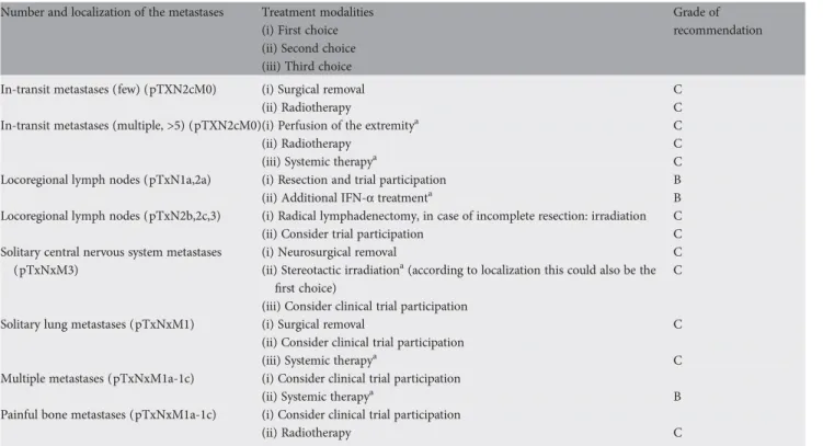
Table 2. Treatment modalities for melanoma metastases.
Number and localization of the metastases Treatment modalities
(i) First choice (ii) Second choice (iii) Third choice
Grade of recommendation
In-transit metastases (few) (pTXN2cM0) (i) Surgical removal C
(ii) Radiotherapy C
In-transit metastases (multiple, >5) (pTXN2cM0)(i) Perfusion of the extremitya C
(ii) Radiotherapy C
(iii) Systemic therapya C
Locoregional lymph nodes (pTxN1a,2a) (i) Resection and trial participation B
(ii) Additional IFN-α treatmenta B
Locoregional lymph nodes (pTxN2b,2c,3) (i) Radical lymphadenectomy, in case of incomplete resection: irradiation C
(ii) Consider trial participation C
Solitary central nervous system metastases (pTxNxM3)
(i) Neurosurgical removal C
(ii) Stereotactic irradiationa(according to localization this could also be the
first choice)
C (iii) Consider clinical trial participation
Solitary lung metastases (pTxNxM1) (i) Surgical removal C
(ii) Consider clinical trial participation
(iii) Systemic therapya C
Multiple metastases (pTxNxM1a-1c) (i) Consider clinical trial participation
(ii) Systemic therapya B
Painful bone metastases (pTxNxM1a-1c) (i) Consider clinical trial participation
(ii) Radiotherapy C
aThese therapies should be preferentially carried out at specialized centers.
patients with BRAF-mutant metastatic melanoma; however emerging data suggest that BRAF inhibition is effective even following immunotherapy. Currently, ipilimumab is only approved by EMA as second-line therapy for patients with advanced disease. Selective BRAF inhibitors can be safely used in patients with brain metastases and show promising efficacy in this compartment. Patients treated with vemurafenib should be carefully followed with special attention to skin [19,21] and other secondary neoplasms.
In patients with BRAF wild-type melanomas ipilimumab, an agent blocking cytotoxic T-lymphocyte-associated protein 4 (CTLA4) and thus activating T-lymphocytes to mount an immune response against tumor cells, is a recommended second-line to first-(FDA) or second-line (FDA and EMA) therapeutic option. In the context of new developments and medical progress, there are continuously new experimental treatment options for patients with advanced metastatic melanoma. Therefore, these patients should be referred to tertiary centers providing a comprehensive clinical trial program. There are early signals that patients with metastatic melanomas carrying NRAS mutation will profit from MEK kinase inhibitor therapy [26].
If clinical trials or the approved new targeted compounds are not available, cytotoxic drugs such as dacarbazine (DTIC), temozolomide, taxanes, fotemustine, platin derivatives or others, cytokines ( IFN, IL-2) or combinations may be applied. Dacarbazine is still considered a reference drug in this situation. In aggressive metastastic disease, multi-agent polychemotherapy containing paclitaxel and carboplatin or cisplatin, vindesine and dacarbazine produce partial response rates and stabilizations in a meaningful number of patients. Despite a better initial control rate, no survival benefit has been shown with polychemotherapy when compared with monochemotherapy, and therefore, polychemotherapies are not considered an establishedfirst-line therapy. Despite a minor increase in progression-free survival, bevacizumab therapy is rarely used in metastatic melanoma [31,32].
There are no randomized clinical trials for IL-2
monotherapy. Some centers still use IL-2 asfirst-line therapy when disease burden is low [33,34]. Several randomized trials did not show any survival benefit for the very intensive biochemotherapy including IL-2.
Surgery of visceral metastases may be appropriate for selected cases with good performance status and isolated tumor manifes-tations. In principal, the goal is R0-resections in these patients.
Palliative radiotherapy should be considered, especially for symptomatic brain or localized and painful bone metastases. Stereotactic irradiation is preferred in case of few brain metastases [23].
In general, stage IV melanoma patients need to be treated and discussed in an interdisciplinary tumor board at centers with broad experience in this disease.
patient information and follow-up
Melanoma patients should be instructed on avoidance of sunburns, extended unprotected solar or artificial UV exposure and on lifelong regular self-examinations of the skin and peripheral lymph nodes. Patients must be aware that familymembers have an increased melanoma risk (III, B). There is no necessity for genetic testing.
During melanoma follow-up, patients are clinically monitored in order to detect a relapse and to recognize additional skin tumors, especially secondary melanomas, as early as possible [4] (III, B). However, it is unproven if this policy leads to improved survival rates. Eight percent of all melanoma patients develop a secondary melanoma within 2 years of their initial diagnosis [35]. Melanoma patients also have increased risks of other skin tumors. In patients with lentigo maligna melanomas, 35% of the patients developed another cutaneous malignancy within 5 years [22].
There is currently no consensus on the frequency of follow-up and the use of imaging techniques. In recent series, most relapses have been detected by the patients themselves, questioning the
Table 3. Summary of recommendations
Diagnosis should be based on a full thickness excisional biopsy with a small side margin (II, A)
The histology report should include at least information on the type of melanoma, actinic damage, maximum thickness in millimeters (Breslow), information on the mitotic rate in case of pT1, presence of ulceration, presence and extent of regression and clearance of the surgical margins (II,A)
Physical examination with special attention to other suspicious pigmented lesions, tumor satellites, in-transit metastases, regional lymph node and systemic metastases is mandatory. In low-risk melanomas (pT1a), no other investigations are necessary. In higher tumor stages, imaging is recommended in order to allow proper staging (III, C)
Wide excision of primary tumors with safety margins of 0.5 cm forin situ
melanomas, of 1 cm for tumors with a Breslow thickness of up to 2 mm and 2 cm for thicker tumors is recommended (II, B)
Sentinel lymph node biopsy in melanoma with a tumor thickness of >1 mm and/or ulceration is necessary for precise staging (II, B) Patients with resected stage III melanomas should be evaluated for
adjuvant therapy with either high-dose IFN-α2b for 1 year or with
pegylated weekly PegIFN-α for up to 5 years (II, B). Subgroup analyses suggest that patients with microscopic regional nodal involvement and/ or ulcerated primaries are the ones most likely to benefit from adjuvant
IFN-α. Participation in clinical trials should be encouraged
Surgical removal of locoregional recurrence or single distant metastasis
should be considered infit patients as a therapeutic option offering
potential for long-term disease control (III, C)
Patients with metastatic melanoma should have a metastasis (preferably) or the primary screened for the presence of BRAF V600 mutation.
Treatment options for thefirst- and second-line setting include
ipilimumab, an anti-CTLA4 antibody, for all patients and vemurafenib, a BRAF inhibitor, for patients with BRAF-mutant melanoma (II, B). Ipilimumab is approved only as 2nd line therapy by EMA
If clinical trials or the approved new targeted compounds are not available, cytotoxic drugs such as DTIC, temozolomide may be applied, with modest activity shown (II, C)
Melanoma patients should be instructed on avoidance of sunburns,
extended unprotected solar or artificial UV exposure and on lifelong
regular self-examinations of the skin and peripheral lymph nodes (III, B) There is no consensus on optimal schedule, frequency of follow-up visits
neither on the utility of imaging and blood tests for patients with resected melanoma
usefulness and cost-effectiveness of follow-up visits every 3 months during thefirst 3 years and every 6–12 months thereafter. The above recommendations were solely based on the relapse-risk profile over time [4]. Intervals between controls tailored according to individual risk may reduce false-positivefindings and suffice for psychological support of the patients [36].
Since patients with a thin primary melanoma have only a small risk of relapse, routine imaging techniques are definitively not recommended for this patient population. In high-risk patients,, e.g. those with thick primary tumors or following treatment of metastases ultrasound of lymph nodes, computed tomography (CT) or whole body positron emission tomography scans (PET)/PET–CT scans may lead to an earlier diagnosis of regional or systemic relapses [37]. However, an impact of radiological exams upon survival has not been demonstrated so far [38]. Rising serum S-100 has a higher specificity for disease progression than lactate dehydrogenase (LDH) and therefore is the most accurate blood test in the follow-up of melanoma patients [39], if any blood test is recommended at all (IV, D).
con
flict of interest
Prof. Dummer has reported: research funding from AstraZeneca, Novartis, Cephalon, Merck Sharp & Dohme, Transgene, Bristol-Myers Squibb, Roche, GlaxoSmithKline, Bayer; consultant or advisory board relationship with AstraZeneca, Novartis, Cephalon, Merck Sharp & Dohme, Transgene, Genta, Bayer, Roche, Bristol-Myers Squibb, GlaxoSmithKline, Spirig. Prof. Hauschild has reported: consultancy/honoraria for advisory board, speakers’ bureau and lecture support from Amgen, AstraZeneca, Biovex, Bristol-Myers Squibb, Boehringer Ingelheim, Celgene, Eisai,
GlaxoSmithKline, IGEA, Lilly, Medec, MelaSciences, Merck Sharpe & Dohme/Merck, Novartis, Roche Pharma, SOBI, Vical, Janssen. Prof. Keilholz has reported: speakers’ bureau for GlaxoSmithKline and Bristol-Myers Squibb.
Other authors have reported no potential conflicts of interest.
references
1. MacKie RM, Bray C, Vestey J et al. Melanoma incidence and mortality in
Scotland 1979–2003. Br J Cancer 2007; 96(11): 1772–1777.
2. Hollestein LM, van den Akker SA, Nijsten T et al. Trends of cutaneous melanoma in The Netherlands: increasing incidence rates among all Breslow thickness categories and rising mortality rates since 1989. Ann Oncol 2012; 23(2):
524–530.
3. Green AC, Williams GM, Logan V et al. Reduced melanoma after regular sunscreen use: randomized trial follow-up. J Clin Oncol 2011; 29(3):
257–263.
4. Dummer R, Guggenheim M, Arnold AW et al. Updated Swiss guidelines for the treatment and follow-up of cutaneous melanoma. Swiss Med Wkly 2011; 141: w13320.
5. Bono A, Tolomio E, Trincone S et al. Micro-melanoma detection: a clinical study on 206 consecutive cases of pigmented skin lesions with a diameter < or = 3
mm. Br J Dermatol 2006; 155(3): 570–573.
6. Grob JJ, Bonerandi JJ. The‘ugly duckling’ sign: identification of the common
characteristics of nevi in an individual as a basis for melanoma screening. Arch
Dermatol 1998; 134(1): 103–104.
7. Balch CM, Gershenwald JE, Soong SJ et al. Final version of 2009 AJCC
melanoma staging and classification. J Clin Oncol 2009; 27(36): 6199–6206.
8. Whiteman DC, Pavan WJ, Bastian BC. The melanomas: a synthesis of epidemiological, clinical, histopathological, genetic, and biological aspects, supporting distinct subtypes, causal pathways, and cells of origin. Pigment Cell
Melanoma Res 2011; 24(5): 879–897.
9. Schoenewolf NL, Bull C, Belloni B et al. Sinonasal, genital and acrolentiginous melanomas show distinct characteristics of KIT expression and mutations. Eur J
Cancer 2012; 48: 1842–1852.
10. Hirakawa S, Kodama S, Kunstfeld R et al. VEGF-A induces tumor and sentinel lymph node lymphangiogenesis and promotes lymphatic metastasis. J Exp Med
2005; 201(7): 1089–1099.
11. Thompson JF, Scolyer RA, Kefford RF. Cutaneous melanoma. Lancet 2005; 365
(9460): 687–701.
12. Morton DL, Thompson JF, Cochran AJ et al. Sentinel-node biopsy or nodal
observation in melanoma. N Engl J Med 2006; 355(13): 1307–1317.
13. Eggermont AM, Suciu S, Testori A et al. Ulceration and stage are predictive of
interferon efficacy in melanoma: results of the phase III adjuvant trials EORTC
18952 and EORTC 18991. Eur J Cancer 2012; 48(2): 218–225.
14. Kirkwood JM, Ibrahim JG, Sondak VK et al. High- and low-dose interferon
alfa-2b in high-risk melanoma:first analysis of intergroup trial E1690/S9111/C9190.
J Clin Oncol 2000; 18(12): 2444–2458.
15. Kirkwood JM, Strawderman MH, Ernstoff MS et al. Interferon alfa-2b adjuvant therapy of high-risk resected cutaneous melanoma: the Eastern Cooperative
Oncology Group Trial EST 1684. J Clin Oncol 1996; 14(1): 7–17.
16. Mocellin S, Pasquali S, Rossi CR et al. Interferon alpha adjuvant therapy in patients with high-risk melanoma: a systematic review and meta-analysis. J Natl
Cancer Inst 2010; 102(7): 493–501.
17. Eggermont AM, Suciu S, Santinami M et al. Adjuvant therapy with pegylated
interferon alfa-2b versus observation alone in resected stage III melanoma:final
results of EORTC 18991, a randomised phase III trial. Lancet 2008; 372(9633):
117–126.
18. Kleeberg UR, Suciu S, Brocker EB et al. Final results of the EORTC 18871/DKG
80–1 randomised phase III trial. rIFN-alpha2b versus rIFN-gamma versus
ISCADOR M versus observation after surgery in melanoma patients with either high-risk primary (thickness >3 mm) or regional lymph node metastasis. Eur J
Cancer 2004; 40(3): 390–402.
19. Oberholzer PA, Kee D, Dziunycz P et al. RAS mutations are associated with the development of cutaneous squamous cell tumors in patients treated with RAF
inhibitors. J Clin Oncol 2012; 30(3): 316–321.
20. Su F, Viros A, Milagre C et al. RAS mutations in cutaneous squamous-cell carcinomas in patients treated with BRAF inhibitors. N Engl J Med 2012; 366(3):
207–215.
21. Zimmer L, Hillen U, Livingstone E et al. Atypical melanocytic proliferations and new primary melanomas in advanced melanoma patients undergoing selective
BRAF inhibition. J Clin Oncol 2012; 30: 2375–2383.
22. Farshad A, Burg G, Panizzon R et al. A retrospective study of 150 patients
with lentigo maligna and lentigo maligna melanoma and the efficacy of
radiotherapy using Grenz or soft X-rays. Br J Dermatol 2002; 146(6):
1042–1046.
23. Hong A, Fogarty G. Role of radiation therapy in cutaneous melanoma. Cancer J
2012; 18(2): 203–207.
24. Burmeister BH, Henderson MA, Ainslie J et al. Adjuvant radiotherapy versus
observation alone for patients at risk of lymph-nodefield relapse after therapeutic
lymphadenectomy for melanoma: a randomised trial. Lancet Oncol 2012; 13(6):
589–597.
25. Flaherty KT, Robert C, Hersey P et al. Improved survival with MEK inhibition in
BRAF-mutated melanoma. N Engl J Med 2012; 367: 107–114.
26. Ascierto PA, Berking C, Agarwala SS et al. Efficacy and safety of oral MEK162 in
patients with locally advanced and unresectable or metastatic cutaneous melanoma harboring BRAFV600 or NRAS mutations. J Clin Oncol 2012; 30 (Suppl); Abstr 8511.
27. Chapman PB, Hauschild A, Robert C et al. Improved survival with vemurafenib in melanoma with BRAF V600E mutation. N Engl J Med 2011; 364(26):
2507–2516.
28. Guo J, Si L, Kong Y et al. Phase II, open-label, single-arm trial of imatinib mesylate in patients with metastatic melanoma harboring c-Kit mutation or
amplification. J Clin Oncol 2011; 29(21): 2904–2909.
29. Hodi FS, O’Day SJ, McDermott DF et al. Improved survival with ipilimumab
in patients with metastatic melanoma. N Engl J Med 2010; 363(8):
711–723.
30. Robert C, Thomas L, Bondarenko I et al. Ipilimumab plus dacarbazine for previously untreated metastatic melanoma. N Engl J Med 2011; 364(26):
2517–2526.
31. von Moos R, Seifert B, Simcock M et al. First-line temozolomide combined with bevacizumab in metastatic melanoma: a multicentre phase II trial (SAKK 50/07).
Ann Oncol 2011; 23(2): 531–536.
32. Kim KB, Sosman JA, Fruehauf JP et al. BEAM: a randomized phase II study evaluating the activity of bevacizumab in combination with carboplatin plus paclitaxel in patients with previously untreated advanced melanoma. J Clin Oncol
2012; 30(1): 34–41.
33. Petrella T, Quirt I, Verma S et al. Single-agent interleukin-2 in the treatment of metastatic melanoma: a systematic review. Cancer Treat Rev 2007; 33(5):
484–496.
34. Hamm C, Verma S, Petrella T et al. Biochemotherapy for the treatment of metastatic malignant melanoma: a systematic review. Cancer Treat Rev 2008;
34(2): 145–156.
35. Titus-Ernstoff L, Perry AE, Spencer SK et al. Multiple primary melanoma: two-year results from a population-based study. Arch Dermatol 2006; 142(4):
433–438.
36. Turner RM, Bell KJ, Morton RL et al. Optimizing the frequency of follow-up visits for patients treated for localized primary cutaneous melanoma. J Clin Oncol
2011; 29(35): 4641–4646.
37. Bastiaannet E, Wobbes T, Hoekstra OS et al. Prospective comparison of [18F] fluorodeoxyglucose positron emission tomography and computed tomography in patients with melanoma with palpable lymph node metastases: diagnostic accuracy and impact on treatment. J Clin Oncol 2009; 27(28):
4774–4780.
38. Nieweg OE, Kroon BB. The conundrum of follow-up: should it be abandoned?
Surg Oncol Clin N Am 2006; 15(2): 319–330.
39. Beyeler M, Waldispuhl S, Strobel K et al. Detection of melanoma relapse:first
comparative analysis on imaging techniques versus S100 protein. Dermatology
2006; 213(3): 187–191.
![Table 1. AJCC Staging system of melanoma [7].](https://thumb-eu.123doks.com/thumbv2/123doknet/14885570.646636/2.918.87.451.111.720/table-ajcc-staging-system-of-melanoma.webp)