Thymic epithelial tumours: ESMO Clinical Practice
Guidelines for diagnosis, treatment and follow-up
†
N. Girard
1, E. Ruf
fini
2, A. Marx
3, C. Faivre-Finn
4& S. Peters
5, on behalf of the ESMO Guidelines
Committee*
1
Department of Respiratory Medicine, Expert Centre for Thymic Malignancies, Reference Centre for Orphan Pulmonary Diseases, Hôpital Louis Pradel, Hospices Civils de Lyon, Lyon, France;2
Department of Thoracic Surgery, University of Torino, Turin, Italy;3
Institute of Pathology, University Medical Centre Mannheim, University of Heidelberg, Mannheim, Germany;4
Institute of Cancer Sciences, The University of Manchester, Manchester Academic Health Science Centre, The Christie NHS Foundation Trust, Manchester, UK;5
Department of Medical Oncology, Centre Hospitalier Universitaire Vaudois (CHUV), Lausanne, Switzerland
incidence and epidemiology
Thymic epithelial tumours represent a heterogeneous group of
rare thoracic cancers, with reported annual incidence ranging
from 1.3 to 3.2 per million [
1
]. Thymic epithelial tumours are
classified according to the World Health Organization (WHO)
histopathological classification, which distinguishes thymomas
from thymic carcinomas.
thymomas
Thymomas are further subdivided into different types (called
A, AB, B1, B2, B3 and rare others) based upon the morphology of
epithelial tumour cells, the relative proportion of the non-tumoural
lymphocytic component (decreasing from type B1 to B3) and
resemblance to normal thymic architecture (Table
1
) [
2
,
3
]. The
term
‘benign thymoma’ should be avoided. Thymomas are far
more frequent than thymic carcinomas, which have an incidence
of 0.2 to 0.5 per million [
3
].
thymic carcinomas
Thymic carcinomas are similar to their extrathymic counterpart,
the most frequent subtype being squamous cell carcinoma.
Neuroendocrine tumours may occur in the thymus, and will not
be discussed in these guidelines; while localised primary thymic
neuroendocrine tumours may benefit from surgical resection,
similar to other thymic carcinomas, the prognosis is poor given
frequent recurrences; for recurrent, advanced and metastatic
tumours, the management actually follows that of extra-thoracic
neuroendocrine tumours.
epidemiology
Mean age at diagnosis is 50
–60 years of age, but thymic tumours
may actually be diagnosed in children as well as in elderly
patients. There is no consistent gender predilection in thymomas
overall, even if a slight female preponderance has been reported
for type A, AB and B1 subtypes in most studies, and a male
pre-dominance in carcinomas [
2
–
7
].
No environmental or infectious factors have been
demon-strated to play a role in the pathogenesis of thymic epithelial
tumours. Reports on development of thymoma after radiation,
solid-organ transplantation and immunosuppression, including
the context of human immunode
ficiency virus infection, are
rare; differential diagnosis with thymic rebound hyperplasia
may be discussed in this setting (see below).
Genetic risk factors, such as multiple endocrine neoplasia
1 (MEN1), may in
fluence the development of thymomas, as well
as thymic carcinoids, given their reported familial occurrence as
well as their association with cancer susceptibility syndromes [
8
].
Moreover, extrathymic haematopoietic cancers (mostly diffuse
large B-cell lymphoma and leukaemia) and a broad spectrum of
solid cancers (stomach, pancreas, colon and thyroid) have been
reported to occur more frequently in thymoma patients,
particu-larly subsequently [
9
]. This might be related to a shared unknown
oncogenic trigger, a thymoma-associated immune de
ficiency or
(less likely) to adverse effects of treatments.
diagnosis
imaging and laboratory tests
Standard imaging for thymic tumours is i.v. contrast-enhanced
computed tomography (CT) scan of the thorax, allowing a
com-plete exploration of the mediastinum and the pleura from the
apex to the costodiaphragmatic recesses [IV, A]. CT is equal or
superior to magnetic resonance imaging (MRI) for the diagnosis
of mediastinal anterior masses, except in the setting of cystic
lesions [IV, B] [
10
].
One-third of patients with thymoma present with autoimmune
disorders (Table
2
), mainly myasthenia gravis which is
particular-ly common in type AB, B1 and B2 thymomas and almost always
associated with anti-acetylcholine receptor antibodies (Table
1
)
[
11
]. Other frequent disorders include pure red cell aplasia (5% of
cases) and hypogammaglobulinaemia (Good syndrome: 5% of
cases) [
12
].
†Approved by the ESMO Guidelines Committee: July 2015.
*Correspondence to: ESMO Guidelines Committee, ESMO Head Office, Via L. Taddei 4, CH-6962 Viganello-Lugano, Switzerland. E-mail: [email protected]
clinical
pr
a
ctice
guidelines
© The Author 2015. Published by Oxford University Press on behalf of the European Society for Medical Oncology. All rights reserved. For permissions, please email: [email protected].
In addition to recording a complete history and conducting a
full clinical examination (looking especially at neurological
signs), systematic immunological check-up is recommended
when a diagnosis of thymic epithelial tumour is suspected,
in-cluding complete blood cells count with reticulocytes and serum
protein electrophoresis, as well as anti-acetylcholine receptor
and anti-nuclear antibodies tests [V, A]. Indeed, frequent
immune disorders associated with thymoma may impact the
course of all therapeutic interventions including surgery,
radio-therapy as well as chemoradio-therapy.
diagnosis approach
The diagnosis of any thymic epithelial tumour relies on making
the differential diagnosis with other anterior mediastinal tumours
and non-malignant thymic lesions [
13
]. CT is the imaging
mo-dality of choice. The need for pretreatment biopsy depends on
the resectability of the tumour [
14
–
16
].
Thymic epithelial tumours are the most frequent cause of
an-terior mediastinal mass, accounting for 35% of cases; the most
relevant differential diagnoses include lymphomas (Hodgkin
’s
or non-Hodgkin’s) in ∼25% of cases and germ-cell tumours
(teratoma or seminoma/non-seminomatous tumours) in
∼20%
of cases [
13
]. Thymic carcinoma must be differentiated from
lung carcinoma, as well as from rarer entities, such as NUT
car-cinomas [
17
].
Clinical judgement based on a complete history and physical,
especially neurological, examination, correlated with laboratory
tests and radiological features, helps to develop a presumptive
diagnosis. Thymoma is the most likely diagnosis when facing a
mediastinal mass associated with one of the above autoimmune
diseases, while thymic carcinoma patients typically have
unspe-ci
fic local symptoms [IV, A]. Lymphoma may be considered in
case of rapid onset of B-signs, coexistent lymphadenopathy or
elevated lactate dehydrogenase. Teratoma usually shows a
het-erogeneous morphology on imaging, with fat and cystic pattern
[
18
]. Seminomas and non-seminomatous germ-cell tumours may
be large and have a fulminant onset. Elevated serum
β-human
chorionic gonadotropin may be observed in seminomas, along
with elevated alphafetoprotein in non-seminomatous germ-cells
tumours.
Differentiating thymic malignancy from hyperplasia or
non-involuted thymus may be challenging. Thymic rebound
hyperplasia should be considered after stress, injuries,
chemo-therapy,
radiotherapy,
anti-hormonal
treatment
or
corticosteroids. Thymic lymphoid hyperplasia is most
common-ly observed in myasthenia gravis, but also in the setting of
hyperthyroidism, connective tissue or vascular disease. CT
Table 1. Histological subtypes of thymic epithelial tumours: relative frequency, frequency of myasthenia gravis and correlation with stageRelative frequency Myasthenia gravis Masaoka stage
I II III IVA IVB Type A 12% (3%–26%) 15% (0%–35%) 60% 31% 8% <1% <1% Type AB 28% (15%–43%) 20% (5%–42%) 67% 26% 6% 1% 1% Type B1 18% (6%–53%) 40% (5%–69%) 50% 37% 9% 3% 1% Type B2 26% (8%–41%) 50% (23%–73%) 32% 29% 28% 8% 3% Type B3 16% (3%–35%) 50% (25%–65%) 19% 36% 27% 15% 3% Carcinoma 18% (1%–28%) <5% 10% 10% 45% 15% 20% Data are based on references [5–8].
Table 2. Autoimmune disorders associated with thymoma [11,12]
Neuromuscular Myasthenia gravis Myotonic dystrophy Limbic encephalitis Peripheral neuropathy Autonomic neuropathy Acquired neuromyotonia
Morvan syndrome (neuromyotonia and encephalitis)
Stiff person syndrome Cerebellar degeneration Polymyositis (carcinomas) Haematological disorders Red cell aplasia
Pernicious anaemia Erythrocytosis Pancytopoenia Haemolytic anaemia Leukaemia Multiple myeloma Collagen and autoimmune
disorders
Systemic lupus erythematosus Rheumatoid arthritis Sjogren’s syndrome Scleroderma Interstitial pneumonitis Immune deficiency disorders Hypogammaglobulinaemia (Good syndrome)
T-cell deficiency syndrome Endocrine disorders Multiple endocrine neoplasia
Cushing’s syndrome Thyroiditis Dermatological disorders Pemphigus
Lichen planus
Chronic mucosal candidiasis Alopecia areata
Miscellaneous Giant cell myocarditis Nephrotic syndrome Ulcerative colitis
Hypertrophic osteoarthropathy
Volume 26 | Supplement 5 | September 2015 doi:10.1093/annonc/mdv277 | v
features include low-attenuation, symmetric and fatty pattern
maintaining the bi-pyramidal shape of the thymus [
18
]. In
equivocal cases at CT, chemical-shift MRI may detect
microscop-ic fatty infiltration by showing homogeneous signal decrease on
opposed phase images relative to in-phase images, which is not
observed in thymoma [IV, B] [
19
]. Therapeutic intervention is
usually not required if the lesion is <30 mm, given a low risk of
progression or thymic malignancy [III, D] [
20
].
18-Fluorodeoxyglucose positron emission tomography (PET)
scan is generally not recommended to assess thymic masses [IV, C].
Standard uptake values may be higher in type B3 thymomas
and thymic carcinomas; however, thymic hyperplasia may also
present with hypermetabolism [
21
]. PET scan is optional in the
case of tumours with aggressive histology and an advanced stage
to complete the staging work-up or further characterise lesions
suspicious for recurrences.
need for biopsy
Pretreatment biopsy is not required if the diagnosis of thymic
tumour is highly probable and upfront surgical resection is
achievable (see below, de
finition of resectability) [IV, E]. Biopsy is
required in all other clinical situations [IV, A]: approaches may
consist of percutaneous core-needle biopsy or incisional
surgi-cal biopsy through mediastinotomy or mini-thoracotomy, with
sensitivity rates ranging from 40% to 93% [
22
]. Biopsies that
are deep and multiple are preferred. Pleural spaces should be
respected to avoid tumour cell seeding. Fine-needle aspiration
is generally not recommended [IV, D].
thymomas. Although designed for surgical resection specimens,
the WHO classi
fication may be used for small biopsies [V, A].
However, thymoma subtyping on small biopsies is usually not
needed for the therapeutically relevant distinction between
lymphoma and solid tumour. In any case, diagnostic discrepancies
between core-needle and resection specimen histology can be
anticipated, given the frequent occurrence of histological
tumour heterogeneity that may be missed due to sampling error
[
23
]. The recent proposal of major and minor morphological
and immunohistochemical criteria to better individualise each
thymic epithelial tumour entity aims at addressing those issues,
and has been integrated in the revised WHO classification
[
3
,
24
]. Immunohistochemical markers may be helpful,
including cytokeratins and p63 expression for normal and
neoplastic epithelial cells, and terminal deoxynucleotidyl
transferase expression in immature T cells (usually observed
in types AB, B1, B2 and B3 thymomas, and absent in
carcinomas and type A thymomas) [
3
].
thymic carcinomas. Immunohistochemistry with anti-CD117/
KIT and anti-CD5 antibodies helps to establish the thymic
origin in
∼80% of mediastinal carcinomas [V, A]. Since these
markers are not absolutely specific, correlation with the clinical
setting is always recommended, and is mandatory in the subset
of 20% of thymic carcinomas without expression of CD117/KIT
and CD5 [
3
].
In thymic tumours showing more than one histological
pattern, each component should be listed (starting with the
pre-dominant one) and be quanti
fied in 10% increments; a thymic
carcinoma component should always be mentioned
first [V, C].
In case of dif
ficult diagnosis, it is recommended to consult a
second pathologist or refer the case to a thymic tumour
path-ology panel.
staging and risk assessment
staging
Thymic epithelial tumours are routinely staged according to the
Masaoka-Koga staging system (Table
3
) [III, A] [
25
–
27
], which is
correlated with overall survival (OS) [
4
,
28
,
29
]. Masaoka-Koga
staging is a surgical pathology system that is assessable only after
surgical resection of the tumour. A typical feature of thymic
epi-thelial tumours is the correlation between the WHO classi
fication
and stage at diagnosis (Table
1
), which may explain its reported
prognostic value [
4
–
6
] (Figures
1
–
3
).
The International Association for the Study of Lung Cancer
(IASLC) Staging Prognostic Factors Committee, together with
the International Thymic Malignancy Interest Group (ITMIG),
recently proposed a Tumour–Node–Metastasis (TNM)-based
staging system for thymic malignancies, based on OS analyses of
a retrospective international database of more than 10 000 cases
(Table
4
) [
30
]. The TNM-based approach has the advantage of
being more appropriate both for thymoma and thymic
carcin-omas, which present with a higher propensity toward nodal and
distant metastatic invasion. The IASLC/ITMIG TNM system of
thymic tumours will be incorporated as the official thymic tumour
staging system into the 8th edition of the TNM staging system of
thoracic malignancies expected in 2016
–2017. From our
stand-point, the Masaoka-Koga staging should remain the standard for
the routine management of patients, pending the approval of the
American Joint Committee on Cancer (AJCC) and Union for
International Cancer Control (UICC) [III, A]. Moreover, given the
major switch that the TNM system represents and the limited
amount of fair level of evidence data to support our current
treat-ment strategies (especially postoperative radiotherapy), the value of
the TNM system to drive the therapeutic strategy has to be assessed.
Correlative clinical data based on this system may be encouraged in
a research setting.
The assessment of resectability is mostly based on the surgeon’s
expertise; it is recommended to discuss indications for surgery in a
multidisciplinary tumour board setting [V, B]. There is no
recog-nised clinical staging system, and the treatment strategy for thymic
epithelial tumours is primarily based on whether the tumour may
be resected upfront or not [IV, A], as complete resection has been
identi
fied as the most consistent and significant prognostic factor of
disease-free survival and OS [
5
,
6
,
29
]. Correlation between clinical
and surgical pathology stage is higher in advanced stages, given the
identi
fication of vessel invasion, enlarged lymph nodes, pleural/
pericardial lesions or even systemic metastases [
28
]. Preoperative
CT
findings reported to be associated with tumour invasiveness
and/or completeness of resection include: tumour size (>5/7/8 cm,
depending on studies), lobulated or irregular contours, calci
fica-tions, in
filtration of surrounding fat, lung infiltration, great vessel
invasion or encirclement [
31
–
33
]. The new TNM staging may even
provide more help in formalising resectability: T1–3 level of
inva-sion refers to structures amenable to surgical resection, while T4
level of invasion includes unresectable structures (Table
4
).
Table 3. Staging of thymic epithelial tumours: Masaoka-Koga-based staging system [25,26], International Thymic Malignancy Interest Group refinements [27] and overall survival and recurrence-free survival (range)a[28]
Masaoka-Koga, 1994 International Thymic Malignancy Interest Group, 2011 10-year overall
survival
10-year cumulative incidence of recurrence
Thymoma Thymic carcinoma
Stage I Grossly and microscopically completely encapsulated tumour – Invasion into but not through the capsule – In the absence of capsule, absence of invasion into
surrounding tissues
84% (81%–86%)
8% (7%–8%) 25% (22%–29%)
Stage IIA Microscopic transcapsular invasion – Microscopic transcapsular invasion (<3 mm) 83% (79%–87%)
Stage IIB Macroscopic invasion into thymic or surrounding fatty tissue, or grossly adherent to but not breaking through the mediastinal pleura or pericardium
– Gross extension into normal thymus or perithymic fat surrounding the tumour (microscopically confirmed) – Adherence to pleura or pericardium, with microscopic
confirmation of perithymic invasion Stage III Macroscopic invasion into neighbouring organ (i.e.
pericardium, great vessel or lung)
– Microscopic invasion of the mediastinal pleura (either partial or penetrating the elastin layer)
– Microscopic invasion of the pericardium (either partial in the fibrous layer or penetrating through to the serosal layer) – Microscopically confirmed direct penetration into the outer
elastin layer of the visceral pleura or into the lung parenchyma
– Invasion into the phrenic or vagus nerves (microscopically confirmed)
– Invasion into or penetration through major vascular structures (microscopically confirmed)
– Adherence (i.e. fibrous attachment) of lung or adjacent organs only if there is mediastinal pleural or pericardial invasion (microscopically confirmed)
70% (64%–75%) 29% (27%–31%) 59% (44%–76%)
Stage IVA Pleural or pericardial metastasis – Microscopically confirmed separate nodules in the visceral
or parietal pleural, pericardial or epicardial surfaces
42% (26%–58%) 71% (34%–100%) 76% (58%–100%)
Stage IVB Lymphogenous or haematogenous metastasis – Lymphogenous or haematogenous metastasis 53% (32%–73%) 57% (24%–90%) 54% (37%–67%)
a
Information reprinted from [27] with permission of John Wiley & Sons, Inc.
V olume 26 | Supplement 5 | September 2015 doi:10.1093/annonc/mdv277 | v Annals of Oncology
clinical
pr
a
ctice
guidelines
risk assessment
Prognostic assessment is challenging, as the impact of tumour
stage and histology on OS is superseded by the achievement of a
complete resection in reported series [
4
–
7
,
28
–
30
]. Moreover,
depending on stage, up to 50%
–60% of patients actually do not
die of progression of the thymic tumour [
34
]. Autoimmune
disorders have been reported as the cause of death in 25% of
thymomas, especially those with early-stage tumours. The
evo-lution of these alterations, which are related to the abnormal
intrathymomatous selection of constitutively autoreactive
lym-phocytes, does not parallel that of the tumour. This is contrary
to what is observed in paraneoplastic syndromes that are
caused by tumour cell-derived cytokines or hormones or by
cross-reactive antibodies. The management of autoimmune
syndromes will not be discussed in these guidelines, but should
be integrated in the oncological management of these patients
[V, A].
management of resectable disease
The treatment strategy is based on the resectability of the tumour.
If complete resection is deemed to be achievable upfront, as it is
the case in Masaoka-Koga stage I/II and some stage III tumours
(classi
fied as stage I, II, IIIA/T3 in the IASLC/ITMIG TNM
pro-posed system), surgery represents the
first step of the treatment
[IV, A], possibly followed by postoperative radiotherapy and, less
frequently, chemotherapy (Table
5
).
surgical principles
The standard approach is median sternotomy [IV, A], which
allows the wide opening of the mediastinum and both pleural
cavities, followed by evaluation of macroscopic capsular
inva-sion, infiltration of perithymic and mediastinal fat, peritumoural
and pleural adherences and involvement of surrounding
struc-tures [
14
,
15
,
35
,
36
]. Generally, complete thymectomy
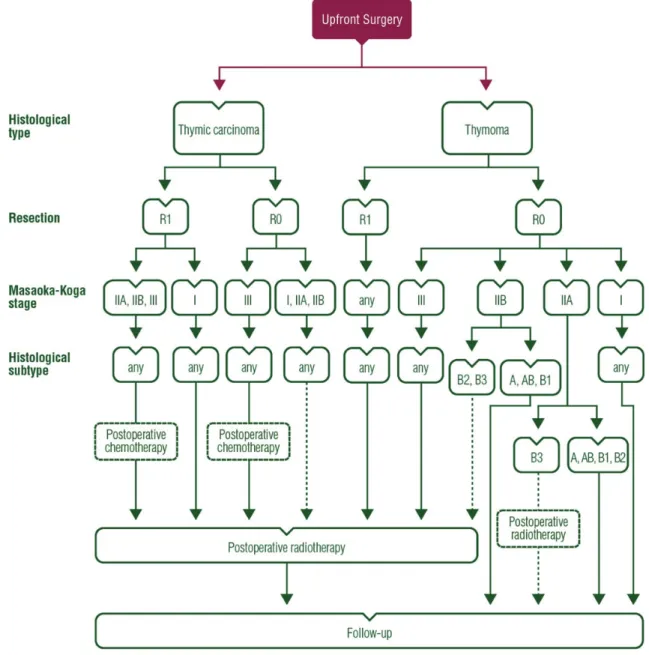
Figure 1. Treatment algorithm for resectable thymic tumour (Masaoka-Koga stage I–III, TNM stage I–IIIA).
including the tumour, the residual thymus gland and perithymic
fat is preferred because local recurrences have been observed
after partial thymectomy when part of the thymus gland is left
behind [IV, B]. Thymomectomy—leaving residual thymic tissue
and perithymic fat behind—alone is an option in stage I
tumours in non-myasthenic patients [IV, C] [
14
,
37
]. If the
tumour is widely invasive (stage III/IV), en bloc removal of all
affected structures, including lung parenchyma (usually through
limited resection), pericardium, great vessels, nerves and pleural
implants, should be carried out [IV, A]. Resection of venous
vascular structures (innominate vein(s) and superior vena
cava) include partial resection with suturing or complete
resec-tion and vessel reconstrucresec-tion using vascular prosthesis. Areas
of uncertain resection margins are marked with clips to allow
precise delivery of postoperative radiotherapy [IV, B]; those
areas are also designated on the resection specimen, as
dis-cussed below. Phrenic nerve preservation does not affect OS
but increases the risk of local recurrence [IV, C], and should be
balanced with the achievement of a complete resection,
espe-cially in patients with severe myasthenia gravis [
38
,
39
]. Frozen
sections to assess tumour involvement of resection margins are
not recommended [V, D], given the high risk of false-negative
results [
36
].
Minimally invasive surgery is an option for presumed stage I
and possibly stage II tumours in the hands of appropriately
trained thoracic surgeons [IV, C] [
14
,
35
,
40
]. This includes
transcervical, extended transcervical, video-assisted
thoraco-scopy (VATS) and robotic approaches (right or left, right and
left, right and cervical, left and cervical, subxiphoid and right
and left, cervical and subxiphoid); robotic surgery may allow a
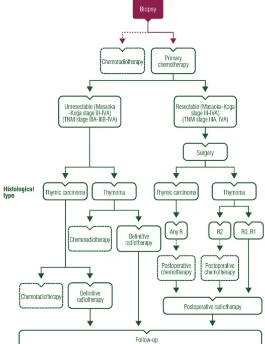
Figure 2. Treatment algorithm for unresectable thymic tumour (Masaoka-Koga stage III–IVA, TNM stage IIIA–IIIB–IVA).
Volume 26 | Supplement 5 | September 2015 doi:10.1093/annonc/mdv277 | v
better visualisation of the tumour when compared with VATS.
The choice for minimally invasive resection should not
jeopardise or change the principles that are deemed
appropri-ate for an open approach, especially the achievement of
com-plete resection that may ultimately require switching to an
open procedure [V, A]. Minimally invasive surgery is not
recommended for stage III tumours, given the absence of
long-term follow-up data [IV, D].
Lymphadenectomy has historically rarely been carried out
after resection of thymic tumours. The new IASLC/ITMIG
TNM staging system of thymic tumours, however, leads to the
recommendation that locoregional lymphoadenectomy should
be carried out during resection of all types of thymic tumours. A
proposed nodal map is available from ITMIG [
41
]. The
pro-posed N descriptor in the staging system includes:
•
anterior region (N1), which involves the anterior mediastinal
nodes ( prevascular, para-aortic, ascending aorta, superior and
inferior phrenic and supradiaphragmatic) and the anterior
cervical nodes (low anterior cervical); and
•
the deep region (N2), which includes the middle mediastinal
(internal mammary, upper and lower paratracheal, subaortic,
subcarinal and hilar) and the deep cervical (lower jugular and
supraclavicular).
Routine removal of anterior mediastinal nodes and anterior
cer-vical nodes is recommended [IV, A]. Systematic sampling of
other intrathoracic sites is encouraged (i.e. paratracheal,
aorto-pulmonary window and subcarinal areas, depending on tumour
location) in stage III/IV tumours [V, B] [
36
]. Systematic
lym-phadenectomy (N1 + N2) is strongly recommended in case of
Figure 3. Treatment algorithm for metastatic thymic tumour (Masaoka-Koga stage IVB, TNM stage IVB).
Table 4. Proposed Tumour–Node–Metastasis staging (International Association for the Study of Lung Cancer Prognostic Factors Committee- International Thymic Malignancy Interest Group) [30] and corresponding Masaoka-Koga stage
Stage Descriptors Tumour
T1 T1a Encapsulated or unencapsulated, with or without extension into the mediastinal fat T1b Extension into the mediastinal pleura
T2 Direct invasion of the pericardium (partial or full-thickness)
T3 Direct invasion of the lung, the brachiocephalic vein, the superior vena cava, the chest wall, the phrenic nerve and/or hilar (extrapericardial) pulmonary vessels
T4 Direct invasion of the aorta, arch vessels, the main pulmonary artery, the myocardium, the trachea or the oesophagus
Node
N0 N0 No nodal involvement
N1 N1 Anterior (perithymic) nodes (IASLC levels 1, 3a, 6 and/or supradiaphragmatic/inferior phrenics/ pericardial)
N2 N2 Deep intrathoracic or cervical nodes (IASLC levels 2, 4, 5, 7, 10 and/or internal mammary nodes) Metastasis
M0 No metastatic pleural, pericardial or distant sites M1 M1a Separate pleural or pericardial nodule(s)
M1b Pulmonary intraparenchymal nodule or distant organ metastasis Stage grouping Corresponding Masaoka-Koga stage
I T1N0M0 I, IIA, IIB, III II T2N0M0 III
IIIA T3N0M0 III IIIB T4N0M0 III IVA T any N0,1 M0,1a IVA, IVB IVB T any N0-2 M0-1b IVB
thymic carcinoma due to the high rate of lymphatic spread (20%
versus 3% in thymomas) [V, B].
surgical pathology principles
Communication between surgeons and pathologists is required to
accurately stage thymic epithelial tumours [V, A] [
36
]. The
proper orientation of the specimen and the designation of
involved structures, organs or areas of likely residual microscopic
or macroscopic disease are the primary responsibility of the
oper-ating surgeon, and may be done using a mediastinal board [V, B].
The
final pathological examination leads to a final histological
diagnosis and staging of the tumour, based on the WHO
classi-fication and the Masaoka-Koga system, respectively (Tables
1
and
3
). Staging according to the proposed IASLC/ITMIG TNM
system is optional [V, C]. Given the potential heterogeneity of
Table 5. Stage-matched therapeutic strategyMasaoka-Koga stage
Thymoma Thymic carcinoma
Stage I Upfront surgery [IV, A] No biopsy [IV, E]
If complete resection (R0): no postoperative radiotherapy [II, E] If incomplete resection (R1): postoperative radiotherapy
(50–54 Gy) [IV, B]
Upfront surgery [IV, A] No biopsy [IV, E]
If resection complete (R0): consider postoperative radiotherapy (45–50 Gy) [V, C]
If incomplete resection (R1): postoperative radiotherapy (50–54 Gy) [IV, B]
Stage IIA Upfront surgery [IV, A] No biopsy [IV, E] If complete resection (R0):
– Type A–B2: no postoperative radiotherapy [IV, C] – Type B3: consider postoperative radiotherapy
(45–50 Gy) [IV, C] If incomplete resection (R1):
– Postoperative radiotherapy (50–54 Gy) [IV, B]
Upfront surgery [IV, A] No biopsy [IV, E] If complete resection (R0):
– Consider postoperative radiotherapy (45–50 Gy) [IV, B] If incomplete resection (R1):
– Postoperative radiotherapy (50–54 Gy) [IV, B] – Consider postoperative chemotherapy
Stage IIB Upfront surgery [IV, A] No biopsy [IV, E] If complete resection (R0):
– Type A–B1: no postoperative radiotherapy [IV, C] – Type B2–B3: consider postoperative radiotherapy
(45–50 Gy) [IV, C] If incomplete resection (R1):
– Postoperative radiotherapy (50–54 Gy) [IV, B]
Upfront surgery [IV, A] No biopsy [IV, E] If complete resection (R0):
– Consider postoperative radiotherapy (45–50 Gy) [IV, B] If incomplete resection (R1):
– Postoperative radiotherapy (50–54 Gy) [IV, B] – Consider postoperative chemotherapy
Stage III–IVA Resectable tumour (TNM I–IIIA, i.e. T1–3): – Upfront surgery [IV, A]
– Postoperative radiotherapy (45–50 Gy), with boost on areas of concern [IV, B]
Unresectable tumour (TNM IIIA-B, i.e. T3-T4, IVA): – Biopsy
– Primary chemotherapy (prefer anthracycline-based) [III, A] – If the tumour becomes resectable:
– Surgery [III, A]
– Postoperative radiotherapy (45–50 Gy), with boost on areas of concern (R0, R1 resection) [IV, B]
– If the tumour remains unresectable or R2: – Definitive radiotherapy (60 Gy) [IV, B] – Option: chemoradiotherapy
– Option: concurrent chemoradiotherapy (platin and etoposide, 60 Gy) [III, B]
Resectable tumour (TNM I–IIIA, i.e. T1–3): – Upfront surgery [IV, A]
– Postoperative radiotherapy (40–50 Gy), with boost on areas of concern [IV, B]
– Consider postoperative chemotherapy
Unresectable tumour (TNM IIIA-B, i.e. T3-T4, IVA): – Biopsy
– Primary chemotherapy (prefer anthracycline-based) [III, A] – If the tumour becomes resectable:
– Surgery [III, A]
– Postoperative radiotherapy (45–50 Gy), with boost on areas of concern (R0, R1 resection) [IV, B]
– Consider postoperative chemotherapy (R0, R1 resection) – If the tumour remains unresectable or R2:
– Definitive radiotherapy (60 Gy) [IV, B] – Option: chemoradiotherapy
– Option: concurrent chemoradiotherapy (platin and etoposide, 60 Gy)
Stage IVB Definitive chemotherapy [III, A]
– If the tumour becomes resectable, consider: – Surgery and postoperative radiotherapy – Definitive radiotherapy
Definitive chemotherapy [III, A]
Volume 26 | Supplement 5 | September 2015 doi:10.1093/annonc/mdv277 | v
thymic epithelial tumours, a sufficient number of representative
sections should be examined regardless of the tumour diameter
(at least
five sections up to a diameter of 5 cm, with one
add-itional block per addadd-itional centimetre of maximal diameter); if
the margin is <1 mm, at least three additional sections through
this area should be obtained [V, B]. Completeness of resection
should be assessed, making the distinction between tissues that
have been cut or dissected, and a surface bounded by a space
(such the mediastinal pleura, pericardium or endothelium of the
innominate veins), which should not be designated as a positive
margin [V, A].
postoperative radiotherapy
Current practices for postoperative mediastinal radiotherapy are
highly variable and there is paucity of prospective, multicentre
evidence. The global trend over the past years has been towards
a less frequent use of postoperative radiotherapy in thymoma,
and to keep it in reserve for high-risk cases (Table
5
) [
14
–
16
].
This is based on recent reports from large databases [
6
,
7
,
42
–
45
],
as well as pooled analyses of retrospective studies [
46
], indicating:
•
the absence of survival bene
fit after radiotherapy in stage I
thymoma, or after R0/1 resection of stage II
–III thymoma
[
7
,
42
,
43
];
•
a similar rate of recurrence in patients who received
post-operative radiotherapy or not, after complete resection of
thymoma [
46
]; and
•
a recurrence-free survival (RFS) and OS bene
fit with postoperative
radiotherapy after resection of thymic carcinoma [
7
,
44
,
45
].
Stage and completeness of resection are thus the most relevant
criteria in the decision making, followed by histology [IV, B].
Those factors are the most significant predictors of RFS [
4
–
7
,
28
–
29
]; however, one must take into account that retrospective
analyses are likely to be biased, since postoperative radiotherapy
is most likely administered in patients with incomplete resection
or high-grade tumours. Therefore, the absence of survival
differ-ences may then suggest that postoperative radiotherapy reduced
or overcame the risk of recurrence in those patients. Another
point to consider is that recurrences of thymic epithelial tumours
occur outside the mediastinum in more than 60% of cases [
47
].
The development of the TNM system leads to redefinition of
subsets of patients, based especially on the T descriptors, that
may help to clarify which patients bene
fit from postoperative
radiotherapy. Meanwhile, the grouping of Masaoka-Koga stage I,
IIA, IIB and some stage III tumours in one single T1 category
must be assessed in dedicated studies.
Current evidence for postoperative radiotherapy in thymic
epithelial tumours support the use of:
•
3D conformal radiotherapy or intensity-modulated radiation
therapy targeted to the tumour bed, keeping at-risk thoracic
organs within accepted safe constraints [IV, A];—clinical
target volume includes the whole thymic space, the tumour
and its extensions and the anterior, superior and middle
mediastinum [IV, A].
•
a total dose of 45–50 Gy after complete resection, 50–54 Gy
after R1 resection, with a boost to areas of likely residual
disease (as mentioned above, surgical clips may then be useful
to define the target volume) [IV, B]; however, the optimal
postoperative dose/fractionation is still to be de
fined; and
•
conventional fractionation scheme consisting of daily doses of
1.8–2 Gy over a 4- to 6-week period [IV, A] [
48
]. The
field
may encompass involved nodes [IV, B] and the site of a
resected pleural implant [V, C].
Prophylactic irradiation of supraclavicular nodes is not
recom-mended [V, E]. Low-dose entire hemithoracic radiotherapy is
also not recommended [IV, C] [
49
]. Ideally, postoperative
radio-therapy should start within 3 months of the surgical procedure
[V, B].
thymomas. Postoperative radiotherapy is not indicated after
complete resection of Masaoka-Koga stage I thymoma [II, E]. One
randomised trial including 29 patients compared postoperative
radiotherapy versus surgery alone in this setting, and failed to show
any differences in patient outcome [
50
].
Postoperative radiotherapy is not recommended after
com-plete resection of stage II thymoma [IV, C]. In the ITMIG
data-base, cumulative incidence of mediastinal or extramediastinal
recurrence was only 8% at 10 years (Table
2
) [
28
]. Postoperative
radiotherapy may be considered in case of aggressive histology
(type B2, B3) or extensive transcapsular invasion (stage IIB) [IV,
C] [
51
–
53
].
Postoperative radiotherapy is recommended after complete
resection of stage III/IVA thymoma, in an effort to prolong RFS
and OS [IV, B] [
54
].
thymic carcinoma. After complete resection of thymic carcinoma,
postoperative radiotherapy is optional for stage I tumours [V, C],
should be considered for stage II tumours [IV, B] and is
recom-mended for stage III/IVA tumours [IV, B] [
7
,
44
,
45
].
Postoperati-ve radiotherapy is recommended in case of microscopically
(R1) or macroscopically incomplete (R2) resection [IV, B], to a
total dose of 50
–54 and 60 Gy, respectively, with a 10-Gy boost
directed to areas of likely residual disease.
postoperative chemotherapy
thymomas. Postoperative chemotherapy is not recommended
after R0
–R1 resection of a thymoma [III, E] [
6
,
14
–
16
,
55
].
thymic carcinomas. Since thymic carcinomas do present with
frequent and early locoregional and systemic recurrences after
incomplete surgery [
3
,
7
,
28
,
44
,
45
], postoperative chemotherapy
may be considered as an option in stage II/III/IV thymic
carcinomas, especially if not delivered as induction treatment
(Table
5
) [
14
–
16
].
management of advanced disease
If complete resection is deemed not to be achievable upfront on
the basis of imaging studies, as it is frequently the case in
Masaoka-Koga stage III/IVA tumours (classi
fied as stage IIIA/
T3, IIIB/T4, /IVA in the IASLC/ITMIG TNM proposed system),
a biopsy should be carried out, followed by primary/induction
chemotherapy as part of a curative-intent sequential strategy
that integrates subsequent surgery or radiotherapy [
14
–
16
].
Patients not eligible for local treatment should receive palliative
chemotherapy only.
primary chemotherapy
Primary/induction chemotherapy is standard in non-resectable
advanced thymic epithelial tumours [III, A] [
56
–
58
].
Cisplatin-based combination regimens should be administered; combinations
of cisplatin, doxorubicin and cyclophosphamide, and cisplatin
and etoposide, are the recommended options (Table
6
) [III, A] [
59
,
60
]. Primary chemoradiotherapy with platin and etoposide is an
option, especially for thymic carcinomas [III, B] [
33
,
61
].
Usually, two to four cycles are administered before imaging is
carried out to reassess resectability of the tumour [III, A]. Surgery
should be offered to patients for whom complete resection is
deemed achievable, according to principles discussed above [III, A];
extended resection may be required [
62
]. Hyperthermic
intra-pleural chemotherapy, as well as extra-intra-pleural pneumonectomy,
may be discussed in case of stage IVA tumour [IV, C] [
63
,
64
].
Postoperative radiotherapy delivery should follow.
Subtotal resection, so-called debulking resection, is an option
in selected cases of thymoma, aiming at facilitating subsequent
definitive radiotherapy [IV, C] [
65
]. Debulking is not
recom-mended in thymic carcinoma [V, D]. Postoperative
chemora-diotherapy (including cisplatin, etoposide chemotherapy and a
total dose of radiation of 60 Gy in 30 fractions) may be
consid-ered after debulking/R2 resection [IV, B].
de
finitive radiotherapy
If the patient is not deemed a surgical candidate
—either because
R0 resection is not thought to be achievable, or because of poor
performance status or co-existing medical conditions—definitive
radiotherapy is recommended as part of a sequential
chemora-diotherapy strategy [III, A] [
56
]. Combination with
chemother-apy (including cisplatin, etoposide chemotherchemother-apy and a total dose
of radiation of 60–66 Gy in 30–33 fractions) may be considered
[V, C].
de
finitive chemotherapy
Chemotherapy should be offered as single modality treatment in
advanced, non-resectable, non-irradiable or metastatic (stage
IVB) thymic epithelial tumours. The aim is to relieve
tumour-related symptoms by eliciting tumour shrinkage, while
pro-longed survival is uncertain [III, A]. Cisplatin-based
combin-ation regimens should be administered [III, A] (Table
6
) [
66
–
71
]. No randomised studies have been conducted and which
regimen should be considered standard remains unknown.
Multiagent combination regimens and anthracycline-based
regi-mens appear to have improved response rates compared with
etoposide-based regimens [
59
,
60
,
72
]. Combination of
cispla-tin, doxorubicin and cyclophosphamide is preferred [III, B].
Combination of carboplatin and paclitaxel is an option for
thymic carcinoma [
69
,
71
] [III, B]. Surgery or radiotherapy is
possible in rare and selected metastatic cases, without proven
outcome benefit [IV, C].
Response Evaluation Criteria in Solid Tumours (RECIST)
v1.1 criteria should be used to assess response to chemotherapy
[V, A]; adapted criteria for pleural lesions include the use of the
short axis as the measurement plane and the unidimensional
meas-urement of two pleural tumour sites at three different levels [
73
].
recurrences
Recurrences of thymic epithelial tumours are not uncommon
(
∼10%–15% of all-stage resected tumours) and should be managed
according to the same strategy as newly diagnosed tumours [IV,
A]. The average time-to-recurrence in completely resected thymic
tumours is 5 years (with a range of 3
–7 years). Complete resection
of recurrent lesions represents a major predictor of favourable
outcome [
74
–
76
], and surgery is then recommended in the case
of resectable lesion. Of note, histological switch from lymphocytic
lesions to more epithelial tumours has been reported, and may be
related to tumour heterogeneity, as well as the effect of previous
corticosteroid and chemotherapy treatment [
74
].
In non-resectable recurrences, several consecutive lines of
chemotherapy may be administered when the patient presents
with tumour progression (Table
6
). The re-administration of a
previously effective regimen should be considered [IV, B],
espe-cially in case of previous response, late occurring recurrence
and, for anthracyclines, a patient in a good medical condition
who has not received cumulative doses precluding the safe
deliv-ery of at least three additional cycles [
77
]. Of note, the risk of
cardiac toxicity is further increased in patients having received
previous mediastinal radiotherapy. Participation to clinical trials
is recommended.
Preferred regimens for second-line treatment include
carbo-platin plus paclitaxel [
69
], and platin plus etoposide [
67
] [III,
B]; capecitabine plus gemcitabine is an option (Table
6
) [III, B].
These regimens were evaluated in dedicated phase II trials.
Table 6. Selected chemotherapy regimens for advanced thymicepithelial tumours assessed in phase II trials (adapted from [60] with permission of Informa Healthcare)
Regimen Agents Doses
ADOC Doxorubicin 40 mg/m2/3 weeks Cisplatin 50 mg/m2/3 weeks
Vincristine 0.6 mg/m2/3 weeks Cyclophosphamide 700 mg/m2/3 weeks
CAP Cisplatin 50 mg/m2/3 weeks Doxorubicin 50 mg/m2/3 weeks
Cyclophosphamide 500 mg/m2/3 weeks PE Cisplatin 60 mg/m2/3 weeks
Etoposide 120 mg/m2× 3 days/3 weeks VIP Etoposide 75 mg/m2× 4 days/3 weeks
Ifosfamide 1.2 g/m2× 4 days/3 weeks Cisplatin 20 mg/m2× 4 days/3 weeks
CODE Cisplatin 25 mg/m2/1 week Vincristin 1 mg/m2/2 weeks
Doxorubicin 40 mg/m2/2 weeks Etoposide 80 mg/m2× 3 days/2 weeks
Carbo-Px Carboplatin AUC 5–6/3 weeks Paclitaxel 200–225 mg/m2/3 weeks
CAP-GEM Capecitabine 650 mg/m2b.i.d. 14 days/3 weeks Gemcitabine 1000 mg/m2× 2 days/3 weeks
Volume 26 | Supplement 5 | September 2015 doi:10.1093/annonc/mdv277 | v
Table 7. Summary of recommendations
Diagnosis
– Thymic epithelial tumours are classified according to the WHO histopathological classification.
– Although designed for surgical resection specimen, the WHO classification may be used for small biopsies [V, A].
– Immunohistochemistry with anti-CD117/KIT and anti-CD5 antibodies is useful to establish the thymic primary nature of a mediastinal carcinoma [V, A]. – Each component of heterogeneous tumours may be quantified by 10% increments [V, C].
– Consultation with a second pathologist or referral of the case to a thymic tumour pathology panel is recommended whenever there is any diagnostic difficulty. – Oncogenetic assessment should be carried out in case of familial thymic epithelial tumour, looking especially at MEN1.
Imaging and diagnostic tests
– Thymoma is the first diagnosis to consider when facing a mediastinal mass associated with autoimmune disease [IV, A].
– The diagnosis of any thymic epithelial tumour relies on making the differential diagnosis with other anterior mediastinal tumours and non-malignant thymic lesions.
– Standard imaging for thymic tumours is i.v. contrast-enhanced (CT) scan of the thorax [IV, A].
– MRI is recommended to differentiate thymic tumour from hyperplasia whenever CT scan is doubtful, or in case of cystic lesion [IV, B]. – PET scan is generally not recommended to assess thymic masses [IV, C].
– Therapeutic intervention is usually not required if the lesion is <30 mm, given a low risk of progression or thymic malignancy [III, D].
– Systematic immunological check-up is recommended, including complete blood cells count with reticulocytes and serum protein electrophoresis, as well as anti-acetylcholine receptor and anti-nuclear antibodies tests [V, A].
Need for a biopsy
– Pretreatment biopsy is not required if the diagnosis of thymic epithelial tumour is highly suspected and upfront surgical resection is achievable [IV, E]. – Biopsy is required in all other clinical situations [IV, A]; approaches may consist of percutaneous core-needle biopsy or incisional surgical biopsy through
mediastinotomy or mini-thoracotomy. Fine-needle aspiration is not recommended [IV, D]. Staging
– Thymic epithelial tumours are routinely staged according to the Masaoka-Koga staging system [III, A]. Masaoka-Koga staging is a surgical pathology system that is assessable only after surgical resection of the tumour.
– Staging according to proposed IASLC/ITMIG TNM system is optional [V, C].
– The Masaoka-Koga staging system should remain the standard for the routine management of patients, pending the approval of the AJCC and UICC [III, A]. Risk assessment
– The management of autoimmune syndromes should be integrated in the oncological management of these patients [V, A]. Management of resectable disease
– The treatment strategy for thymic epithelial tumour is primarily based on whether the tumour may be resected upfront or not [IV, A].
– The assessment of resectability is mostly based on the surgeon’s expertise; it is recommended to discuss indications for surgery in a multidisciplinary tumour board setting [V, B].
– If complete resection is deemed to be achievable upfront, surgery represents the first step of the treatment [IV, A]. Surgery principles
– Standard approach is median sternotomy [IV, A].
– Complete thymectomy including the tumour, the residual thymus gland and perithymic fat, is preferred [IV, B].
– Thymomectomy alone—leaving residual thymic tissue and perithymic fat behind—is an option in stage I tumours in non-myasthenic patients [IV, C]. – If the tumour is widely extensive invasive (stage III/IV), en bloc removal of all affected structures, including lung parenchyma (usually through limited
resection), pericardium, venous great vessels, nerves and pleural implants, should be carried out [IV, A].
– Areas of uncertain margins are marked with clips to allow precise delivery of postoperative radiotherapy [IV, B]: those areas are also designated on the resection specimen.
– Phrenic nerve preservation does not affect OS but increases the risk of local recurrence [IV, C]. – Frozen sections to assess tumour involvement of resection margins are not recommended [V, D].
– Minimally invasive surgery is an option for presumed stage I–II tumours in the hands of appropriately trained thoracic surgeons [IV, C].
– The choice for minimally invasive resection should not jeopardise or change the principles that are deemed appropriate for an open approach, especially the achievement of complete resection that may ultimately require switching to an open procedure [V, A].
– Minimally invasive surgery is not recommended for stage III tumours, given the absence of long-term follow-up data [IV, D]. – Routine removal of anterior mediastinal and anterior cervical nodes is recommended [IV, A].
– Systematic sampling of intrathoracic sites is encouraged in stage III/IV tumours [V, B].
– Systematic lymphadenectomy (N1 + N2) is strongly recommended in case of thymic carcinoma due to the high rate of lymphatic spread [V, B].
Continued
Table 7. Continued
Surgical pathology principles
– Communication between surgeons and pathologists is required to accurately stage thymic epithelial tumours [V, A].
– The proper orientation of the specimen and the designation of involved structures, organs or areas of likely residual microscopic or macroscopic disease are the primary responsibility of the operating surgeon and may be done using a mediastinal board [V, B].
– A sufficient number of representative sections should be examined regardless of the tumour diameter; if the margin is <1 mm, at least three additional sections through this area should be obtained [V, B].
– Completeness of resection should be assessed, making the distinction between tissues that have been cut or dissected, and a surface bounded by a space, which should not be designated as a positive margin [V, A].
Postoperative radiotherapy
– Postoperative radiotherapy should start within 3 months of the surgical procedure [V, B].
– The use of 3D conformal radiotherapy or intensity-modulated radiation therapy targeted to the tumour bed is recommended [IV, A].
– Clinical target volume includes the whole thymic space, the tumour and its extensions and the anterior, superior and middle mediastinum [IV, A]. – Standard dose constraints for thoracic radiotherapy should be used.
– Total dose of 45–50 Gy after complete resection, 50–54 Gy after R1 resection, with a boost to areas of likely residual disease are recommended (surgical clips may then be useful to define the target volume) [IV, B].
– The use of a conventional fractionation scheme is recommended in daily doses from 1.8 to 2 Gy over a 4- to 6-week period [IV, A]. – The field may encompass involved nodes [IV, B], and the site of a resected pleural implant [V, C].
– Prophylactic irradiation of supraclavicular nodes is not recommended [V, E]. – Low-dose entire hemithoracic radiotherapy is not recommended [IV, C]. – After complete resection of thymoma:
• Postoperative radiotherapy is not indicated after complete resection of Masaoka-Koga stage I thymoma [II, E].
• Postoperative radiotherapy is not recommended after complete resection of stage II thymoma [IV, C], but may be considered in case of aggressive histology (type B2, B3) or transcapsular invasion (stage IIB) [IV, C].
• Postoperative radiotherapy is recommended after complete resection of stage III/IVA thymoma [IV, B]. – After complete resection of thymic carcinoma:
• postoperative radiotherapy is optional for stage I tumours [V, C], • it should be considered for stage II tumours [IV, B] and • it is recommended for stage III/IVA tumours [IV, B].
– Postoperative radiotherapy is recommended in case of microscopically (R1) or macroscopically incomplete (R2) resection [IV, B], to a total dose of 50–54 and 60 Gy, respectively, with a 10-Gy boost to areas of likely residual disease.
Postoperative chemotherapy
– Postoperative chemotherapy is not recommended after R0–R1 resection of a thymoma [III, E].
– Postoperative chemotherapy may be considered as an option in stage II/III/IV thymic carcinomas, especially if not delivered as induction treatment. Management of advanced disease
– If complete resection is deemed not to be achievable upfront, primary/induction chemotherapy is administered, part of curative-intent sequential strategy integrating subsequent surgery or radiotherapy. Cases not eligible for local treatment receive palliative chemotherapy only.
Primary/induction chemotherapy
– Primary/induction chemotherapy is the standard in non-resectable locally advanced thymic epithelial tumours [III, A].
– Cisplatin-based combination regimens should be administered; combinations of cisplatin, doxorubicin and cyclophosphamide, and cisplatin and etoposide are recommended options [III, A].
– Primary chemoradiotherapy with platin and etoposide chemotherapy is an option for thymic carcinomas [III, B]. – Usually, two to four cycles are administered before imaging is carried out to reassess resectability of the tumour [III, A].
– Surgery should be offered to patients for whom complete resection is deemed achievable after primary chemotherapy, according to principles discussed in the text [III, A].
– Hyperthermic intrapleural chemotherapy, as well as extra-pleural pneumonectomy, may be discussed in case of stage IVA tumour [IV, C]. – Postoperative radiotherapy is recommended.
– Subtotal resection, so-called debulking resection, is an option in selected cases of thymoma, aiming at facilitating subsequent definitive radiotherapy [IV, C]. – Debulking is not recommended in thymic carcinoma [V, D].
– Postoperative chemoradiotherapy (including cisplatin, etoposide chemotherapy and a total dose of radiation of 60 Gy) may be considered after debulking/ R2 resection [IV, B].
Definitive radiotherapy
– When the patient is not deemed to be a surgical candidate, definitive radiotherapy is recommended as part of a sequential chemoradiotherapy strategy [III, A]. – Combination with chemotherapy (including cisplatin, etoposide chemotherapy and a total dose of radiation of 60–66 Gy) may be considered [V, C].
Continued
Volume 26 | Supplement 5 | September 2015 doi:10.1093/annonc/mdv277 | v
Options for subsequent lines include pemetrexed [
78
] [III, B]
and oral etoposide. In patients with octreoscan-positive thymoma
not eligible to receive additional chemotherapy, octreotide alone or
with prednisone may represent a valuable option [III, B] [
79
,
80
].
personalised medicine
Molecular characterisation of thymic epithelial tumours
indi-cates the occurrence of chromosomal aberrations, altered DNA
methylation and deregulated expression of cancer-related genes,
such as CDKN2, MGMT, FOXC1, IGF-1R [
81
]; some of those
alterations and gene expression signatures were reported to
asso-ciate preclinically with the ef
ficacy of some targeted agents, or to
predict recurrence or survival of patients [
81
,
82
].
Personalised medicine approaches in thymic malignancies are
ultimately hampered by the limited amount of available research
to identify reliable, validated molecular markers with prognostic
or predictive value. However, the recent identi
fication of
mo-lecular alterations occurring in the KIT, vascular endothelial
growth factor receptors (VEGFRs) and mammalian target of
rapamycin (mTOR) signalling pathways, may lead to
consider-ation
—in an off-label setting—of the use of targeted agents for
the treatment of refractory thymic malignancies.
targeting of KIT
While KIT is overexpressed in 80% of thymic carcinomas, KIT
gene mutations are found only in 9% of cases, consisting of
mutations observed in gastrointestinal stromal tumours or
mela-nomas (V560del, L576P), or restricted to thymic carcimela-nomas
(H697Y, D820E) [
81
]. Responses were reported with the use of
KIT tyrosine kinase inhibitors (TKIs) imatinib, sunitinib or
sor-afenib, mostly in single-case observations [
59
]. KIT-mutant
tumours are not uniformly sensitive to imatinib, based on the
clinical and/or the preclinical evidence in thymic carcinoma
Table 7. Continued
Definitive chemotherapy
– Chemotherapy should be offered as the single modality treatment in advanced, non-resectable, non-irradiable or metastatic (stage IVB) thymic epithelial tumour [III, A].
– Cisplatin-based multiagent combination regimens should be administered [III, A]. – Combination of cisplatin, doxorubicin and cyclophosphamide is preferred [III, B]. – Combination of carboplatin and paclitaxel is an option for thymic carcinoma [III, B].
– Surgery or radiotherapy is possible in rare and selected metastatic cases without proven outcome benefit [IV, C]. – RECIST v1.1 criteria should be used to assess response to chemotherapy [V, A].
Recurrences
– Recurrences of thymic epithelial tumours should be managed according to the same strategy as newly diagnosed tumours [IV, A]. – Complete resection of recurrent lesions, when achievable, is recommended.
– Several consecutive lines of chemotherapy may be administered when the patient presents with tumour progression. The re-administration of a previously effective regimen should be considered [IV, B].
– Preferred regimens for second-line treatment include carboplatin plus paclitaxel, and platin plus etoposide [III, B]; capecitabine plus gemcitabine is an option [III, B].
– Options for subsequent lines include pemetrexed [III, B] and oral etoposide.
– In patients with octreoscan-positive thymoma not eligible to receive additional chemotherapy, octreotide alone or with prednisone may represent a valuable option [III, B].
Targeted agents
– KIT sequencing (exons 9–17) is an option for refractory thymic carcinomas in the setting of potential access to specific inhibitors, particularly in the context of clinical trials [IV, B].
– It is not recommended to administer imatinib in the absence of a KIT-sensitising mutation [III, E]. – Sunitinib is an option as second-line treatment of thymic carcinomas independently from KIT status [III, A]. – Everolimus may represent an option for refractory tumours [III, B].
Follow-up
– Baseline thoracic CT scan should be carried out 3–4 months after surgery [V, C].
– For completely resected stage I/II thymomas: CT scan should be done every year for 5 years, then every 2 years [V, C].
– For stage III/IV thymomas, thymic carcinoma or after R1–2 resection: CT scan should be done every 6 months for 2 years, then annually [V, C]. – Follow-up may be continued for 10–15 years [V, C].
– Patients with clinical myasthenia gravis, or even isolated positive anti-acetyl choline receptor antibodies, should be informed and educated about the risks of myasthenic crisis in specific situations such as stress or the administration of certain drugs [V, A].
WHO, World Health Organization; MEN1, multiple endocrine neoplasia 1; CT, computed tomography; MRI, magnetic resonance imaging; PET scan, positron emission tomography scan; AJCC, American Joint Committee on Cancer; UICC, Union for International Cancer Control; OS, overall survival; RECIST, Response Evaluation Criteria in Solid Tumours; IASLC, International Association for the Study of Lung Cancer; ITMIG, International Thymic Malignancy Interest Group; TNM, tumour node metastasis.
and/or other KIT-mutant malignancies. KIT sequencing (exons
9
–17) is an option for refractory thymic carcinomas in the
setting of potential access to such inhibitors, particularly in the
context of clinical trials [IV, B].
It is not recommended to administer imatinib in the absence
of a KIT-sensitising mutation, following the report showing the
absence of activity in two phase II trials conducted in this
setting [III, E] [
83
,
84
].
The use of KIT inhibitors is off-label in thymic malignancies.
targeting of angiogenesis
Available KIT TKIs also potently inhibit other kinases, including
VEGFRs and platelet-derived growth factor receptors activated in
thymic malignancies [
85
,
86
]. A phase II trial recently
demon-strated the ef
ficacy of sunitinib in terms of response and disease
control rate (DCR) in thymic epithelial tumours, including thymic
carcinomas [objective response rate (ORR) 26%; DCR: 91%] and,
to a lesser extent, thymomas (ORR: 6%; DCR: 81%) [
82
].
Sunitinib may then represent an off-label option as second-line
treatment of thymic carcinomas, independently from KIT status
[III, A]. There is no reliable clinical data reporting on anti-tumour
ef
ficacy of other anti-angiogenic drugs, including aflibercept and
bevacizumab.
targeting mammalian target of rapamycin
mTOR is emerging as a potential target in thymic epithelial
tumours, following tumour responses observed in phase I trials
[
87
]. Everolimus was evaluated in thymic epithelial tumours in
a recently reported phase II trial reporting on a 22% response
rate, as well as a 93% DCR [
88
]. Everolimus may, therefore,
re-present an off-label option for refractory tumours [III, B].
follow-up and long-term implications
No prospective data are available to build recommendations
about post-treatment oncological follow-up of patients. While a
relapse might still be treatable in a curative-intent, patients should
benefit from a regular radiological assessment. Based on expert
consensus, and data of cumulative incidence of recurrences over
time [
28
], the proposals are the following [V, C]:
•
baseline thoracic CT scan 3–4 months after surgery
•
for completely resected stage I/II thymomas: CT scan every
year for 5 years, then every 2 years
•
for stage III/IV thymomas, thymic carcinoma or after R1–2
resection: CT scan every 6 months for 2 years, then annually
•
continuation of follow-up for 10
–15 years
Secondary tumours may occur. Besides oncological follow-up,
clinicians should be aware of the possible late onset of new
auto-immune disorders. Patients with clinical myasthenia gravis, or
even isolated positive anti-acetyl choline receptor antibodies,
should be informed and educated about the risks of myasthenic
crisis in specific situations such as stress or the administration of
certain drugs [V, A].
Participation in collaborative research initiatives, including
regional and international databases, and clinical trials when
available, is recommended.
methodology
These clinical practice guidelines were developed in accordance
with the ESMO standard operating procedures for clinical
prac-tice guidelines development. The relevant literature has been
selected by the expert authors, using the Medline database, as
well as abstracts lists from major surgery, medical oncology and
thoracic oncology meetings, over the past 20 years. A summary
of recommendations is presented in Table
7
. Levels of evidence
and grades of recommendation have been applied using the
system shown in Table
8
[
89
]. Statements without grading were
considered justified standard clinical practice by the experts and
the ESMO faculty. This manuscript has been subjected to an
an-onymous peer review process.
acknowledgements
The authors thank Benjamin Besse (Gustave Roussy, Villejuif,
France) for reviewing the manuscript. NG thanks all
investiga-tors of the RYTHMIC network for their input on the French
recommendations, which was valuable for the subsequent
devel-opment of the present clinical practice guidelines.
con
flict of interest
NG has reported consultancy from Amgen, Astra-Zeneca,
Bristol-Myers Squibb, Boehringer Ingelheim, Eli-Lilly, Hoffmann-La
Roche, Novartis, P
fizer, Teva. SP has provided consultancy, attended
Table 8. Levels of evidence and grades of recommendation(adapted from the Infectious Diseases Society of America-United States Public Health Service Grading Systema)
Levels of evidence
I Evidence from at least one large randomised, controlled trial of good methodological quality (low potential for bias) or meta-analyses of well-conducted randomised trials without heterogeneity
II Small randomised trials or large randomised trials with a suspicion of bias (lower methodological quality) or meta-analyses of such trials or of trials with demonstrated heterogeneity
III Prospective cohort studies
IV Retrospective cohort studies or case–control studies V Studies without control group, case reports, experts opinions Grades of recommendation
A Strong evidence for efficacy with a substantial clinical benefit, strongly recommended
B Strong or moderate evidence for efficacy but with a limited clinical benefit, generally recommended
C Insufficient evidence for efficacy or benefit does not outweigh the risk or the disadvantages (adverse events, costs,…), optional
D Moderate evidence against efficacy or for adverse outcome, generally not recommended
E Strong evidence against efficacy or for adverse outcome, never recommended
a
By permission of the Infectious Diseases Society of America [89].
Volume 26 | Supplement 5 | September 2015 doi:10.1093/annonc/mdv277 | v

![Table 3. Staging of thymic epithelial tumours: Masaoka-Koga-based staging system [25, 26], International Thymic Malignancy Interest Group refinements [27] and overall survival and recurrence-free survival (range) a [28]](https://thumb-eu.123doks.com/thumbv2/123doknet/14931381.665003/4.1186.288.1092.199.724/staging-epithelial-masaoka-international-malignancy-refinements-survival-recurrence.webp)
![Table 4. Proposed Tumour–Node–Metastasis staging (International Association for the Study of Lung Cancer Prognostic Factors Committee- International Thymic Malignancy Interest Group) [30] and corresponding Masaoka-Koga stage](https://thumb-eu.123doks.com/thumbv2/123doknet/14931381.665003/7.918.80.839.614.1079/metastasis-international-association-prognostic-committee-international-malignancy-corresponding.webp)
![Table 6. Selected chemotherapy regimens for advanced thymic epithelial tumours assessed in phase II trials (adapted from [60] with permission of Informa Healthcare)](https://thumb-eu.123doks.com/thumbv2/123doknet/14931381.665003/10.918.90.446.131.551/selected-chemotherapy-regimens-advanced-epithelial-assessed-permission-healthcare.webp)

