HAL Id: hal-03170665
https://hal.sorbonne-universite.fr/hal-03170665
Submitted on 16 Mar 2021
HAL is a multi-disciplinary open access
archive for the deposit and dissemination of
sci-entific research documents, whether they are
pub-lished or not. The documents may come from
teaching and research institutions in France or
abroad, or from public or private research centers.
L’archive ouverte pluridisciplinaire HAL, est
destinée au dépôt et à la diffusion de documents
scientifiques de niveau recherche, publiés ou non,
émanant des établissements d’enseignement et de
recherche français ou étrangers, des laboratoires
publics ou privés.
Obestatin signalling counteracts glucocorticoid-induced
skeletal muscle atrophy via NEDD4/KLF15 axis
Tania Cid-díaz, Saúl Leal-lópez, Fátima Fernández-barreiro, Jessica
González-sánchez, Icía Santos-zas, Luis Andrade-bulos, Manuel
Rodríguez-fuentes, Carlos Mosteiro, Vincent Mouly, Xesús Casabiell, et al.
To cite this version:
Tania Cid-díaz, Saúl Leal-lópez, Fátima Fernández-barreiro, Jessica González-sánchez, Icía
Santos-zas, et al..
Obestatin signalling counteracts glucocorticoid-induced skeletal muscle atrophy via
NEDD4/KLF15 axis. Journal of Cachexia, Sarcopenia and Muscle, Wiley Open Access/Springer
Verlag, In press, �10.1002/jcsm.12677�. �hal-03170665�
Obestatin signalling counteracts
glucocorticoid-induced skeletal muscle atrophy via NEDD4/KLF15 axis
Tania Cid-Díaz1, Saúl Leal-López2, Fátima Fernández-Barreiro1, Jessica González-Sánchez1, Icía Santos-Zas3,Luis J. Andrade-Bulos1, Manuel E. Rodríguez-Fuentes1, Carlos S. Mosteiro1, Vincent Mouly5, Xesús Casabiell4, Jose Luis Relova1,4, Yolanda Pazos2& Jesus P. Camiña1*
1Laboratorio de Endocrinología Celular, Instituto de Investigación Sanitaria de Santiago (IDIS), Complejo Hospitalario Universitario de Santiago (CHUS), Servicio Gallego de Salud (SERGAS), Trav. Choupana s/n, Santiago de Compostela,15706, Spain,2Laboratorio de Patología Digestiva, Instituto de Investigación Sanitaria de Santiago (IDIS), Complejo Hospitalario Universitario de Santiago (CHUS), Servicio Gallego de Salud (SERGAS), Trav. Choupana s/n, Santiago de Compostela,15706, Spain,3Paris Cardiovascular Research Center-PARCC, Université Paris Descartes, Sorbonne Paris Cité, INSERM UMRS970, Paris, France,4Departamento de Fisiología, Universidade de Santiago de Compostela (USC), Santiago de Compostela, Spain,5Center of Research in Myology, Sorbonne Universités, UPMC Univ Paris06, INSERM UMRS 974, Paris, France
Abstract
Background A therapeutic approach for the treatment of glucocorticoid-induced skeletal muscle atrophy should be based on the knowledge of the molecular mechanisms determining the unbalance between anabolic and catabolic processes and how to re-establish this balance. Here, we investigated whether the obestatin/GPR39 system, an autocrine signalling system acting on myogenesis and with anabolic effects on the skeletal muscle, could protect against chronic glucocorticoid-induced muscle atrophy.
Methods In this study, we used an in vivo model of muscle atrophy induced by the synthetic glucocorticoid dexamethasone to examine the liaison molecules that define the interaction between the glucocorticoid receptor and the obestatin/GPR39 systems. Thefindings were extended to in vitro effects on human atrophy using human KM155C25 myotubes.
Results KLF15 and FoxO transcription factors were identified as direct targets of obestatin signalling in the control of proteostasis in skeletal muscle. The KLF15-triggered gene expression program, including atrogenes and FoxOs, was regulated via KLF15 ubiquitination by the E3 ubiquitin ligase NEDD4. Additionally, a specific pattern of FoxO post-translational modifica-tion, including FoxO4 phosphorylation by Akt pathway, was critical in the regulation of the ubiquitin–proteasome system. The functional cooperativity between Akt and NEDD4 in the regulation of FoxO and KLF15 provides integrated cues to counteract muscle proteostasis and re-establish protein synthesis.
Conclusions The effective control of FoxO activity in response to glucocorticoid is critical to counteract muscle-related pathologies. These results highlight the potential of the obestatin/GPR39 system tofine-tune the effects of glucocorticoids on skeletal muscle wasting.
Keywords Skeletal muscle; Glucocorticoid-induced muscle atrophy; Obestatin signalling; KLF15; NEDD4; FoxO
Received:21 July 2020; Revised: 16 December 2020; Accepted: 30 December 2020
*Correspondence to: Jesus P. Camiña, Laboratorio de Endocrinología Celular, Instituto de Investigación Sanitaria de Santiago (IDIS), Complejo Hospitalario Universitario de Santiago (CHUS), Servicio Gallego de Salud (SERGAS), Trav. Choupana s/n,15706 Santiago de Compostela, Spain. Phone: 0034981955072,
Email: jesus.perez.camina@sergas.es; jesus.perez@usc.es
Introduction
The actions of adrenal glucocorticoids (GCs) are mediated by glucocorticoid receptors (GRs), prototypic member of the nuclear receptor superfamily, which act as a
ligand-dependent transcription factor.1 Upon binding GCs, GRs translocate into the nucleus and bind to the glucocorti-coid response element (GRE) in the promoters of target genes.1The binding of liganded receptors to target DNA is followed by the recruitment of mediators and coactivators
Journal of Cachexia, Sarcopenia and Muscle (2021)
to the proximity of GRE, resulting in the recruitment of RNA polymerase II to adjacent transcription start sites and the activation of transcription.1–3In skeletal muscle, GCs elicit a variety of biological actions in the metabolism of lipids, glucose, and proteins and contribute to metabolic homoeostasis.4 On the other hand, the prolonged over-secretion or exogenous administration of GCs results in undesirable effects including muscle atrophy.5,6 Although many studies addressed the mechanism of GC-induced mus-cle atrophy, the exact mechanism by which the GC–GR sys-tem participates to the functional coupling between metabolic regulation and fibre size adjustment in skeletal muscle remains unsolved. A crosstalk between GR and mam-malian target of rapamycin (mTOR) has been proposed as a mechanism for the fine-tuning of the muscle volume.7 Of note, many pathological conditions characterized by muscle atrophy, for example, cachexia, sepsis, starvation, metabolic acidosis, and severe insulinopenia, are associated with an in-crease in circulating GC levels. Adrenalectomy or treatment with the GR antagonist RU486 attenuates muscle atrophy in various metabolic conditions and in GC-induced myopathy.8,9 Furthermore, GCs were shown to be required to stimulate muscle atrophy in acute diabetic rodents.10Therefore, under-standing the GC-mediated regulation of metabolism–volume coupling in skeletal muscle is important for the treatment of not only muscle wasting but also these metabolic disorders.
In the last decade, the obestatin/GPR39 system has gained relevance as an autocrine pathway in the myogenic program.11,12Obestatin, a 23-amino acid peptide derived from a polypeptide called preproghrelin, exerts an autocrine func-tion through the G-protein-coupled receptor GPR39 to control the myogenic differentiation program.11 Obestatin is expressed in healthy skeletal muscle, and this expression is strikingly increased upon muscle injury.11,12 Obestatin is co-ordinately up-regulated during the early stages of myogenesis, and its level remains sustained throughout termi-nal differentiation. The obestatin/GPR39 system up-regulates the satellite cells marker Pax7, myogenic factors, and embryonic myosin heavy chain, thus participating to the regu-lation of the different stages of myogenesis: proliferation, migration, fusion, and myofibre growth.12More generally, it enhances the regeneration process and the formation of larger-calibrefibres in mouse skeletal muscle.12In a human context, the obestatin/GPR39 system exerts an autocrine effect on the skeletal myogenic process promoting myogenic differentiation and fusion of human myoblasts.13 The molecular mechanisms underlying the action of obestatin in-volve both G-protein-dependent and G-protein-independent mechanisms linking the activated GPR39 receptor with distinct sets of effector proteins, thereby controlling the specificity and efficiency of the signals. The obestatin-associated mito-genic action is determined by G-protein-dependent activation leading to intricate pathways related to the ERK1/2 and JunD
axis. The transactivation of epidermal growth factor receptor (EGFR) through theβ-arrestin signal complex determines the cell cycle exit and the triggering and progression of obestatin-dependent differentiation through a kinase hierar-chy determined by the Akt, CAMKII, c-Jun, and p38 axes.13 β-Arrestins arbitrate cell fate between G-protein-dependent andβ-arrestin-dependent signalling pathways to regulate the progression of the myogenic program. Added to its role in myogenesis and muscle regeneration, obestatin participates in the specification of muscle fibre identity by inducing skeletal muscle remodelling towards an oxidative phenotype and en-hancing muscle strength. Obestatin acts through both class II histone deacetylases (HDAC)/myocyte enhancer factor-2 (Mef2) and peroxisome proliferator-activated receptor-gamma coactivator 1α (PGC1α), thereby controlling the estab-lishment of oxidative muscle fibres.14 These data position obestatin as a potential therapeutic candidate not only for muscle injury but also for skeletal muscle myopathies involv-ing muscle degeneration/regeneration. Recent studies have demonstrated that obestatin can restore to some extent mus-cle integrity and function in a Duchenne muscular dystrophy (DMD) murine model: obestatin-treated mdx mice display skeletal muscle remodelling towards an oxidative phe-notype, with improved muscle strength and reduced skeletal muscle pathology.15 The potential of obestatin as a therapeutic approach is not only limited to an ameliorative treatment to slow the muscle damage but also as part of com-binatorial treatment strategies. In cell transplantationtherapy, for example, myoblast-based therapy by xenotransplanting primary human myoblasts into immunodeficient mice, obestatin not only enhances the efficiency of engraftment but also facilitates an even distribution of myoblasts within the host muscle, probably by enhancing migration.14Of note, the obestatin/GPR39 system is able to counteract deregula-tions in proteostasis, for example, those associated with GC-induced myotube atrophy, and to restore efficient basal homoeostasis.16We provided evidence in vitro that obestatin triggers an antiatrophic signalling pathway, thereby protecting from experimentally induced atrophy, by regulating Akt, PKD/ PKCμ, CAMKII, and AMPK signalling and its downstream targets in the control of protein synthesis, ubiquitin– proteasome system, and autophagy–lysosome system in mouse and human cells.16 These findings emphasize the function of the obestatin/GPR39 system in co-ordinating a variety of pathways involved in the regulation of protein degradation during catabolic conditions.
In the present study, we have explored the role of the obestatin/GPR39 system in the GC-induced muscle atrophy in both in vivo in mice and in vitro in human models. We identified FoxO transcription factors as key targets of the obestatin/GPR39 system to counteract the catabolic processes provoked by GCs. The activity of FoxOs is regulated at both transcriptional and post-translational levels. At tran-scriptional levels, the expression of FoxO transcription factors
is regulated by obestatin signalling via up-regulation of the E3 ligase NEDD4 that determines the ubiquitin-dependent deg-radation of Krüppel-like factor 15 (KLF15). On the other hand, specific patterns of post-translational modifications of FoxO transcription factors, including FoxO4 phosphorylation, play a key role in directing FoxO activity in response to obestatin. Both processes determine protein degradation and autoph-agy via the regulation of, for example, MAFbx, MuRF1, LC3, p62, and cathepsin L. Simultaneously, obestatin activates Akt/mTOR signalling and positively modulates myofibre size, leading to a substantial restoration of muscle phenotype in GC-treated mice. We propose that the co-ordinated crosstalk between GR and obestatin signalling can regulate the fine-tuning of muscle mass and thus could represent a poten-tial therapeutic target for muscle wasting.
Materials and methods
Materials
Rat/mouse obestatin and human obestatin were obtained from BCN Peptides (Barcelona, ES). The rat/mouse haemagglutinin (HA)-tagged obestatin (HA-obestatin) was ob-tained from Biomedal (Sevilla, ES). Antibodies used are listed in Supporting Information, Table S1. All other chemical
re-agents were from Sigma Chemical Co. (St. Louis, MO, USA).
Animals
This study used 6-week-old female Swiss mice (25 g) obtained from the breeding animal facilities of the University of Santiago de Compostela (Santiago de Compostela, ES). Mice were housed in 12 h light/12 h dark cycles with free access to standard mice chow diet and water. Experiments were per-formed in accordance with the University of Santiago de Compostela guidelines for animal handling and animal care as determined by the University of Santiago de Compostela Animal Care Committee according to the guidelines of the Spanish Royal Decree 53/2013, Directive 2010/63/EU, and FELASA guidelines.
Dexamethasone and obestatin dosing
For systemic obestatin administration and dispersal study, mice were injected intraperitoneally with saline [vehicle control, 0.9% NaCL (w/v), 100 μL] or HA-obestatin solution in saline (500 nmol/kg body weight, 100 μL) inserting the needle of a 29-gauge syringe (BD Micro-Fine insulin sy-ringe) into the peritoneal cavity. Mice were euthanized 2 h after treatment, and tibialis anterior (TA), gastrocne-mius, soleus, and diaphragm muscles were excised
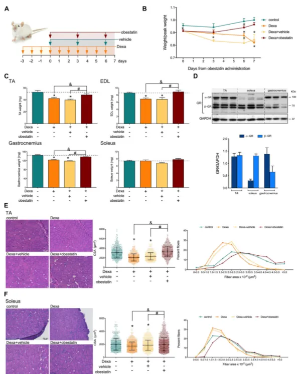
and processed for subsequent immunofluorescence analyses. For studies in the dexamethasone (Dexa; dexamethasone-water soluble)-induced skeletal muscle at-rophy model, mice were assigned to one of the following experimental groups (n = 8 per group): (i) control group, (ii) Dexa-treated group, (iii) Dexa + vehicle-treated group, and (iv) Dexa + obestatin-treated group. The control group was not exposed to any experimental intervention, thus serving as a neutral comparison for study groups. The Dexa-treated group was injected intraperitoneally with Dexa, dissolved in water (15 mg/kg body weight, 100 μL), every 24 h during 10 days. The Dexa + vehicle group was injected intraperitoneally with Dexa under the same conditions (15 mg/kg body weight/24 h, 10 days) and, as from the fourth day, received intraperitoneal injections of saline [vehicle control, 0.9% NaCL (w/v), 100 μL] every 72 h for 7 days. The obestatin-treated group was injected with Dexa (100 μL, 15 mg/kg body weight each 24 h for 10 days), and, as from the fourth day, intraperitoneal injections of obestatin solution in saline (500 nmol/kg body weight, 100 μL) were co-administered every 72 h for 7 days. Mice were euthanized 10 days after the Dexa treat-ment, and skeletal muscle samples [TA, extensor digitorum longus (EDL), gastrocnemius, soleus, and diaphragm mus-cles] were excised and processed for subsequent analyses. A schematic representation of this protocol is presented in Figure 2A.
Cell culture and differentiation
Human myogenic cell line, KM155C25 Clone 48 (KM155C25 cells), was obtained from the platform for immortalization of human myoblasts of the Center of Research in Myology in Paris (France), who developed it from a biopsy obtained through MYOBANK, a partner in the EU network EuroBioBank (gracilis muscle, donor age 25 years), following the protocols previously described.16,17 Immortalized human myoblasts
maintain their capacity to differentiate both in vitro and
in vivo after transplantation into the regenerating muscles
of immunodeficient mice.17,18 KM155C25 cells were
culti-vated in GM-containing Medium 199:DMEM (1:5, v/v; Lonza, Pontevedra, ES) supplemented with 20% foetal bovine serum (v/v), 25μg/μL fetuin, 5 ng/mL human epidermal growth fac-tor, 0.5 ng/mL basic fibroblast growth factor, and 50 μg/mL gentamicin (Invitrogen; CA, USA) as described previously (Cid-Díaz T et al. 2017). Differentiation into myotubes was ini-tiated at 90% confluence by switching to DM [Medium 199: DMEM (1:5, v/v) supplemented with 50μg/mL gentamicin] for 7 days unless otherwise stated. Human myotubes were treated with Dexa (1μM) for 24 h in the presence or absence of obestatin (10 nM) or with insulin (100 nM) as a positive control of atrophy protection.
Histological analysis
Muscle samples were mounted in tissue freezing medium (tragacanth gum) and snap frozen in nitrogen-cooled isopentane. The sections, 5 μm thick, were mounted on HistoBond adhesion microslides (Marienfeld, Lauda-Königshofen, DE). Skeletal muscle sections were stained with haematoxylin and eosin (HE) to quantify myofibre area using NIH Image Software ImageJ64 (National Institutes of Health, Bethesda, MD, USA). For immunofluorescence analysis, skele-tal muscle sections were permeabilized and blocked with PBST [1% Triton X-100, 1% Tween 20, 5% heat-inactivated goat se-rum, 0.2% bovine serum albumin in phosphate-buffered saline (PBS)] for 30 min and incubated with primary antibodies di-luted in PBST overnight at 4°C, washed with PBS, and then in-cubated with a mixture of appropriate secondary antibodies for 45 min at 37°C. 4′,6-Diamidino-2-phenylindole (DAPI) was used to counterstain the nuclei (Invitrogen). The digital images of the cell cultures were acquired with a Leica TCS SP8 spectral confocal microscope (Leica Microsystems, Heidelberg, DE).
Small interfering RNA silencing of gene expression
To ablate KLF15 or Nedd4 expression in human KM155C25 myotubes, small interfering RNA (siRNA) specifically targeting human KLF15 (sc-45567, Santa Cruz ogy, CA, USA) or Nedd4-1 (sc-41079, Santa Cruz Biotechnol-ogy) was used. siRNA duplexes targeting FoxO4 were selected from ON-TARGETplus SMARTpool siRNA from Thermo Fisher Scientific (Dharmacon; human FoxO4: AGUCAUGCCUGGAAGCUUU, GAGAAGCGACUGACACUUG, GGAAAUACCAGCUUCAGUC, CAACGAGGCCACCGGCAAA). An ON-TARGETplus nontargeting siRNA was used as a control for all siRNA experiments. Human KM155C25 myotubes were transfected at Day 6 post-differentiation with Lipofec-tamine 2000 (Invitrogen), following the manufacturer’s instructions.SDS-PAGE and western blot analysis
Cell or tissue samples were directly lysed in ice-cold RIPA buffer [50 mM Tris-HCl (pH 7.2), 150 mM NaCl, 1 mM EDTA, 1% (v/v) NP-40, 0.25% (w/v) Na-deoxycholate, protease inhib-itor cocktail, phosphatase inhibinhib-itor cocktail]. The lysates were clarified by centrifugation (14 000× g for 15 min at 4°C), and the protein concentration was quantified using the QuantiProTM BCA assay kit. For immunoblotting, equal amounts of protein were fractionated by SDS-PAGE and transferred onto nitrocellulose membranes. Immunoreactive bands were detected by enhanced chemiluminescence (Pierce ECL Western Blotting Substrate; Thermo Fisher Scientific, Pierce, Rockford, IL, USA).
Co-immunoprecipitation assays
Following treatment [DM, DM + Dexa (1 μM, 24 h), DM + Dexa (1 μM, 24 h) + obestatin (10 nM, 24 h), DM + obestatin (10 nM, 24 h) in the absence or presence of 15 μM MG132 for 6 h], KM155C25 myotube cells were washed twice with ice-cold PBS and lysed in co-immunoprecipitation lysis buffer (50 mM Tris, 100 mM NaCl, 5 mM EDTA, 50 mM NaF, 1% Triton X-100, 10 mM glyc-erol phosphate, 200 μM sodium orthovanadate, 2.5 mM sodium pyrophosphate plus protease inhibitors). KLF15 was immunoprecipitated using mouse monoclonal anti-KLF15 an-tibody coupled to protein A Dynabeads according to the instructions provided by the manufacturer (Thermo Fisher Scientific, Life Technologies, Rockford, IL, USA). The washed immunoprecipitates were subjected to western blot analysis using the indicated antibodies.
Statistics
All values are presented as mean ± standard error of the mean (SEM). A Shapiro–Wilks normality test was performed for each data set. t-tests were carried out for comparisons between two samples. Unpaired t-test was used to assess the statistical significance of one-way or two-way analysis when the test statistic followed a normal distribution.
P < 0.05 was considered as statistically significant
(*,&,#P< 0.05).
Study approval
All animal experiments were approved by the animal experimentation committee of the University of Santiago de Compostela (Santiago de Compostela, Spain; No. 15010/17/005).
Results
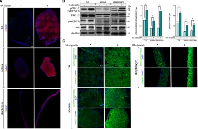
To examine the effect of obestatin signalling on GC-induced muscle atrophy, we first tested the adequate delivery of obestatin peptide to body musculature. Intraperitoneal injec-tion of HA-tagged obestatin in mice resulted in marked increase of HA-tag in TA, soleus, and diaphragm muscles (Figure 1A). Consistent with this, HA-tagged obestatin treatment resulted in elevated phosphorylation levels of ERK1/2 [pERK1/2(T202/Y204)] and Akt [pAkt(S473)] 2 h after the onset of peptide administration in TA, soleus, and dia-phragm muscles (Figure 1B and 1C). We then utilized a mouse model of GC-induced muscle wasting by exogenous administration of high dose of Dexa to determine the interac-tion of GR and obestatin signalling in the regulainterac-tion of
metabolism–volume coupling in skeletal muscle. In particular, skeletal muscle decline was promoted prior to obestatin ad-ministration by treatment with Dexa (15 mg/kg body weight, each 24 h) for 3 days prior to intraperitoneal injection of obestatin (500 nmol/kg body weight, each 72 h) for 7 days (Figure 2A). Indeed, the Dexa-treated mice displayed a signif-icant decrease of relative body weight compared with control mice 3 days after Dexa administration (designated as Day 0;
Figure 2B). The divergence in the Dexa-treated population
oc-curred about 6 days following obestatin treatment, with the Dexa-treated or Dexa + vehicle-treated groups showing a sig-nificant loss of total body weight, compared with the control group. No loss of weight was observed, however, in the Dexa + obestatin-treated group (Figure 2B). The increase of total body weight included an increase in skeletal muscle mass, as demonstrated by significant increase in type II-rich TA, EDL, and gastrocnemius muscle mass in the Dexa + obestatin-treated mice compared with the Dexa-treated or Dexa + vehicle-Dexa-treated groups (Figure 2C). In clear contrast, the mass of the type I-rich soleus muscle was un-changed (Figure 2C). Cross-sectional area (CSA) analysis
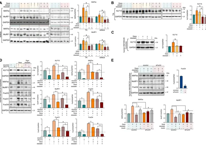
showed a shift towards smaller myofibre CSA in the TA mus-cle and, to a lesser extent, in the soleus musmus-cle in both the Dexa-treated and Dexa + vehicle-treated groups. The Dexa + obestatin group revealed a significant recovery of muscle when compared with Dexa groups in TA muscle and, to a minor extent, in the soleus muscle (Figure 2E and 2F, re-spectively). CSA distribution of myofibres supported this fact; the leftward shift in myofibre size was observed in the Dexa-treated and Dexa + vehicle-Dexa-treated groups, but not in the Dexa + obestatin group showing values closely related to the control mice in TA muscles. In contrast, the difference in the size was far less pronounced in the soleus fibres, highlighting the effect of obestatin treatment in myofibre size. The mechanism of this specificity can be related to the relatively lowα-isoform of the GR (α-GR) levels observed in the type I-rich soleus muscle compared with the type II-rich TA muscles (Figure 2D).19–21 Indeed, Dexa induction of the E3 ubiquitin ligases MAFbx and MuRF1 was observed in α-GR-rich TA muscles with reduced or no impact on soleus fibres (Figure 3A). When obestatin was administrated, the levels of MAFbx and MuRF1 were efficiently
Figure 1 Analysis of delivery of obestatin peptide administration via intraperitoneal injection to body musculature. Haemagglutinin (HA)-tagged obestatin (500 nmol/kg body weight each; n = 5) or vehicle (control; n = 5) was administrated to mice via intraperitoneal for 2 h as described in the Materials and methods section. (A) Representative immunostaining for HA in transverse cryosections of TA, soleus, and diaphragm muscles after 2 h of treatment with HA-tagged obestatin or vehicle. (B) Analysis of ERK1/2 and Akt activity in TA, soleus, and diaphragm muscles 2 h after HA-tagged obestatin administration. Immunoblots are representative of the mean value (n = 5 per group). ERK1/2 and Akt activities are in arbitrary units (au). Data are expressed as mean ± SEM obtained from intensity scans (*P< 0.05). (C) Representative immunostaining for pERK1/2 and pAkt in transverse cryosections of TA, soleus, and diaphragm muscles after 2 h of treatment with HA-tagged obestatin or vehicle.
Figure 2 Obestatin treatment restores muscle phenotype in dexamethasone-induced skeletal muscle atrophy model. (A) Schematic diagram of the intraperitoneal administration of dexamethasone (Dexa; 15 mg/kg body weight every 24 h, 10 days; n = 8), obestatin (500 nmol/kg body weight every 72 h, 7 days; n = 8), or vehicle (PBS every 72 h, 7 days; n = 8) in Swiss mice. (B) Time course of body weight of control, Dexa-treated, Dexa + vehicle-treated, and Dexa + obestatin-treated mice. (C) TA, EDL, gastrocnemius, and soleus muscle weight in control, Dexa-vehicle-treated, Dexa + vehicle-vehicle-treated, and Dexa + obestatin-treated mice. (D) Immunoblot analysis of GR levels in mouse TA, soleus, and gastrocnemius muscle. GR data were expressed relative to GAPDH as arbitrary units (mean ± SEM) obtained from intensity scans. (E) Left panel, representative haematoxylin and eosin staining of cryosections of TA muscle from control, Dexa-treated, Dexa + vehicle-treated, and Dexa + obestatin-treated mice. Middle panel, cross-sectional area (CSA) of muscle fibres from TA muscles obtained from control, Dexa-treated, Dexa + vehicle-treated, and Dexa + obestatin-treated mice. Right panel, distribution of fibre diameter from TA muscles obtained from control, Dexa-treated, Dexa + vehicle-treated, and Dexa + obestatin-treated mice. Data are expressed as % of myofibres per class of fibre diameter. (F) Left panel, representative haematoxylin and eosin staining of cryosections of soleus muscle from con-trol, Dexa-treated, Dexa + vehicle-treated, and Dexa + obestatin-treated mice. Middle panel, cross-sectional area (CSA) of musclefibres from TA mus-cles from control, Dexa-treated, Dexa + vehicle-treated, and Dexa + obestatin-treated mice. Right panel, distribution offibre diameter from TA muscles from control, Dexa-treated, Dexa + vehicle-treated, and Dexa + obestatin-treated mice. Data are expressed as % of myofibres in each class of fibre diameter. In (E) and (F), data are expressed as mean ± SEM (n = 8 animals per group). *,&,#P< 0.05.
down-regulated in TA muscles (Figure 3A). In this model, obestatin signalling also suppressed the Dexa-induced ex-pression of KLF15 in TA muscle, a transcription factor induced by various stressors, including GCs, to regulate muscle catab-olism via the regulation of the E3 ubiquitin ligases MAFbx and MuRF1 (Figure 3B).7,22–24 Similarly, we identified the Dexa-mediated up-regulation of KLF15 protein expression and con-firmed its down-regulation by obestatin signalling in human KM155C25 myotubes (Figure 3C). We then asked whether the transcription network activated by GRs was dependent on KLF15. For that purpose, we tested the effect of the down-regulation of KLF15 by specific siRNA (siKLF15) on
protein expression of MAFbx, MuRF1, FoxO3, FoxO1, and FoxO4 (Figure 3D). In human KM155C25 myotubes, KLF15 knockdown diminished the Dexa-dependent protein induc-tion of all these GR target proteins (Figure 3D). Note that Dexa + obestatin vs. Dexa resulted in additive effect on siKLF15 (Figure 3D). MAFbx and MuRF1 were originally identified as FoxO target genes,25,26and KLF15 was shown to regulate FoxO expression.7 Indeed, the siRNA against FoxO4, the main obestatin-responsive FoxO isoform,16 significantly decreased the MuRF1 and MAFbx expression in Dexa-stimulated cells (Figure 3E). Moreover, siFoxO4 experiments partially restored the MuRF1 and MAFbx
Figure 3 Regulation of atrogenes and FoxOs by KLF15 in dexamethasone-induced skeletal muscle atrophy. (A) Immunoblot analysis of MAFbx and Murf1 levels in mouse TA and soleus muscle after intraperitoneal administration of Dexa (15 mg/kg body weight every 24 h, 10 days; n = 8), obestatin (500 nmol/kg body weight every 72 h, 7 days; n = 8), or vehicle (PBS every 72 h, 7 days; n = 8) in Swiss mice. (B) Immunoblot analysis of KLF15 levels in mouse TA muscle after intraperitoneal administration of Dexa (15 mg/kg body weight every 24 h, 10 days; n = 8), obestatin (500 nmol/kg body weight every 72 h, 7 days; n = 8), or vehicle (PBS every 72 h, 7 days; n = 8) in Swiss mice. (C) Immunoblot analysis of KLF15 in human KM155C25 myotubes under DM, DM + Dexa (1μM, 24 h), DM + Dexa (1 μM, 24 h) + obestatin (10 nM, 24 h), or DM + Dexa (1 μM, 24 h) + insulin (100 nM, 24 h) (n = 4). (D) After transfection with specific KLF15 siRNA, KM155C25 myotubes were treated with Dexa (1 μM) or Dexa (1 μM) + obestatin (10 nM) for 24 h. The effect of KLF15 siRNA was evaluated on MAFbx, Murf1, FoxO3, FoxO1, and FoxO4 expression (n = 4). (E) Immunoblot analysis of MAFbx, Murf1, and FoxO4 in human KM155C25 myotubes transfected with specific FoxO4 siRNA under DM, DM + Dexa (1 μM, 24 h), or DM + Dexa (1 μM, 24 h) + obestatin (10 nM, 24 h). In (A) and (B), protein level is expressed as fold of control. Data are expressed as mean ± SEM obtained from intensity scans. *,#,&P< 0.05. In (C)–(E), immunoblots are representative of the mean value (n = 4). Data were expressed as mean ± SEM obtained from intensity
expression in Dexa + obestatin-treated cells (Figure 3E). These results strongly indicate the critical involvement of obestatin signalling on the GR-KLF15 cascade to regulate the Dexa-mediated up-regulation of E3 ubiquitin ligase expression.
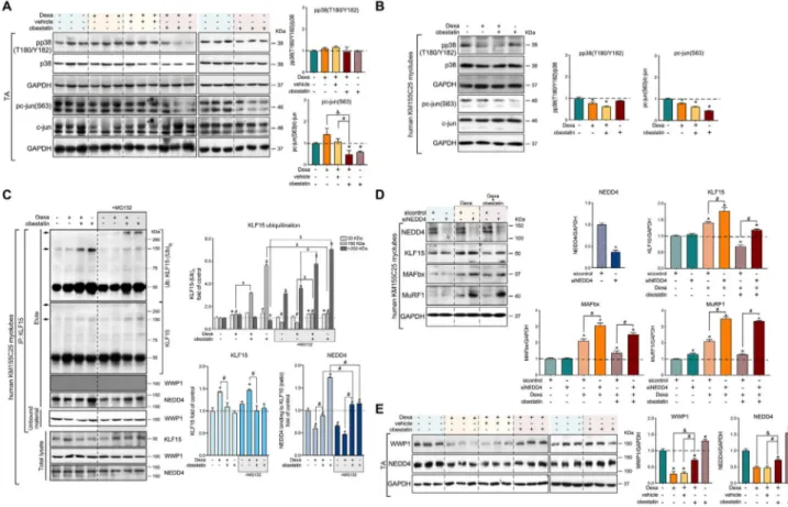
An important question is how KLF15 can be shut down by obestatin signalling in skeletal muscle. p38 MAPK27 and c-Jun28 were identified as negative modulators of the GR-mediated KLF15 transcription. However, obestatin suppressed Dexa-mediated phosphorylation of c-Jun at S63 in TA muscles, without a significant alteration in p38 phos-phorylation at T180/Y182 (Figure 4A). These effects were
confirmed in human KM155C25 myotubes (Figure 4B). Post-translational modification of KLF15 protein by the E3 ubiquitin ligases is one of the key regulatory steps that guides protein degradation through regulation of proteasome activity. WW domain-containing E3 ubiquitin protein ligase 1 (WWP1) and neural precursor cell-expressed developmen-tally down-regulated 4 (NEDD4) were previously shown to interact with other members of the KLF family.29 Co-immunoprecipitation analysis revealed that NEDD4, but not WWP1, interacted with KLF15 in human KM155C25 myotubes (Figure 4C). Dexa increased the protein expression but failed to modify the polyubiquitination of KLF15
Figure 4 Obestatin signalling regulates the KLF15-triggered expression program via the E3 ubiquitin ligase NEDD4. (A) Immunoblot analysis of pp38 (T180/Y182) and pc-jun(S63) in TA muscle after intraperitoneal administration of Dexa (15 mg/kg body weight each 24 h, 10 days; n = 8), obestatin (500 nmol/kg body weight each 72 h, 7 days; n = 8), or vehicle (PBS each 72 h, 7 days; n = 8) in Swiss mice. Protein level is expressed as fold of control. Data are expressed as mean ± SEM obtained from intensity scans (*,#,&P< 0.05). (B) Immunoblot analysis of pp38(T180/Y182) and pc-jun(S63) in
hu-man KM155C25 myotubes under DM, DM + Dexa (1μM, 24 h), or DM + Dexa (1 μM, 24 h) + obestatin (10 nM, 24 h). Immunoblots are representative of the mean value (n = 4). Data were expressed as mean ± SEM obtained from intensity scans. (*P< 0.05). (C) Co-immunoprecipitation assays used to determine the KLF15-interacting proteins in human KM155C25 myotubes under DM, DM + Dexa (1μM, 24 h), DM + Dexa (1 μM, 24 h) + obestatin (10 nM, 24 h), or DM + obestatin (10 nM, 24 h) stimulation in the absence or presence of MG132 (15 μM). The KLF15 complex was co-immunoprecipitated with anti-KLF15 affinity beads and analysed by western blot using anti-ubiquitin, anti-KLF15, anti-WWP1, and anti-NEDD4 an-tibodies. Arrows indicate the main polyubiquitinated forms of KLKF15, for example, 150 and 250 kDa. Immunoblots are representative of the mean value (n = 4). Data are expressed as mean ± SEM obtained from intensity scans (*,#,&,$P< 0.05). (D) After transfection with specific NEDD4 or control
siRNA, KM155C25 myotubes were treated with Dexa (1μM) or Dexa (1 μM) + obestatin (10 nM) for 24 h. The effect of NEDD4 siRNA was evaluated on KLF15, MAFbx, and Murf1 (n = 4). Data are expressed as mean ± SEM obtained from intensity scans (*,#P< 0.05). (E) Immunoblot analysis of the
WWP1 and NEDD4 protein levels in TA muscle after intraperitoneal administration of dexamethasone (Dexa; 15 mg/kg body weight each 24 h, 10 days;
n = 8), obestatin (500 nmol/kg body weight each 72 h, 7 days; n = 8), or vehicle (PBS each 72 h, 7 days; n = 8) in Swiss mice. Protein level is expressed as
[KLF15-(Ub)n]. In clear contrast, obestatin inhibited
Dexa-mediated KLF15 expression by promoting the polyubiquitination of KLF15-(Ub)n (mw ≅ 150 kDa; Figure
4C). Moreover, treatment of the cells with the proteasome in-hibitor MG132 enhanced the amount of KLF15 ubiquitin con-jugates in both Dexa + obestatin-stimulated and obestatin-stimulated human myotubes [KLF15-(Ub)n
mw > 250 kDa; Figure 4C]. Such an enhancement was paralleled by NEDD4 recruitment onto KLF15 (Figure 4C). Thus, obestatin negatively modulates KLF15 function, most possibly by altering the protein ubiquitination profile, the linkage type, and length of ubiquitin chains through NEDD4 and linking the substrates to downstream processes.
We next examined the effect of knocking down the expression of NEDD4 by siRNA (siNEDD4) on KLF15 activity from human KM155C25 myotubes. NEDD4 siRNA-mediated knockdown increased the response to Dexa of a number of GC-inducible proteins, including KLF15, MAFbx, and MuRF1, when compared with control siRNA (Figure 4D). Treatment with obestatin clearly inhibited the induction by DEXA of KLF15, MAFbx, and MuRF1 proteins in human myotubes transfected with control siRNA, and such a process was signif-icantly reversed by siNEDD4 (Figure 4D). In TA muscle, pro-tein expression of NEDD4 was suppressed by Dexa, and obestatin suppressed at least partly, but significantly, this Dexa-mediated down-regulation of NEDD4 (Figure 4E). This positive modulation by obestatin was also observed on WWP1 protein expression profile (Figure 4E). Taken together, these data indicate that obestatin regulates the liaison mole-cule for GR in the induction of atrogenes, KLF15, through the control of NEDD4.
We then examined the effects of Dexa and obestatin on protein levels and phosphorylation status of AMPK and Akt, the upstream activator of mTOR, and mTOR downstream ef-fectors S6 and 4E-BP1, in the mouse GC-induced atrophy model. In TA muscle, treatment with Dexa increased the phosphorylation of AMPK [pAMPK(T172)], without significant alteration in the phosphorylation of Akt [pAkt(S473)], FoxO family [pFoxO3(T32), pFoxO1(T24), pFoxO4(T28)], S6 [pS6 (S240/244)], and 4E-BP1 [p4E-BP1(T37/41)] (Figure 5A). In clear contrast, the levels of pAkt(S473), pFoxO4(T24), pS6 (S240/244), and p4E-BP1(T37/41) were significantly increased when obestatin was administrated, while pAMPK(T172) was inhibited (Figure 5A). In this model, Dexa administration in-creased the expression of lysosomal enzyme cathepsin L and the conversion rate of the microtubule-associated pro-tein 1 light chain 3 isoform I (LC3I) to LC3 isoform II (LC3II), with significant decline in the amount of p62 protein indicat-ing autophagic degradation activity (Figure 5B). In clear con-trast, treatment with obestatin reversed the effects of Dexa (Figure 5B). Significant accumulation of p62 protein was seen compared with Dexa treatment alone indicating a block in au-tophagy (Figure 5B). Furthermore, obestatin treatment was associated with a reduction in the expression of LC3II and
cathepsin L, further supporting an inactivation of autophagy (Figure 5B). Histological examination of the TA muscle dem-onstrated type II fibre-dominant atrophy in the Dexa and Dexa + vehicle groups. This atrophic effect was also charac-terized by fast-twitch type IIb/IIx glycolytic musclefibre loss and reduced impact on slow-twitch type I and fast-twitch type IIa oxidative muscle fibres (Figure 5C). In contrast, the Dexa + obestatin group showed less impairment in the TA muscle by preventing of atrophy and type IIfibre loss (Figure 5C). CSA analysis of myofibres supported this observation, because a leftward shift in the fast-twitch type IIb/IIx glyco-lytic myofibre size was observed in the Dexa and Dexa + vehicle groups, but not in the Dexa + obestatin group (Figure 5C). Dexa-treated muscles showed limited or no im-pact on fast-twitch type IIa oxidative and slow-twitch type I oxidativefibres, respectively (Figure 5C). Of note, there were significant differences in the size of slow-twitch type I, fast-twitch type IIa oxidative, and fast-twitch IIb glycolytic muscle fibres in the obestatin-treated group compared with the treatment with Dexa alone or Dexa + vehicle and even to the control group (Figure 5C). Therefore, we conclude that the interplay among Akt, mTOR, KLF15, and FoxO4 regulates two of the main proteolytic systems, the ubiquitin –protea-some and autophagy–lysosome systems, and the signalling associated with protein synthesis in response to the obestatin/GPR39 system to counteract the GR-dependent catabolic processes.
Discussion
In skeletal muscle, anabolic and catabolic pathways arefinely tuned in a co-ordinated manner to regulate myofibre size and total muscle mass. Pathological conditions associated with in-creased levels of circulating GC distort this balance, leading to a generalized decrease in myofibre size and to muscle atro-phy. In this report, we show that the activation of the obestatin/GPR39 system protects from GC-induced atrophy by regulation of Akt and KLF15 signalling and its downstream targets in the control of protein synthesis, ubiquitin –protea-some system, and autophagy–lysosome system. FoxO tran-scription factors are common elements that act as critical liaison molecules for protein degradation and autophagy. The pattern of post-translational modifications of FoxO tran-scription factors, including specific FoxO4 phosphorylation, triggers FoxO activity in response to obestatin. At transcrip-tional levels, the expression of FoxO family is regulated by the ubiquitin-dependent degradation of KLF15 via up-regulation of the E3 ligase NEDD4. Moreover, obestatin activates Akt/mTOR signalling and modulates positively myofibre size, leading to a substantial restoration of muscle phenotype in GC-treated atrophic mice. The co-ordinated crosstalk between GR and the obestatin/GPR39 system
represents a novel mechanism for thefine-tuning of muscle mass and, in this way, a potential therapeutic target to coun-teract muscle wasting.
The GC-induced muscle atrophy is a result of the functional cooperativity among GR, FoxOs, and KLF15 as critical liaison molecules aimed at increasing protein degradation.1,7 A sig-nificant question raised in this study is how the GR-driven proteolytic cascades can be shut down by obestatin signal-ling. In our model, obestatin signalling negatively modulates GR-mediated gene expression, and this action derives from the regulation of FoxO transcription factors at both transla-tional and post-transcriptransla-tional levels. At post-translation level, FoxO family is negatively regulated by the Akt pathway
in response to obestatin. Phosphorylation of FoxO factors by Akt causes the sequestration of FoxO in the cytoplasm, thereby preventing FoxO factors from transactivating their target genes in the nucleus.30–32Our results show that nei-ther FoxO1 nor FoxO3 is involved in the response to obestatin via phosphorylation by Akt activation. In contrast, FoxO4 is the main obestatin-responsive isoform, thus supporting an isoform-specific mode of regulation previously described in human myotubes.16At transcriptional level, the effect of obestatin on FoxO factors is mediated by a down-regulation of KLF15 expression. It is worth to underline that the E3 ubiquitin ligase responsible for KLF15 polyubiquitination in response to obestatin is NEDD4. This
Figure 5 Obestatin treatment protects from atrophy with induction of protein synthesis signalling. (A) Immunoblot analysis of pAkt(S473), Akt, pAMPK (T172), AMPK, pS6(S240/244), S6, p4E-BP1(T37/46), 4E-BP1, pFoxO3a(T32), pFoxO1(T24), and pFoxO4(T28) in TA muscle after intraperitoneal admin-istration of Dexa (15 mg/kg body weight each 24 h, 10 days; n = 8), obestatin (500 nmol/kg body weight every 72 h, 7 days; n = 8), or vehicle (PBS every 72 h, 7 days; n = 8) in Swiss mice. Protein level is expressed as fold of control. Data are expressed as mean ± SEM obtained from intensity scans (*,&,#P< 0.05). (B) Immunoblot analysis of the LC3, p62, and cathepsin L in TA muscle after intraperitoneal administration of Dexa (15 mg/kg body
weight every 24 h, 10 days; n = 8), obestatin (500 nmol/kg body weight every 72 h, 7 days; n = 8), or vehicle (PBS every 72 h, 7 days; n = 8) in Swiss mice. Protein level is expressed as fold of control. Data are expressed as mean ± SEM obtained from intensity scans (*,&,#P< 0.05). (C) Left panel,
representative images of control-treated, Dexa-treated, Dexa + vehicle-treated, and Dexa + obestatin-treated TA muscles showing MHC expression. Serial muscle cross sections were incubated with a primary antibody against MHCI, MHCIIa, MHCIIb, or MHCIIx. Right panel, frequency distribution histograms of cross-sectional areas (CSA;μm2). Data are shown as mean ± SEM (n = 8 per group). (D) Schematic model of the crosstalk between the GC-associated catabolic processes and the obestatin-triggered anabolic processes in skeletal muscle.
fact is supported by the results of the immunoprecipitation assays that demonstrate that NEDD4 interacts specifically with KLF15 and that this interaction is increased in response to obestatin. Moreover, siRNA targeting NEDD4 suppresses the potential of obestatin to regulate KLF15 and the atrogenes during GC treatment. Interestingly, in skeletal mus-cle, NEDD4 has been reported to be involved in the genera-tion of atrophy under very specific conditions in skeletal muscle such as in disuse (unloading or denervation)-induced muscle atrophy.33–38However, the NEDD4 expression under obestatin signalling correlates with a regression of the atro-phic process, concomitant with a decreased expression of KLF15 protein. Thus, we propose that NEDD4 controls the ubiquitination, or stability, or both, of KLF15. This protein complex determines the KLF15 activity, for example, control of FoxO and atrogenes expression. In contrast to changes in subcellular localization achieved by Akt activation, which are rapidly reversible, changes in FoxO protein levels are more permanent, which may have profound impacts on FoxO functions in the progress of muscle wasting. Within the GR-triggered proteostasis network, the activation of obestatin signalling blocks the lysosome–autophagy system. In fact, the reduction of LC3 lipidation (LC3II) and the accu-mulation of p62 together with a reduced cathepsin L protein level limit the final degradation steps of up-regulated autophagy. It is well established that FoxO proteins induce autophagy not only through transactivation of autophagic genes but also by interacting with autophagic proteins and by epigenetically regulating autophagic activity.39–42 In this way, the post-translational modification of FoxOs may down-regulate autophagy. Thus, in skeletal muscle, the lyso-somal and protealyso-somal pathways for protein degradation can be co-ordinately regulated upon activation of obestatin signalling. The simultaneous inhibition of these two proteo-lytic pathways by obestatin signalling presumably plays the role of safeguards against the loss of different cell compo-nents, for example, myofibrillar proteins, and organelles, es-pecially mitochondria. Both modes of regulating FoxO activity through obestatin signalling with the enhancement of the protein synthesis pathway, for example, the activation of S6K1 and the eIF4E availability, through phosphorylation of 4E-BP1,43restore basal homoeostasis conditions. Indeed, we show in this report that in obestatin-treated mice, the type II-rich skeletal muscle mass increases as well as the size of musclefibres in the presence of atrophy-promoting GC, main-taining proteome integrity.
In skeletal muscle, KLF15 is a critical regulator of nutrient catabolism and a key determinant of exercise capacity7,22,23; however, its role on atrogene expression is debated. KLF15 is described as a GR target gene acting as a catabolic modu-lator by regulating the expression of atrogenes under stress-ful conditions associated with excess levels of GCs.7 Paradoxically, GC steroid treatment is used in muscular dys-trophy, for example, DMD, where their use exerts ergogenic
muscle performance effects.44In mdx mice, a genetic model of DMD, the GC treatment improves muscle regeneration and specific force.45–47 It has been shown that GC steroids rely on KLF15 to mediate ergogenic muscle performance ef-fects. Indeed, deletion of KLF15 in the mdx mouse exacer-bated the mdx muscle phenotype, and muscle-specific overexpression of KLF15 improved muscle performance. In addition, it appears that low dose and/or transient presence of GC induces KLF15 that controls metabolic genes without inducing atrogenes.48A separate study comparing the effects of daily and weekly administrations of GCs found that KLF15 expression is increased by weekly administration of GCs, but not by chronic administration, which elicited muscle wasting.49–51Additionally, intermittent exposure to GCs is as-sociated with epigenetic programming that enhances nutri-ent utilization by the co-ordinated action of GR and the transcription factors KLF15 and MEF2C.52Intriguingly, similar effects can be observed in other models of neuromuscular diseases, such as the mouse model of spinal muscular atrophy.53Thus, the regulatory effects of GRs on KLF15 and muscle gene transcription seem to be context specific. An in-teresting point is the differential effects of GCs, whereby at-rophy signalling was induced in healthy animals,7,54 while ergogenic effects occur in pathological models, for example, mouse models of DMD, limb-girdle muscle dystrophy, or spi-nal muscular atrophy.48,50–53As such, the down-regulation of KLF15 leads to decreased expression of atrophy-related genes. Further studies will be needed to clarify the functional involvement of KLF15 in muscle wasting.
In conclusion, the present study reveals that activation of the obestatin/GPR39 system blocks FoxO action in multiple, and possibly sequential, ways (Figure 5D): it promotes post-translational modifications on the FoxO proteins, includ-ing phosphorylation, by Akt and modifies FoxO protein levels by NEDD4-dependent ubiquitination of KLF15. This dual ac-tion of obestatin to trigger FoxO inactivaac-tion by obestatin highlights the importance of turning off FoxO in wasting disorder-related skeletal muscle atrophy, possibly to ensure a tight response to obestatin to counteract catabolic activity. These findings, combined with previous human studies, highlight the potential therapeutic relevance of the obestatin/GPR39 system for fine-tuning of muscle mass in conditions characterized by muscle atrophy associated with increased circulating GC levels.
Acknowledgements
The platform for immortalization of human cells of the Center of Research in Myology is acknowledged, particu-larly K. Mamchaoui, as well as the MYOBANK-AFM in the Myology Institute (code BB-0033-00012). The authors cer-tify that they comply with the ethical guidelines for
authorship and publishing of the Journal of Cachexia,
Sarcopenia and Muscle.55
Funding
This work was supported by grants from Instituto de Salud Carlos III in co-financing with Fondo Europeo de Desarrollo Re-gional (ISCIII-FEDER; Ministerio de Ciencia e Innovación (MICINN), Spain; PI17/01707 and PI18/00760), Axencia de Coñecemento en Saúde (ACIS), Servicio Galego de Saúde (SERGAS; Xunta de Galicia; PRIS Program), and Axencia Galega de Innovación (GAIN; Xunta de Galicia; IN607B2019/06).
Online supplementary material
Additional supporting information may be found online in the Supporting Information section at the end of the article.
Table S1. Primary antibodies
Con
flict of interest
The authors declare no competing interests.
References
1. Patel R, Williams-Dautovich J, Cummins CL. Minireview: new molecular mediators of glucocorticoid receptor activity in meta-bolic tissues. Mol Endocrinol 2014;28: 999–1011.
2. Evans RM. The nuclear receptor superfam-ily: a rosetta stone for physiology. Mol
Endocrinol 2005;19:1429–1438.
3. Meijsing SH, Pufall MA, So AY, Bates DL, Chen L, Yamamoto KR. DNA binding site sequence directs glucocorticoid receptor structure and activity. Science 2009;324: 407–410.
4. Zierath JR, Wallberg-Henriksson H. From receptor to effector: insulin signal transduction in skeletal muscle from type II diabetic patients. Ann N Y Acad Sci 2002;967:120–134.
5. Kuo T, Harris CA, Wang JC. Metabolic functions of glucocorticoid receptor in skeletal muscle. Mol Cell Endocrinol
2013;380:79–88.
6. Watson ML, Baehr LM, Reichardt HM, Tuckermann JP, Bodine SC, Furlow JD. A cell-autonomous role for the glucocorticoid receptor in skeletal muscle atrophy in-duced by systemic glucocorticoid exposure.
Am J Physiol Endocrinol Metab 2012;302:
E1210–E1220.
7. Shimizu N, Yoshikawa N, Ito N, Maruyama T, Suzuki Y, Takeda S, et al. Crosstalk be-tween glucocorticoid receptor and nutri-tional sensor mTOR in skeletal muscle.
Cell Metab 2011;13:170–182.
8. Menconi M, Fareed M, O’Neal P, Poylin V, Wei W, Hasselgren PO. Role of glucocorti-coids in the molecular regulation of muscle wasting. Crit Care Med 2007;35:S602–S608. 9. Schakman O, Gilson H, Thissen JP. Mecha-nisms of glucocorticoid-induced myopathy.
J Endocrinol 2008;197:1–10.
10. Hu Z, Wang H, Lee IH, Du J, Mitch WE. Endogenous glucocorticoids and impaired insulin signaling are both required to stim-ulate muscle wasting under pathophysio-logical conditions in mice. J Clin Invest 2009;119:3059–3069.
11. Gurriarán-Rodríguez U, Santos-Zas I, Al-Massadi O, Mosteiro CS, Beiroa D, Nogueiras R, et al. The obestatin/GPR39 system is up-regulated by muscle injury and functions as an autocrine regenerative
system. J Biol Chem 2012;287:
38379–38389.
12. Gurriarán-Rodríguez U, Santos-Zas I, González-Sánchez J, Beiroa D, Moresi V, Mosteiro CS, et al. Action of obestatin in skeletal muscle repair: stem cell expansion, muscle growth, and microenvironment
remodeling. Mol Theraphy 2015;23:
1003–1021.
13. Santos-Zas I, Gurriarán-Rodríguez U, Cid-Díaz T, Figueroa G, González-Sánchez J, Bouzo-Lorenzo M, et al. β-Arrestin scaffolds and signaling elements essential for the obestatin/GPR39 system that deter-mine the myogenic program in human myo-blast cells. Cell Mol Life Sci 2016;73: 617–635.
14. Santos-Zas I, Negroni E, Mamchaoui K, Mosteiro CS, Gallego R, Butler-Browne GS, et al. Obestatin increases the regenerative capacity of human myoblasts transplanted intramuscularly in an immunodeficient
mouse model. Mol Ther 2017;25:
2345–2359.
15. González-Sánchez J, Sánchez-Temprano A, Cid-Díaz T, Pabst-Fernández R, Mosteiro CS, Gallego R, et al. Improvement of Duchenne muscular dystrophy phenotype following obestatin treatment. J Cachexia
Sarcopenia Muscle 2018;9:1063–1078.
16. Cid-Díaz T, Santos-Zas I, González-Sánchez J, Gurriarán-Rodríguez U, Mosteiro CS, Casabiell X, et al. Obestatin controls the ubiquitin-proteasome and autophagy-lysosome systems in glucocorticoid-induced muscle cell atrophy. J Cachexia Sarcopenia
Muscle 2017;8:974–990.
17. Mamchaoui K, Trollet C, Bigot A, Negroni E, Chaouch S, Wolff A, et al. Immortalized pathological human myoblasts: towards a universal tool for the study of neuromuscu-lar disorders. Skelet Muscle 2011;1:34. 18. Thorley M, Duguez S, Mazza EMC, Valsoni
S, Bigot A, Mamchaoui K, et al. Skeletal muscle characteristics are preserved in hTERT/cdk4 human myogenic cell lines.
Skelet Muscle 2016;6:43.
19. Hollenberg SM, Weinberger C, Ong ES, Cerelli G, Oro A, Lebo R, et al. Primary structure and expression of a functional human glucocorticoid receptor cDNA.
Na-ture 1995;318:635–641.
20. Hinds TD Jr, Ramakrishnan S, Cash HA, Stechschulte LA, Heinrich G, Najjar SM, et al. Discovery of glucocorticoid receptor-beta in mice with a role in metab-olism. Mol Endocrinol 2010;24:1715–1727. 21. Kino T, Manoli I, Kelkar S, Wang Y, Su YA, Chrousos GP. Glucocorticoid receptor (GR) β has intrinsic, GRα-independent transcrip-tional activity. Biochem Biophys Res Commun 2009;381:671–675.
22. Gray S, Wang B, Orihuela Y, Hong EG, Fisch S, Haldar S, et al. Regulation of gluconeo-genesis by Krüppel-like factor 15. Cell
Metab 2007;5:305–312.
23. Haldar SM, Jeyaraj D, Anand P, Zhu H, Lu Y, Prosdocimo DA, et al. Kruppel-like factor 15 regulates skeletal muscle lipidflux and exercise adaptation. Proc Natl Acad Sci U
24. Jeyaraj D, Scheer FA, Ripperger JA, Haldar SM, Lu Y, Prosdocimo DA, et al. Klf15 or-chestrates circadian nitrogen homeostasis.
Cell Metab 2010;15:311–323.
25. Sandri M, Sandri C, Gilbert A, Skurk C, Calabria E, Picard A, et al. Foxo transcrip-tion factors induce the atrophy-related ubiquitin ligase atrogin-1 and cause skele-tal muscle atrophy. Cell 2004;117:399–412. 26. Waddell DS, Baehr LM, van den Brandt J, Johnsen SA, Reichardt HM, Furlow JD, et al. The glucocorticoid receptor and FOXO1 synergistically activate the skeletal muscle atrophy-associated MuRF1 gene.
Am J Physiol Endocrinol Metab 2008;295:
E785–E797.
27. Leenders JJ, Wijnen WJ, Hiller M, van der Made I, Lentink V, van Leeuwen RE, et al. Regulation of cardiac gene expression by KLF15, a repressor of myocardin activity. J
Biol Chem 2010;285:27449–27456.
28. Lee DS, Choi H, Han BS, Kim WK, Lee SC, Oh KJ, et al. c-Jun regulates adipocyte differentiation via the KLF15-mediated mode. Biochem Biophys Res Commun 2016;469:552–558.
29. Hirata Y, Nomura K, Senga Y, Okada Y, Kobayashi K, Okamoto S, et al. Hyperglyce-mia induces skeletal muscle atrophy via a WWP1/KLF15 axis. JCI Insight 2019;4: e124952.
30. Stitt TN, Drujan D, Clarke BA, Panaro F, Timofeyva Y, Kline WO, et al. The IGF-1/ PI3K/Akt pathway prevents expression of muscle atrophy-induced ubiquitin ligases by inhibiting FOXO transcription factors.
Mol Cell 2004;14:395–403.
31. Calnan DR, Brunet A. The FoxO code.
Onco-gene 2008;27:2276–2288.
32. Tia N, Singh AK, Pandey P, Azad CS, Chaudhary P, Gambhir IS. Role of forkhead box O (FOXO) transcription factor in aging and diseases. Gene 2018;648:97–105. 33. Koncarevic A, Jackman RW, Kandarian SC.
The ubiquitin-protein ligase Nedd4 targets Notch1 in skeletal muscle and distin-guishes the subset of atrophies caused by reduced muscle tension. FASEB J 2007;21: 427–437.
34. Cao XR, Lill NL, Boase N, Shi PP, Croucher DR, Shan H, et al. Nedd4 controls animal growth by regulating IGF-1 signaling. Sci
Signal 2008;1:ra5.
35. Liu Y, Oppenheim RW, Sugiura Y, Lin W. Ab-normal development of the neuromuscular
junction in Nedd4-deficient mice. Dev Biol 2009;330:153–166.
36. Fouladkou F, Lu C, Jiang C, Zhou L, She Y, Walls JR, et al. The ubiquitin ligase Nedd4-1 is required for heart development and is a suppressor of thrombospondin-1. J
Biol Chem 2010;285:6770–6780.
37. Nagpal P, Plant PJ, Correa J, Bain A, Takeda M, Kawabe H, et al. The ubiquitin
ligase Nedd4-1 participates in
denervation-induced skeletal muscle atro-phy in mice. PLoS ONE 2012;7:e46427. 38. Huang X, Chen J, Cao W, Yang L, Chen Q,
He J, et al. The many substrates and functions of NEDD4. Cell Death Dis
2019;10:904.
39. Zhao J, Brault JJ, Schild A, Cao P, Sandri M, Schiaffino S, et al. FoxO3 coordinately acti-vates protein degradation by the autopha-gic/lysosomal and proteasomal pathways in atrophying muscle cells. Cell Metab 2007;6:472–483.
40. Webb AE, Brunet A. FOXO transcription factors: key regulators of cellular quality control. Trends Biochem Sci 2014;39: 159–169.
41. Milan G, Romanello V, Pescatore F, Armani A, Paik JH, Frasson L, et al. Regulation of autophagy and the ubiquitin-proteasome system by the FoxO transcriptional net-work during muscle atrophy. Nat Commun 2015;6:6670.
42. Cheng Z. The FoxO-autophagy axis in health and disease. Trends Endocrinol
Metab 2019;30:658–667.
43. Laplante M, Sabatini DM. mTOR signaling in growth control and disease. Cell
2012;149:274–293.
44. McDonald CM, Henricson EK, Abresch RT, Duong T, Joyce NC, Hu F, et al. Long-term effects of glucocorticoids on function, quality of life, and survival in patients with Duchenne muscular dystrophy: a prospec-tive cohort study. Lancet 2018;391: 451–461.
45. Anderson JE, McIntosh LM, Poettcker R. Deflazacort but not prednisone improves both muscle repair and fiber growth in diaphragm and limb muscle in vivo in the mdx dystrophic mouse. Muscle Nerve 1996;19:1576–1585.
46. St-Pierre SJ, Chakkalakal JV, Kolodziejczyk SM, Knudson JC, Jasmin BJ, Megeney LA. Glucocorticoid treatment alleviates dystro-phic myofiber pathology by activation of
the calcineurin/NF-AT pathway. FASEB J 2004;18:1937–1939.
47. Baltgalvis KA, Call JA, Nikas JB, Lowe DA. Ef-fects of prednisolone on skeletal muscle contractility in mdx mice. Muscle Nerve 2009;40:443–454.
48. Morrison-Nozik A, Anand P, Zhu H, Duan Q, Sabeh M, Prosdocimo DA, et al. Glucocorti-coids enhance muscle endurance and ameliorate Duchenne muscular dystrophy through a defined metabolic program.
Proc Natl Acad Sci U S A 2015;112:
E6780–E6789.
49. Guerron AD, Rawat R, Sali A, Spurney CF, Pistilli E, Cha HJ, et al. Functional and molecular effects of arginine butyrate and prednisone on muscle and heart in the mdx mouse model of Duchenne Muscular Dystrophy. PLoS ONE 2010;5: e11220.
50. Quattrocelli M, Barefield DY, Warner JL, Vo AH, Hadhazy M, Earley JU, et al. Intermit-tent glucocorticoid steroid dosing en-hances muscle repair without eliciting
muscle atrophy. J Clin Invest
2017a;127:2418–2432.
51. Quattrocelli M, Salamone IM, Page PG, Warner JL, Demonbreun AR, McNally EM. Intermittent glucocorticoid dosing im-proves muscle repair and function in mice with limb-girdle muscular dystrophy. Am J
Pathol 2017b;187:2520–2535.
52. Quattrocelli M, Zelikovich AS, Jiang Z, Peek CB, Demonbreun AR, Kuntz NL, et al. Pulsed glucocorticoids enhance dystrophic muscle performance through epigenetic-metabolic reprogramming. JCI Insight 2019;
4:e132402.
53. Walter LM, Deguise MO, Meijboom
KE, Betts CA, Ahlskog N, van Westering TLE, et al. Interventions targeting glucocor-ticoid-Krüppel-like factor 15-branched-chain amino acid signaling improve disease phenotypes in spinal muscular atrophy mice. EBioMedicine 2018;31:226–242. 54. van Raalte DH, Ouwens DM, Diamant M.
Novel insights into
glucocorticoid-mediated diabetogenic ef-fects: towards expansion of therapeutic options? Eur J Clin Invest 2009;39:81–93. 55. von Haehling S, Morley JE, Coats AJS, Anker
SD. Ethical guidelines for authorship and publishing in the Journal of Cachexia,
Sarcopenia and Muscle. J Cachexia Sarcop Muscle 2015;4:315–316.