HAL Id: inserm-01170440
https://www.hal.inserm.fr/inserm-01170440v2
Submitted on 15 Apr 2020
HAL is a multi-disciplinary open access
archive for the deposit and dissemination of
sci-entific research documents, whether they are
pub-lished or not. The documents may come from
teaching and research institutions in France or
abroad, or from public or private research centers.
L’archive ouverte pluridisciplinaire HAL, est
destinée au dépôt et à la diffusion de documents
scientifiques de niveau recherche, publiés ou non,
émanant des établissements d’enseignement et de
recherche français ou étrangers, des laboratoires
publics ou privés.
Mesenchymal-epithelial interactions during digestive
tract development and epithelial stem cell regeneration
Ludovic Le Guen, Stéphane Marchal, Sandrine Faure, Pascal de Santa Barbara
To cite this version:
Ludovic Le Guen, Stéphane Marchal, Sandrine Faure, Pascal de Santa Barbara.
Mesenchymal--epithelial interactions during digestive tract development and Mesenchymal--epithelial stem cell regeneration:
Mesenchymal-epithelial interactions.. Cellular and Molecular Life Sciences, Springer Verlag, 2015,
72 (20), pp.3883-96. �10.1007/s00018-015-1975-2�. �inserm-01170440v2�
R E V I E W
Mesenchymal–epithelial interactions during digestive tract
development and epithelial stem cell regeneration
Ludovic Le Guen1•Ste´phane Marchal1•Sandrine Faure1•Pascal de Santa Barbara1
Received: 30 April 2015 / Revised: 17 June 2015 / Accepted: 18 June 2015 / Published online: 1 July 2015 ! Springer Basel 2015
Abstract The gastrointestinal tract develops from a simple and uniform tube into a complex organ with specific differentiation patterns along the anterior–posterior and dorso-ventral axes of asymmetry. It is derived from all three germ layers and their cross-talk is important for the regulated development of fetal and adult gastrointestinal structures and organs. Signals from the adjacent mesoderm are essential for the morphogenesis of the overlying epithelium. These mesenchymal–epithelial interactions govern the development and regionalization of the different gastrointestinal epithelia and involve most of the key morphogens and signaling pathways, such as the Hedge-hog, BMPs, Notch, WNT, HOX, SOX and FOXF cascades. Moreover, the mechanisms underlying mesenchyme dif-ferentiation into smooth muscle cells influence the regionalization of the gastrointestinal epithelium through interactions with the enteric nervous system. In the neonatal and adult gastrointestinal tract, mesenchymal– epithelial interactions are essential for the maintenance of the epithelial regionalization and digestive epithelial homeostasis. Disruption of these interactions is also asso-ciated with bowel dysfunction potentially leading to epithelial tumor development. In this review, we will dis-cuss various aspects of the mesenchymal–epithelial interactions observed during digestive epithelium devel-opment and differentiation and also during epithelial stem cell regeneration.
Keywords Mesenchymal–epithelial interactions! Anterior-posterior axis! Gastrointestinal tract ! Epithelial cell ! Smooth muscle cell !
Enteric nervous system ! Myofibroblast ! Stem cell ! Regeneration! Metaplasia ! Colorectal cancer ! Hedgehog ! Homeotic HOX genes ! BMP pathway ! Notch pathway ! SOX9! NKX2.5 ! FOXF
Abbreviations
aSMA Alpha smooth muscle actin AIP Anterior intestinal portal AP Anterior-posterior
BMP Bone morphogenetic protein CDX Caudal type homeobox CIP Caudal intestinal portal CRC Colorectal cancer ECM Extracellular matrix ENS Enteric nervous system FGF Fibroblast growth factor GAS1 Growth arrest specific gene 1 GI Gastrointestinal
GSEMF Gastric subepithelial myofibroblast IHH Indian hedgehog
ISEMF Intestinal subepithelial myofibroblast miR microRNA
P-SMAD1 Phosphorylated SMAD1/5/8 SHH Sonic hedgehog
SOX9 Sry-containing box gene 9 SM22 Smooth muscle protein 22 SMC Smooth muscle cell
TGF-b Transforming growth factorb vENCC Vagal enteric neural crest cell
& Pascal de Santa Barbara
1
INSERM U1046, UMR CNRS 9214, Universite´ de Montpellier, 34295 Montpellier, France
Introduction
The vertebrate gastrointestinal (GI) tract is a vital and specialized organ system that is located behind the body wall and is characterized by its exceptional length and its morphological and functional regionalization. The GI tract starts as a uniform tube without any difference along the anterior–posterior (AP) axis. During development, each region of the GI tract will acquire its unique mesodermal and endodermal morphology that are easily discernable by gross and microscopic examination. Specifically, this uni-form tube will differentiate along the AP axis into the pharynx, esophagus, stomach (foregut), small intestine (midgut) and large intestine (hindgut).
These regional morphological and functional differences are maintained throughout life and are essential for normal GI function. Briefly, the stomach secretes acid and enzymes necessary for food digestion and possesses a hypertrophic muscular structure involved in the mechanical digestion of food. Conversely, the small intestine and colon have a thin muscular layer necessary for the transit and elimination of feces. Other functions ensured by the small intestine and colon are the absorption of nutrients and water and the immune defense. Histologically, the GI tract is composed of four functional layers (mucosa, submucosa, muscularis propria and adventitia or serosa) that present morphological features specific to each part of the GI tract. The mucosa is the innermost layer, in contact with the intestinal lumen; it is composed of epithelial cells with a supporting layer of connective tissue (the lamina propria) and a thin smooth muscle layer (the muscularis mucosae). Underneath the mucosa lays the submucosa, a sheet of loose connective tissue involved in its support. This is followed by the muscularis propria that is involved in the mechanical breakage of food intake, especially in the stomach, and is responsible for its transit along the AP axis by contracting in a phasic manner under the regulation of the autonomous enteric nervous system (ENS). Finally, the GI tract is surrounded by the adventitia or serosa (de-pending on its AP position) to prevent frictions between the GI tract and other tissues/organs.
The specific intrinsic epithelial molecular pathways involved in GI tract regionalization and maintenance have already been reviewed elsewhere (see [1, 2]). Over the last five decades, many studies have shown that reciprocal mes-enchymal–epithelial interactions drive and control the development and regionalization of the GI tract. These pat-terning events are remarkably well conserved across vertebrate species [3], and patterning anomalies during development have been associated with a number of human GI diseases. Recently, new molecular and cellular players in GI tract mesenchymal–epithelial interactions have been
identified and our review will summarize and discuss older and newer studies that may help understanding these mech-anisms and how their interactions could provide insights into disease-associated epithelial differentiation perturbations.
Epithelial–mesenchymal interactions during early
development of the digestive tract
During early embryogenesis, the GI tract develops from two endoderm invaginations at the anterior (anterior intestinal portal, AIP) and posterior (caudal intestinal portal, CIP) ends of the embryo. The AIP structure forms first and it is closely followed by the CIP. Both structures elongate mir-ror-wise, while the subjacent lateral plate splanchnic mesoderm, which will give rise to smooth muscle, is recruited. The AIP and CIP fuse together around the con-nection with the yolk sac in the middle of the embryo body, forming a straight and uniform primary tube that closely associates endoderm and visceral splanchnic mesoderm. The AIP and CIP invaginations are thought to arise through an active endoderm-specific mechanism [4]. Factors that are specifically expressed in each of these two structures could be involved in their formation. This hypothesis is supported by the finding that when AIP endoderm is grafted into the CIP area, hindgut development is impaired [5]. Several transcription factors are expressed in the early AIP and CIP endoderm and their mutant phenotypes suggest roles in endoderm specification and early patterning. For instance, Gata4 (a member of the GATA family of transcription factors) is expressed very early in the definitive AIP endo-derm. Gata4-/- mouse embryos display multiple anomalies, including malformed AIP and absence of fore-gut [6, 7]. Foxa2 (a forkhead domain/winged helix transcription factor, previously called Hnf3b) is also expressed in the definitive endoderm [8,9] and Foxa2 -/-mouse embryos do not develop foregut and midgut endo-derm [9–12]. Moreover, Gata4 is a direct transcriptional target of FOXA2 during early endoderm specification [13]. The atypical small GTPase RhoU, a WNT response gene involved in cell adhesion and migration [14, 15], is also expressed in the AIP and in the foregut endoderm of E8.0 mouse embryos [16, 17]. In RhoU-/- mouse embryos, endoderm cells in the foregut lose their proper columnar epithelial organization and the gut displays a deflated shape [16, 17]. In addition, F-actin distribution is no longer strongly polarized apically and microvilli are absent at the cell apical surface. Moreover, the expression of genes that are specifically found in the foregut endoderm (Pyy, Igfbp5, Pax9 and Apom) is reduced. These results demonstrated that RhoU is required for regulating epithelial morphogenesis and endoderm differentiation.
Some members of the homeobox (HOX) family gene also are expressed in the visceral endoderm [18]. For instance, Hoxa13 is detected in the CIP and the most caudal part of the GI endoderm [5,18] and mutations in humans and mice result in anorectal malformations [19–
24]. In chick embryos, the epithelial-specific expression of Hoxa13 is essential for anorectal patterning through its involvement in endodermal–mesenchymal interactions. Hoxa13 expression in the tailgut and cloaca endoderm regulates probably through epithelial–mesenchymal inter-action, the expression of essential mesenchymal factors, such Fgf8 and Hoxd13 [5]. The three vertebrate caudal type homeobox (Cdx) genes are expressed in the CIP and pos-terior part of the visceral endoderm and in the developing hindgut endoderm [25–27]. Specific inactivation of Cdx2 in the endoderm leads to blunt ending of the GI tract at the cecum, demonstrating its function in the expansion of the posterior endoderm [28]. In addition, Cdx2 mutant mice that harbor anorectal and cloacal malformations show early expression of Hoxa13, Hoxb13, Hoxc13 and Hoxd13 genes [29], a finding suggestive of aberrant gut endoderm formation.
Sonic hedgehog (SHH) is a signaling morphogen involved in the patterning of different organs and tissues, and its binding to the Patched receptor induces expression of target genes, such as the GLI1-3 transcription factors. Shh is initially expressed in the AIP [30] and CIP [18] endoderm and then in the whole endoderm after AIP/CIP fusion. Shh-/- mouse embryos exhibit multiple GI tract malfor-mations, such as anorectal and duodenal atresia and also midgut malrotation [31,32]. Interestingly, the complexity of these abnormalities point towards mesenchymal defects and can be explained by the mesenchymal expression of the receptor Patched and of the transcriptional activator GLI1 [32]. Early ectopic activation of Shh in the visceral meso-derm generates an anterior shift of Hoxd11 and Hoxd13 expression in the mesoderm, supporting SHH function in the regulation of the early Hox expression domains that define the GI morphologic boundaries [18]. Altogether, these examples highlight the importance of Hedgehog signaling essentially through epithelial–mesenchymal interactions, and demonstrate the tight collaborations between these two adjacent tissue layers.
Mesenchymal–epithelial interactions
during the AP regionalization of GI epithelia
After AIP and CIP fusion, the visceral endoderm is uniform along its AP axis. However, as differentiation takes place, morphological differences appear, leading to the formation of region-specific epithelium types. These mechanisms involve reciprocal (bi-directional) signals between
epithelial cells and the adjacent mesoderm that are required for normal homeostasis. Many studies have demonstrated the importance of the interactions between endoderm and mesoderm during GI tract development/regionalization [33–35]. For instance, when skin fibroblasts or somitic mesoderm are co-cultured with gut endoderm, they dif-ferentiate into smooth muscle rather than fibroblasts or skeletal muscle [36] through the induction of the expres-sion of visceral mesodermal proteins, such as tenascin [37] and alpha-smooth muscle actin (aSMA) [38]. Reciprocally, in vitro experiments have demonstrated that the primitive foregut endoderm needs to be co-cultured with mesodermal tissues to differentiate [39]. Interestingly, while heterolo-gous splanchnic mesenchyme generally allows the survival and correct differentiation along the AP axis, somitic or cephalic mesoderm and limb buds are relatively inefficient [33–35]. This finding supports the idea that the actors governing the regionalization of digestive epithelia are specifically found in the splanchnic mesenchyme.
Moreover, there is a developmental window after which the primitive gut endoderm, although still morphologically uniform and undifferentiated, is committed to differentiate into its regional-specific epithelial types even if cultured with a variety of heterotypic mesodermal tissues [40–43]. Conversely, before this commitment, the primitive undif-ferentiated endoderm is flexible and its future shape and function rely on signals sent from the adjacent mes-enchyme, and thus on its location along the AP axis. Many studies reported evidence that in the gut, the mesoderm dictates the ultimate epithelial pattern [44–46]. For instance, in chicken embryos, early gizzard endoderm co-cultured with proventricular mesoderm differentiates into proventricular epithelium [39]. However, there is one exception to the endoderm morphological/cytological plasticity upon exposure to regional-specific mesoderm. Indeed, co-culture of midgut epithelium with heterologous mesenchymal tissues does not influence its specification, as indicated by the retained expression of midgut epithelial digestive enzymes [43, 47–49]. This difference in the ability of midgut and foregut endoderm to undergo com-plete heterologous differentiation demonstrates that the mechanisms involved in the gut epithelium cyto-differen-tiation are different along the AP axis, with midgut endoderm displaying cell-autonomous features.
Signaling pathways and transcription factors
involved in GI tract mesenchymal–epithelial
interactions
Mesenchymal–epithelial interactions during GI tract development are regulated by several signaling pathways and transcription factors involved in its specification
(Fig.1). As previously commented, Roberts and colleagues demonstrated that ectopic Shh expression in the mesoderm induces Hoxd11 and Hoxd13 in the visceral mesoderm [18]. Hox genes are homeobox-containing transcription factors that regulate pattern formation during development (for review see [50]). A number of Hox genes are expressed very early in the developing gut (mainly in the mesoderm) before the onset of regionalization. Their early mesodermal expression plays an important role in gut morphogenesis by regulating its regionalization along the AP axis and the mesenchymal–epithelial interactions that are required later for normal epithelium differentiation [5, 49]. During gut
development, the expression of AbdB class Hox genes is spatially and temporally regulated in the posterior gut mesoderm, from the post-umbilical portion of the midgut through the hindgut [5,18,49,51]. For example, Hoxd13 is expressed in the distal-most region of the hindgut meso-derm (anorectum in the mouse and cloaca in the chick), where it controls the differentiation of the underlying endoderm [5, 49]. Experiments in mouse and chick embryos indicate that in the hindgut, Hoxd13 plays a major role in the mesoderm to endoderm signaling that drives the final epithelial phenotype. Indeed, Hoxd13 ectopic expression in the midgut mesoderm is sufficient to induce the differentiation of the underlying endoderm towards a hindgut type [5,49]. Moreover, Hoxd13-/-mouse mutants show malformations in the muscular and epithelial layers of the rectum [24]. Hoxa5 is expressed in a dynamic fashion in the mesenchymal compartment of the develop-ing gut [18,52]. In Hoxa5-/–mice, cell specification during stomach development is perturbed, resulting in gastric epithelial and enzymatic defects. This indicates that Hox-a5-driven mesenchymal–epithelial signaling is required for the stomach regional specification [52]. Indeed, in these mutants, the expression of genes encoding signaling molecules, such as SHH, Indian hedgehog (IHH), trans-forming growth factor b (TGF-b) family members and Fibroblast growth factor 10 (FGF10), is altered. Other Hox genes (such as Hoxd8) are expressed in the small and large intestine mesenchyme; however, no functional studies have been undertaken so far [53–55].
Another important factor involved in visceral mes-enchyme development is Bone Morphogenetic Protein 4 (BMP4) the expression of which is induced by SHH (Fig.1) [49]. Bmp4 is detected in the whole early primitive GI mesenchyme with the exception of the stomach [49,
56]. BMP ligands are members of the TGF-b superfamily of signaling molecules and play major roles during embryogenesis and organogenesis. BMP signaling is quite complex. Indeed, it integrates positive and negative signals coming from many different ligands, inhibitors and receptors that are widely expressed in the gut, ultimately leading to activating phosphorylation of SMAD1, 5 and 8. Activated SMAD proteins are translocated into the nucleus where they induce their transcriptional targets [57]. Thus, detection of phosphorylated SMAD1/5/8 (P-SMAD1) is commonly used to monitor BMP activity in vertebrates [58,
59]. As expected from BMP4 expression, active BMP signaling is observed in all developing digestive mes-enchymal tissues, but not in the stomach. Surprisingly, mesodermal BMP4 can induce BMP activity in the adja-cent endoderm [56,60] and high BMP activity is detected in the developing small intestine endoderm. Given the restricted expression of BMP ligands in the mesoderm layer at this stage, BMP activation in the endoderm
Fig. 1 Molecular control of the mesenchymal–epithelial interactions in the developing GI tract in vertebrates. Throughout the AP axis, epithelial Shh induces Bmp4 expression in the adjacent mesenchyme, with the exception of the distal stomach where Bapx1 represses Bmp4 and Wnt5a expression. In the distal stomach, Barx1, which is upstream of Bapx1, regulates the mesenchymal expression of the WNT antagonists sFRP1 and sFRP2 that inhibit WNT activity and Cdx expression in the gastric epithelium. In the small intestine, Foxf1 and Foxf2 activate Bmp4, leading to BMP activity in both mesoderm and endoderm. In the pyloric sphincter, Bmp4 activates the expression of Nkx2.5 and Sox9 that induce the pyloric epithelial phenotype through modulation of mesenchymal–epithelial signaling. In addition, Bapx1, Gata3 and Isl1 regulate Sox9 expression in the pyloric structure. SOX9 controls Gremlin expression in the pyloric sphincter mesenchyme. Gremlin, a diffusible factor, modulates the activation of the endodermal BMP pathway to induce the specific pyloric epithelium differentiation
probably is regulated by signaling molecules present in the mesoderm. In the colon, P-SMAD1 detection is delayed compared to the small intestine, probably due to transient expression of the transcription factor Bapx1, a BMP inhi-bitor [61–63]. Inhibition of BMP activity through sustained Bapx1 expression in the hindgut mesoderm results in per-sistence of undifferentiated endoderm associated with stenosis of the hindgut lumen [64]. These data demonstrate that BMP activity must be tightly regulated for the correct development and differentiation of colon epithelium. Recently, a similar stenosis phenotype was observed upon misexpression of a dominant negative form of LEF1 (a protein involved in WNT signaling) in the hindgut/cecal mesoderm [65], suggesting a connection between the BMP and WNT pathways in this process.
The forkhead transcription factors FOXF1 and FOXF2 are detected early in the splanchnic mesoderm and their expression, which is induced by SHH, persists in the intestinal mesenchyme (Fig.1) [66–69]. Foxf2-/- and Foxf2?/-mouse embryos show multi-visceral abnormali-ties, including colorectal muscle hypoplasia. In these animals, the mesenchymal expression of Bmp4 and of extracellular matrix components is reduced, whereas Wn-t5a, which is normally inhibited by BMP4 in the mesenchyme, is up-regulated [70]. This is associated with increased proliferation and resistance to apoptosis of intestinal epithelial cells, leading to epithelial disorgani-zation [70].
Many pathways are at play during the development/ differentiation of the intestinal epithelia and these data highlight the importance of the cross-talk between endo-derm and mesoendo-derm for epithelial integrity. Other transcription factors have been described as specific intestinal mesenchymal markers, such as the homeotic factor NKX2.3, but their exact function and involvement in mesenchymal–epithelial interactions have not been eluci-dated yet [71–73].
In all vertebrates, the pyloric sphincter connects the stomach to the duodenum [3]. This sphincter is a meso-dermal structure that holds food in the stomach when contracted and controls the gastric content flow into the duodenum. The molecular events required for the estab-lishment and the differentiation of the stomach/duodenum boundary are regulated by the BMP signaling pathway (Fig.1) [56,64,74,75]. Mesodermal BMP4 activates the specific mesenchymal pyloric expression of the transcrip-tion factors NKX2.5 [56,75] and SOX9 [74,76]. Ectopic expression of BMP4, NKX2.5 and SOX9 in the stomach mesenchyme is sufficient to redirect differentiation of the underlying epithelial layer towards a pyloric sphincter type, suggesting a cross-talk between these genes (Fig.1) [56,
74–76]. Interestingly, SOX9 misexpression in the stomach had no effect on BMP4 or NKX2.5 expression, suggesting
that SOX9 and NKX2.5 present distinct, but complemen-tary features [74]. This is in agreement with the finding that the Drosophila homologs of genes of the Sox and Nkx2 families act in concert to regulate cell fate in the fly neu-roectoderm [77]. Expression of WNT ligands, such as WNT11 and Gremlin (a modulator of the BMP pathway), is restricted to the pyloric sphincter mesoderm, and is essential for the establishment of the pyloric epithelium through mesenchymal–epithelial interactions (Fig.1) [65,
75]. The function of SOX9 in this mechanism was inves-tigated because it is a potent regulator of diffusible WNT and BMP proteins and their related inhibitors. Wnt11 expression is not affected by gain or loss of SOX9 func-tion; conversely, Gremlin expression is controlled by SOX9 [74]. Moreover, ectopic expression of Gremlin in the stomach mesenchyme partially phenocopies SOX9 misex-pression, leading to the differentiation of the stomach epithelium into a pyloric sphincter-type epithelium [74]. In addition, BMP activation in the pyloric sphincter epithe-lium is tightly regulated and Gremlin certainly acts as a modulator of BMP activity. Indeed, pyloric epithelium patterning needs low levels of BMP activity, whereas for-mation of the gizzard (the chick muscular stomach) is associated with complete absence of BMP activity [64,74] (Fig.1). Other transcription factors involved specifically in the pyloric sphincter development, such as GATA3 and the LIM homeodomain transcription factor ISL1, have been recently described and connected with the BMP4/NKX2.5 and SOX9 pathways [78–80]. Their requirement or involvement in mesenchymal–epithelial interactions has not been addressed yet.
The development of the vertebrate stomach also is controlled by inductive and reciprocal interactions between the endoderm and the adjacent mesoderm. The homeotic transcription factor Bapx1 (previously named Nkx3.2) is expressed in the mesenchyme of the developing gizzard in chicken embryos and in the distal stomach in mouse embryos [61,63,81] (Fig.1). Ectopic expression of Bapx1 in the mesenchyme of the developing proventriculus (glandular stomach in chicken embryos) results in impor-tant morphological defects (thicker mesenchymal layer and loss of proventricular glands associated with Wnt5a down-regulation) [63]. The stomach of Bapx1-/-mouse embryos is modestly reduced in size and loses the constriction of the pyloric sphincter. Moreover, the stomach epithelium does not have antral gland units, leading to a significant hind stomach truncation [81]. Barx1, another homeodomain-containing protein, is specifically expressed in the mes-enchyme of the entire developing stomach in vertebrates [3,82]. Bapx1 expression is lost in the absence of Barx1 [81], suggesting that Barx1 is upstream of Bapx1. In Barx1-/-mice, the stomach is reduced in size and shows homeotic transformation of the epithelium associated with
aberrant expression of the intestinal marker CDX2 [83,84]. Indeed, Barx1 is required for mesenchymal expression of secreted frizzled-related protein 1 and 2 (sFRP1 and sFRP2), two WNT antagonists that attenuate the activity of the WNT pathway and contribute to the normal develop-ment and regionalization of the stomach epithelium [83,
84] (Fig.1).
Influence of smooth muscle differentiation
and vagal enteric neural crest cells
on mesenchymal–epithelial interactions
In addition to its regionalization along the AP axis, the GI tract differentiates through the radial axis, giving rise (from the outer to the inner part of the gut wall) to the longitu-dinal and circular muscle layers, the submucosa and the muscularis mucosae, close to the epithelial lining [32,64,
85–87]. The elongation and clustering of digestive mes-enchymal cells are the first sign of their differentiation into
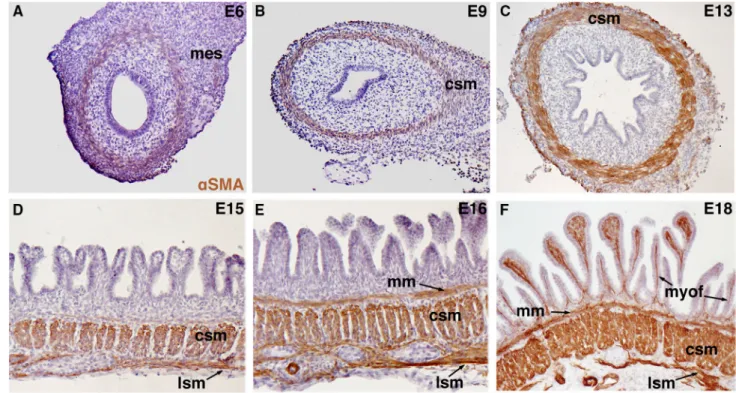
smooth muscle cells (SMCs). Expression of SMC-specific lineage markers, such asaSMA, smooth muscle protein 22 (SM22) and Calponin, is followed by the acquisition of contractile capacities [88, 89]. In the colon and small intestine, SMCs organize sequentially into three different muscle layers (Fig.2). The first to form is the circular smooth muscle layer, located in the middle of the intestinal mesenchyme. It is quickly followed by the formation of the longitudinal smooth muscle layer in the outer part of the mesenchyme. Finally, around week 14–20 in humans, the muscularis mucosae forms and constitutes the third smooth muscle layer, close to the epithelium (Fig.2) [90–92]. Concomitantly with the smooth muscle layer differentia-tion, the uniform and multi-stratified intestinal endoderm forms villi that project into the lumen. Specifically, the endoderm is transformed into epithelial ridges that are then organized into parallel zigzags and finally into intestinal villi [93]. The link between epithelial morphogenesis and smooth muscle layer differentiation was nicely investigated step by step by Shyer and colleagues [93]. By chemical and
Fig. 2 Smooth muscle cell differentiation in the small intestine of chicken embryos. Smooth muscle cells (SMC) were detected by immunostaining with an antibody against the SMC-specific marker aSMA. a Non-organized and undifferentiated mesenchymal cells in the small intestine of a 6-day/old (E6) chick embryo. baSMA-positive cells are present in the smooth muscle layer in the small intestine of an E9 chick embryo. a, b During early embryonic development, the visceral endoderm is uniform with stratified layers of cells. c aSMA-positive cells in the circular smooth muscle layer of the small intestine of an E13 chick embryo. The ongoing intestinal epithelial cytodiffer-entiation leads to the formation of an epithelial monolayer. d
aSMA-positive cells in the longitudinal and circular smooth muscle layers of the small intestine of an E15 chick embryo. eaSMA-positive cells in the longitudinal, circular and submucosal smooth muscle layers in the small intestine of an E16 chick embryo. f aSMA-positive cells (myofibroblasts) are detected also in the lamina propria of the small intestine in a E18 chick embryo. d–f Intestinal epithelial cytodiffer-entiation is marked by mesodermal growth into the lumen and villi formation, characterized by specific long and thin villi in the small intestine. Abbreviations: mes mesenchyme, csm circular smooth muscle, lsm longitudinal smooth muscle, myof myofibroblast,aSMA alpha smooth muscle actin, mm muscularis mucosae
physical ablation of the different smooth muscle layers, they showed that the differentiation of the circular smooth muscle layer is necessary for the development and main-tenance of the epithelial ridges, probably by enforcing the mechanical constraint to limit mesenchymal and endoder-mal growth. The longitudinal smooth muscle layer and muscularis mucosae are required, respectively, for the development of the zigzags and of the villi. Villi formation also requires localized changes in endodermal and mes-enchymal cell proliferation. Altogether, these authors demonstrated that the intestine epithelial morphogenesis is dependent on the sequential differentiation of the three smooth muscle layers present in the gut [93].
Concomitantly with these early patterning events, the primitive GI tract is colonized by neural crest precursor cells that give rise to the ENS, the intrinsic innervation of the GI tract. In vertebrates, the ENS is predominantly derived from vagal enteric neural crest cells (vENCCs) that are initially located adjacent to somites 1 to 7 [94–98]. These cells migrate away from the neural tube and enter the esophageal mesenchyme. Their AP migration in the GI tract is promoted and regulated through interactions with extracellular matrix proteins produced by mesenchymal cells [99,100]. Ultimately, vENCCs populate the entire GI tract, from the esophagus to the terminal colon, but also the lungs and pancreas, two associated GI organs. During their migration along the GI tract, neural crest cells proliferate
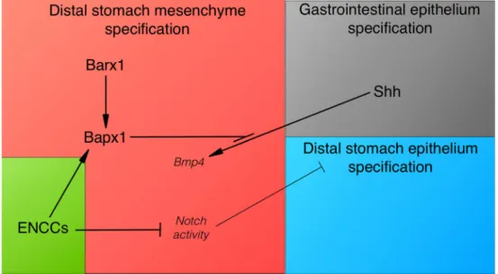
and differentiate into neurons and glial cells, the main ENS components. These cells form two concentric ganglionic plexuses localized within the muscle layers of the gut wall [101], while in the pancreas, ENCCs are found close to the epithelium [102]. Surprisingly, ENCC genetic ablation leads to aberrant morphogenesis of the pancreas epithe-lium, suggesting the existence of instructive signals from ectoderm-derived cells to the endoderm-derived develop-ing pancreas [102]. In the stomach, ENS influence on the developing mesenchyme and epithelium was recently demonstrated. Indeed, vENCC ablation in chick embryos impairs smooth muscle development and induces trans-differentiation of both stomach mesenchymal and epithelial layers into a mixed stomach-intestine structure [72]. Con-stitutive activation of Notch signaling is sufficient to induce the expression of intestinal mesenchymal and epithelial markers in the stomach, suggesting an additional role for Notch signaling in the regulation of intestinal development [72]. vENCCs contribute to the inhibition of the Notch pathway in the mesenchyme, which is a pre-requisite for the establishment of gastric identity (Fig.3). These findings support the involvement of specific tissue and molecular interactions in the establishment of gastric features [72]. Altogether, these observations are in line with previous studies demonstrating that intestinal speci-fication is the default state of the gut endoderm [83] and that reciprocal mesenchymal–epithelial interactions are
Fig. 3 Model of vENCC role in reciprocal epithelial–mesenchymal interactions during distal stomach development. The schematic shows the molecular pathways and their potential interactions during stomach patterning in vertebrates. Shh from the gastrointestinal epithelium induces Bmp4 expression in the adjacent mesenchyme, with the exception of the distal stomach, where Bapx1 prevents its expression. Bapx1 expression in the distal stomach mesenchyme requires Barx1. vENCCs are required for Bapx1 expression in the
distal stomach mesenchyme, independently of Barx1. Importantly, inhibition of Notch activity in the distal stomach mesenchyme is essential for conferring stomach identity to the mesenchymal and epithelial layers. Indeed, ectopic activation of the Notch pathway in the stomach leads to a mixed stomach–intestinal phenotype. In conclusion, in the vertebrate stomach, mesenchymal–epithelial inter-actions involve also vENCCs required for stomach patterning during development
crucial for its morphological and functional regionaliza-tion. They also demonstrate ENS role in these interactions, a contribution that was previously unappreciated.
Mesenchymal–epithelial interactions in stem cell
regeneration and GI tract pathologies
In adults, the main morphological patterning of the intestinal epithelium occurs along the crypt-villus axis. Proliferative progenitor cells are at the bottom and differentiated/functional/ apoptotic cells in the lumen [2,103]. In the gastric mucosa, epithelial cells are organized in vertical flask-shaped structures [104,105] with progenitor/proliferative cells at the bottom and differentiated/functional/apoptotic cells in the lumen [2,103]. The organization of these structures is established during embryogenesis and maintained throughout life, despite the continuous renewal of the digestive epithelium from stem cells [2, 93,106, 107]. The genetic control of the adult digestive epithelium homeostasis (reviewed in [108]) involves recipro-cal interactions between epithelial cells and also between epithelium and mesenchyme [109,110]. However, the influ-ence of mesenchymal–epithelial interaction mechanisms on stem cell renewal and epithelial architecture has only been partially elucidated. Disruption of this morphological organi-zation results in bowel dysfunction, potentially leading to tumor development. Many factors that are involved in intes-tine morphogenesis during embryonic life also contribute to the maintenance of its pattern in adult life. In this section, we will describe and discuss the different pathways and factors that control cell fate decision and cell differentiation through mesenchymal–epithelial interactions, during digestive epithe-lium regeneration and in GI tract pathologies.
Myofibroblasts are a component of the neonate and adult mesenchymal layer in close contact with the digestive epithelium. Intestinal subepithelial myofibroblasts (ISEMFs) reside within the mesenchymal layer, just below the epithelium of both villi and crypts and next to the muscularis mucosae, along the whole length of the intestine (colon and small intestine). In humans, they start devel-oping from week 21 of gestation [111] and form a sheath between the epithelium and the muscularis mucosae, where they contribute to the composition of the extracellular matrix (ECM) and basal membrane [112, 113]. ISEMFs express vimentin and aSMA, but not (or weakly) desmin [114,115]. Although still debated, they may originate from neuroepithelial cells [116], bone-marrow-derived stem cells [117, 118], or trans-differentiation of resident fibroblasts [119] or of intestinal SMCs [120]. In mouse embryos, ISEMFs are detected from E18.5 [121] and in avian embryos from E16 (Fig.2) [122]. Similar to epithelial cells, ISEMFs differentiate as they migrate from the crypt along the villus axis [123]. There are increasing
evidences that ISEMFs are major players in GI epithelium regeneration in normal and pathological conditions. ISEMFs envelop the intestinal crypts [124] and support the intestinal epithelium growth ex vivo and in tissue xeno-grafts [113, 125]. It was suggested that growth factors essential for the development of intestinal stem cells are in part secreted by ISEMFs [126]. For example, in the intestine, epimorphin, a membrane-associated protein that regulates late epithelial morphogenesis [127], is produced by ISEMFs [127,128]. The intestine length is increased in adult Epimorphin-/- mice due to enhanced crypt cell proliferation and crypt fission during the neonatal (suck-ling) period. This is mediated at least in part through changes in the BMP and WNT/beta-catenin signaling pathways [129]. In Epimorphin-/-mice, ISEMFs are less responsive to Hedgehog signals, leading to decreased BMP activity associated with strong expression of Chordin (an inhibitor of the BMP pathway) and reduction of stromal IL-6 secretion [127,130].
Recent studies also highlighted key roles of microRNAs (miRs) in the control of intestinal epithelial regeneration through mesenchymal–epithelial interactions. miR-143/ 145 are strongly expressed in the colon and are down-regulated in colorectal cancer [131]. In the absence of miR-143/145, colon epithelial development occurs normally. However, following injury, epithelial regeneration is strongly impaired [132]. Moreover, miR-145 is a positive regulator of intestinal SMC differentiation and maturation through its capacity to directly target Gata6 mRNA [133]. In agreement, miR-143 and miR-145 are also detected in the intestine mesenchyme, mostly in smooth muscles and ISEMFs [132]. Interestingly, miR-143/145 ablation exclu-sively in the mesenchyme phenocopies the defective epithelial regeneration observed in miR-143/145 knockout animals, while epithelial loss of miR-143/145 does not. This demonstrates that the control of epithelial regenera-tion is governed in part through mesenchymal–epithelial interactions in an miR-143/145-dependent manner.
Less is known about the potential differences in the subepithelial myofibroblast populations along the AP axis. Comparison of the expression of genes associated with the HOX, Hedgehog, Notch, BMP, FDF and WNT pathways in ISEMFs and gastric subepithelial myofibroblasts (GSEMFs) did not highlight any significant difference in the expression of activators and inhibitors of the WNT pathway [134]. This is in agreement with the finding that WNT secretion by ISEMFs is not required for the intestinal stem cell niche homeostasis [135]. In contrast, growth arrest-specific gene 1 (Gas1), a SHH receptor [136], is up-regulated in GSEMFs, while Hoxc8 and Notch1 are strongly expressed in ISEMFs, suggesting that the Notch pathway is more active in ISEMFs. These features are reminiscent of the recent finding that Notch pathway activation is required during the
development of the intestinal, but not of the gastric mes-enchyme [72]. They also demonstrate the existence of GSEMF- and ISEMF-specific intrinsic molecular pathways and raise the question of whether these specific differences modulate their paracrine influence on the renewal of diges-tive epithelial stem cells.
Epithelial metaplasia is frequently observed in the adult GI tract [104]. This corresponds to a change of the epithelium characteristics towards a different type of epithelium. For instance, the esophagus epithelium shows features of intestinal epithelium and pyloric-type gastric glands in patients with Barrett’s esophagus syndrome or following Helicobacter pylori infection [104, 137, 138]. Epithelial metaplasia is the result of homeotic transfor-mations (i.e., the aberrant differentiation of one region into another, implying misregulation of homeotic genes) [139]. The mechanisms ensuring the maintenance of the regional epithelial differentiation in the GI tract are partially known and involve the same factors found in early GI patterning. Many studies on the intestinal metaplasia of the stomach have focused on the misregulation and function of key actors of intestinal epithelial patterning and differentiation, such as the homeotic transcription factors CDX2 and SOX9 [140, 141]; for review [104, 142]. Interestingly, a switch from gastric to intestinal morphology is observed in both epithelium and mesenchyme in the stomach of patients with intestinal metaplasia and in Cdx2-/- mice [143]. In addition, Helicobacter pylori infection in the stomach induces gastric-intestinal metaplasia associated with CDX2 misexpression and an increase in aSMA-positive myofi-broblasts [144, 145]. Although no molecular analysis of these myofibroblasts was done, we can hypothesize that stromal myofibroblats participate in or at least maintain this tissue-specific epithelial transformation through the same mechanisms based on mesenchymal–epithelial interactions used during GI tract development.
Conclusion and perspectives
The importance of mesenchymal–epithelial interactions has been first demonstrated and studied during the devel-opment of the chicken GI tract by dissociating and recombining the mesenchymal and endodermal layers. These pioneering approaches were followed by the iden-tification of the molecular mechanisms involved in the communication between these interdependent tissues. Several major signaling pathways (BMP, WNT, Notch and FGF) and transcription factors (HOX, SOX, FOXF) are involved in the interactions between these tissues along the AP axis. Moreover, recent studies have shown the impact of smooth muscle differentiation on the regulation of
intestinal epithelium morphogenesis and on the regional-ization of the gastric epithelium.
In adults, the reciprocal influence of mesenchyme and epithelium has been hypothesized for a long time, but investigations on these interactions have really started only recently. It has been already demonstrated that mes-enchyme-derived cells, such as subepithelial myofibroblats, actively participate in epithelial stem cell regeneration and could also be involved in the maintenance of the digestive epithelial regionalization. Different studies have found a correlation between epithelial and mesenchymal alterations in metaplasia. Moreover, genes expressed by stromal cells rather than by epithelial tumor cells define subtypes of colorectal cancer (CRC) with poor prognosis [146], sup-porting the mesenchyme influence in GI tract pathologies. During development, the digestive mesenchyme is colo-nized by vENCCs, endothelial and lymphatic cells [147–
150]. Unexpectedly, the number of vENCCs contributes to the maintenance of stomach identity and differentiation through inhibition of the Notch signaling pathway [72]. This demonstrates that by regulating mesenchyme identity, vENCCs act as a new mediator of mesenchymal–epithelial interactions in the control of gastric epithelial regionalization. The close association of endothelial and lymphatic cells with ENCCs during the establishment of their respective networks and also in adults [148–150] opens the way for investigating new potential actors in the regulation of mesenchymal–ep-ithelial interactions during development, but also in adults. Besides smooth muscle, enteric and endothelial cells, the GI wall contains many other cell types (for instance, fibroblasts, lymphocytes and leukocytes) [124] that could be involved in mesenchymal–epithelial interaction mechanisms to maintain the regionalization and homeostasis of digestive epithelia in adults. Their real involvement needs to be investigated and the underlying mechanisms identified.
Altogether these challenging questions must be addres-sed in the context of GI tract development, stem cell and pathology research, and collaborative efforts will give us a better understanding of these complex and exciting three-dimensional structures.
Acknowledgments The work in de Santa Barbara’s lab is supported by the Association pour la Recherche Contre le Cancer (ARC) foundation, the Association Franc¸aise contre les Myopathies (AFM) and the French Patients’ Association POIC. LLG is supported by an ARC post-doctoral fellowship. PdSB thanks Drucilla Jane Roberts for her contribution in the field, inspiration and continuous support.
References
1. Buller NV, Rosekrans SL, Westerlund J, van den Brink GR (2012) Hedgehog signaling and maintenance of homeostasis in the intestinal epithelium. Physiology (Bethesda) 27(3):148–155. doi:10.1152/physiol.00003.2012
2. de Santa Barbara P, van den Brink GR, Roberts DJ (2003) Development and differentiation of the intestinal epithelium. Cell Mol Life Sci 60(7):1322–1332. doi: 10.1007/s00018-003-2289-3
3. Smith DM, Grasty RC, Theodosiou NA, Tabin CJ, Nascone-Yoder NM (2000) Evolutionary relationships between the amphibian, avian, and mammalian stomachs. Evol Dev 2(6):348–359
4. Narita N, Bielinska M, Wilson DB (1997) Wild-type endoderm abrogates the ventral developmental defects associated with GATA-4 deficiency in the mouse. Dev Biol 189(2):270–274. doi:10.1006/dbio.1997.8684
5. de Santa Barbara P, Roberts DJ (2002) Tail gut endoderm and gut/genitourinary/tail development: a new tissue-specific role for Hoxa13. Development 129(3):551–561
6. Kuo CT, Morrisey EE, Anandappa R, Sigrist K, Lu MM, Par-macek MS, Soudais C, Leiden JM (1997) GATA4 transcription factor is required for ventral morphogenesis and heart tube formation. Genes Dev 11(8):1048–1060
7. Molkentin JD, Lin Q, Duncan SA, Olson EN (1997) Require-ment of the transcription factor GATA4 for heart tube formation and ventral morphogenesis. Genes Dev 11(8):1061–1072 8. Ang SL, Wierda A, Wong D, Stevens KA, Cascio S, Rossant J,
Zaret KS (1993) The formation and maintenance of the defini-tive endoderm lineage in the mouse: involvement of HNF3/forkhead proteins. Development 119(4):1301–1315 9. Kaestner KH, Lee KH, Schlondorff J, Hiemisch H, Monaghan
AP, Schutz G (1993) Six members of the mouse forkhead gene family are developmentally regulated. Proc Natl Acad Sci USA 90(16):7628–7631
10. Dufort D, Schwartz L, Harpal K, Rossant J (1998) The tran-scription factor HNF3beta is required in visceral endoderm for normal primitive streak morphogenesis. Development 125(16):3015–3025
11. Weinstein DC, Ruiz i Altaba A, Chen WS, Hoodless P, Prezioso VR, Jessell TM, Darnell JE Jr (1994) The winged-helix tran-scription factor HNF-3 beta is required for notochord development in the mouse embryo. Cell 78(4):575–588 12. Zaret K (1999) Developmental competence of the gut
endo-derm: genetic potentiation by GATA and HNF3/fork head proteins. Dev Biol 209(1):1–10. doi:10.1006/dbio.1999.9228
13. Rojas A, Schachterle W, Xu SM, Martin F, Black BL (2010) Direct transcriptional regulation of Gata4 during early endoderm specification is controlled by FoxA2 binding to an intronic enhancer. Dev Biol 346(2):346–355. doi:10.1016/j.ydbio.2010. 07.032
14. Fort P, Guemar L, Vignal E, Morin N, Notarnicola C, de Santa Barbara P, Faure S (2011) Activity of the RhoU/Wrch1 GTPase is critical for cranial neural crest cell migration. Dev Biol 350(2):451–463. doi:10.1016/j.ydbio.2010.12.011
15. Notarnicola C, Le Guen L, Fort P, Faure S, de Santa Barbara P (2008) Dynamic expression patterns of RhoV/Chp and RhoU/ Wrch during chicken embryonic development. Dev Dyn 237(4):1165–1171. doi:10.1002/dvdy.21507
16. Loebel DA, Studdert JB, Power M, Radziewic T, Jones V, Coultas L, Jackson Y, Rao RS, Steiner K, Fossat N, Robb L, Tam PP (2011) Rhou maintains the epithelial architecture and facilitates differentiation of the foregut endoderm. Development 138(20):4511–4522. doi:10.1242/dev.063867
17. Loebel DA, Tam PP (2012) Rho GTPases in endoderm devel-opment and differentiation. Small GTPases 3(1):40–44. doi:10. 4161/sgtp.18820
18. Roberts DJ, Johnson RL, Burke AC, Nelson CE, Morgan BA, Tabin C (1995) Sonic hedgehog is an endodermal signal inducing Bmp-4 and Hox genes during induction and regional-ization of the chick hindgut. Development 121(10):3163–3174
19. Dolle P, Izpisua-Belmonte JC, Brown J, Tickle C, Duboule D (1993) Hox genes and the morphogenesis of the vertebrate limb. Prog Clin Biol Res 383A:11–20
20. Goodman FR, Scambler PJ (2001) Human HOX gene mutations. Clin Genet 59(1):1–11
21. Imagawa E, Kayserili H, Nishimura G, Nakashima M, Tsurusaki Y, Saitsu H, Ikegawa S, Matsumoto N, Miyake N (2014) Severe manifestations of hand-foot-genital syndrome associated with a novel HOXA13 mutation. Am J Med Genet A 164A(9):2398–2402. doi:10.1002/ajmg.a.36648
22. Mortlock DP, Innis JW (1997) Mutation of HOXA13 in hand-foot-genital syndrome. Nat Genet 15(2):179–180. doi:10.1038/ ng0297-179
23. Post LC, Innis JW (1999) Altered Hox expression and increased cell death distinguish Hypodactyly from Hoxa13 null mice. Int J Dev Biol 43(4):287–294
24. Warot X, Fromental-Ramain C, Fraulob V, Chambon P, Dolle P (1997) Gene dosage-dependent effects of the Hoxa-13 and Hoxd-13 mutations on morphogenesis of the terminal parts of the digestive and urogenital tracts. Development 124(23):4781–4791
25. Deschamps J, van Nes J (2005) Developmental regulation of the Hox genes during axial morphogenesis in the mouse. Develop-ment 132(13):2931–2942. doi:10.1242/dev.01897
26. Ishii Y, Fukuda K, Saiga H, Matsushita S, Yasugi S (1997) Early specification of intestinal epithelium in the chicken embryo: a study on the localization and regulation of CdxA expression. Dev Growth Differ 39(5):643–653
27. van den Akker E, Forlani S, Chawengsaksophak K, de Graaff W, Beck F, Meyer BI, Deschamps J (2002) Cdx1 and Cdx2 have overlapping functions in anteroposterior patterning and posterior axis elongation. Development 129(9):2181–2193
28. Gao N, White P, Kaestner KH (2009) Establishment of intestinal identity and epithelial-mesenchymal signaling by Cdx2. Dev Cell 16(4):588–599. doi:10.1016/j.devcel.2009.02.010
29. van de Ven C, Bialecka M, Neijts R, Young T, Rowland JE, Stringer EJ, Van Rooijen C, Meijlink F, Novoa A, Freund JN, Mallo M, Beck F, Deschamps J (2011) Concerted involvement of Cdx/Hox genes and Wnt signaling in morphogenesis of the caudal neural tube and cloacal derivatives from the posterior growth zone. Development 138(16):3451–3462. doi:10.1242/ dev.066118
30. Echelard Y, Epstein DJ, St-Jacques B, Shen L, Mohler J, McMahon JA, McMahon AP (1993) Sonic hedgehog, a member of a family of putative signaling molecules, is implicated in the regulation of CNS polarity. Cell 75(7):1417–1430
31. Litingtung Y, Lei L, Westphal H, Chiang C (1998) Sonic hedgehog is essential to foregut development. Nat Genet 20(1):58–61. doi:10.1038/1717
32. Ramalho-Santos M, Melton DA, McMahon AP (2000) Hedge-hog signals regulate multiple aspects of gastrointestinal development. Development 127(12):2763–2772
33. Le Douarin N (1964) Induction of prehepatic endoderm by mesoderm of the cardiac region in the Chick Embryo. J Embryol Exp Morphol 12:651–664
34. Le Douarin N (1964) The experimental isolation of the mes-enchyme of the liver and the role of the mesodermal component of the liver in Its organogenesis. J Embryol Exp Morphol 12:141–160 35. Le Dourain N, Bussonnet C (1966) Early determination and inductive role of the pharyngeal endoderm in the chick embryo. C R Acad Sci Hebd Seances Acad Sci D 263(17):1241–1243 36. Andrew A, Rawdon BB (1992) Can a non-gut mesenchyme
support differentiation of gut endocrine cells? Anat Embryol (Berl) 185(5):509–516
37. Aufderheide E, Ekblom P (1988) Tenascin during gut devel-opment: appearance in the mesenchyme, shift in molecular
forms, and dependence on epithelial-mesenchymal interactions. J Cell Biol 107(6 Pt 1):2341–2349
38. Kedinger M, Simon-Assmann P, Bouziges F, Arnold C, Alexandre E, Haffen K (1990) Smooth muscle actin expression during rat gut development and induction in fetal skin fibrob-lastic cells associated with intestinal embryonic epithelium. Differentiation 43(2):87–97
39. Koike T, Yasugi S (1999) In vitro analysis of mesenchymal influences on the differentiation of stomach epithelial cells of the chicken embryo. Differentiation 65(1):13–25. doi:10.1046/j. 1432-0436.1999.6510013.x
40. Rawdon BB (2000) Can gastric endoderm change the regionally specific inducing ability of presumptive small intestinal meso-derm? Dev Dyn 219(3):402–416. doi: 10.1002/1097-0177(2000)9999:9999\:AID-DVDY1068[3.0.CO;2-1
41. Sumiya M, Mizuno T (1974) Differentiation of the endoderm in digestive tract of the chick embryo cultured in vitelline mem-brane, in absence of mesenchyma. C R Acad Sci Hebd Seances Acad Sci D 278(11):1529–1532
42. Matsushita S, Ishii Y, Scotting PJ, Kuroiwa A, Yasugi S (2002) Pre-gut endoderm of chick embryos is regionalized by 1.5 days of development. Dev Dyn 223(1):33–47. doi:10.1002/dvdy.1229
43. Yasugi S, Mizuno T (2008) Molecular analysis of endoderm regionalization. Dev Growth Differ 50(Suppl 1):S79–S96. doi:10.1111/j.1440-169X.2008.00984.x
44. Haffen K, Lacroix B, Kedinger M, Simon-Assmann PM (1983) Inductive properties of fibroblastic cell cultures derived from rat intestinal mucosa on epithelial differentiation. Differentiation 23(3):226–233
45. Kedinger M, Simon-Assmann P, Bouziges F, Haffen K (1988) Epithelial-mesenchymal interactions in intestinal epithelial dif-ferentiation. Scand J Gastroenterol Suppl 151:62–69
46. Kedinger M, Simon-Assmann PM, Lacroix B, Marxer A, Hauri HP, Haffen K (1986) Fetal gut mesenchyme induces differen-tiation of cultured intestinal endodermal and crypt cells. Dev Biol 113(2):474–483
47. Duluc I, Freund JN, Leberquier C, Kedinger M (1994) Fetal endoderm primarily holds the temporal and positional infor-mation required for mammalian intestinal development. J Cell Biol 126(1):211–221
48. Hayashi K, Yasugi S, Mizuno T (1988) Pepsinogen gene tran-scription induced in heterologous epithelial-mesenchymal recombinations of chicken endoderms and glandular stomach mesenchyme. Development 103(4):725–731
49. Roberts DJ, Smith DM, Goff DJ, Tabin CJ (1998) Epithelial-mesenchymal signaling during the regionalization of the chick gut. Development 125(15):2791–2801
50. Montavon T, Soshnikova N (2014) Hox gene regulation and timing in embryogenesis. Semin Cell Dev Biol 34:76–84. doi:10.1016/j.semcdb.2014.06.005
51. Yokouchi Y, Sakiyama J, Kuroiwa A (1995) Coordinated expression of Abd-B subfamily genes of the HoxA cluster in the developing digestive tract of chick embryo. Dev Biol 169(1):76–89
52. Aubin J, Dery U, Lemieux M, Chailler P, Jeannotte L (2002) Stomach regional specification requires Hoxa5-driven mes-enchymal–epithelial signaling. Development 129(17):4075–4087
53. Beck F, Tata F, Chawengsaksophak K (2000) Homeobox genes and gut development. BioEssays 22(5):431–441. doi:10.1002/ (SICI)1521-1878(200005)22:5\431:AID-BIES5[3.0.CO;2-X
54. Kondo T, Dolle P, Zakany J, Duboule D (1996) Function of posterior HoxD genes in the morphogenesis of the anal sphincter. Development 122(9):2651–2659
55. Sekimoto T, Yoshinobu K, Yoshida M, Kuratani S, Fujimoto S, Araki M, Tajima N, Araki K, Yamamura K (1998)
Region-specific expression of murine Hox genes implies the Hox code-mediated patterning of the digestive tract. Genes Cells 3(1):51–64
56. Smith DM, Nielsen C, Tabin CJ, Roberts DJ (2000) Roles of BMP signaling and Nkx2.5 in patterning at the chick midgut-foregut boundary. Development 127(17):3671–3681
57. Whitman M (1998) Smads and early developmental signaling by the TGFbeta superfamily. Genes Dev 12(16):2445–2462 58. Faure S, de Santa Barbara P, Roberts DJ, Whitman M (2002)
Endogenous patterns of BMP signaling during early chick devel-opment. Dev Biol 244(1):44–65. doi:10.1006/dbio.2002.0579
59. Faure S, Lee MA, Keller T, ten Dijke P, Whitman M (2000) Endogenous patterns of TGFbeta superfamily signaling during early Xenopus development. Development 127(13):2917–2931 60. Goldstein AM, Brewer KC, Doyle AM, Nagy N, Roberts DJ
(2005) BMP signaling is necessary for neural crest cell migra-tion and ganglion formamigra-tion in the enteric nervous system. Mech Dev 122(6):821–833. doi:10.1016/j.mod.2005.03.003
61. Faure S, Georges M, McKey J, Sagnol S, de Santa Barbara P (2013) Expression pattern of the homeotic gene Bapx1 during early chick gastrointestinal tract development. Gene Expr Pat-terns 13(8):287–292. doi:10.1016/j.gep.2013.05.005
62. Murtaugh LC, Zeng L, Chyung JH, Lassar AB (2001) The chick transcriptional repressor Nkx3.2 acts downstream of Shh to promote BMP-dependent axial chondrogenesis. Dev Cell 1(3):411–422
63. Nielsen C, Murtaugh LC, Chyung JC, Lassar A, Roberts DJ (2001) Gizzard formation and the role of Bapx1. Dev Biol 231(1):164–174. doi:10.1006/dbio.2000.0151
64. De Santa Barbara P, Williams J, Goldstein AM, Doyle AM, Nielsen C, Winfield S, Faure S, Roberts DJ (2005) Bone mor-phogenetic protein signaling pathway plays multiple roles during gastrointestinal tract development. Dev Dyn 234(2):312–322. doi:10.1002/dvdy.20554
65. Theodosiou NA, Tabin CJ (2003) Wnt signaling during devel-opment of the gastrointestinal tract. Dev Biol 259(2):258–271 66. Aitola M, Carlsson P, Mahlapuu M, Enerback S, Pelto-Huikko
M (2000) Forkhead transcription factor FoxF2 is expressed in mesodermal tissues involved in epithelio-mesenchymal inter-actions. Dev Dyn 218(1):136–149. doi: 10.1002/(SICI)1097-0177(200005)218:1\136:AID-DVDY12[3.0.CO;2-U
67. Mahlapuu M, Ormestad M, Enerback S, Carlsson P (2001) The forkhead transcription factor Foxf1 is required for differentiation of extra-embryonic and lateral plate mesoderm. Development 128(2):155–166
68. McLin VA, Shah R, Desai NP, Jamrich M (2010) Identification and gastrointestinal expression of Xenopus laevis FoxF2. Int J Dev Biol 54(5):919–924. doi:10.1387/ijdb.092916vm
69. Ormestad M, Astorga J, Carlsson P (2004) Differences in the embryonic expression patterns of mouse Foxf1 and -2 match their distinct mutant phenotypes. Dev Dyn 229(2):328–333. doi:10.1002/dvdy.10426
70. Ormestad M, Astorga J, Landgren H, Wang T, Johansson BR, Miura N, Carlsson P (2006) Foxf1 and Foxf2 control murine gut development by limiting mesenchymal Wnt signaling and pro-moting extracellular matrix production. Development 133(5):833–843. doi:10.1242/dev.02252
71. Buchberger A, Pabst O, Brand T, Seidl K, Arnold HH (1996) Chick NKx-2.3 represents a novel family member of vertebrate homologues to the Drosophila homeobox gene tinman: differ-ential expression of cNKx-2.3 and cNKx-2.5 during heart and gut development. Mech Dev 56(1–2):151–163
72. Faure S, McKey J, Sagnol S, de Santa Barbara P (2015) Enteric neural crest cells regulate vertebrate stomach patterning and differentiation. Development 142(2):331–342. doi:10.1242/dev. 118422
73. Pabst O, Schneider A, Brand T, Arnold HH (1997) The mouse Nk2–3 homeodomain gene is expressed in gut mesenchyme during pre- and postnatal mouse development. Dev Dyn 209(1):29–35. doi:10.1002/(SICI)1097-0177(199705)209:1\29: AID-AJA3[3.0.CO;2-Z
74. Moniot B, Biau S, Faure S, Nielsen CM, Berta P, Roberts DJ, de Santa Barbara P (2004) SOX9 specifies the pyloric sphincter epithelium through mesenchymal–epithelial signals. Develop-ment 131(15):3795–3804. doi:10.1242/dev.01259
75. Smith DM, Tabin CJ (1999) BMP signalling specifies the pyloric sphincter. Nature 402(6763):748–749. doi:10.1038/ 45439
76. Theodosiou NA, Tabin CJ (2005) Sox9 and Nkx2.5 determine the pyloric sphincter epithelium under the control of BMP signaling. Dev Biol 279(2):481–490. doi:10.1016/j.ydbio.2004.12.019
77. Zhao G, Skeath JB (2002) The Sox-domain containing gene Dichaete/fish-hook acts in concert with vnd and ind to regulate cell fate in the Drosophila neuroectoderm. Development 129(5):1165–1174
78. Li Y, Pan J, Wei C, Chen J, Liu Y, Liu J, Zhang X, Evans SM, Cui Y, Cui S (2014) LIM homeodomain transcription factor Isl1 directs normal pyloric development by targeting Gata3. BMC Biol 12:25. doi:10.1186/1741-7007-12-25
79. Prakash A, Udager AM, Saenz DA, Gumucio DL (2014) Roles for Nk2–5 and Gata3 in the ontogeny of the murine smooth muscle gastric ligaments. Am J Physiol Gastrointest Liver Physiol 307(4):G430–G436. doi:10.1152/ajpgi.00360.2013
80. Udager AM, Prakash A, Saenz DA, Schinke M, Moriguchi T, Jay PY, Lim KC, Engel JD, Gumucio DL (2014) Proper development of the outer longitudinal smooth muscle of the mouse pylorus requires Nkx2-5 and Gata3. Gastroenterology 146(1):157–165 e110. doi:10.1053/j.gastro.2013.10.008
81. Verzi MP, Stanfel MN, Moses KA, Kim BM, Zhang Y, Schwartz RJ, Shivdasani RA, Zimmer WE (2009) Role of the homeodomain transcription factor Bapx1 in mouse distal stomach development. Gastroenterology 136(5):1701–1710. doi:10.1053/j.gastro.2009.01.009
82. Tissier-Seta JP, Mucchielli ML, Mark M, Mattei MG, Goridis C, Brunet JF (1995) Barx1, a new mouse homeodomain tran-scription factor expressed in cranio-facial ectomesenchyme and the stomach. Mech Dev 51(1):3–15
83. Kim BM, Buchner G, Miletich I, Sharpe PT, Shivdasani RA (2005) The stomach mesenchymal transcription factor Barx1 specifies gastric epithelial identity through inhibition of transient Wnt sig-naling. Dev Cell 8(4):611–622. doi:10.1016/j.devcel.2005.01.015
84. Kim BM, Miletich I, Mao J, McMahon AP, Sharpe PA, Shiv-dasani RA (2007) Independent functions and mechanisms for homeobox gene Barx1 in patterning mouse stomach and spleen. Development 134(20):3603–3613. doi:10.1242/dev.009308
85. Kosinski C, Stange DE, Xu C, Chan AS, Ho C, Yuen ST, Mifflin RC, Powell DW, Clevers H, Leung SY, Chen X (2010) Indian hedgehog regulates intestinal stem cell fate through epithelial-mesenchymal interactions during development. Gastroenterol-ogy 139(3):893–903. doi:10.1053/j.gastro.2010.06.014
86. Mao J, Kim BM, Rajurkar M, Shivdasani RA, McMahon AP (2010) Hedgehog signaling controls mesenchymal growth in the developing mammalian digestive tract. Development 137(10):1721–1729. doi:10.1242/dev.044586
87. Sukegawa A, Narita T, Kameda T, Saitoh K, Nohno T, Iba H, Yasugi S, Fukuda K (2000) The concentric structure of the developing gut is regulated by Sonic hedgehog derived from endodermal epithelium. Development 127(9):1971–1980 88. Gabella G (2002) Development of visceral smooth muscle.
Results Probl Cell Differ 38:1–37
89. Notarnicola C, Rouleau C, Le Guen L, Virsolvy A, Richard S, Faure S, De Santa Barbara P (2012) The RNA-binding protein
RBPMS2 regulates development of gastrointestinal smooth muscle. Gastroenterology 143(3):687–697, e681–689. doi:10. 1053/j.gastro.2012.05.047
90. Johnson FP (1913) The development of the mucous membrane of the large intestine and vermiform process in the human embryo. Am J Anat 14:187–233
91. Fu M, Tam PK, Sham MH, Lui VC (2004) Embryonic devel-opment of the ganglion plexuses and the concentric layer structure of human gut: a topographical study. Anat Embryol (Berl) 208(1):33–41. doi:10.1007/s00429-003-0371-0
92. Wallace AS, Burns AJ (2005) Development of the enteric ner-vous system, smooth muscle and interstitial cells of Cajal in the human gastrointestinal tract. Cell Tissue Res 319(3):367–382. doi:10.1007/s00441-004-1023-2
93. Shyer AE, Tallinen T, Nerurkar NL, Wei Z, Gil ES, Kaplan DL, Tabin CJ, Mahadevan L (2013) Villification: how the gut gets its villi. Science 342(6155):212–218. doi:10.1126/science.1238842
94. Burns AJ, Champeval D, Le Douarin NM (2000) Sacral neural crest cells colonise aganglionic hindgut in vivo but fail to compensate for lack of enteric ganglia. Dev Biol 219(1):30–43. doi:10.1006/dbio.1999.9592
95. Burns AJ, Le Douarin NM (1998) The sacral neural crest con-tributes neurons and glia to the post-umbilical gut: spatiotemporal analysis of the development of the enteric ner-vous system. Development 125(21):4335–4347
96. Le Douarin NM, Teillet MA (1973) The migration of neural crest cells to the wall of the digestive tract in avian embryo. J Embryol Exp Morphol 30(1):31–48
97. Yntema CL, Hammond WS (1954) The origin of intrinsic ganglia of trunk viscera from vagal neural crest in the chick embryo. J Comp Neurol 101(2):515–541
98. Fairman CL, Clagett-Dame M, Lennon VA, Epstein ML (1995) Appearance of neurons in the developing chick gut. Dev Dyn 204(2):192–201. doi:10.1002/aja.1002040210
99. Breau MA, Dahmani A, Broders-Bondon F, Thiery JP, Dufour S (2009) Beta1 integrins are required for the invasion of the caecum and proximal hindgut by enteric neural crest cells. Development 136(16):2791–2801. doi:10.1242/dev.031419
100. Akbareian SE, Nagy N, Steiger CE, Mably JD, Miller SA, Hotta R, Molnar D, Goldstein AM (2013) Enteric neural crest-derived cells promote their migration by modifying their microenvi-ronment through tenascin-C production. Dev Biol 382(2):446–456. doi:10.1016/j.ydbio.2013.08.006
101. Furness JB (2006) The organisation of the autonomic nervous system: peripheral connections. Auton Neurosci 130(1–2):1–5. doi:10.1016/j.autneu.2006.05.003
102. Nekrep N, Wang J, Miyatsuka T, German MS (2008) Signals from the neural crest regulate beta-cell mass in the pancreas. Development 135(12):2151–2160. doi:10.1242/dev.015859
103. Potten CS (1997) Epithelial cell growth and differentiation. II. Intestinal apoptosis. Am J Physiol 273(2 Pt 1):G253–G257 104. Barros R, Freund JN, David L, Almeida R (2012) Gastric
intestinal metaplasia revisited: function and regulation of CDX2. Trends Mol Med 18(9):555–563. doi:10.1016/j.molmed.2012. 07.006
105. Mills JC, Shivdasani RA (2011) Gastric epithelial stem cells. Gastroenterology 140(2):412–424. doi:10.1053/j.gastro.2010. 12.001
106. Grapin-Botton A (2005) Antero-posterior patterning of the vertebrate digestive tract: 40 years after Nicole Le Douarin’s Ph.D. thesis. Int J Dev Biol 49(2–3):335–347. doi:10.1387/ijdb. 041946ag
107. McLin VA, Henning SJ, Jamrich M (2009) The role of the visceral mesoderm in the development of the gastrointestinal tract. Gastroenterology 136(7):2074–2091. doi:10.1053/j.gastro. 2009.03.001
108. Barker N (2014) Adult intestinal stem cells: critical drivers of epithelial homeostasis and regeneration. Nat Rev Mol Cell Biol 15(1):19–33. doi:10.1038/nrm3721
109. Kedinger M, Lefebvre O, Duluc I, Freund JN, Simon-Assmann P (1998) Cellular and molecular partners involved in gut mor-phogenesis and differentiation. Philos Trans R Soc Lond B Biol Sci 353(1370):847–856. doi:10.1098/rstb.1998.0249
110. Simon-Assmann P, Spenle C, Lefebvre O, Kedinger M (2010) The role of the basement membrane as a modulator of intestinal epithelial-mesenchymal interactions. Prog Mol Biol Transl Sci 96:175–206. doi:10.1016/B978-0-12-381280-3.00008-7
111. Sappino AP, Dietrich PY, Skalli O, Widgren S, Gabbiani G (1989) Colonic pericryptal fibroblasts. Differentiation pattern in embryogenesis and phenotypic modulation in epithelial prolif-erative lesions. Virchows Arch A Pathol Anat Histopathol 415(6):551–557
112. Islam MS, Kusakabe M, Horiguchi K, Iino S, Nakamura T, Iwanaga K, Hashimoto H, Matsumoto S, Murata T, Hori M, Ozaki H (2014) PDGF and TGF-beta promote tenascin-C expression in subepithelial myofibroblasts and contribute to intestinal mucosal protection in mice. Br J Pharmacol 171(2):375–388. doi:10.1111/bph.12452
113. Lahar N, Lei NY, Wang J, Jabaji Z, Tung SC, Joshi V, Lewis M, Stelzner M, Martin MG, Dunn JC (2011) Intestinal subepithelial myofibroblasts support in vitro and in vivo growth of human small intestinal epithelium. PLoS One 6(11):e26898. doi:10. 1371/journal.pone.0026898
114. Powell DW, Mifflin RC, Valentich JD, Crowe SE, Saada JI, West AB (1999) Myofibroblasts. II. Intestinal subepithelial myofibroblasts. Am J Physiol 277(2 Pt 1):C183–C201 115. Powell DW, Mifflin RC, Valentich JD, Crowe SE, Saada JI,
West AB (1999) Myofibroblasts. I. Paracrine cells important in health and disease. Am J Physiol 277(1 Pt 1):C1–C9
116. Bockman DE, Sohal GS (1998) A new source of cells con-tributing to the developing gastrointestinal tract demonstrated in chick embryos. Gastroenterology 114(5):878–882
117. Andoh A, Bamba S, Fujiyama Y, Brittan M, Wright NA (2005) Colonic subepithelial myofibroblasts in mucosal inflammation and repair: contribution of bone marrow-derived stem cells to the gut regenerative response. J Gastroenterol 40(12):1089–1099. doi:10.1007/s00535-005-1727-4
118. Mifflin RC, Pinchuk IV, Saada JI, Powell DW (2011) Intestinal myofibroblasts: targets for stem cell therapy. Am J Physiol Gastrointest Liver Physiol 300(5):G684–G696. doi:10.1152/ ajpgi.00474.2010
119. Gabbiani G (1996) The cellular derivation and the life span of the myofibroblast. Pathol Res Pract 192(7):708–711. doi:10. 1016/S0344-0338(96)80092-6
120. Ronnov-Jessen L, Petersen OW, Koteliansky VE, Bissell MJ (1995) The origin of the myofibroblasts in breast cancer. Recapitulation of tumor environment in culture unravels diver-sity and implicates converted fibroblasts and recruited smooth muscle cells. J Clin Invest 95(2):859–873. doi:10.1172/ JCI117736
121. Plateroti M, Rubin DC, Duluc I, Singh R, Foltzer-Jourdainne C, Freund JN, Kedinger M (1998) Subepithelial fibroblast cell lines from different levels of gut axis display regional characteristics. Am J Physiol 274(5 Pt 1):G945–G954
122. Thomason RT, Bader DM, Winters NI (2012) Comprehensive timeline of mesodermal development in the quail small intes-tine. Dev Dyn 241(11):1678–1694. doi:10.1002/dvdy.23855
123. Hall PA, Coates PJ, Ansari B, Hopwood D (1994) Regulation of cell number in the mammalian gastrointestinal tract: the importance of apoptosis. J Cell Sci 107(Pt 12):3569–3577 124. Powell DW, Pinchuk IV, Saada JI, Chen X, Mifflin RC (2011)
Mesenchymal cells of the intestinal lamina propria. Annu Rev
Physiol 73:213–237. doi:10.1146/annurev.physiol.70.113006. 100646
125. Lei NY, Jabaji Z, Wang J, Joshi VS, Brinkley GJ, Khalil H, Wang F, Jaroszewicz A, Pellegrini M, Li L, Lewis M, Stelzner M, Dunn JC, Martin MG (2014) Intestinal subepithelial myofi-broblasts support the growth of intestinal epithelial stem cells. PLoS One 9(1):e84651. doi:10.1371/journal.pone.0084651
126. Yeung TM, Chia LA, Kosinski CM, Kuo CJ (2011) Regulation of self-renewal and differentiation by the intestinal stem cell niche. Cell Mol Life Sci 68(15):2513–2523. doi:10.1007/ s00018-011-0687-5
127. Fritsch C, Swietlicki EA, Lefebvre O, Kedinger M, Iordanov H, Levin MS, Rubin DC (2002) Epimorphin expression in intesti-nal myofibroblasts induces epithelial morphogenesis. J Clin Invest 110(11):1629–1641. doi:10.1172/JCI13588
128. Andoh A, Fujino S, Hirai Y, Fujiyama Y (2004) Epimorphin expression in human colonic myofibroblasts. Int J Mol Med 13(1):57–61
129. Wang Y, Wang L, Iordanov H, Swietlicki EA, Zheng Q, Jiang S, Tang Y, Levin MS, Rubin DC (2006) Epimorphin(-/-)mice have increased intestinal growth, decreased susceptibility to dextran sodium sulfate colitis, and impaired spermatogenesis. J Clin Invest 116(6):1535–1546. doi:10.1172/JCI25442
130. Shaker A, Swietlicki EA, Wang L, Jiang S, Onal B, Bala S, DeSchryver K, Newberry R, Levin MS, Rubin DC (2010) Epi-morphin deletion protects mice from inflammation-induced colon carcinogenesis and alters stem cell niche myofibroblast secretion. J Clin Invest 120(6):2081–2093. doi:10.1172/JCI40676
131. Michael MZ, O’Connor SM, van Holst Pellekaan NG, Young GP, James RJ (2003) Reduced accumulation of specific micro-RNAs in colorectal neoplasia. Mol Cancer Res 1(12):882–891 132. Chivukula RR, Shi G, Acharya A, Mills EW, Zeitels LR,
Anandam JL, Abdelnaby AA, Balch GC, Mansour JC, Yopp AC, Maitra A, Mendell JT (2014) An essential mesenchymal function for miR-143/145 in intestinal epithelial regeneration. Cell 157(5):1104–1116. doi:10.1016/j.cell.2014.03.055
133. Zeng L, Carter AD, Childs SJ (2009) miR-145 directs intestinal maturation in zebrafish. Proc Natl Acad Sci USA 106(42):17793–17798. doi:10.1073/pnas.0903693106
134. Katano T, Ootani A, Mizoshita T, Tanida S, Tsukamoto H, Ozeki K, Kataoka H, Joh T (2015) Gastric mesenchymal myofibroblasts maintain stem cell activity and proliferation of murine gastric epithelium in vitro. Am J Pathol 185(3):798–807. doi:10.1016/j.ajpath.2014.11.007
135. San Roman AK, Jayewickreme CD, Murtaugh LC, Shivdasani RA (2014) Wnt secretion from epithelial cells and subepithelial myofibroblasts is not required in the mouse intestinal stem cell niche in vivo. Stem Cell Reports 2(2):127–134. doi:10.1016/j. stemcr.2013.12.012
136. Biau S, Jin S, Fan CM (2013) Gastrointestinal defects of the Gas1 mutant involve dysregulated Hedgehog and Ret signaling. Biol Open 2(2):144–155. doi:10.1242/bio.20123186
137. DeMeester SR, DeMeester TR (2000) Columnar mucosa and intestinal metaplasia of the esophagus: fifty years of contro-versy. Ann Surg 231(3):303–321
138. Lavery DL, Nicholson AM, Poulsom R, Jeffery R, Hussain A, Gay LJ, Jankowski JA, Zeki SS, Barr H, Harrison R, Going J, Kadirkamanathan S, Davis P, Underwood T, Novelli MR, Rodriguez-Justo M, Shepherd N, Jansen M, Wright NA, McDonald SA (2014) The stem cell organisation, and the pro-liferative and gene expression profile of Barrett’s epithelium, replicates pyloric-type gastric glands. Gut 63(12):1854–1863. doi:10.1136/gutjnl-2013-306508
139. Slack JM (1985) Homoeotic transformations in man: implica-tions for the mechanism of embryonic development and for the organization of epithelia. J Theor Biol 114(3):463–490