Design and Synthesis of Inhibitors of dTDP-D-glucose 4,6~
dehydratase (RmlB), an Enzyme Required for dTDP-L-Rhamnose
Production in M.Tuberculosis
by
Neena Sujata Kadaba S.B. Chemistry
Massachusetts Institute of Technology, 2002
SUBMITFED TO THE BIOLOGICAL ENGINEERING DIVISION IN PARTIAL FULFILLMENT OF THE REQUIREMENTS FOR THE DEGREE OF
MASTER OF SCIENCE IN MOLECULAR AND SYSTEMS TOXICOLOGY
AT THE
MASSACHUSETTS INSTITUTE OF TECHNOLOGY JUNE 2003
© 2003 Neena S. Kadaba. All rights reserved.
The author hereby grants to MIT permission to reproduce and to distribute publicly paper and electronic copies of this thesis document in whole or in part.
Signature of A uthor... V ...
Neena S. Kadaba Biological Engineering Division May 9, 2003
Certified By... ... . ...
*
*
\ jjJohn
M. Essigmann Professor of Chemistry and ToxicologyThesis Advisor
A ccep ted By ... 2 ... Ram Sasisekharan Chair, Biological Engineering Graduate Committee
Design and Synthesis of Inhibitors of dTDP-D-glucose
4,6-dehydratase (RmlB), an Enzyme Required for dTDP-L-Rhamnose
Production in M.Tuberculosis
by
Neena Sujata Kadaba
Submitted to the Biological Engineering Division on May 9, 2003 in Partial Fulfillment of the Requirements for the
Degree of Master of Science in Molecular and Systems Toxicology
Abstract
The purpose of this work is to probe the dTDP-L-rhamnose pathway in an effort to develop small molecule inhibitors that could act as therapeutics for Mycobacterium
tuberculosis. The necessity for newer, more effective treatments for tuberculosis is growing, as the bacteria evolve resistance to traditional treatments.
In an effort to develop more effective and perhaps more abbreviated courses of treatment, a plan was developed to investigate a pathway involved in cell wall biosynthesis as a promising target: the dTDP-L-rhamnose pathway. This pathway plays an essential role in linking the peptidoglycan and arabinogalactan portions of the mycolic acid-arabinogalactan-peptidoglycan complex, a significant part of the
mycobacterial cell wall. The mounting level of biochemical understanding of this pathway and its importance in bacterial cell wall biosynthesis indicates that it is not only a relevant target but also an accessible one. Of the four enzymes crucial to this biosynthetic pathway, one was chosen as the primary focus: dTDP-D-glucose-4,6-dehydratase (RmlB). There are 3 steps in the reaction mechanism of RmlB: oxidation of the C4 position of dTDP-D-glucose to form a 4-keto structure, dehydration of the C6 position via the elimination of water and a subsequent reduction to result in a 6-deoxy product. Crystal structures of this particular enzyme, dTDP-D-glucose 4,6-dehydratase
(RmlB), complexed with single substrates or substrate analogs have provided a
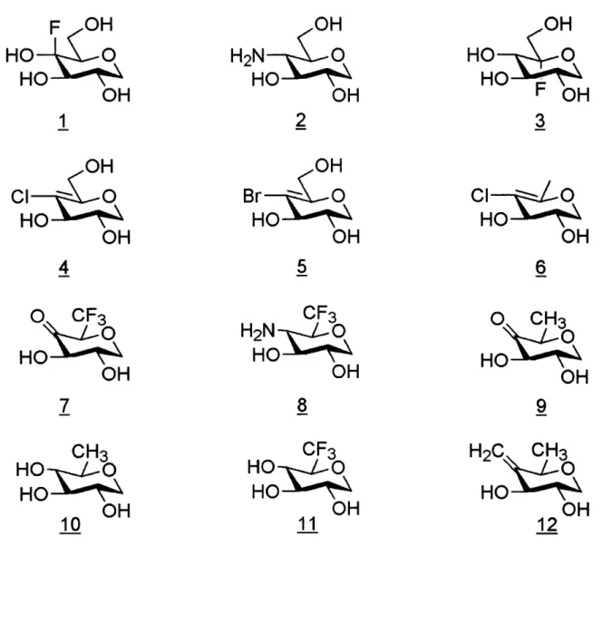
foundation for these studies, enabling the rational design of a small library of potential inhibitors. Twelve mechanism-based inhibitors of RmlB are proposed. These
compounds reflect the current understanding of the mechanism and mimic the sugar portion of the sugar-nucleotide substrate at various steps throughout the reaction mechanism. Each of the proposed inhibitors is designed to inhibit one of the specific steps of the mechanism. While the intention of this project is to synthesize each compound in this library from commercially available starting materials in 15 steps or less, the primary goal of this particular dissertation is to synthesize 3 of the 12 proposed inhibitors from the commercially available starting material 1,5-anhydro-D-glucitol. The long term goal of this work is to produce these compounds in significant amounts
in order to test their efficacy in an animal model of mycobacterial infection. Thesis Supervisor: John M. Essigmann
Acknowledgements
Above all, I would like to take the time to sincerely thank my advisor, John Essigmann, for everything he has done for me over the past 4 years. I had the good fortune of having John as my undergraduate advisor, and from that time, he has been a consistent source of information and support. In the past year and a half in particular, he has given me a number of opportunities that I greatly appreciate-it is thanks to him that I had a wonderful time helping to teach the Biotechnology and Engineering course in Bangkok, and the amazing experience of spending a few months in Thailand. It was also John who had the great idea to attempt this research project this year, and I have only become even more excited about the project as the year progressed; all of these experiences have been a big part of my professional and personal growth over the past year, and I am grateful for his influences.
I am eternally indebted to A. Nicole Dinaut, whose constant support and
encouragement over the past months has been invaluable. She has not only provided guidance with every one of my chemistry dilemmas, but has also been a close friend, always celebrating our successes and dismissing our setbacks with smiling optimism. I am grateful to Kaushik Mitra for sharing his practical expertise, Denise Rodrigues for her help with my syntheses and 2D NMRs, Uday Sharma for synthetic words of wisdom and his assistance with hydrogenations and Will Neeley for sharing suggestions on technical matters. Many other lab members were a pleasure to work with, and I thank them for their moral support and friendship.
I would like to thank the students of BEH.214 during the spring of 2002 for
their assistance in putting the project together, and, in particular, Hector Hernandez for working with me on the final project for the class. I would like to thank Drs. Mathuros Ruchirawat and Somsak Ruchirawat for providing me the opportunity to stay and work
at the Chulabhorn Research Institute in Bangkok, Thailand and Kim Bond Schaefer for continual administrative support while I was away. I appreciate the financial support from the NIH Training Grant in Environmental Toxicology along with help from Professor Douglas Lauffenburger of the Biological Engineering Division; both helped make my trip to Thailand a reality.
Finally, I thank my family for their unconditional love and support. My mother and grandparents have encouraged every one of my endeavors, often making personal sacrifices for my benefit. They have instilled in me a love of learning, the confidence to pursue my goals relentlessly, and the desire to do something meaningful.
Table of Contents
Design and Synthesis of Inhibitors of dTDP-D-glucose 4,6-dehydratase (RmlB), an
Enzyme Required for dTDP-L-Rhamnose Production in M .Tuberculosis... 1
A b stract ... 2
Acknowledgements ... 3
Table of Contents... 5
List of Fig u res... 7
List of Abbreviations ... 8
Chapter 1. Introduction... 10
1.1 Overview ... 10
1.2 History and Epidemiology of TB Infection W orldwide ... 11
1.3 Infection with Mycobacterium Tuberculosis... 15
1.3.1 Characteristics of Mycobacterium Tuberculosis... 15
1.3.2 The M ycobacterial Cell W all ... 16
1.3.3 Transmission, Infection and Disease Progression ... 17
1.4 Host-Pathogen Interactions and the Immune Response ... 18
1.4.1 M acrophage Activation and Response ... 19
1.4.2 Factors increasing susceptibility...24
1.5 Current Treatments and the BCG Vaccine... 25
1.5 .1 Ison iazid ... 2 5 1.5.2 Rifampin ... 26
1.5.3 Ethambutol ... 26
1.5.4 Pyrazinamide... 27
1.5.5 Streptomycin ... 27
1.5.6 Second line drugs... 27
1.5.7 Bacillus Calmette-Gudrin Vaccine ... 28
1.5.8 The Need for New Therapeutics ... 28
Chapter 2. Target Selection and Project Design ... 30
2.1 Target Exploration: Narrowing Down the Possibilities... 30
2.2 Target Exploration: Focus on Cell Wall Biosynthesis and the MAPc ... 33
2.2.1 Peptidoglycan Biosynthesis ... 33
2.2.2 M ycolic Acid Biosynthesis... 34
2.2.3 Arabinogalactan Biosynthesis ... 34
2.3 Target Identification: The d-TDP-L-Rhamnose Pathway... 35
2 .4 R m lB ... 36
2.4.1 The Structure of RmlB ... 36
2.4.2 The Active Site ... 37
2.4.3 M echanism of Action... 38
2.5 Design of Inhibitors... 39
2.5.1 Inhibition of Oxidation ... 41
2.5.2 Inhibition of Dehydration ... 41
2.5.3 Inhibition of Reduction... 42
2.5.4 Selection of Experimental Focus ... 42
Chapter 3. Experimental M ethods... 43
3 .1 G en eral... 4 3 3.2 Synthesis of 6-deoxy- 1,5-Anhydro-D-glucitol (10). ... 43
2,3-O-Acetyl-4,6-O -Benzylidene- 1,5-Anhydro-D-glucitol (19). ... 43
2,3-O-Acetyl-4-O-Benzyl-1,5-Anhydro-D-glucitol (20) ... 44
2,3-O-Acetyl-6-Deoxy~ 1,5-Anhydro-D-glucitol (22) ... 45
6-Deoxy- 1,5-Anhydro-D-glucitol (10). ... 46
Chapter 4. Results and Discussion... 47
4.1 Retrosynthetic Analysis ... 47
4.2 Reduction of Methyl ax-D-Glucopyranoside ... 49
4.3 Benzylidene Acetal Formation and Acetyl Protection... 50
4.4 Assignment of NMR Spectra and the Regioselective Ring Opening Reaction...51
4 .5 lod in ation ... 5 5 4.6 Hydrogenation ... 56
4.7 Final Deprotection and Peak Assignment of Inhibitor 10 ... 57
4.10 Conclusions and Future Studies... 58
R eferen ces ... 6 0
List of Figures
Figure 1. WHO statistics on global tuberculosis infection. ... 64
Figure 2. Scanning electron micrographs of Mycobacterin tuberculosis... 65
Figure 3. Schematic representation of the cell wall of M. TB. ... 66
Figure 4. Stages of the immune response... 67
Figure 5. The immune response against M. TB. ... 68
Figure 6. Chemical structures of first-line drugs... 69
Figure 7. The glyoxylate pathway as it relates to the citric acid cycle... 70
Figure 8. Biosynthesis of the mycolic acid-arabinogalactan-peptidoglycan complex... 71
Figure 9. Biosynthesis of dTDP-L-rhamnose from glucose- I -phosphate... 72
Figure 10. The Structure of RmlB. ... 73
Figure 11. The active site of Rm lB. ... 74
Figure 12. Proposed mechanism of action of RmlB. ... 75
Figure 13. The twelve inhibitors designed for RmlB... 76
Figure 14. Inhibitors designed against the oxidation step of the mechanism ... 77
Figure 15. Inhibitors designed against the dehydration step of the mechanism... 78
Figure 16. Inhibitors designed against the reduction step of the mechanism... 79
Figure 17. Retrosynthetic analysis using a protected glucal as the starting material...80
Figure 18. Retrosynthetic analysis using D-glucose as the starting material...81
Figure 19. Synthesis of inhibitors 10, 9 and 12. ... 82
Figure 20. Suggested mechanism for the regioselective benzylidene ring opening reactio n ... 8 3 Figure 21. 'H Spectrum of Compound 19, 2,3-O-acetyl-4,6-O-benzylidene- 1,5-anhydro-D -glucitol. ... 84
Figure 22. 13 C Spectrum of Compound 19... 85
Figure 23. gCOSY spectra of Compound 19. ... 86
Figure 24. HETCOR spectrum of Compound 19... 87
Figure 25. IH spectrum of Compound 20, 2,3-O-acetyl-4-O-benzyl- 1,5-anhydro-D-g lu cito l...8 8 Figure 26. 13C spectrum of Compound 20. ... 89
Figure 27. gCOSY spectra of Compound 20. ... 90
Figure 28. HETCOR spectrum of Compound 20... 91
Figure 29. 1H Peak assignments for compounds 19 and 20. ... 92
Figure 30. IH spectrum of Compound 21, 2,3-O-acetyl-4-0-benzyl-6-deoxy-6-iodo-1,5-anhydro-D -glucitol ... 93
Figure 31. 13C spectrum of Compound 21. ... 94
Figure 32. 'H spectrum of Compound 22, 2,3-O-acetyl-6-deoxy- 1,5-anhydro-D-g lu cito l...9 5 Figure 33. 13C spectrum of Compound 22. ... 96
Figure 34. 1 H spectrum of Compound 10, 6-deoxy- 1 ,5-anhydro-D-glucitol...97
Figure 35. 13C spectrum of Compound 10. ... 98
Figure 36. gCOSY spectrum of Compound 10... 99
List of Abbreviations
BCG bacillus Calmette-GudrinCTLs cytotoxic T-lymphocytes
DOTS Directly Observed Treatment, Short-course dTTP deoxythymidine triphosphate
EMB ethambutol
FAS fatty acid synthase ICL isocitrate lyase
IFN-y interferon gamma
IL- 12 interleukin 12
INH isoniazid
KO knock out
M. TB Mycobacterium Tuberculosis
MAPc mycolic acid-arabinogalactan-peptidoglycan complex MDR-TB Multi-Drug Resistant Tuberculosis
MS malate synthase NO nitric oxide
PI phosphatidyl inositol POA pyrazinoic acid
PPD purified protein derivative PZA pyrazinamide
RIF rifampin
RmlB dTDP-D-glucose 4,6-dehydratase ROI reactive oxygen intermediates RNI reactive nitrogen intermediates
SDR short-chain dehydrogenase/reductase
TACO tryptophan aspartate containing coat
TB Tuberculosis
TLRs Toll-Like Receptors
TNF-a Tumor Necrosis Factor alpha
UDP-Galf UDP-galactofuranose UDP-Galp UDP-galactopyranose
UDP-MurNAc UDP-N-acetyl muramic acid WHO World Health Organization
-9-Chapter 1. Introduction
1. 1 Overview
Tuberculosis (TB), the disease caused by Mycobacterium tuberculosis (M.TB), is currently a serious health problem in the developing world, while its threat in
developed countries continues to grow every year. Each year, over two million people die from TB, including a large number of HIV-positive individuals who are unable to fight the infection.1 Approximately two billion people are currently infected with M. TB, a number that is constantly growing, as around nine million new cases are identified each year.2 This growing rate of infection translates into a new person infected with TB every second; almost 1% of the total world population is newly infected each year. Of the one-third of the world population infected with TB, 5-10% will become sick or infectious during their lifetime.' The rates of infection are growing at over 2% per year, despite implementation of health strategies worldwide.
Complicating matters is the sheer difficulty in treating the infection. Current therapy consists of a six to nine month barrage of four or more different medications, with different dosages and side effects. Despite this rigorous regimen, treatment can still leave latent TB infection in the lungs. Adding to the current crisis is the growing concern of multi-drug resistant (MDR) TB, which is defined as strains that are resistant to both isoniazid and rifampin, the two most effective current TB drugs.
The World Health Organization (WHO), along with numerous health experts, believe that the world is currently experiencing a public health crisis with respect to TB, and that this problem will continue to persist unless measures are taken to better control the spread of this disease. Unfortunately, 60% of TB-related deaths occur in the
poorest 20% of the global population, and, consequently, there is a serious dearth in the amount of funding put towards profit-driven diagnostic and therapeutic development.
This project is derived from a novel approach to the development of therapeutics for TB. Instead of focusing on an established project in an existing lab, the problem of TB was analyzed in terms of identifying a new "ideal" target and developing a viable method for altering its biological activity. To achieve this goal, the deficits in natural and synthetic means to fight off the infection were examined. This was accomplished through an analysis of both the mechanisms by which the mycobacteria are able to overcome the human immune system as well as the shortcomings of existing therapeutics. There are a large number of possible drug targets in mycobacteria.
While certain targets could be considered for further study, the main focus of this project will be the selection of one particular enzyme as a target, and the remainder of this dissertation will discuss the rational design of small molecule inhibitors of this
protein. In an effort to develop more effective and perhaps more abbreviated courses of treatment, the target of interest is a particular enzyme, dTDP-D-glucose
4,6-dehydratase (RmlB), involved in the dTDP-L-rhamnose pathway in cell wall biosynthesis. Using the crystal structures of the enzyme complexed with single substrates or substrate analogs as a foundation for the studies, the intentions of this project were to conduct the rational design, synthesis and screening of a small library of sugar analogs that could be potential therapeutics for TB.
1.2 History and Epidemiology of TB Infection Worldwide
The problem of TB is nothing new. TB is clearly a significant worldwide killer, and has been for centuries; in fact, it has been referred to as the "white plague" due to the pale appearance of its victims. Robert Koch, who discovered the TB bacilli, wrote in
1880, "If the number of victims which a disease claims is the measure of its
1-significance, then all diseases, particularly the most dreaded infectious disease, must rank far behind tuberculosis. Statistics teach that one-seventh of all human beings die of tuberculosis, and that, if one considers only productive middle-age groups,
tuberculosis carried away one-third and often more of these..." As it was in the 19th century and before, TB continues to be a problematic infection.
While cases of TB consistently dropped once therapeutics were introduced in the 1950's and 1960's, since the 1980's the number has been growing once again. There are numerous possible reasons for this increase, but one of the main contributing
factors to an increase in TB infections worldwide is the HIV/AIDS epidemic, as those
afflicted with HIV/AIDS are much more susceptible to active TB infections than other individuals. About one-third of those afflicted with HIV/AIDS are infected with TB. Due to their compromised immune systems, HIV/AIDS victims are unable to fight the infection, resulting in high rates of reactivation TB and high rates of fatalities (raised by 5-20% over the normal 15%) due to TB.3 Other possibilities for the increase in TB
infections worldwide could include increased poverty, higher levels of global travel, and significantly higher levels of crowding in big cities, as well as institutions such as
prisons and hospitals. Lack of available treatment is another concern in the developing
world, as there is a 50% fatality rate for untreated cases-a number which rises to 80% in HIV/AIDS patients. Whatever the reasons for this increase, it is clear that the
problem is only getting worse at the status quo. During the 1990's, it was
approximated that there were ninety million new cases and around thirty million deaths due to TB. However, it is estimated that over the next twenty years,
approximately one billion more people will become infected, with more than a hundred and fifty million people experiencing symptoms and over thirty six million of those succumbing to the infection. The infection is currently the leading cause of death due to infectious disease among people older than five.4
Regionally, the distribution of TB is quite striking, as the rates of TB infection worldwide vary tremendously. The prevalence of TB is much higher in developing countries, as is the death rate due to the infection. TB accounts for 20% of adult deaths and 6% of infant deaths, and is considered to be the cause of 26% of all avoidable deaths in the developing world.' It is hypothesized that the developing world is home to 95% of the world's TB, and 98% of the deaths caused by the disease; overall,
approximately 7% of all deaths in the developing world are attributed to TB. There is a relationship between lower standards of living and higher rates of TB, as it has a much higher incidence in poorer countries and large, crowded cities. Of the twenty two countries with the highest occurrence of TB, only one (Brazil) was not classified as a low income or low middle income country, according to the per capita income designations set by WorldBank.6
The continents of Asia, Africa and South America are hardest hit, yet the top five infected countries (India, China, Indonesia, Bangladesh and Nigeria) are almost all in Asia (Figure 1). 1 About three million cases per year occur in south-east Asia, while around two million cases per year are present in sub-Saharan Africa, an area of increasing concern because of the growing relationship between HIV/AIDS and TB. Forty percent of the global incidence occurs in India and China, the two countries with the largest populations worldwide. India has an estimated 1.8 million cases while
China reports approximately 1.4 million. Both countries claim significantly high levels of treatment success (79% and 96%, respectively), yet it is clear their National TB programs are not as extensive and treatment is not as readily available as it should be.'
DOTS or Directly Observed Treatment, Short-course is the primary strategy
endorsed by the WHO to achieve high levels of treatment success. The strategy encompasses several goals that involve government commitment in TB control activities, case detection by sputum smear microscopy, standardized chemotherapy
~13-regimens for eight weeks, an uninterrupted supply of essential anti-TB drugs, and finally, a recording/reporting system allowing for assessment of each individual patient and the TB program overall. It has been rated as an extremely cost-effective way of efficiently treating large numbers of patients, yet the distribution of DOTS enforcement is not as high as desired.! The countries listed above are not the countries with the highest rates of incidence per 100,000 people. Cambodia, Zimbabwe, South Africa, Afghanistan and Uganda are the countries with very high rates of disease and these countries have very low therapeutic effectiveness. The enforcement of DOTS in most of these countries is minimal or non-existent, due to funding and personnel shortages, leading to the further spread of disease.'
Problems with effective TB therapy and DOTS enforcement have led to the spread of multi-drug resistant (MDR) forms of TB. Due to the long course of treatment, patient compliance is a serious issue, as patients tend to feel better after two months of treatment. However, discontinuing treatment at that stage leads to the development of strains of MDR-TB. Poor availability of therapeutics along with incorrect single-drug treatment regimens also contribute to the growing problem of MDR-TB. There are cases of MDR-TB on every continent, yet there are certain countries with particularly
high levels. Latvia (14.4%), Estonia (10.2%), Dominican Republic (6.6%), Ivory Coast
(5.3%), Argentina (4.4%) and Russia (4.0%) are among those with the most significant
incidences of MDR~TB. In countries not implementing DOTS, rates of MDR-TB greater than 2% were much more common-54% in non-DOTS countries versus 22% in DOTS countries. It is estimated that up to fifty million people may already be infected with MDR strains of TB, and this number continues to rise.7
-14-1.3 Infection with Mycobacterium Tuberculosis
Infections due to M.TB pose a significant public health concern, yet it is the lack of understanding of the complexities of the bacterium itself that frustrates the efforts to contain the disease. The unique features of the pathogen pose as much of a challenge in the development of therapeutics now as they have for the past 100 years. Despite the vast knowledge base currently available regarding M. TB, there are a considerable number of questions that remain. However, there have been recent developments in understanding the interactions between the pathogen and the host that present a beautiful scientific problem.
1.3.1 Characteristics of Mycobacterium Tuberculosis
Mycobacterium Tuberculosis possesses a variety of unusual characteristics that contribute to its virulence as a pathogen. The bacterium has a Gram-positive type cell wall, yet cannot be detected by the conventional Gram stain due to the waxy outer
coating of numerous lipids. The rod-shaped bacteria (ranging between 1 to 4 tm in
length and 0.3 to 0.6 pm in width) can be detected using an acid-fast stain, a harsh
stain that involves heating the bacteria. The primary method of diagnosis in most of the world is through the culture of sputum samples and detection using this stain (Figure 2). The slow generation time of the bacterium (18-24 hours) is attributed to the hydrophobic cell surface, which may cause the clumping together of bacteria, thereby preventing the entry of nutrients into the cell.2 The organism is an obligate aerobe,
and, as expected, it prefers the high oxygen content of organs such as the lungs, despite having the capacity to change its metabolism and survive in a microaerophilic
environment.
-15-1.3.2 The Mycobacterial Cell Wall
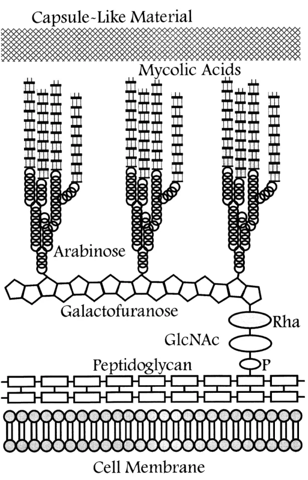
While M. TB is considered Gram-positive, the organism possesses cell-wall features that are characteristic of both Gram-positive and Gram-negative bacteria, resulting in a particularly unique structure. The mycobacterial cellular envelope consists of three components: 1) a plasma membrane, 2) a covalently linked mycolic acid-arabinogalactan-peptidoglycan complex (MAPc) and 3) a polysaccharide-rich capsule-like material (Figure 3). The lipid content of the cellular envelope is so dramatic that it is estimated to make up 30-40% of the total weight of the bacilli and could account for characteristics such as low permeability, growth in clumps, physical strength and a decreased response to certain toxic substances.9
The plasma membrane serves as the initial protection of the mycobacterium from the outside environment. It appears to have characteristics quite similar to normal bacterial membranes, as does the peptidoglycan component of the MAPc. The MAPc consists of the cross-linked peptidoglycan which is bound covalently to
arabinogalactan chains through a phosphoryl-N-acetylglucosaminosyl-rhamnosyl
linker. The arabinogalactan is additionally esterified to a variety of ca-alkyl and
p-hydroxy mycolic acids.10 In particular, the arabinogalactan-peptidoglycan component of the cell wall has been implicated in the viability of mycobacteria; the relatively recent elucidation of biosynthesis pathways has resulted in a number of targets for small molecule enzyme inhibition. Certain pathways are becoming increasingly well-characterized, but the specific roles of many enzymes involved in these pathways remain elusive. There are a number of lipids that are non-covalently attached to the MAPc, and beyond this layer of lipids lies a loosely attached layer of polysaccharides and proteins, forming the outer capsule around the mycobacteria.9 Overall, this extensive cell wall decoration plays a role in the behavior of mycobacteria in the host
environment, and could affect processes ranging from intracellular growth, to the immune response, to reactions of the bacilli to therapeutics.
1.3.3 Transmission, Infection and Disease Progression
The disease itself is spread primarily by aerosolized droplets of tubercle bacilli containing three or less organisms; these droplet nuclei float in the air, and can remain airborne for long periods of time after the fluid evaporates. A patient with pulmonary TB can infect another while coughing, exhaling, sneezing, talking or spitting; it is estimated that 10- 15 people each year are infected by one infected individual.4
Once inhaled, the bacteria travel to the alveoli, as infection in the upper respiratory tract is unlikely. Smaller droplets of bacilli can reach the alveoli, where they are taken up by alveolar macrophages which attempt to phagocytose the bacteria. Once the bacteria are taken up into the macrophages in a phagosome, the macrophage attempts to lyse the bacteria by fusing the phagosome and the lysosome. This fusion is
often blocked by mycobacterial mechanisms, resulting in a pulmonary infection that
consists of active bacteria living within inactivated macrophages."I The mycobacteria
are able to grow and spread, and can even enter systemic venous circulation at times, so they may re~enter the lungs and be deposited at additional locations within the lungs or other organs. At this phase there are three possibilities for the mycobacteria and the
host effector cell: the macrophage can kill the mycobacteria, the mycobacteria can
cause the cell death of the macrophage, or the macrophage can become a host cell for the mycobacteria and propagate infection." It is at this point that an active immune
response is key in determining the outcome of infection. Poor T-cell immunity at this phase can lead to a serious progression of the disease, as well as transmission to others. The symptoms of an infection consist primarily of a fever, coughing for over three weeks (often with bloody sputum), chest pain, loss of energy and weight loss, in
~17-addition to progressive (irreversible) lung destruction. In most cases, the immune response fails to completely clear the bacteria, which can continue to grow within alveolar macrophages.2
When phagocytes fail to clear the infection, other T-cells and macrophages amass around the bacterial growth. Macrophages can fuse to form giant cells, resulting in a layer consisting of macrophages and T-cells that surrounds the bacteria. This layer often forms a self-contained lesion, consisting of a mass of bacteria surrounded by a coat of fibrin.2 This lesion, called a tubercle or granuloma, has a cheese-like
consistency (caseous necrosis) at first, yet as bacterial growth continues and more phagocytes surround the area, the lesion becomes more liquid. Latent TB consists of mycobacteria contained in these lesions, and these liquefied lesions can form infectious aerosols, burst to reactivate an active TB infection or travel to other organs, resulting in miliary TB, which is often fatal. The chances of reactivation TB are 2-23% over a lifetime, but this number can increase depending on immune system factors.2
1. 4 Host-Pathogen Interactions and the Immune Response
When encountering any pathogen, the human immune system attempts to mount an effective response to kill the invader. The case of M. TB is no different; there are a number of mechanisms by which the innate and adaptive immune systems can clear the infection. However, what makes mycobacteria perhaps more complex than certain other pathogens is their ability to evade damage by these intervention systems and thus render immune system processes ineffective. This section will discuss some of the interactions between mycobacteria and various human immune system components in order to highlight the major players at the cellular and molecular levels.
1.4.1 Macrophage Activation and Response
Once phagocytosed bacteria are living within a macrophage, there are a
number of pathways that determine the fate of both the mycobacteria and macrophage. The complexity of M. TB lies in its ability to modulate normal immunological activity in its favor, despite a variety of mechanisms that are intended to clear the infection
through the various stages of infection (Figure 4).
Oxidative burst
Reactive oxygen intermediates (ROI) and reactive nitrogen intermediates (RNI) comprise the two major sets of effector molecules important in the macrophage defense against mycobacteria (Figure 5). These reactive intermediates are synthesized in
parallel by two related pathways. Beginning with 0z and L-arginine, the macrophage is able to form radicals and other damaging agents to use against the bacteria. ROI are a product descendent from 02, as the enzyme phagocyte oxidase (phox) catalyzes the formation of superoxide, which is used in the synthesis of peroxide by superoxide dismutase. Hydrogen peroxide then undergoes radical mechanisms to result in
hydroxyl and ferryl radicals. The importance of ROI in defense against mycobacteria is slightly controversial, yet they do seem to play some kind of role, despite the number of bacterial resistance genes and products that are used to neutralize this response. RNI are formed in a similar manner from guanidine-L-arginine, which is transformed into nitric oxide (NO) radicals due to nitric oxide synthase (iNOS or NOS2). These nitric oxide radicals can go on to form peroxynitrite (along with superoxide) or they can
form N02O, N02, S-nitrosothiols, dinitrosyl iron complexes and other reactive
molecules. Similar to ROI, mycobacteria have developed a number of mechanisms involving molecular scavengers, antioxidant enzymes, repair systems and antioxidant regulons."I Studies of inducible nitric oxide synthase (iNOS or NOS2) and phagocyte
NADPH oxidase (phox) suggest that RNI are significantly more important in the defense
against TB. Macrophages from actively infected individuals have been found to have high levels of NOS2, and the levels of NO exhaled in TB patients are higher than that of non-infected individuals. There appear to be more mechanisms by which ROI can be evaded, as many cell wall components such as lipoarabinomannan and
phenolicglycolipid I are oxygen radical scavengers. However, despite the controversy over the relative importance of these two mechanisms, it is clear that they play a role in the macrophage defense.
Control of phagolysosome fusion
The response of macrophages to mycobacteria is to form a phagosome around the bacteria, and once the bacteria have been taken up into the cell, fuse the
mycobacteria-containing phagosome with the acidic lysosome, thereby ensuring the death of the bacteria. However, one of the most complex mechanisms of mycobacterial survival is its ability to block this phagosome-lysosome fusion, allowing the bacilli to
survive indefinitely within the macrophage.
There are a number of biochemical reasons for the arrest in phagolysosome maturation. The ability of the lysosome to degrade particles depends upon hydrolases that must be maintained at low pH levels, yet it has been demonstrated that
mycobacteria-infected phagosomes possess a lack of vacuolar ATPases, which are usually responsible for acidification; the less acidic environment could be responsible for the lower amount of degradation by the vacuole.II Another possible mechanism is the role of Rab GTPases; Rab5 helps in early endosome fusion while Rab7 assists in late endosomal membrane trafficking, yet it has been found that mycobacterial phagosomes utilize Rab5 while they exclude Rab 7.13 Calcium-binding calmodulin may be involved in the regulatory functions of membrane fusion, as phagocytosis usually leads to an
increase in calcium concentrations, yet mycobacteria are able to alter the signaling cascade and prevent this increase.'I Another important protein could be the tryptophan aspartate containing coat (TACO), an actin-binding protein that is implicated in the early phagosome membrane. However, the TACO-membrane
association is expected to be transient, yet the retention of this association is postulated to prevent phagolysosome fusion by delaying maturation of the phagosome;
mycobacteria that are somehow able to prevent the detachment of TACO are able to survive longer within macrophages.12 There are other mechanisms that may explain the lack of degradation of mycobacteria by macrophages; certain mycobacterial
sulfatides may be able to inhibit phagolysosomal fusion, while it is also possible that high levels of ammonia production by mycobacteria may change saltatory movements and even raise the pH of the compartment.1 3
CD8+ Cells
The activities of CD8+ cells, in protecting against mycobacteria are two-fold; the cytotoxic functions of the cells enable them to kill intracellular mycobacteria directly in addition to serving as a facilitator in lysing macrophages containing live bacteria (Figure 5). Initially, the CD8+ cells are presented with mycobacterial lipid or peptide antigens by antigen-presenting cells via molecules such as MHC Class I or CD 1. 14 Once these CD8+ cells come into contact with an infected macrophage, they can activate the macrophage to kill the mycobacteria through the production of interferon-gamma and tumor necrosis factor-alpha, two crucial cytokines that can activate enzymes such as nitric oxide synthase. CD8+ cells are able to function as cytotoxic T lymphocytes (CTLs) in order to facilitate lysing of infected macrophages. This lysing can occur via two pathways: a perforin-dependent pathway and a Fas/FasL pathway. The perforin dependent pathway involves granulysin: perforin forms a pore that
enables the uptake of granulysin, a cytolytic T cell granule protein, into the macrophage, resulting in the death of both the macrophage and its mycobacteria. However, if the mechanism of macrophage lysis is via the Fas/Fas ligand system, the
macrophage is destroyed but the live mycobacteria are released.14
CD4+
CD4+ cells are important in the presentation of mycobacterial antigens by MHC class II molecules. However, the primary effector function of CD4+ cells is the active role in IFN-y production, which is crucial in macrophage activation. These cells are also involved with NOS2 expression in the early phases of infection, but these cells play a role in later stages of the infection as well, as they have been found to be important in the priming and maintenance of CD8+ T cell effector and memory functions. The influence of these cells in the immune response has been studied extensively. For example, it has been found that M. TB infected mice treated with an anti-CD4+ antibody experienced a reactivation of active disease, demonstrating the key role of these cells. M. TB has been able to alter the activity of these cells through disrupting the macrophage-CD4+ cell interaction. M. TB-infected macrophages are unable to present antigens to CD4+ T cells, and infection by mycobacteria may inhibit recognition
of macrophages by CD4+ cells by down regulating the surface expression of MHC Class II molecules. This interference with normal function is yet one more mechanism by which mycobacteria can survive within the host successfully.13
Interferon-gamma
Interferon-gamma (IFN-y) is perhaps the most important cytokine involved in
M.TB infection. It plays a crucial role in macrophage activation by enhancing the Th 1 response and increasing IL- 12 production.'3 It is produced by CD4+ and CD8+ cells,
along with NK cells, and has been found to be important in antigen-specific T-cell
immunity.13 The functions of IFN-y are quite diverse; it has been known to promote the production of opsonising antibodies, increase CTL activation as well as NK cell activity and increase macrophage MHC Class II activity.' 6 It may also trigger the movement of T cells into infected areas by facilitating a bond between endothelial cells and T cells and serve as a trigger to differentiate macrophages into active effector cells.17 It has
been found that IFN-y knock-out (KO) mice are most susceptible to virulent TB;
additionally, it was found that KO mice had a defective macrophage response and low
NOS2 expression.13
Tumor Necrosis Factor-a
Tumor Necrosis Factor-ca (TFN-a) is a cytokine that plays a role in the mediation
of macrophage activation.' 3 During acute infection of M. TB, TNF-ca expression is increased. Along with IFN-y, TNF-a initiates an increase in NOS2 expression, enabling a stronger response to M. TB infection. TNF-a may also coordinate the migration and
localization of cells within particular tissues, and it appears to be essential in granuloma formation, as it regulates expression of certain adhesion molecules.
However, it has also been found that TNF-cL is responsible for lung tissue damage, as
well as the symptoms of weight loss experienced by TB patients.
Interleukin- 12
Interleukin- 12 (IL- 12) is a pro-inflammatory regulatory cytokine which is primarily produced by phagocytes.I7 The release of IL- 12 is induced after
phagocytosis, and is thought to initiate a Th 1 response by stimulating IFN-y
pr oT 1 i not completely u s yet i s
production.'3 ThLC wCAct role 0f IL- Iz Is 1ot compieLtIy 4IiLUL;-LVULL Y%,L ItL i ,I
-23-found that IL~ 12 KO mice are highly susceptible to M.TB infections,17 while treating TB infected mice with IL- 12 in the early phase of infection increased the mean survival time and decreased the bacterial load.13 IL- 12 serves as a mediator that can connect
the response of phagocytes to that of T cells through its control over IFN~y.
Toll-Like Receptors
Adaptive Toll-like receptors (TLRs) facilitate the activation of the immune response against M.TB. TLRs can recognize a number of mycobacterial antigens; the
antigens most important in the immune response are the 19kDa lipoprotein,
lipoarabinomannan and phosphatidylinositolmannan, all of which interact with TLR2. TLR2 is proposed to have a more significant role in defense against TB than TLR4;
activation of TLR2 can result in an induction of macrophage apoptosis and the down regulation of MHC class II, two important anti-mycobacterial functions.18
1.4.2 Factors increasing susceptibility
Most characteristics that have been identified as increasing a particular individual's susceptibility towards TB infection involve the response of the immune system; whether it is in the form of a genetic variation or other factors that decrease
normal immune functions. A defect in IL- 12 or IFN-y causes an individual to be at a
significantly heightened risk of TB, while other mutations, such as ones in the promoter
regions of TNF-c, or the natural resistance-associated macrophage protein 1 (nRAMP), result in only slight increases in susceptibility.19
A normal person infected with TB bacilli incurs a 10% risk of developing active
TB throughout a lifetime. A weakening of the immune system, however, can increase this risk. The single largest factor to increase the chances of reactivation is HIV
to the immunocompromised state of the patients.4 Other factors include silicosis, renal disease, malignancy, diabetes, malnutrition or long-term treatment with
immunosuppressive drugs.19
1.5 Current Treatments and the BCG Vaccine
From the above description of mycobacteria, it is quite clear that there are a number of complexities associated with successfully fighting infections of M. TB. There are a number of anti-mycobacterial agents in use, yet none are perfect. In fact, only four can be considered "first-line" treatments, while others are only used on an "as-needed" basis due to the rapid resistance that is developed by the mycobacteria. TB therapy is currently made up of an initial two-month phase of four drugs daily
(Isoniazid, Rifampin, Pyrazinamide and Ethambutol) after which time the regimen
solely consists of Isoniazid and Rifampin. In cases of MDR-TB, the so-called
"DOTS-plus" strategy is employed, and the use of second-line drugs along with any viable first-line options is used. This section will summarize the currently available treatments and the problems associated with each.
1.5.1 Isoniazid
Isoniazid (INH) has been the most important anti-mycobacterial agent in use since the 1950's, and it continues to dominate therapeutic regimens, aside from cases of MDR-TB (Figure 6). It is hypothesized that INH is a prodrug which is transformed into a number of reactive radical species by the mycobacterial catalase-peroxidase KatG. These reactive molecules can react with a number of mycobacterial targets, but the two most well-understood enzymes are InhA (an enoyl ACP reductase) and KasA (a beta-ketoacyl ACP synthase), both of which are involved in the cell wall mycolic acid biosynthesis. Resistance often arises to INH. Resistance to INH can be attributed to
mutations in katG; it is proposed that a particular serine is mutated into a threonine, resulting in a form of the catalase-peroxidase that does not react with INH but maintains enough normal activity to continue detoxification against antibacterial radicals.2 0 Another gene, ndh (NADH dehydrogenase), which increases the ratio of
NADH/NAD+ in order to prevent a reaction between the isonicotinic acyl radical and
an NAD radical, has also been implicated in INH resistance.2 1 Despite the fact that the activated INH can react with a number of mycobacterial cellular components, drug resistance is becoming a serious problem.zi
1.5.2 Rifampin
Rifampin (RIF), a compound with a very complex macrocyclic ring structure, works to inhibit transcription in mycobacteria via an interaction with the DNA-dependent RNA polymerase (Figure 6). The mode of action is fairly well understood, and crystallographic studies confirm the tight binding of the drug to RNA polymerase. The means by which mycobacteria develop resistance to rifampin is based upon mutations in an 81 -base pair region of the gene encoding the B-subunit of RNA polymerase (rpoB).20
1.5.3 Ethambutol
Ethambutol (EMB), a diamine derivative, is another first-line therapeutic used in defense against TB (Figure 6). The precise mechanism of action is not yet known, yet it is clear it is involved in the inhibition of cell wall biosynthesis. It has been shown to inhibit the incorporation of mycolic acid into the mycobacterial cell wall,2 and it has more recently been suggested that EMB blocks some part of the polymerization process involved in arabinogalactan and lipoarabinomannan biosynthesis.23 More support for an interaction between EMB and arabinosyl transferases is demonstrated by the gene
which confers resistance to EMB; mutations in embCAB, an operon encoding arabinosyl transferases, have been implicated in most cases of resistance to EMB.2 4
1.5.4 Pyrazinamide
Pyrazinamide (PZA) is a nicotinamide-like prodrug that reacts with
pyrazinamidase to form pyrazinoic acid (POA), the active form of the drug, that can accumulate in the mycobacterial cytoplasm (Figure 6). Large amounts of POA can cause a lowering of the intracellular pH, and perhaps inactivates pH sensitive enzymes such as fatty acid synthase. When resistance to PZA occurs, it is usually the case that mycobacteria stop using pyrazinamidase. Most PZA-resistant mycobacteria possess a mutation in the promoter region of the gene that encodes pyrazinamidase (pncA).20
1.5.5 Streptomycin
Streptomycin, an aminocyclitol glycoside antibiotic, is considered as another first-line drug against TB, yet its use is limited by serious toxicity and its intramuscular delivery method (Figure 6). It inhibits protein synthesis by somehow interfering with translational proofreading, perhaps by interacting with the ribosomal S12 protein and
16SrRNA.2 1 Streptomycin increases the missense rate by suppressing proofreading and possibly disrupts the decoding of aminoacyl-tRNA, leading to incorrect translation. Resistance is attributed to point mutations in the S12 ribosomal protein encoded by RpsL, and mutations in the rrs operon encoding the 16S rRNA.25
1.5.6 Second line drugs
There are a number of second-line drugs that are used in the treatment of TB. These therapeutics are classified as second-line because of decreased efficacy against the bacilli, increased toxicity, increased rates of resistance or unfavorable
pharmacokinetics. These drugs include ethionamide, kanamycin, cycloserine,
-27-aminosalicylic acid, capreomycin, amikacin and the fluoroquinolones, all of which are used primarily in cases where resistance is developed against the first-line therapeutics. However, none of these are optimal therapeutic options for the treatment of TB
infections.2
1.5.7 Bacillus Calmette-Gudrin Vaccine
The vaccine available for TB, called bacillus Calmette-Gudrin (BCG), is an attenuated strain of Mycobacterium bovis that is the most widely administered vaccine
in the WHO Expanded Programme for Immunization. However, the efficacy of this vaccine is quite controversial. Protection from disseminated and meningeal TB in
infants and children is quite reliable, but its value in preventing pulmonary TB in adults is questionable; studies on the rates of vaccine protection show dramatically different efficiencies, ranging from 77% in the UK to 0% in India.8 As it stands, the BCG vaccine does not do a successful job of providing immunity against TB, and is currently
contraindicated in HIV patients, as it is a live vaccine.26 Additionally, vaccination with
BCG results in a positive response to the purified protein derivative (PPD), and
therefore a false-positive on the Mantoux diagnostic skin test, one of the most effective diagnostic tools used to identify TB-infected persons.2
1.5.8 The Need for New Therapeutics
In 1993, the WHO declared that TB was a global emergency, yet over the past ten years there has not been a significant increase in practical solutions to this problem. There has not been a new drug approved for TB in over thirty years, yet the current health crisis demonstrates a significant need for new developments in order to eradicate TB. The effective therapies currently in use are not optimal; due to nine-month long regimens, patient non-compliance is a serious issue, leading to the emergence of newer
drug-resistant strains. New anti-TB therapies are an absolute necessity for MDR-TB as well as latent TB infection, and the development of such novel therapies is the focus of the remainder of this thesis.
-29-Chapter 2. Target Selection and Project Design
2.1 Target Exploration: Narrowing Down the Possibilities
In the process of choosing the dTDP-L-rhamnose pathway in TB as the
therapeutic objective for rational drug design, a range of possibilities were considered based upon the current literature. The field of TB research is constantly evolving, and the validity of targets is constantly changing. The purpose of this section is to briefly explore various other possible targets and to explain reasons why the dTDP-L~ rhamnose pathway was viewed most favorably as a target for novel TB therapeutics.
Isocitrate Lyase and Malate Synthase
The glyoxylate cycle is a shunt that is used to effectively convert acetate
(degraded from fatty acids, amino acids or ethanol) into carbohydrates by bypassing the
loss of CO2 in the citric acid cycle in M. TB (Figure 7). The two enzymes involved in
the cycle, isocitrate lyase (ICL) and malate synthase (MS) catalyze the formation of malate from isocitrate, via the intermediate glyoxylate; the cycle facilitates the
formation of succinate from two acetate molecules (as acetyl-Coenzyme A), conserving carbon that would otherwise be released as carbon dioxide via the normal Krebs cycle enzymes. In conditions of latency, fatty acids may be the primary source of carbon and energy for mycobacteria, and it has been found that the ability of M. TB to persist in macrophages is dependent upon ICL but not necessarily MS.2 7
The use of ICL as a possible target for anti-mycobacterial development bears further investigation, as not only is it an enzyme not present in humans, but it has been examined extensively through mechanistic and crystallographic studies. However, it was believed to an imperfect target because of the similarities between the substrate
and end products of the enzyme as compared to endogenous human substrates from the citric acid cycle. Any inhibitors would most likely lead to undesired side effects.
Galactofuranose pathway
Another particular pathway of interest was the pyranose pathway consisting of UDP-galactopyranose (UDP~Galp) mutase and galactofuranose (Galf) transferase, which together function to transform an galactopyranose into
UDP-galactofuranose and transfer UDP-galactofuranose molecules onto the growing chain of the arabino-galactan complex, a component of the cell wall. The UDP-Galp mutase has been crystallized, although the mechanism by which the mutase achieves the
unprecedented ring contraction of a nonreducing sugar is unclear. Galf transferase is even more complex; it has been suggested that there are two to four separate
transferases involved, with the overall activity being significantly complicated.28 Nevertheless, the pathway of core galactofuran synthesis has been found to be essential
for the viability of mycobacteria, and these galactofuranose units are not present in
human tissues.29 Despite these important details, the lack of a clear mechanism renders these enzymes a less attractive target for the rational design of inhibitors.30
Antgen 85 complex
The Antigen 85 complex is made up of three related mycolyl transferases, Antigen A, B and C. These three proteins are important in transferring a mycolic acid molecule between one trehalose monomycolate molecule and another,3' which leads to the formation of trehalose dimycolate, also known as the "cord factor," and free
trehalose, two important factors that are incorporated into the mycobacterial cell wall.3 2 The cord factor has been implicated in cell wall integrity, while the Antigen 85 complex itself may also foster interaction between the mycobacteria and the
macrophage.-" However, while there are some structures for this complex, the role of these proteins is not completely understood; the value of this complex as a drug target
has been controversial, and thus it was decided to pursue alternatives.
Phosphatidylinositol synthase
Phosphatidylinositol (PI), along with other lipoglycan molecules such as PI mannosides, lipomannan and lipoarabinomannan, have been implicated in the cell wall integrity of M. TB, and may additionally be important in the response of the
mycobacterium to the host immune system. Studies of PI synthase mutants have revealed that this enzyme is essential to mycobacterial survival and the mechanism is somewhat well understood, suggesting that it could be a viable therapeutic target. However, as humans also possess a version of PI synthase, inhibitors of this enzyme may lack specificity and could be quite harmful to human tissues.33
Other Targets
The sheer number of possibilities for TB drug development is quite
overwhelming. Due to the recent sequencing of the M.TB genome, there have been a number of targets identified, yet the mechanistic and structural knowledge about many of these is still in the early phases of discovery. Among targets that could be important are: the gated mechanosensitive ion channel, which has been implicated in ionic and osmotic regulation; InhA, the enoyl-acyl carrier protein reductase; glutamine synthase, which helps to control ammonia levels and thereby modulate phagosome-lysosome
fusion; and the
p-ketoacyl-ACP
synthase, which is important in mycolic acid biosynthesis. Numerous other pathways such as those involving peptidoglycansynthesis specific to mycobacteria, and the
p-oxidation
processes important in fatty acidinvolves the modulation of the host-pathogen interaction, in order to augment the immune response, enabling the body to successfully fight the infection. It is clear, however, that the knowledge base for this type of drug development is currently lacking; not enough is known about such interactions to design appropriate
therapeutics effectively. Since further development is required before these strategies can be successfully pursued, a more well-established pathway became the focus of this project.
2.2 Target Exploration:
Focus on Cell Wall Biosynthesis and the MAPc
As previously mentioned, the composition of the cellular envelope consists of three components: 1) a plasma membrane, 2) a covalently linked mycolicacid-arabinogalactan-peptidoglycan complex (MAPc) and 3) a polysaccharide-rich capsule like material. The MAPc in particular is assembled via a complex set of biosynthetic pathways; the three components are synthesized in parallel and joined together as the final step.
2.2.1 Peptidoglycan Biosynthesis
Since peptidoglycan structures are fairly homologous between various bacterial species, it is assumed that the peptidoglycan layer of M. TB is no different. Two enzymes, MurA and MurB, are important in the synthesis of UDP-N-acetyl muramic acid (MurNAc) from N-acetyl-glucose and enoylpyruvate. The UDP-MurNAc is subsequently used to form a UDP-UDP-MurNAc-pentapeptide through the linkage of an L-alanine, a D-glutamic acid residue, a diaminopimelic acid molecule and finally a D-alanyl-D-alanine dipeptide, reactions catalyzed by MurC, MurD, MurE and MurF. At this point, this peptide molecule is linked to an undecaprenyl phosphate and bound to a N-acetyl glucose molecule to form a
-33-diphosphoryl-undecaprenol, also known as Lipid II, by MurG. This fragment is subsequently ready for transport to the inner cell membrane where the undecaprenyl diphosphate is released and the peptide crosslinks are formed. Each of these enzymes are potential targets for anti-bacterial agents, yet one disadvantage is that there would be no specificity for mycobacteria in particular.10
2.2.2 Mycolic Acid Biosynthesis
The mycolic acids decorating the surface of mycobacteria can be classified in a variety of different ways, yet most possess a linear carbon chain (between 60 and 90 carbons), with vicinal carboxyl and hydroxyl groups on carbons 23 and 24 (±2), and often contain cyclopropane rings in place of double bonds.34 Both fatty acid synthase
(FAS) type I and type II systems are present in mycobacteria, and the synthesis of these
mycolic acids proceeds like much of fatty acid synthesis; the fatty acid synthase system processes-p-ketoacyl synthase,
p-ketoacyl
reductase,p-hydroxyacyl
dehydratase andenoyl reductase-perform the cyclic functions of condensation, ketone reduction, dehydration and enoyl reduction. However, while the system is relatively
well-characterized, not only are structural details not fully known, but there is additionally a lack of specificity with regards to other bacteria (FAS II) or even humans (FAS I).
2.2.3 Arabinogalactan Biosynthesis
The portion of the MAPc that was of most interest was the branched-chain arabinogalactan component, which links together the peptidoglycan and mycolic acid layers. Two residues, a N-acetyl-glucose and a rhamnose, link the peptidoglycan and arabinogalactan layers together; the galactose moieties are added on to this rhamnose residue in succession, followed by the branching out of the layer by the addition of the arabinose groups (Figure 8). The synthesis of the galactose molecules has been
~34-described earlier, and it is hypothesized that the arabinose sugars originate from the pentose phosphate pathway/hexose monophosphate shunt. While the synthesis of this layer has been somewhat characterized, the majority of the enzymes involved have not been crystallized or even distinctly isolated; it was decided that the optimal target for this project would be a structurally and mechanistically understood pathway, and for this reason many of the arabinogalactan component enzymes were eliminated.'0
2.3 Target Identification: The d-TDP-L-Rhamnose Pathway
The arabinogalactan component of the cell wall is linked to the peptidoglycan part via an N-acetylglucosamine and a rhamnose. As rhamnose is not a sugar produced by humans, it is synthesized within the bacteria and transferred onto the growing chain. L-Rhamnose is a 6-deoxyhexose that is synthesized from glucose-I-phosphate and deoxythymidine triglucose-I-phosphate (dTTP) and found in the cell walls of numerous pathogenic bacteria. This pathway represents an interesting therapeutic target, as no analogs of this pathway have been elucidated in humans.35
The first enzyme in the pathway is RmlA (glucose- 1-phosphate
thymidylyltransferase), which transfers a dTDP on to glucose-I-phosphate to make glucose. RmlB, a D-glucose 4,6-dehydratase, next converts the dTDP-glucose into dTDP-4-keto-6-deoxy-D-dTDP-glucose. The third enzyme in the pathway, RmlC or dTDP-4 -keto-6-deoxy-D-glucose, epimerizes the dTDP-4-keto-6-deoxy-D-glucose into dTDP-L-lyxo-6-deoxy-4-hexulose. Finally, RmlD catalyses the reduction of the C4 keto group to form dTDP-L-rhamnose (Figure 9).3 The dTDP-L-rhamnose is then transferred onto the N-acetylglucosamine that will become the arabinogalactan component. As an essential linker between the peptidoglycan and the arabinogalactan molecule, rhamnose serves an important purpose; the inhibition of this sugar pathway would theoretically prevent the assembly of the complex and prevent bacterial
survival.10 It has been recently found that the formation of dTDP-L-rhamnose is essential for the growth of M.TB.37
2.4 RmIB
RmlB, the second enzyme in the dTDP-L-Rhamnose pathway, is perhaps the best characterized in terms of structure and mechanism of the four enzymes. Of the four, it presents the best therapeutic target because of the high level of understanding of its
function. Distinct crystal structures of the enzyme with its cofactor and the substrate have been solved, resulting in considerable insight into the positioning of these molecules within the active site.
Mechanistic studies have confirmed the presence of proposed intermediates, and the convergence of both mechanistic and structural analyses have resulted in general acceptance of the currently proposed mechanism. Additionally, the intermediates and product of this conversion are distinct from any human substrates, suggesting that selective targeting of the bacterial enzyme is possible. Therefbre, structure-based inhibitor design will be used to produce small-molecule inhibitors that are hoped to
selectively act upon this target enzyme.
2.4.1 The Structure of RmlB
RmlB is a member of the short-chain dehydrogenase/reductase extended family. It is a homodimeric enzyme; monomer association occurs through hydrophobic interactions via a four-helix bundle. Each monomer has two domains: a larger
N-terminal Rossmann fold with seven
p-strands
and ten a-helices and a smallerC-terminal domain with six a-helices and four


![Le co-activateur T1F1b[beta] dans la transcription du gène de la pro-opiomélanocortine](data:image/gif;base64,R0lGODlhAQABAIAAAP///wAAACH5BAEAAAAALAAAAAABAAEAAAICRAEAOw==)