HAL Id: tel-01744183
https://tel.archives-ouvertes.fr/tel-01744183
Submitted on 27 Mar 2018HAL is a multi-disciplinary open access archive for the deposit and dissemination of
sci-L’archive ouverte pluridisciplinaire HAL, est destinée au dépôt et à la diffusion de documents
Hydrogenated nanodiamond as radiosensitizer : chemical
and physical investigations of the involved mechanisms
Magdalena Kurzyp
To cite this version:
Magdalena Kurzyp. Hydrogenated nanodiamond as radiosensitizer : chemical and physical investiga-tions of the involved mechanisms. Other. Université Paris Saclay (COmUE), 2017. English. �NNT : 2017SACLN060�. �tel-01744183�
Hydrogenated nanodiamond as
radiosensitizer: chemical and
physical investigations of the
involved mechanisms
Thèse de doctorat de l'Université Paris-Saclay préparée à l’École Normale Supérieure de Cachan (École Normale Supérieure Paris-Saclay)
École doctorale n°575: Electrical, Optical, Bio-physics and Engineering (EOBE)
Spécialité de doctorat: Physique
Thèse présentée et soutenue à Saclay, le 20 décembre 2017, par
Magdalena Kurzyp
Composition du Jury :
Anke KRUEGER Rapporteur
Professeure, Université de Wurtzbourg, Allemagne
Jean-François HOCHEPIED Rapporteur
Maître de Recherche, MINES ParisTech
Jean-Louis MARIGNIER Examinateur
Chargé de Recherche, Université Paris-Saclay, LCP
Johan ALAUZUN Examinateur
Maître de Conférences, Institut Charles Gerhardt Montpellier
Michel MERMOUX Président
Directeur de Recherche, Université Grenoble Alpes, CNRS
Jean-Charles ARNAULT Directeur de thèse
Directeur de Recherche, CEA-LIST
Hugues A. GIRARD Encadrant
Ingénieur-Chercheur, CEA-LIST NNT : 20 17 S A C L N 0 6 0
Résumé
Les nanodiamants (NDs) sont des candidats pertinents envisagés pour plusieurs domaines d’applications : les composites à base de polymères, les lubrifiants, les capteurs basés sur la luminescence des centres colorés, la catalyse et les applications en biologie. En plus de leur petite taille (5 nm) [1], les nanodiamants de détonation possèdent des propriétés intéressantes pour la nanomédecine avec des applications potentielles pour la thérapie (délivrance de médicaments en utilisant la chimie de greffage du carbone [2]) ou le diagnostic (centres photoluminescents NV [3]).
Parmi les applications en biologie, il a été démontré très récemment que le nanodiamant pouvait agir comme une nanoparticule active avec un effet thérapeutique ajustable. En effet, T. Petit et ses collègues du Laboratoire Capteurs Diamant (CEA-DRT-LIST) ont mis en évidence une activité radiosensibilisante des nanodiamants hydrogénés (H-NDs) en collaboration avec des biologistes du laboratoire de Cancérologie Expérimentale (CEA-DSV-iR2CM) [4]. L’étude
in vitro a montré que les H-NDs pouvaient générer des radicaux libres à l’intérieur de cellules
cancéreuses radiorésistantes exposées à une irradiation gamma. L’effet principal conduit à la mise en sénescence de ces cellules qui correspond à un état de vieillissement avec un arrêt de la prolifération cellulaire [5]. Avec cette propriété de surface, les nanodiamants hydrogénés sont donc des agents radiosensibilisants potentiels pour le traitement de tumeurs afin d’augmenter l’effet des traitements de radiothérapie. L’utilisation de nanoparticules à faible Z par une telle application contraste avec les approches actuellement utilisées qui reposent sur des matériaux métalliques à fort Z. L’effet radiosensibilisant des nanodiamants a donné lieu à un brevet international [6].
Bien que les effets biologiques aient été étudiés en détail, les mécanismes physiques et chimiques permettant d’expliquer l’origine de cet effet radiosensibilisant des H-NDs restent très peu compris. Il a été supposé que la production d’espèces radicalaires de l’oxygène (ROS) était liée aux propriétés électroniques spécifiques des H-NDs et à leur capacité à émettre des électrons sous irradiation. Cette interprétation est liée aux propriétés de la surface du diamant massif hydrogéné qui possède une affinité électronique négative (NEA). Ceci correspond à une structure électronique particulière pour laquelle le minimum de la bande de conduction est situé au-dessus du niveau du vide. Par conséquent, l’émission électronique peut être induite par une illumination correspondant à la largeur de la bande interdite. Cette bande interdite est de 5.5 eV pour le diamant, ce qui correspond à une énergie minimale dans le domaine de l’ultraviolet ( 225 nm). Les électrons transférés de la bande de valence à la bande de conduction peuvent ensuite facilement diffuser vers la surface sans barrière énergétique. L’exposition à l’air de la surface de diamant hydrogénée conduit à l’apparition d’une conductivité de surface de type p (SC) [7].
Résumé
effectuées par le groupe de R. Hamers (UW-Madison USA) qui a démontré la possibilité d’utiliser des films de diamant hydrogénés comme des sources solides d’électrons solvatés dans des liquides sous illumination UV [9]. Cette propriété ouvre la voie à des réductions catalytiques comme celle de N2 en NH3 ou de CO2 en CO. Une propriété comparable a été
mise en évidence pour des surfaces de diamant aminées (de terminaison -NH2) [10]. Très
récemment (juin 2017), le même groupe a aussi mis en évidence ces propriétés photocatalytiques induites sous UV pour des particules de diamant hydrogénées de 125 nm [11]. En revanche, l’effet n’a pas été observé pour des NDs de détonation de 5 nm. Ceci pourrait être expliqué par la densité élevée de défauts cristallins présents dans ces NDs ou l’oxydation de la surface par les espèces radicalaires générées sous UV.
L’interrogation principale porte sur la question suivante : l’effet radiosensibilisant observé sous irradiation gamma par T. Petit et ses collègues est-il lié à la production d’électrons solvatés comme ceci est décrit pour le diamant massif ?
La détection d’une activité des H-NDs de détonation dans l’eau est intimement liée à la radiolyse de l’eau. Pour la radiothérapie traditionnelle, l’interaction de la radiation ionisante avec les molécules d’eau conduit à la formation de ROS dans le milieu biologique. Cet effet observé pour les H-NDs pourrait être associé à une interaction spécifique ayant lieu à l’interface diamant / eau pendant l’irradiation. Parmi les espèces réactives de l’oxygène, l’espèce la plus active conduisant à des dommages biologiques importants correspond aux radicaux hydroxyles (HO•). La présence de HO• est accompagnée principalement de celles d’électrons solvatés (eaq), d’anions superoxides (O2•-) et du peroxyde d’hydrogène (H2O2).
Il est donc essentiel de mettre au point des techniques permettant la détection des radicaux HO• et leur quantification. Un protocole de référence a été mis au point par C. Sicard-Roselli et E. Brun de l’Université Paris-Saclay conduisant à la capture de radicaux HO• par la molécule de coumarine en présence de nanoparticules d’or [12]. Cette interaction conduit à la formation de la 7-hydroxycoumarine possédant des propriétés de fluorescence qui est proportionnelle à la concentration de HO• ayant réagi. La réalisation de ce protocole dans une atmosphère différente permet de détecter et de quantifier les électrons solvatés produits sous irradiation.
L’objectif principal de ce travail de thèse est d’étudier les mécanismes physiques et chimiques expliquant l’effet radiosensibilisant des H-NDs de détonation. Pour cela, des
suspensions colloïdales de NDs dans l’eau seront préparées et irradiées. La sonde coumarine permettra d’étudier la photo-activité de ces NDs en incluant :
o La quantification des radicaux HO• et la production d’électrons
o L’effet de la chimie de surface (oxygène, hydrogène et terminée sp2)
o L’effet de l’énergie d’activation (rayons X: 17.5 keV et gamma: 1.17 MeV) o L’effet de la source de NDs de détonation
Le Chapitre 2 présente les méthodes physiques permettant de modifier la chimie de surface de NDs de détonation provenant de plusieurs sources commerciales. Le recuit thermique sous air conduit à une oxydation de la surface, les traitements par plasma micro-onde d’hydrogène et le recuit sous hydrogène favorisent la formation de liaisons C-H à la surface, le recuit sous vide génère des reconstructions de type sp2 à la surface. Chacun de ces traitements a été
optimisé.
Les propriétés de ces NDs modifiés ont été comparées à celles des NDs initiaux en combinant plusieurs méthodes d’analyse pour caractériser :
1. La qualité de la structure crystalline par la microscopie électronique en transmission (TEM) et la spectroscopie Raman
L’étude en TEM montre tout d’abord la polydispersité des nanodiamants de détonation fournis par la société PlasmaChem (entre 3nm et 10 nm). Le cœur diamant a été identifié par TEM en mesurant des distances inter-réticulaires qui correspondent à celle des plans (111) du diamant.
Pour les NDs oxydés, les clichés TEM révèlent une surface dépourvue de carbone amorphe ou de carbone graphitique. Au contraire, une couche de carbone désordonné a été observée à la périphérie des NDs hydrogénés quelle que soit la méthode d’hydrogénation utilisée.
Enfin, pour les NDs recuites sous vide à 750 °C, cette couche périphérique est plus organisée avec des domaines de carbone sp2.
La spectroscopie Raman confirme la présence du diamant quelle que soit la modification de surface effectuée. Le pic du diamant est élargi pour les NDs hydrogénés.
2. La composition chimique par la spectroscopie Infrarouge (FTIR) et la spectroscopie Raman qui constitue une méthode complémentaire moins sensible à la chimie de surface
Le FTIR confirme l’efficacité du recuit sous air pour éliminer les groupes CHx et favoriser la
formation de liaisons C=O. Les deux méthodes d’hydrogénation conduisent à une très forte réduction des liaisons C=O. La forme des structures liées aux vibrations des liaisons CHx diffère
selon la méthode d’hydrogénation. Pour le recuit sous vide, le FTIR ne révèle pas de changement majeur pour des températures de recuit inférieures à 850 °C.
3. La concentration d’impuretés métalliques et non métalliques par ICP-MS (Inductively-coupled Plasma Mass Spectroscopy).
Pour les NDs de détonation (PlasmaChem) utilisées dans cette thèse, l’analyse par ICP-MS montre que la quantité d’impuretés est 5 fois plus faible comparée à une autre source de NDs. Pour la majeure partie des modifications de surface appliquées à ces NDs, une réduction très significative de la concentration d’impuretés est constatée : - 67 % après l’hydrogénation thermique, - 34 % après l’hydrogénation plasma et - 25 % après le recuit sous vide. En
Résumé
les paramètres qui gouvernent la stabilité colloïdale. Tout d’abord, le protocole de préparation des suspensions a été optimisé en ajustant les différents paramètres impliqués. Ensuite, une étude comparative a été menée se focalisant sur les interactions des molécules d’eau avec la surface des NDs. La stabilité colloïdale à court terme (< 24 h) et à long terme (50 – 60 jours) a été étudiée par chacune des chimies de surface en utilisant la même source de NDs de détonation.
Les NDs oxydés sont extrêmement stables en suspension. Après 40 jours, malgré une diminution significative, le potential zeta reste égal à - 40 mV.
Les suspensions de NDs hydrogénés préparées par les deux méthodes (plasma, recuit) présentent des propriétés colloïdales proches pour des suspensions fraîches avec un potentiel zeta positif. Une légère réoxydation de la surface hydrogénée a été observée au FTIR qui favorise l’hydrophilicité des H-NDs. Ces suspensions présentent néanmoins une moins bonne stabilité colloïdale après plusieurs dizaines de jours. Les NDs hydrogénés par plasma sont les moins stables par rapport à ceux préparés par recuit. Une agrégation importante des H-NDs est constatée avec la formation de bulles à la surface des suspensions. Le rôle joué par des nanobulles dans le colloïde est discuté.
Pour les NDs recuits sous vide, les suspensions aqueuses préparées juste après le recuit ont des propriétés colloïdales très similaires avec un potentiel zeta positif. En revanche, leur vieillissement est très différent. En effet, les NDs recuits à des températures supérieures à 750 °C s’agrègent très rapidement tandis que les NDs préparés à 750 °C pendant une heure conservent une bonne stabilité colloïdale avec un potentiel zeta de 45 - 50 mV. Le lien entre la stabilité colloïdale et la chimie de surface des NDs est discuté en détail.
Le Chapitre 4 fait la synthèse des comportements des NDs modifiés en suspension dans l’eau sous irradiation. La production de radicaux HO• a été mesurée sous rayons X et rayons gamma pour les différentes chimies de surface (oxydée, hydrogénée et terminée sp2). Cette détection
a été réalisée avec la coumarine (Cou) qui est sensible à la présence de radicaux HO•. En
parallèle, les électrons solvatés produits seront sondés en utilisant le même protocole sous une atmosphère de N2O/O2.
Les NDs oxydés par recuit sous air en suspension dans l’eau n’induisent pas de surproduction de radicaux HO• par rapport à ceux produits par la radiolyse de l’eau lorsqu’ils sont irradiés
sous rayons X ou rayons gamma quelle que soit la concentration.
Au contraire, les NDs hydrogénés conduisent à une surproduction de radicaux HO• de + 40 %
et + 50 % pour les NDs traités par plasma et par recuit sous hydrogène, respectivement. Cette propriété a été observée pour trois sources différentes de NDs de détonation. Ceci démontre que le phénomène ne dépend pas de la source de NDs. Lorsque les suspensions sont irradiées par des rayons gamma, la surproduction est plus élevée (+ 60 %). Cette production de radicaux HO• a été étudiée en fonction du temps. Les NDs recuites sous hydrogène ont une meilleure
détail. Des électrons solvatés produits sous irradiation ont été détectés pour les H-NDs. La surproduction est de + 80 % et + 100 % sous rayons X et rayons gamma.
De façon surprenante, les NDs recuits sous vide à 750 °C conduisent aussi à une surproduction de radicaux HO•par rapport à la radiolyse de l’eau. Ceci est observé également pour des NDs
recuites à 850 °C. Cependant, le comportement diffère en fonction de la concentration des NDs. Pour atteindre une surproduction de + 50 %, il faut utiliser une concentration trois fois plus importante de NDs recuites à 850 °C.
Pour toutes les chimies de surface exceptée la surface oxydée sous air, une saturation de la production de radicaux HO• est observée pour des concentrations en NDs supérieures à 20
µg.mL-1. Les expériences réalisées permettent d’exclure un effet de « quenching » de la
fluorescence. Cette saturation est plutôt liée à une recombinaison des radicaux HO• produits
à forte concentration de NDs et/ou à une interaction de ces radicaux avec la surface des NDs conduisant à des modifications de leur chimie de surface.
L’origine du mécanisme conduisant à cette surproduction de radicaux HO• et d’électrons
solvatés sous irradiation (rayons X ou gamma) pour les H-NDs et les NDs recuites sous vide est ensuite discutée. Le rôle joué par la structure des molécules d’eau autour de ces NDs est en particulier envisagé.
Références
[1] A. S. Barnard, Analyst, 2009, 134, 1751–64.
[2] E. K. Chow, X.-Q. Zhang, M. Chen, R. Lam, E. Robinson, H. Huang, D. Schaffer, E. Osawa, A. Goga and D. Ho, Sci. Transl. Med., 2011, 3, 73ra21.
[3] R. Schirhagl, K. Chang, M. Loretz and C. L. Degen, Annu. Rev. Phys. Chem, 2014, 65, 83-105. [4] T. Petit, J.-C. Arnault, H. A. Girard, M. Sennour, T.-Y. Kang, C.-L. Cheng, P. Bergonzo, T. Koitaya, J. Yoshinobu, M. Kawai, A. M. Lear, L. L. Kesmodel, S. L. Tait and M. C. Hersam,
Nanoscale, 2012, 4, 6792.
[5] R. Grall, H. Girard, L. Saad, T. Petit, C. Gesset, M. Combis-Schlumberger, V. Paget, J. Delic, J.-C. Arnault and S. Chevillard, Biomaterials, 2015, 61, 290–298.
[6] WO 2014009930 A1, International Pat., 2013.
[7] X. Gao, L. Liu, D. Qi, S. Chen, A. T. S. Wee, T. Ouyang, K. P. Loh, X. Yu and H. O. Moser, J.
Phys. Chem. C, 2008, 112, 2487–2491.
[8] A. Bolker, C. Saguy and R. Kalish, Nanotechnology, 2014, 25, 385702.
[9] D. Zhu, L. Zhang, R. E. Ruther and R. J. Hamers, Nat. Mater., 2013, 12, 836–841. [10] D. Zhu, J. A. Bandy, S. Li and R. J. Hamers, Surf. Sci., 2016, 650, 295–301.
List of Abbreviations
7-OH Cou : 7-hydroxycoumarin chemical compound, also called umbelliferone a-C : Amorphous carbon
AFM : Atomic Force Microscopy AgNP(s) : Silver nanoparticle(s)
Al2O3 : Aluminum(III) oxide chemical compound, also called alumina or alumina crucible
Ar : Argon gas
ATR-FTIR : Attenuated Total Reflectance Fourier Transform Infrared Spectroscopy AuNP(s) : Gold nanoparticle(s) also abbreviated as GNP(s)
BET : Brunauer-Emmett-Teller method of the specific surface area (SSA) analysis with gas CNT(s) : Carbon Nanotube(s)
CO : Carbon oxide gas CO2 : Carbon dioxide gas
Cou : Coumarin chemical compound DLS : Dynamic Light Scattering DMSO : Dimethyl sulfoxide
DSC : Differential Scanning Calorimetry
eaq : Solvated or hydrated electron (electron entrapped by the water structure)
EPS : Electron Paramagnetic Resonance FLR : Fullerene-like reconstruction
FTIR : Fourier Transform Infrared Spectroscopy H• : Hydrogen atom
H2O2 : Hydrogen peroxide
HB : Hydrogen bond
H-ND : Hydrogenated nanodiamond HO• : Hydroxyl radical
List of Abbreviations
IICI : Incoherent Interfacial Coulombic Interactions KFM : Kelvin Force Microscopy
KPM : Kelvin Probe Microscopy
MPCVD : Microwave Plasma Chemical Vapor Deposition or plasma hydrogenation technique NAA : Neutron Activation Analysis
NaCl : Sodium Chloride chemical compound, commonly named as salt ND(s) : Nanodiamond(s)
NH : Secondary amide
NH2 : Amidogen or amide chemical group
NP(s) : Nanoparticle(s) O2•- : Superoxide anion
OLC : Onion-like carbon
Ox-ND : Air annealed or air oxidized nanodiamond PCA : Photon Correlation Spectroscopy
poly-ND : polyfunctional nanodiamond PRF : Pulse Repetition Frequency ROS : Reactive Oxygen Species RS : Raman Spectroscopy
SAUD : Salt-assisted Ultrasonic Deaggregation SAXS : Small Angle X-ray Scattering
SC : Surface conductivity
SDND : Single-digit nanodiamond or single / one primary sized nanodiamond particle SF6 : Sulfur hexafluoride gas
Si : Silicon (wafer)
sp2-ND : Vacuum annealed nanodiamond or surface-graphitised nanodiamond
SPT : Single Particle Tracking SSA : Specific surface area
UDD : Ultra-dispersed detonation diamond powder UV : Ultra-violet wavelength
Table of Contents
Résumé ... iii
List of Abbreviations ... ix
General introduction ... 7
Bibliography ... 11Characterization of nanodiamonds ... 13
2.1 Introduction ... 152.2 Surface modifications of detonation nanodiamonds ... 15
2.2.1 Materials ... 16
2.2.2 Thermal annealing under air ... 16
2.2.3 Microwave plasma hydrogenation ... 17
2.2.4 Thermal annealing under hydrogen ... 18
2.2.5 Vacuum annealing... 19
2.2.6 Summary ... 20
2.3 Fourier Transform Infrared Spectroscopy ... 21
2.3.1 As-received detonation nanodiamonds ... 21
2.3.2 Air annealed nanodiamonds ... 23
2.3.3 Plasma hydrogenated nanodiamonds ... 25
2.3.4 Hydrogen annealed nanodiamonds ... 27
2.3.5 Vacuum annealed nanodiamonds ... 28
2.3.6 Summary ... 30
2.4 Raman Spectroscopy ... 30
2.4.1 Surface-modified nanodiamonds ... 30
2.4.2 Summary ... 32
2.5 Inductively Coupled Plasma Mass Spectrometry ... 32
2.5.1 As-received nanodiamonds ... 33
2.5.2 Surface modified nanodiamonds ... 34
Table of Contents
2.6.3 Plasma hydrogenated nanodiamonds ... 39
2.6.4 Hydrogen annealed nanodiamonds ... 40
2.6.5 Vacuum annealed nanodiamonds ... 41
2.6.6 Summary ... 42
Bibliography ... 43
Colloidal properties of nanodiamonds ... 49
3.1 Introduction ... 52
3.2 Bibliography review ... 53
3.2.1 Colloidal properties of nanodiamonds ... 53
3.2.2 Deaggregation strategies for nanodiamond hydrosol preparation ... 56
3.2.3 Colloidal stability of nanodiamonds versus their surface chemistry ... 56
3.2.4 Interaction of nanodiamonds with solvents ... 60
3.2.5 Summary ... 62
3.3 Nanodiamond hydrosol preparation ... 63
3.3.1 Preparation of colloids: general concept of sonication and centrifugation ... 64
3.3.2 Suspended-solid concentration measurement ... 66
3.3.3 Characterization of colloids: Dynamic Light Scattering ... 70
3.3.4 Representation of the dynamic light scattering results ... 73
3.3.5 Calibration of the DLS equipment ... 77
3.3.6 Optimization of the NDs colloid preparation ... 80
3.4 Colloidal behavior of NDs with different surface chemistries ... 86
3.4.1 Colloidal properties of air annealed nanodiamonds ... 86
3.4.2 Colloidal properties of plasma hydrogenated nanodiamonds ... 88
3.4.3 Colloidal properties of hydrogen annealed nanodiamonds ... 91
3.4.4 Properties of vacuum annealed nanodiamonds ... 93
3.4.5 Summary ... 94
3.5 Stabilisation and aging effect ... 95
3.5.1 Stability over 24 h ... 95
3.5.2 Stability over 50 days ... 100
3.6 FTIR study ... 107
3.6.1 Surface-modified nanodiamond powder ... 107
3.7 Discussion about the colloidal properties of nanodiamonds according to their surface
chemistries ... 111
3.7.1 Air annealed nanodiamonds ... 111
3.7.2 Hydrogenated nanodiamonds ... 111
3.7.3 Vacuum annealed nanodiamonds ... 113
3.7.4 General discussion on the colloidal behavior of cationic nanodiamonds ... 114
Bibliography ... 116
Irradiation of nanodiamonds ... 123
4.1 Introduction ... 125
4.2 Bibliography review ... 125
4.2.1 Water radiolysis ... 126
4.2.2 Fluorescence probes for HO• detection ... 126
4.2.3 Emission of electrons from detonation nanodiamond particles ... 128
4.2.4 Radiosensitization with nanoparticles ... 130
4.3 Establishment of the irradiation protocol ... 132
4.3.1 Properties of coumarin ... 132
4.3.2 Properties of 7-hydroxycoumarin ... 133
4.3.3 Fluorescence properties of nanodiamonds alone ... 136
4.3.4 Colloidal stability of nanodiamonds in coumarin / 7-hydroxycoumarin ... 137
4.3.5 Fluorescence properties of 7-hydroxycoumarin in the presence of nanodiamonds .... 138
4.3.6 Summary ... 142
4.4 Measurements of HO• radicals ... 142
4.4.1 Air annealed nanodiamonds ... 143
4.4.2 Plasma hydrogenated nanodiamonds ... 145
4.4.3 Hydrogen annealed nanodiamonds ... 148
4.4.4 Vacuum annealed nanodiamonds ... 150
4.4.5 Summary ... 151
4.4.6 Aging effect vs. measurements of HO• radicals ... 152
4.5 Gamma irradiation ... 157
4.6 Detection and quantification of solvated electrons via HO• and N 2O / O2 ... 158
4.6.1 Principle of the method ... 158
4.6.2 X-rays irradiation ... 158
Table of Contents
4.7.1 Discussion on the origin of the phenomenon ... 164
Bibliography ... 171
General conclusions and perspectives ... 177
Perspectives ... 179 Bibliography ... 181
Scientific communication ... 183
Publications ... 183 Oral Presentations ... 183 Posters ... 183Appendix ... 185
A.1 PlasmaChem GmbH commercial nanodiamonds powder: quality data sheet ... 186
A.2 Submicron latex beads as a spherical nanoparticles standard for DLS ... 187
A.3 Effect of centrifugal force and speed ... 188
A.4 Colloidal properties of as-received PlasmaChem nanodiamonds ... 188
A.5 Stability over 50 days ... 189
Chapter 1
General introduction
Nanodiamonds (NDs) are pertinent candidates for different fields of applications: polymer composites, lubricants, sensors, catalysts, and bioapplications. More specifically, detonation NDs with a primary diameter of 5 nm have suitable assets for nanomedicine applications including their small size1. NDs are carbon-based materials possessing a wide range of
properties with high potential for therapy (delivery via carbon surface chemistry2) or
diagnosis (stable luminescent Nitrogen-Vacancy – NV centers3).
Among these well-identified bioapplications of nanodiamonds, it was recently shown that NDs can also act as active nanoparticles, with a therapeutic effect which can be triggered. Indeed, T. Petit and his colleagues from the Diamond Sensors Laboratory (CEA-DRT-LIST) have demonstrated a radiosensitizing activity of hydrogen-terminated NDs (H-NDs) in collaboration with biologists from the Experimental Cancerology Laboratory (CEA-DSV-iR2CM). The in vitro investigation has demonstrated that H-NDs can generate free radicals into radioresistant cancer cell lines when exposed to gamma irradiation leading these cells to a senescence state, i. e. an irreversible aging state with stopping of their proliferation4.
Considering this surface-property, H-NDs could potentially act as radiosensitizing agents to treat resistant tumors by enhancing the effect of radiotherapy treatment. The use of carbon nanoparticles for such application is in contrast with the common radiosensitizing strategies where mostly high-Z (e.g. metals) nanoparticles are currently used. This rare idea to use non-metallic H-NDs for generating free radicals for therapeutic purposes under radiation also resulted in an international patent in the radiology field5.
While the in vitro biological effects of H-ND radiosensitization have been investigated in details, the physical and chemical mechanisms behind the effect remains not well understood. It was suggested that the unusual activity toward the production of reactive oxygen species (ROS) in cells was linked to specific electronic properties and irradiation-induced ability to emit electrons from the surface of H-NDs. In other words, important modifications of the electronic band structure may take place when the surface of detonation NDs is terminated with hydrogen, in particular, after exposure to a microwave hydrogen plasma.
Chapter 1 - General introduction
possess unique electronic assets such as a negative electron affinity (NEA). The NEA term corresponds to a particular electronic structure where the position of the vacuum level lies below the conduction band minimum. As a consequence of such band configuration, the emission of electrons can be induced with a bandgap or even sub-bandgap illumination (Egap
= 5.5 eV) with minimum energy corresponding to the UV range (~225 nm). Electrons excited from the valence band into conduction band can then easily diffuse to the surface without a physical barrier. Moreover, the air exposure of H-terminated diamond can confer it a p-type surface conductivity (SC)6.
The connection with diamond properties was made explaining that origin of the ROS production and the radiosensitization should be also assigned to the NEA phenomena occurring for H-NDs. Later in 2014, the same hypothesis was experimentally confirmed for the first time by the work of A. Bolker et al7. Using a combination of Scanning Tunnelling
Spectroscopy (STS) and Kelvin Force Microscopy (KFM), the researchers probed electron affinity of isolated detonation NDs previously exposed to microwave hydrogen plasma for 20 min. It was demonstrated that NDs of different primary size down to 4 nm exhibit NEA due to band bending explicitly observed in the presence of hydrogen on the surface.
All these studies on H-NDs related to the ROS production, radiosensitizing effect, SC and NEA property were also in line with the work of R. Hamers’ group from UW-Madison, USA. The particular interest of Hamers’ group in physical and chemical properties of surface and interfaces resulted in several studies dealing with electron ejection from diamond materials. The pioneering work in that topic showed that photo-illuminated under UV diamond with H-terminations can act as a solid-state source of electrons in liquids8. The paradigm of
diamond’s photoactivity observed in water was attributed to specific surface terminations leading to NEA property and production of solvated electrons under illumination9. The
importance of H-diamond as a solid source of solvated electrons resides in the possibility to have access to highly-energetic electrons10. This is due to the position of the conduction band
which lies above the conduction band of other semiconductors. This unique property of H-terminated surface may drive chemically and biologically important catalytic reductions (e.g. reduction of N2 to NH3 or CO2 into CO) which are non-accessible by other materials.
Research activities were also driven toward other surface functionalities, including amino-terminated (NH2) diamond surface11. The experiments involved UV-activation of
amino-modified diamond followed by emission of electrons into vacuum and water. The results revealed that protonated amide create stable NEA and are equally active as H-diamond under illumination. The studies expanded toward photoemission measured in a gaseous atmosphere (e.g. argon – Ar, air and sulfur hexafluoride - SF6) instead of water. Up to this
particles (125 nm) instead of planar structures12. However, the same effect was not
demonstrated for smaller detonation H-NDs (diameter ~5 nm) which are more suitable for any possible bioapplications due to their ultra-small size. Lack of surface-activity of detonation H-NDs was attributed to their low-crystallographic quality, the presence of structural defects and the fast surface-oxidation induced by reactive oxygen species under UV-range.
The particularity of the studies conducted by T. Petit and co-workers relies on the activation of the H-ND surface with high energy gamma rays (E = 660 keV), whereas experiments performed by R. Hamers et al. involved illumination of H-diamond (bulk and nano) with UV (E > 5.5 eV). Low absorption of carbon in gamma rays compare to UV may strongly limit the phenomenon of spontaneous oxidation seen for detonation NDs under UV. But, at the same time and for the same reason, electron emission is naturally expected much lower in the MeV range compared to UV.
Thus, an important question is raised: is the effect of radiosensitization under γ rays observed by T. Petit and co-workers somehow linked to the production of solvated electrons as described on hydrogenated bulk materials? The current state-of-art did not consider utilization of X-rays (keV) or Gamma-rays (MeV), conventionally used for radiotherapy treatment, to prove the same effect on NDs as seen for H-terminated planar diamond and natural diamond particles of larger size.
Detection of H-terminated activity of detonation NDs in aqueous media is closely linked to water radiolysis mechanism. In traditional radiotherapy, the interaction of ionizing radiation with water molecules leads to the production of ROS in bio-media. The effect previously observed on H-NDs should also be associated with specific interaction taking place on diamond/water interface under irradiation13. Referring back to water radiolysis phenomenon,
the major and most invasive ROS species responsible for biological damage to cells due to oxidative destruction are hydroxyl radicals (HO•). Presence of HO• is accompanied by the production of solvated electrons (eaq), superoxide anions (O2•-) along with hydrogen/oxygen
atoms (H•/O•), and hydrogen peroxide (H2O2).
The importance of HO• in photochemical damage or cellular dysfunctions resulted in many studies dealing with their effective detection and quantification14,15. A reference protocol was
previously developed by C. Sicard-Roselli and E. Brun from Paris Sud University which allows scavenging HO• radical via coumarin (Cou) molecule in the presence of nanoparticles (e.g. gold - Au)16. As a consequence, the probe is converted into 7-hydroxycoumarin with fluorescence
properties proportional to HO• concentration. Apart from HO• radical, the same method can be used for detection of solvated electrons under a specific atmosphere (e.g. N2O/O2).
Thus, following the previous work of T. Petit et al. and the current state-of-art, the aim of this
Chapter 1 - General introduction
o quantification of HO• and electron production
o effect of the surface chemistry (oxygen, hydrogen and sp2-terminated)
o effect of the activation energy (X-ray: 17.5 keV and gamma: 1.17 MeV) o effect of the source of detonation NDs
However, before undertaking any experiments related to photochemical activity of H-NDs, the focus will be on production of modified NDs toward homogeneous surface chemistry.
Chapter 2 will deal with physical methods used to obtain various surface terminations of
commercially produced detonation NDs. Thermal annealing under air, plasma and thermal hydrogenation, and annealing under vacuum will be performed under optimized and controlled conditions.
Following the physical surface activation, properties of such modified NDs with respect to native material will be probed and characterized by various complementary techniques allowing to assess:
4. The quality of crystallographic structure via Transmission Electron Microscopy (TEM) and Raman Spectroscopy
5. The chemical composition via Fourier Transform Infrared Spectroscopy (FTIR)
6. The level of metallic and non-metallic impurities probed by Inductively Coupled Plasma Mass Spectroscopy (ICP-MS).
Hence the experiments with NDs will be conducted in water, Chapter 3 will investigate the colloidal properties of NDs with respect to their surface chemistry to determine which parameters are governing the colloidal stability. Optimization of the hydrosol preparation parameters and characterization protocol allowed to conduct a comparative study focusing on the specific interaction of water molecules with the surface of NDs. Short-term (< 24 h) and long-term (50 – 90 days) colloidal stability of such stabilizer-free ND suspensions will be investigated.
Chapter 4 will present results related to the behavior of modified NDs under irradiation. The
production of HO• radicals will be measured under X-rays and Gamma-rays with respect to their surface chemistry (oxidized, hydrogenated and sp2-C terminated). The detection of HO•
radicals will be realized in the presence of the chemical probe – Cou sensitive to the presence of HO• radicals. In parallel, solvated electrons will be probed by the mean of a gaseous mixture – N2O/O2 acting as electron scavengers.
Bibliography
1. Barnard, A. S. Diamond standard in diagnostics: nanodiamond biolabels make their mark. Analyst 134, 1751–64 (2009).
2. Chow, E. K. et al. Nanodiamond therapeutic delivery agents mediate enhanced chemoresistant tumor treatment. Sci. Transl. Med. 3, 73ra21 (2011).
3. Schirhagl, R., Chang, K., Loretz, M. & Degen, C. L. Nitrogen-Vacancy Centers in Diamond: Nanoscale Sensors for Physics and Biology. Annu. Rev. Phys. Chem 65, 83– 105 (2014).
4. Grall, R. et al. Impairing the radioresistance of cancer cells by hydrogenated nanodiamonds. Biomaterials 61, 290–298 (2015).
5. Petit, T. et al. Use of nanodiamonds for generating free radicals for therapeutic purposes under radiation. WO/2014/009930 International Patent (2013).
6. Gao, X. et al. Water-induced negative electron affinity on diamond (100). J. Phys. Chem.
C 112, 2487–2491 (2008).
7. Bolker, A., Saguy, C. & Kalish, R. Transfer doping of single isolated nanodiamonds, studied by scanning probe microscopy techniques. Nanotechnology 25, 385702 (2014). 8. Zhu, D., Zhang, L., Ruther, R. E. & Hamers, R. J. Photo-illuminated diamond as a
solid-state source of solvated electrons in water for nitrogen reduction. Nat. Mater. 12, (2013).
9. Hamers, R. J. & Bandy, J. Atmospheric-pressure photoelectron emission from H-terminated and amino-H-terminated diamond; Atmospheric-pressure photoelectron emission from H-terminated and amino-terminated diamond. Phys. Status Solidi A 213
213, 2069–2074 (2016).
10. Zhang, L., Zhu, D., Nathanson, G. M. & Hamers, R. J. Selective photoelectrochemical reduction of aqueous CO2 to CO by solvated electrons. Angew. Chemie - Int. Ed. 9746– 9750 (2014).
11. Zhu, D., Bandy, J. A., Li, S. & Hamers, R. J. Amino-terminated diamond surfaces: Photoelectron emission and photocatalytic properties. Surf. Sci. 650, 295–301 (2016). 12. Zhang, L. & Hamers, R. J. Photocatalytic reduction of CO2 to CO by diamond
nanoparticles. Diam. Relat. Mater. 78, 24–30 (2017).
13. Le Caër, S. Water Radiolysis: Influence of Oxide Surfaces on H2 Production under Ionizing Radiation. Water 3, 235–253 (2011).
14. Gomes, A., Fernandes, E. & Lima, J. L. Fluorescence probes used for detection of reactive oxygen species. J. Biochem. Biophys. Methods 65, 45–80 (2005).
Chapter 2
Characterization of nanodiamonds
Summary
2.1 Introduction ... 15 2.2 Surface modifications of detonation nanodiamonds ... 15 2.2.1 Materials ... 16 2.2.2 Thermal annealing under air ... 16 2.2.3 Microwave plasma hydrogenation ... 17 2.2.4 Thermal annealing under hydrogen ... 18 2.2.5 Vacuum annealing... 19 2.2.6 Summary ... 20 2.3 Fourier Transform Infrared Spectroscopy ... 21 2.3.1 As-received detonation nanodiamonds ... 21 2.3.1.1 Effect of sample drying ... 22 2.3.2 Air annealed nanodiamonds ... 23 2.3.3 Plasma hydrogenated nanodiamonds ... 25 2.3.4 Hydrogen annealed nanodiamonds ... 27 2.3.5 Vacuum annealed nanodiamonds ... 28 2.3.6 Summary ... 30 2.4 Raman Spectroscopy ... 30 2.4.1 Surface-modified nanodiamonds ... 30 2.4.2 Summary ... 32 2.5 Inductively Coupled Plasma Mass Spectrometry ... 32 2.5.1 As-received nanodiamonds ... 33 2.5.2 Surface modified nanodiamonds ... 34 2.5.3 Summary ... 36 2.6 Transmission Electron Microscopy ... 37
2.6.3 Plasma hydrogenated nanodiamonds ... 39 2.6.4 Hydrogen annealed nanodiamonds ... 40 2.6.5 Vacuum annealed nanodiamonds ... 41 2.6.6 Summary ... 42 Bibliography ... 43
2.1 Introduction
The following chapter provides information about various types of modifications used to change the physicochemical composition of NDs under well-controlled and reproducible conditions. The detailed description of the apparatus used for thermal annealing under air, plasma and hydrogen annealing, and vacuum annealing of detonation NDs is presented and discussed. The parameters applied were carefully chosen based on literature available in the diamond field including long-term expertise and experimental experience shared among researchers from the Diamond Sensors Laboratory.
Secondly, the physicochemical properties and purity of as-received and surface-modified NDs with respect to different sources of detonation NDs are discussed. Complementary techniques will be used to probe the core and the surface of NDs while assuring optimized experimental parameters. Bearing in mind the nanometric size of NDs, a probability of inducing critical, structural damage through characterization is higher as compared to larger scale materials and needs to be eliminated. To avoid such effects, softer Transmission Electron Microscopy (TEM) and Raman Spectroscopy conditions will be applied. Finally, the interaction between detonation NDs and their external environment (e.g. water, air atmosphere) are probed with respect to their surface chemistry.
Inductively Coupled Plasma Mass Spectrometry (ICP-MS) will be used to estimate the contamination of samples, Raman Spectroscopy, and Transmission Electron Microscopy (TEM) will probe structural properties and hybridization of carbon. Surface chemistry of NDs will be analyzed via Fourier Transform Infrared Spectroscopy (FTIR).
2.2 Surface modifications of detonation nanodiamonds
The inhomogeneous surface chemistry of as-received detonation NDs limits their possible bioapplications e.g. as nanocarriers1, radiosensitizers2 or imaging agents3. As-received NDs
are mostly covered by amorphous carbon, graphite shells4, and various oxygen-containing
groups such as ethers, ketones, hydroxyls, and carboxylic acids5. Their initial surface chemistry
is highly dependent on the purification steps which differ from one source to another. The different reactivity of surface functional groups limits the possibility of direct functionalization due to uncontrolled surface chemistry. The homogenization of the surface terminations helps to overcome these limitations while opening doors toward functionalization of NDs under more controlled conditions which may also limit their toxicity6,7. Different pathways have
been explored by the scientists to better understand and control the complex chemistry of the raw NDs8. Herein, the physical surface modifications via plasma and annealing conditions
2.2 Surface modifications of detonation nanodiamonds
2.2.1 Materials
PlasmaChem GmbH company was chosen as the main supplier of the detonation NDs. Germany-based brand provides extra-pure NDs (grade G-02, purity > 99 %) and low ash content (< 0.1 %). According to the quality certificate given by the producer, the NDs have an average size between 4 – 6 nm with a specific surface area (SSA) > 350 m2.g-1, as measured by
BET, and density: ca. 3.18 g.cm-3. (Appendix, section A.1)
Figure 2.1 – TEM micrograph of PlasmaChem detonation NDs. (source: PlasmaChem GmbH)
The second type of detonation NDs was kindly provided by Olga Shenderova from Adamas Nanotechnologies (USA). Particles (RuD150) had an average diameter of ~5 nm and were oxidized because of post-synthesis purifications. These NDs were not commercially available. The last type of uncommercial source of NDs was given by Eiji Osawa from Nanocarbon Research Institute Co. in Japan. The typical primary size of NDs was within 3 - 6 nm range with well-defined diamond core and amorphous shell9. The same source of detonation NDs was
previously used by the former Ph.D. student from the LCD laboratory - Tristan Petit who also worked on surface modifications of NDs10.
2.2.2 Thermal annealing under air
Annealing of NDs under controlled temperature in the air atmosphere was performed. Such treatment reduces the influence of the outer shell by removing amorphous and sp2 carbon
from the surface while promoting the formation of COOH groups among other oxygen-related functions11,12,13,14.
Technically, NDs provided by PlasmaChem and Adamas Nanotechnologies (100 - 150 mg) were manually milled with mortar and pestle prior to the oxidative treatment. NDs were then
when the oven is set at 550 °C. The temperature and duration of the treatment were carefully chosen after several tests, as the treatment has to be strong enough to oxidize the surface without etching completely the material11,15. We also paid attention to parameters like the
amount of NDs in the crucible and the thickness of the NDs layer. At the end of the process, the crucible containing NDs was removed from the oven and left to cool down. Typically, introducing 150 mg of NDs in the oven, we collected 30 mg at the end of the process. Modified NDs were then transferred into a glass vial, tightly closed and stored in an ambient temperature. The dry powder was used for further experiments.
Figure 2.2 – Picture of Carbolite Gero tube furnace used for thermal annealing of NDs under
air atmosphere.
The air oxidized NDs will be used as a reference type of material for colloidal properties and irradiation experiments presented in Chapter 3 and 4, respectively.
2.2.3 Microwave plasma hydrogenation
The homogenization of the NDs surface with hydrogen termination using a microwave plasma has been studied for a long time in the laboratory, with the first published work in 20108. This
approach is based on a home-made chemical vapor deposition (MPCVD) set-up4,10. Such a
treatment reduces all oxygenated surface terminations while promoting the creation of C-H groups at the NDs surface, as confirmed by the FTIR study (section 2.3). NDs provided by PlasmaChem, Adamas Nanotechnologies and Nanocarbon Research Institute Co., were hydrogenated using the same method.
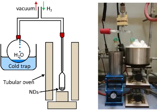
Manually milled NDs powder (40 - 50 mg) was placed in a quartz tube and transferred into the microwave waveguide cavity connected to a microwave generator working at 2.45 GHz produced by Sairem (model: GMP G3, 2 kW) industry. The quartz tube (HSQ300) used is transparent to microwaves and resistant to very high temperatures without degradation. After closing the tube, pumping of the system was carried out for 1 h (down to 1 x 10⁻² mbar).
2.2 Surface modifications of detonation nanodiamonds
to decompose the H2 gas into highly-reactive atomic hydrogen under plasma conditions.
When the plasma conditions were stable (no changes in visual appearance, temperature, density or electric field), hydrogenation of NDs was continued for 20 min followed by 10 min of exposure to hydrogen flow. Post-plasma hydrogen treatment helps to saturate the dangling bonds at the surface of NDs with hydrogen while reducing the possible recombination with oxygen after exposure to air. After the MPCVD treatment, the quartz tube was removed from the microwave cavity and modified NDs were placed in a glass vial, closed and stored in ambient conditions. The same conditions were applied to all types of detonation NDs presented in the manuscript.
Figure 2.3 – Scheme (left) and picture (right) of the MPCVD experimental set-up used for
plasma hydrogenation of detonation NDs.
2.2.4 Thermal annealing under hydrogen
The thermal annealing under hydrogen constitutes an alternative to MPCVD to reduce oxygen groups present at NDs surface16,17,18. This technique is also used by colleagues from the
Tritium Labelling Laboratory (CEA Saclay), who aim to label NDs with hydrogen isotopes like deuterium and tritium19. While hydrogen annealing is usually performed under flow
conditions, they developed an alternative approach to realize the same treatment in a closed system for safety reasons. Hydrogen annealed NDs used in this manuscript were thus provided by Emilie Nehlig from the Tritium Labelling Laboratory. Here is a short description of the system:
Detonation NDs supplied by PlasmaChem and Adamas Technologies were manually milled and used for the thermal hydrogenation. 30 - 35 mg of dry NDs powder were treated each time and placed in a quartz flask. The vacuum was achieved for 3 h of dynamic pumping (1 x
Figure 2.4 – Scheme (left) and picture (right) of the experimental set-up used for thermal
hydrogenation of detonation NDs.
2.2.5 Vacuum annealing
The last type of modification was performed via annealing under secondary vacuum aiming to induce sp2 reconstruction on the surface of detonation NDs. PlasmaChem NDs (140 – 150
mg) were manually milled and placed in a small alumina crucible. The crucible was covered and transferred into a furnace equipped with a turbomolecular vacuum pump used for vacuum annealing (< 10-8 mbar) of a diamond. The vacuum chamber heats up due to a heating
element (brand: Ceramisis) in size of 2 inches made of graphite covered with a ceramic. The heating element temperature is measured by a thermocouple (range of -200 °C to +1350 °C) whereas the sample’s temperature is measured indirectly by an infrared pyrometer (brand: Impac, model: IGA 140) which operates between 250 °C to 1350 °C.
The crucible with NDs was placed on the heating plate next to the piece of silicon which allows the pyrometer to measure the temperature. The NDs were treated via annealing under vacuum (1.0 × 10⁻⁶ mbar) at various temperatures and time conditions: 1 h / 750 °C, 2 h / 750
°C, 1 h / 850 °C and 2 h / 850 °C. Similar studies were previously performed in LCD laboratory by T. Petit and co-workers (2011) investigating early stages of surface graphitization of detonation NDs. Such conditions assure decomposition of oxygen‐containing groups and CHx
groups which decompose at 300 – 900 °C and 700 – 1150 °C, respectively20.
After vacuum annealing, the chamber was brought back to ambient pressure, the NDs sample was cooled down and the crucible was transported into the fume cupboard. The dry powder
2.2 Surface modifications of detonation nanodiamonds
Figure 2.5 – Scheme (left) of the chamber and picture (right) of the experimental set-up used
for thermal annealing of detonation NDs under secondary vacuum.
2.2.6 Summary
Three sources of detonation NDs were modified toward surface chemical homogenization: PlasmaChem GmbH (commercial), Adamas Nanotechnologies and Nanocarbon Research Institute Co. (non-commercial). Air annealing, plasma and thermal hydrogenations, and vacuum annealing treatment were applied using specific conditions listed in the table below.
Surface modification Mass (mg) Time (min) Temperature (°C) Power (W) Pressure (mbar) H2 flow/pressure Air annealing ~150 mg introduced 30 mg collected
90 550 n/a n/a n/a
Plasma
hydrogenation 40 - 50 20 estimated 800
1 250 12 10 sccm
Thermal
hydrogenation 30 - 35 60 550 - 560 n/a 200-250 static
Vacuum annealing 140 - 150 60 750 n/a 1 × 10⁻⁶ n/a 120 750 60 850 120 850
Table 2.1 – Summary of treatment conditions used for surface modifications of detonation
2.3 Fourier Transform Infrared Spectroscopy
Fourier Transform Infrared Spectroscopy (FTIR) provides information about the surface chemistry of the investigated NDs. Their diamond core is transparent to infrared but their rich external chemistry ensures sensitivity to various functional groups (mostly oxygen and hydrogen related), exalted by their high surface area (> 350 m².g-1). With our chosen
experimental approach (transmission through KBr pellet), FTIR technique will only be used qualitatively. However, the technique itself is simple, fast and accurate, thereby allowing monitoring of changes in surface chemical compositions after several different chemical and physical modifications.
The FTIR spectra were measured in transmission mode using a ThermoNicolet 8700 spectrometer. KBr pellets (~150 mg) were prepared with ca. 2 wt.% of NDs, dried or not within the spectrometer by the N2 flow (for technical details see Appendix, section A.6).
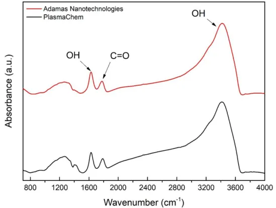
2.3.1 As-received detonation nanodiamonds
Three sources of detonation NDs were characterized via FTIR spectroscopy at room temperature, such as PlasmaChem, Adamas Nanotechnologies, and NanoCarbon Research Institute.
Figure 2.6 – Absorbance spectra of native PlasmaChem (black), Adamas Nanotechnologies
2.3 Fourier Transform Infrared Spectroscopy
To begin with, as the samples were not dried, the signature of adsorbed water on the NDs or in the KBr is clearly visible on all samples with the two contributions of OH stretching and bending modes at 1635 cm-1 and around 3400 cm-1, respectively. The effect of drying will be
presented in the next paragraph.
Apart from water-related bands, some differences between various sources can be observed. Concerning the fingerprint area between 800 and 1400 cm-1, which correspond to the
superposition of structural defects and diverse contributions including C-O related groups like ethers (around 1100 cm-1) or alcohols (1200 - 1300 cm-1), the NDs provided by Adamas differ
slightly compared to the two other sources.
More important is the area above 1635 cm-1 with the contributions related to C=O stretching
modes, associated with ketones, carboxylic acids or anhydrides functionalities. Downshifted to 1720 cm-1 and of weak intensity for particles provided by NanoCarbon Research Institute,
this band is clearly visible and of the same intensity at 1750 cm-1 for particles provided by
PlasmaChem and Adamas Nanotechnologies.
The region between 2800 - 3000 cm-1 with multiple-bands appears for PlasmaChem and
NanoCarbon Res. Inst. but seems not to be present for Adamas particles. These bands come from C-HX stretching modes21,22 of C-H2 and C-H3 and can be attributed to alkyl functionalities
at the surface of the NDs, or surface contamination with amorphous carbon. Note that the presence of water in the sample and the related OH stretching modes around 3400 cm-1
partially hides this part of the spectrum.
To sum up, all sources of detonation NDs have oxygen-containing groups on the surface, in different proportion, which can be seen either in the fingerprint area or around 1750 cm-1. In
addition to that, the PlasmaChem and NanoCarbon Research Institute exhibit C-Hx features
which are not observed for ND particles from Adamas Nanotechnologies.
2.3.1.1 Effect of sample drying
FTIR spectra of NDs often have a high content of water which is quickly adsorbed on the surface when particles are exposed to air atmosphere23,24. The high content of adsorbed
water on the surface can easily disturb or mask the signal coming from other surface functional groups not related to H2O. Even though the affinity toward water molecules of NDs
is high, adsorbed water layer can be progressively reduced via drying procedure which leads to its desorption. A very simple method of drying of the FTIR samples is to keep them at room temperature under a flow of an inert dry gas (e.g. nitrogen25) for a few days. If water is not
completely desorbed by this mean, a spectrum is still ameliorated. Note that a more effective method will be to expose the KBr pellet in-situ at elevated temperature (above 25° C up to 80 °C) and under vacuum4. However, using this technique, some modification of the surface
temperature. The resulting spectrum is plotted together with the one obtained prior to the drying step.
Figure 2.7 – Absorbance FTIR spectra of native PlasmaChem NDs taken immediately after
pellet preparation (black) followed by drying under nitrogen flow (red). Normalization of the fingerprint area has been applied. Spectra were also background-corrected.
It can be noticed that after 48 h there has been a reduction in the signal related to the O-H stretch at 3400 cm-1 and the H
2O bending mode at 1630 cm-1. As a consequence, features
around 1730 - 1750 cm-1 (C=O stretching) and 2800 - 3000 cm-1 (C-H
x vibrations) are more
pronounced. The drying does not affect the fingerprint area (single bonds around 900 - 1500 cm-1) which are specific for each type of NDs (the peak around 2230 cm-1 is due to the
vibration of CO2)26.
For the following experiments, all samples were dried for at least 24 h under nitrogen flow at room temperature.
2.3.2 Air annealed nanodiamonds
The first modification of detonation NDs involved air annealing, performed on PlasmaChem and Adamas Nanotech. The pellets were left for drying inside FTIR spectrometer for 24 h
2.3 Fourier Transform Infrared Spectroscopy
Figure 2.8 – Absorbance FTIR spectra of air annealed PlasmaChem (black) and Adamas
Nanotechnologies (red) detonation NDs. Spectra were background-corrected.
At the first glance, it can be seen that the spectrum of the NDs produced by Adamas Nanotechnologies did not evolve strongly after oxidative treatment (Figure 2.8). The native particles already possess a high content of C=O groups (1730 - 1750 cm-1), as shown in the
first part of the FTIR characterization. The same features as described for the native sample, are still present on the surface of NDs, only the fingerprint area evolves slightly. In terms of PlasmaChem NDs, the suppression of the C-Hx vibrations region (2800 - 3000 cm-1) is observed
after thermal oxidation.
After further drying of PlasmaChem particles (Figure 2.9), the C=O stretching band (peak around 1730 - 1750 cm-1) is enhanced as compared to the water signature (OH bending at
1630 cm-1). Notably, some weak C-H vibrations (2800 - 3000 cm-1) can now be observed, but
much less intense than for native PlasmaChem NDs. The O-H stretching bands at 3400 cm-1
and 1630 cm-1 are present due to the use of an incomplete drying method. However, the
O-H stretch at 3400 cm-1 could also come from the COOH content. The multiple-vibration in the
fingerprint region 900 - 1500 cm-1 are observed whereas the peak at 1100 cm-1 can be related
Figure 2.9 – Absorbance FTIR spectrum of dried (48 h) air annealed PlasmaChem NDs.
Spectrum was background-corrected.
To sum up, air annealing of NDs reduces the C-Hx content originally present on the native NDs.
Normalized to the fingerprint area, the amount of C=O does not seem to significantly increase. Thermal annealing does not modify the broad absorbance in the region 900 - 1500 cm-1
associated with the fingerprint (wavenumber < 1500 cm-1 representing bending vibrations
characteristic for the NDs). O-H groups are detected on the surface coming either from adsorbed water and hydroxylic groups (C-OH).
2.3.3 Plasma hydrogenated nanodiamonds
Plasma treatment of NDs was carried out for 20 min under hydrogen flow of 10 sccm and microwave generator power of 250 W (at 2.45 GHz). The detonation NDs provided by PlasmaChem, Adamas Nanotechnologies, and NanoCarbon Research Institute were modified under the same hydrogenation conditions. KBr pellets were prepared immediately after the modification and dried under nitrogen flow prior the FTIR analysis.
2.3 Fourier Transform Infrared Spectroscopy
Figure 2.10 – Absorbance FTIR spectra of plasma hydrogenated: PlasmaChem (black), Adamas
Nanotechnologies (red), and NanoCarbon Research Institute (blue) detonation NDs. Spectra were taken after drying for 24 h under nitrogen flow, and background-corrected.
At first glance, a few differences between detonation NDs are clearly noticeable (Figure 2.10). The C=O content (1730 - 1750 cm-1) is reduced for all samples. However, the Adamas samples
still have the highest remaining amount of C=O groups present on the surface. The C-Hx
features are observed for all samples, but they are partially masked by the water adsorbed on the surface, which is still present after 24 h of drying.
The PlasmaChem sample was left for further drying under nitrogen flow (Figure 2.11). Indeed, the high content of adsorbed water may partially mask the effect of plasma modification where the hydrogenation is mainly evidenced by the loss of C=O groups (1750 cm-1) and
exaltation of C-Hx stretching bands (2800 - 3000 cm-1)22. Further drying enhances the presence
of C-Hx groups and progressively reduces the O-H bands (3300 and 1050 cm-1). However, the
conditions used were not sufficient to completely remove the water content. These bands may also be related to the presence of hydroxylic groups on the surface27.
Looking at the spectrum of plasma H-NDs (PlasmaChem), two other features can be observed (Figure 2.11). Firstly, the OH band at 1630 cm-1 has a small shoulder around 1718 cm-1 which
distinctive than the one at 2879 cm-1 which is also in agreement with the previous study
focused on plasma hydrogenation mechanism4.
Figure 2.11 – Absorbance FTIR spectrum of dried (72 h) plasma hydrogenated PlasmaChem
NDs. The main bands of interest (oxygen and hydrogen related) are indicated on the spectrum.
In conclusion, plasma hydrogenation reduces the C=O content, whatever the initial source of detonation NDs. The same conditions of MPCVD treatment seem to be insufficient in terms of strongly oxidized Adamas particles (small amount of C=O around 1730 - 1750 cm-1 is still
present). The drying procedure reduces the adsorbed water content whereas the plasma treatment exalts the C-Hx stretching bands. The C-Hx band after plasma treatment has a
unique shape with two local maxima.
2.3.4 Hydrogen annealed nanodiamonds
The as-received PlasmaChem and Adamas Nanotechnologies NDs underwent another hydrogenation treatment via thermal annealing under hydrogen (1 h, 550 °C). The ND powder was mixed with KBr and left to dry for 48 hrs prior to the FTIR acquisition.
2.3 Fourier Transform Infrared Spectroscopy
Figure 2.12 – Absorbance FTIR spectra of hydrogen annealed PlasmaChem (black) and
Adamas Technologies (red) NDs. Samples were dried for 48 h under nitrogen flow and spectra were recorded at room temperature.
The main confirmation of successful modification is the almost complete reduction of C=O groups (1730 - 1750 cm-1) and the enhancement of C-H
x (C-H, C-H2, C-H3) bands (2800 – 3000
cm-1)18 (Figure 2.12). Moreover, the C-H
x bands exhibit two distinctive peaks: around 2879 cm -1 and 2925 cm-1, which have also been observed on plasma H-NDs8. They have previously been
associated with C(111) and C(100) surfaces, respectively. However, the shifted positions (2870 cm-1 and 2940 cm-1) may also come from size domain effect and polydispersity of
detonation NDs22. Another interesting observation can be made by looking at the shape of
the peak in the region between 1500 - 1750 cm-1 with 4 local maxima: 1579 cm-1, 1630 cm-1,
1660 cm-1, and 1695 cm-1. The latter can be attributed to remaining carbonyl or lactone
groups21,29, as previously seen for plasma treated samples. The peak located at 1630 cm-1 is
attributed to the OH bending modes of water, while the feature around 1580 cm-1 may be
associated with some C=C bonds30.
In conclusion, the thermal hydrogenation of detonation NDs results in the reduction of oxygen content (C=O peak) and the modification/increase of C-Hx bands. However, no obvious
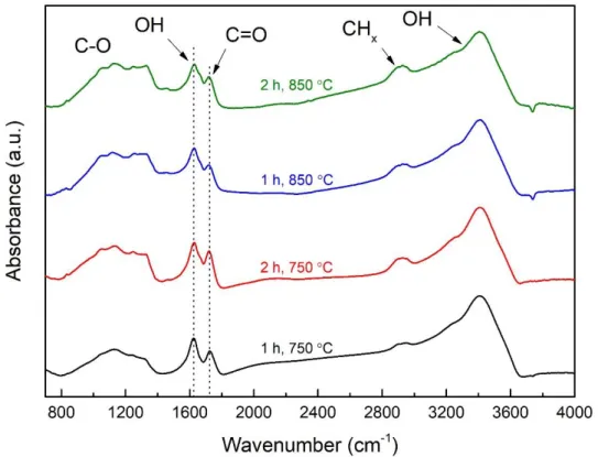
2 h / 850 °C. Immediately after modification, the dry powder was mixed with KBr (2 wt.%) and left for drying prior the FTIR analysis.
Figure 2.13 – Absorbance FTIR spectra of vacuum annealed PlasmaChem NDs for 1 h at 750
°C (black), 2 h at 750 °C (red), 1 h at 850 °C (blue), and 2 h at 850 °C (green). Samples were dried for 48 h under nitrogen flow. Spectra were background-corrected and normalized to the fingerprint area.
Looking at the FTIR spectra obtained, the chemical surface chemistry on NDs treated under vacuum remains similar for all samples (Figure 2.13). The features previously described on the native PlasmaChem NDs are still observed with C-O contributions between 800 and 1300 cm-1, C-H
x features between 2800 - 3000 cm-1, and C=O signature around 1730 cm-1.
Nevertheless, small modifications can be detected by looking at the intensity of the C=O related peak (normalized to the fingerprint), with a slight decrease according to the strength of the treatment. Smooth exaltation of the C-H related bands is also noticeable after treatment in the harshest conditions.
In the literature, it is well-established that graphitization of detonation NDs’ resulting in OLC structure takes place only when the temperature of thermal annealing under vacuum exceeds 1000 °C31. In our case, the temperature remains below 900 °C at which surface graphitic
reconstructions have been reported to affect only the outer shell32 while preserving the
2.4 Raman Spectroscopy
2.3.6 Summary
• Native detonation NDs have similar surface chemistry
• Sample drying under nitrogen flow reduces the water adsorbed on the surface of NDs • Air annealing reduces the C-Hx groups and slightly increases the surface coverage with
C=O, as seen for PlasmaChem and Adamas NDs
• Hydrogenation reduces significantly the C=O content. However, some small amount can still be detected when hydrogenation is performed at the same conditions on different sources of detonation NDs
• The shape of C-Hx bands is different for plasma and thermally hydrogenated samples
• Vacuum annealing below a temperature of 850 °C does not drastically change the surface chemistry of native PlasmaChem NDs
• The findings presented in this part of the chapter are consistent with previously published works on thermally Ox-NDs, plasma and thermally H-NDs as well the vacuum annealed NDs17,33,13,4.
2.4 Raman Spectroscopy
The Raman Spectroscopy is a complementary technique to FTIR providing information about the carbon phase composition (sp2-sp3 content), the crystalline structure, and the
homogeneity of carbon-materials34, e.g. nanodiamonds35. Surface modifications of NDs
toward homogenization change the composition of the surface functional groups and can lead to soft-shell reconstructions (change in the sp2-sp3 ratio)9. The experimental conditions
used for Raman analysis, which minimalizes any possible laser-induced effects (e.g. local sample heating and modification/damage due to high power density) of NDs, have been shown to be essential when monitoring the surface treatment of detonation NDs9. However,
this technique is not fully sensitive to surface-functional groups of NDs and exact interpretation of Raman peaks is not always straightforward after modifications34.
2.4.1 Surface-modified nanodiamonds
Raman spectra of as-received, air annealed (1 h 30 min, 550 °C), plasma hydrogenated (250 W, 12 mbar, 20 min) and hydrogen annealed (1 h, 550 °C) PlasmaChem (ultra-pure, grade G-02) NDs were recorded at room temperature. Ultra-violet (UV) laser excitation (325 nm, HeCd laser) with low power density (< 200 μW) was employed to avoid heating of NDs. Raman analysis was done by Michel Mermoux at LEPMI (Grenoble).