Dev Genes Evol (2005) 215: 165–176 DOI 10.1007/s00427-004-0464-7
O R I G I N A L A RT I C L E
Géza Burghardt . Monika Hediger . ChristinaSiegenthaler . Martin Moser . Andreas Dübendorfer . Daniel Bopp
The
transformer2 gene in Musca domestica is required
for selecting and maintaining the female pathway of development
Received: 4 November 2004 / Accepted: 8 December 2004 / Published online: 21 January 2005# Springer-Verlag 2005
Abstract We present the isolation and functional analysis of a transformer2 homologue Mdtra2 in the housefly Musca domestica. Compromising the activity of this gene by in-jecting dsRNA into embryos causes complete sex reversal of genotypically female individuals into fertile males, re-vealing an essential function of Mdtra2 in female develop-ment of the housefly. Mdtra2 is required for female-specific splicing of Musca doublesex (Mddsx) which structurally and functionally corresponds to Drosophila dsx, the bottom-most regulator in the sex-determining pathway. Since Mdtra2 is expressed in males and females, we propose that Mdtra2 serves as an essential co-factor of F, the key sex-determining switch upstream of Mddsx. We also provide evidence that Mdtra2 acts upstream as a positive regulator of F supporting genetic data which suggest that F relies on an autocatalytic activity to select and maintain the female path of development. We further show that repression of male courtship behavior by F requires Mdtra2. This function of F and Mdtra2 appears not to be mediated by Mddsx, suggesting that bifurcation of the pathway at this level is a conserved feature in the genetic architecture of Musca and Drosophila.
Keywords Transformer2 . Musca domestica . Sex determination . Splicing regulation
Introduction
Among insect species, a variety of seemingly different cues have been found to determine sex (Nöthiger and Steinmann-Zwicky 1985). In an attempt to understand how and why different cues have evolved, we are
con-ducting a comparative study between the sex-determina-tion pathway of Drosophila melanogaster and that of a distant dipteran relative, the housefly Musca domestica. In D. melanogaster, the ratio of X chromosomes to sets of autosomes (X:A ratio) serves as the primary signal in sex determination (reviewed in Cline and Meyer 1996; Parkhurst and Meneeley 1994; Schutt and Nothiger
2000). This signal defines the state of activity of Sex-lethal (Sxl), which is the top switch in the pathway. Sxl controls all aspects of sexual dimorphic development through a short cascade of subordinate genes. In XX:AA zygotes, Sxl is activated and keeps its active state by an autoregulatory function (Bell et al.1991). Its product, SXL protein, regulates the splicing of the transformer gene (tra), which results in the production of functional TRA protein (Inoue et al. 1990; Sosnowski et al. 1989). TRA, together with TRA2, a co-factor that is expressed in both sexes, promotes female-specific splicing of the bottom-most element in the pathway, dsx (Amrein et al. 1988; Baker and Wolfner 1988; Burtis and Baker 1989; Hoshijima et al.1991). As a consequence, female-specific dsx products (DSXF) are generated which act as tran-scriptional regulators to direct female differentiation. In X:AA zygotes, Sxl remains inactive and, in the absence of functional SXL products, tra pre-mRNA is spliced into the non-functional male mode. When TRA protein is absent, dsx is spliced into the male-specific mode and produces mRNAs that encode the male-specific activity (DSXM) of this gene. The pathway bifurcates downstream of tra as courtship behavior is controlled by a different target than dsx, namely by fruitless (fru). In the absence of an active TRA and TRA2 complex, male-specific splice variants of fru are produced which are essential for normal male courtship (Ito et al. 1996; Ryner et al.1996).
The Drosophila transformer2 gene encodes a protein with an RNA-recognition motif (RRM), flanked by two arginine-rich/serine-rich regions (RS-domains). It is a mem-ber of the family of splicing regulator proteins (SR-proteins) that control the utilization of alternative splice sites (Goralski et al.1989; Manley and Tacke1996). RS-domains mediate protein-protein interactions to facilitate Edited by D. Tautz
G. Burghardt . M. Hediger . C. Siegenthaler . M. Moser . A. Dübendorfer . D. Bopp (*)
Zoological Institute, University of Zürich, Winterthurerstrasse 190,
8057 Zürich, Switzerland e-mail: [email protected] Tel.: +41-1-6354869 Fax: +41-1-6356823
the formation of both spliceosomal and regulatory splicing complexes (Amrein et al.1994; Kohtz et al.1994; Wu and Maniatis 1993). In Drosophila females, TRA2 together with TRA and other RS-proteins build up a splice-enhancing complex that promotes the use of a 3′ splice site upstream of female-specific exon 4 of the dsx gene (Lynch and Maniatis1995; Tian and Maniatis1993). The TRA/TRA2 complex binds to six nearly identical copies of an exonic splicing enhancer (ESE) and a single purine-rich element (PRE) allowing the assembly of a functional spliceosome at the adjacent 3′ splice site (Lynch and Maniatis 1996; Tian and Maniatis 1993). Additionally, TRA2 is involved in 5′ splice site choice of fru pre-mRNA. Similar to dsx, this regulation depends on binding to a specific set of ESEs present in the fru pre mRNA (Heinrichs et al.1998; Lam et al.2003).
In M. domestica, sex is determined by the presence or absence of a dominant male-determining factor M (Perje
1948). When M is present in the zygote, it prevents the activation of the female-promoting F gene and thereby imposes male development. M is usually located on the Y chromosome, but it can also be found on different auto-somes in natural populations (Rubini et al. 1972). In the absence of M, F is active and directs female development (Dubendorfer et al.2002). F was mapped to chromosome 4 and is genetically defined by two mutant alleles. The gain-of-function allele FD behaves as a dominant female de-terminer since it cannot be repressed by M (McDonald et al.
1978; Rubini et al.1972). The recessive allele Fman, on the other hand, has features of a loss-of-function mutation. Fman homozygous individuals develop as males, even in the absence of M (Schmidt et al.1997). To become zygotically active, F requires its own maternal activity, suggesting that this gene relies on an autoregulatory function to maintain a female-promoting active state (Dubendorfer and Hediger
1998). Though F in many regards behaves as Sxl in Dro-sophila, the Musca homologue of Sxl is an unlikely candidate for F because it is equally expressed in both sexes (Meise et al.1998). It appears that the components at the top of the sex-determining pathway are different in Drosophila and Musca. However, the dsx homologue of Musca (Mddsx) corresponds not only structurally but also functionally to Drosophila dsx, the bottom most regulator in the pathway (Hediger et al. 2004). Furthermore, sex-specific regulation of Mddsx is based on a differential splicing mechanism that is very similar to the one described for Drosophila dsx. In Musca females, an alternative 3′ splice site is utilized in the Mddsx pre-mRNA (Hediger et al.
2004). The female-specific exon contains several putative ESEs and a PRE, suggesting that conserved trans-acting factors are involved in promoting the use of this female-specific acceptor site. This finding prompted us to isolate the Musca homologue of tra2 since this component of the complex is thought to interact directly with the ESEs and PRE (Lynch and Maniatis 1996). Furthermore, previous characterization of tra2 homologues in Drosophila virilis (Dvtra2) and in humans (tra2α and tra2β) suggested that this gene has been well preserved in structure and function
(Amrein et al.1988; Beil et al.1997; Chandler et al.1997; Dauwalder et al.1996).
We here present the isolation and characterization of the tra2 homologue of M. domestica (Mdtra2), and we pro-vide epro-vidence for an essential function of this gene in female development of the housefly, not only in regulating targets of F but also in maintaining the female state.
Materials and methods
PCR with degenerate primers
The 5′ primers correspond to sequences located in the middle of the RRM; 3′ primers correspond to sequences in the extended homology region downstream of the RRM. The following primers were used:
f orward MAR25 :
50 TGY CTI GGN GTN TTY GGS YT R 30 MAR5 :
50MGN TCI CGI GGN TTY TGY TTY R 30 reverse
MAR17 :
50GT RTG IGS ICG YTK NGT DAT NGA 30 Genomic DNA templates were prepared from adult males and females of a wild-type standard XX/XY strain. A first PCR round was performed with MAR25 and MAR17, followed by a second amplification with MAR5 (nested) and MAR17. We used standard concentrations of Mg and nucleotides. An amount of 50 ng DNA template was amplified in a total volume of 50μl with 100 pmol each primer. PCR conditions for first amplification were: denaturation at 95°C for 2 min, 35 cycles (denaturation at 95°C for 30 s, annealing at 42°C for 1 min and extension at 72°C for 1 min) and extension at 72°C for 5 min. Second amplification with nested primers involved: dena-turation at 95°C for 2 min, 5 cycles (denadena-turation 95°C for 30 s, annealing at 42°C for 1 min and extension at 72°C for 1 min), then 25 cycles (denaturation 95°C for 30 s, annealing at 62°C for 1 min and extension at 72°C for 1 min) and a final extension at 72°C for 5 min. Fragments from 100 to 700 bp were gel-eluted, some reamplified, using the same conditions as with nested PCR. Subcloning and sequencing of the candidate fragments were carried out by standard procedures.
Rearing of Musca strains
Strains were reared as described previously (Schmidt et al.
1997). Since small populations of larvae are difficult to raise on standard medium, larvae obtained from injected embryos were raised on pig dung.
Strains of M. domestica
(1) Wild-type strain: females XX, males XY; (2) autoso-mal M strain: feautoso-males XX; pw bwb/pw bwb, autoso-males XX; MIIIpw+bwb+/pw bwb; (3) FD strain: females MIII/MIII; FDBa/F+Ba+, males MIII/MIII; F+Ba+/F+Ba+; (4) Fman strain: females XX; Fman/F+, males XX; Fman/Fman (Schmidt et al. 1997); (5) Fman of FD strain: females XX; FDBa/FmanBa+, males FmanBa+/FmanBa+.
To obtain a pure female progeny, females of strain 1 were crossed to males of strain 4. Pure male progeny was ob-tained by crossing females of strain 1 with males of strain 3.
In situ hybridizations
In situ hybridizations were carried out according to the protocol of Tautz and Pfeifle (1989).
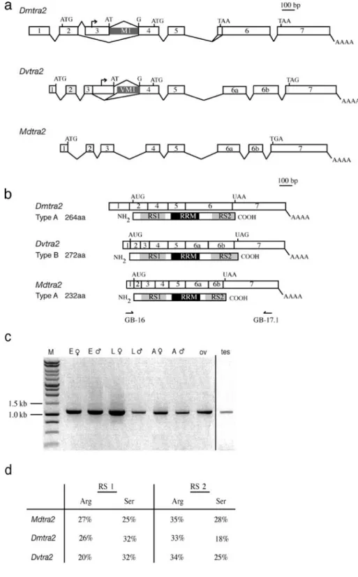
Fig. 1 tra2 genes in Drosophila melanogaster, D. virilis and Musca domestica. a Genomic organization of Dmtra2, Dvtra2 and Mdtra2. Testis-specific transcription start sites in Dmtra2 and Dvtra2 are marked by small arrows. M1 and VM1 are alternatively spliced introns. b Comparison of the structures of the major splice variant of Dmtra2 and Dvtra2 (Chandler et. al.1997) with the Mdtra2 transcript and the corresponding protein (below). The RNA-rec-ognition motif (RRM) is marked in black and the arginine-rich/ serine-rich (RS) domains are in grey. c RT-PCR amplifications with primers in exon 1 (GB-16 in b) and exon 7 (GB-17.1 in b) of Mdtra2 that encompass the complete ORF (E♀ female em-bryos, E♂ male embryos, L♀ female larvae, L♂ male larvae, A♀ female adults, A♂ male adults, ov ovaries, tes testes). d Percentages of arginine and serine residues present in the RS 1 and RS 2 domains of DmTRA2, DvTRA2 and MdTRA2
Injection of dsRNA and RT-PCR analysis
A cDNA fragment of the Mdtra2 gene was amplified with primers each flanked by T7 promoter sequences at the 5′ end. The resulting 560-bp fragment extends from 14 bp upstream to 385 bp downstream of the RRM. This template was used to produce dsRNA fragments by in vitro tran-scription with T7 RNA polymerase. The dsRNA was precipitated with ethanol and resuspended in injection buff-er to a final concentration of 1 μg/μl. Embryos were col-lected within 1 h of egg laying and dechorionated prior to injection (Hediger et al. 2001). Injected embryos were allowed to develop at room temperature.
For transcript detection by RT-PCR, total RNA of single adult flies was extracted according to the RNeasy Mini protocol of Qiagen. RT-PCR reactions were performed using the Titan One Tube RT-PCR Kit (Roche).
Results
Isolation of a tra2 homologue in Musca
A unique structural feature among previously isolated homologues of tra2 is the presence of an extended stretch of 57 bp of homology downstream of the RRM. We designed degenerate primers that hybridize within the RRM (5′) and
within the extended homology block (3′) and used these in various combinations to amplify genomic DNA from a wild-type laboratory strain of M. domestica. Two of 35 sequences recovered were identical and displayed the same type of extended RRM homology when compared to tra2 sequences of D. melanogaster (Dmtra2) and D. virilis (Dvtra2). We therefore referred to this sequence as Mdtra2. 5′ and 3′ RACEs on poly (A)+RNA prepared from Musca embryos resulted in the assembly of a transcript of 1.4 kb in length (GenBank Accession No AY847518). Genomic fragments covering the 1.4-kb transcribed region were isolated by screening a Musca genomic library and by PCR from genomic templates. From aligning genomic and cDNA sequences we deduced that the assembled Mdtra2 transcript is composed of eight exons (Fig.1a).
An alignment of genomic and cDNA sequences of Dmtra2, Dvtra2 and Mdtra2 revealed that at least two introns are located at the exact same position, namely between exons 4 and 5 and between exons 5 and 6 (Fig.1a). In addition, exon 6 of Dmtra2 is split by an intron in Dvtra2 and Mdtra2 at exactly the same location. We refer to these exons as 6a and 6b (Fig. 1a; Chandler et al. 1997). In Dmtra2, the translational start codon is located in exon 2, whereas in Dvtra2 the last three nucleotides of exon 1 represent the putative start codon for translation (Fig.1a). Likewise, we found that the upstream-most translational start site in the assembled Mdtra2 transcript is also located
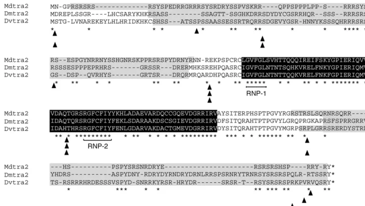
Fig. 2 Sequence alignment of the major protein variants of DmTRA2, DvTRA2 and MdTRA2. The RNA recognition motif (RRM) is in black; RS domains are grey. Asterisks indicate the positions of amino acids that are identical in all three sequences. Arrowheads below sequences show the position of introns. Note the
high amount of identical amino acids within the RRM, whereas both RS domains are less conserved but abundant in arginine and serine. RNP-1 and RNP-2 indicate the positions of two ribonucleoprotein identifier sequences, which are highly conserved between RRM proteins
at the very 3′ end of exon 1. Based on these features Mdtra2 seems to be structurally more closely related to Dvtra2 than to Dmtra2. Different from Dvtra2 and Dmtra2, however, we did not detect alternatively spliced products in Mdtra2. We also found no evidence for the presence of an internal testis-specific promoter (see below). It thus appears that regulation of Mdtra2 is less complex at the level of transcription and splicing than that of the corresponding gene in D. virilis and D. melanogaster (Fig.1a).
Mdtra2 produces a single transcript in soma and germ line of both sexes
In D. melanogaster, three distinct TRA2 protein variants (TRA2264, TRA2226 and TRA2179) arise as a result of alternative splicing and the use of different transcription start sites (Amrein et al.1990; Mattox et al. 1990). In the soma, TRA2264 and TRA2226 function redundantly to regulate female-specific splicing of dsx (Mattox et al.1996). In the male germline, TRA2226originates from a different transcription start site and is necessary and sufficient for
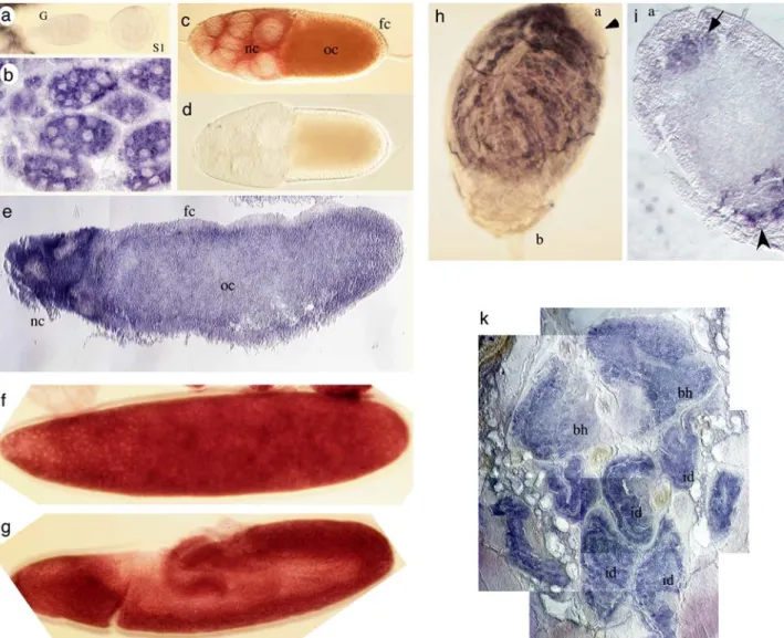
Fig. 3 Distribution of Mdtra2 transcripts in different tissues of Musca. a–e Localization of transcripts at different stages of oo-genesis. a Mdtra2 transcripts are not detected in the germarium (G) and in stage 1 cysts (S1). b Mdtra2 transcripts first accumulate in nurse cells at stage 4. c At stage 7, Mdtra2 transcripts accumulate in the cytoplasm of nurse cells (nc) and in the growing oocyte (oc), but are not detected in the somatic follicle cells (fc). d No signal is observed with a Mdtra2 sense probe at the same stage 7. e Section through a late egg chamber with uniform distribution of Mdtra2 transcripts in the oocyte (oc) and in the degenerating nurse cells (nc). f, g Distribution of Mdtra2 transcripts in embryos. High levels of uniform staining are found at the blastoderm stage (f) and at the
germband extension stage (g). h, i Distribution of Mdtra2 transcripts in adult testes. h Whole-mount staining of a testis: transcripts are prominently expressed in differentiating spermatids, but excluded from the area beneath the apical end which contains stem cells and early stages of spermatogenic development (a, arrowhead). j Section through a testis: transcripts are detected in groups of cells at the apical end (a) which are presumed to be clusters of primary sper-matocytes (arrow). At the basal end (b), transcripts are found in cells which are presumed to be somatically derived (arrowhead). k In situ hybridization on section through the anterior part of a third instar larva. High levels of transcripts are detected in cells of the brain hemispheres (bh) and in the epithelial cells of the imaginal discs (id)
male fertility, ensuring correct processing of exuperantia pre-mRNA (Hazelrigg and Tu 1994) and controlling the splicing of its own pre-mRNA (Mattox et al.1990).
In Mdtra2 only a single transcript of about 1.4 kb in size was detected in male and female embryos by northern blot analysis (not shown). Using a pair of primers in exons 1 and 7 (Fig. 1b), we tested cDNA samples prepared from dif-ferent tissues and stages for the presence of alternatively spliced products. A single amplification product was re-covered in all cases (Fig.1c). RT-PCR amplification with primers located at different positions in the intron between exon 3 and 4, which corresponds to M1 in Dmtra2 and VM1 in Dvtra2, did not yield any products in poly (A)+ samples of dissected testes. These results argue against the presence of male germline-specific transcripts in Mdtra2. Rather they suggest that Mdtra2 is expressed as a single transcript of 1.4 kb in all tissues. The longest ORF of this transcript encodes a protein of 232 aa and shows a best fit with the DmTRA2264and DvTRA2272isoforms (Fig.1b).
Alignment of MdTRA2232 protein sequence with DmTRA2264shows an overall similarity of 57%, and of 87% within the RRM. Two regions of low-complexity, RS 1 and RS 2 which are rich in arginine and serine, flank the RRM (Fig. 2). Most identities in these regions consist of repeated Arg/Ser residues (Fig.1d). Most importantly, the predicted MdTRA2 protein contains a highly conserved linker region between the RRM and the RS 2 domain (76% similarity). This structural feature appears to be specific to tra2 homologues within the class of RRM-containing genes.
Northern blot analysis and RT-PCR experiments in-dicated that Mdtra2 is transcribed continuously throughout the life cycle of the housefly. However, transcripts are more abundant in females and early embryos (data not shown), suggesting that there is a maternal deposit in embryos. This was tested by in situ hybridization with a DIG-labeled Mdtra2 RNA probe. We confirmed that levels of transcripts accumulate in previtellogenic and late stages of oogenesis and in early embryos (Fig.3a–g). The uniform expression pattern gradually declines during embryonic development. At late larval stages, we observe an accumulation of Mdtra2 transcripts specifically in cells of imaginal tissues and the CNS (Fig. 3k). In testes of the adult male, tran-scripts are prominently located in the compartment
con-taining differentiating germ cells (Fig.3h). Notably, tran-scripts are absent in the most apical region where germline stem cells and early dividing spermatogonial cells reside (arrow in Fig.3h).
Compromising Mdtra2 activity causes female-to-male sex reversal
In D. melanogaster, tra2 acts as an indispensable cofactor of TRA in the female-specific splicing of dsx (Tian and Maniatis 1993). Animals with a female XX genotype are sex reverted and develop into sterile males when mutant for tra2 (Amrein et al.1990). We tested the function of Mdtra2 in Musca by applying RNAi (McGregor et al. 2001). We used a 384-bp cDNA fragment derived from an embryonic Mdtra2 cDNA clone to synthesize Mdtra2 dsRNA. This fragment includes the RRM, the linker region and part of the RS 2 domain and was injected into pre-blastoderm stage embryos of the autosomal M strain (strain 2, see
Materials and methods) which allowed us to distinguish between genotypically female and male animals. The male-determining factor M on chromosome 3 is linked to the dominant wild-type alleles of bwb (brown body) and pw (pointed wings) whereas the non- M carrying chromosome 3 is marked with mutant alleles of these loci. Since cross-ing-over is virtually absent in male flies, these phenotypical markers are reliable indicators for presence or absence of M, i.e., for the genetic sex: males (M pw+bwb+/pw bwb) have a wild-type body color and normal shaped wings, whereas females (pw bwb/pw bwb) are brown and have pointed wings.
Of about 800 injected embryos, 34 individuals hetero-zygous and 35 homohetero-zygous for bwb and pw survived to adulthood. All 34 heterozygous flies (male genotype) had a normal male phenotype (Table1). Single fertile flies of this type when crossed to untreated bw pw females produced offspring with a normal 1:1 sex ratio, confirming the presence and transmission of a functional M factor. Of the 35 bwb pw mutant survivors (female genotype), 20% displayed male-like external genitalia and eye-distance (Fig.4g, h). Of the bwb pw individuals 68% were intersex-ual composed of a mixture of male and female structures (Fig.4e, f) and only 11% developed into normal-looking
Table 1 Sexual phenotypes of Mdtra2 dsRNA injectees (N.D not determined)
a
N Total number of flies examined
b
Flies with male phenotype were tested for presence of M by crossing to wild-type females (with M mixed progeny, without M female only progeny)
c
ix Intersexual composed of male and female structures
Genotype Percentage (Na) Interocular width Genitalia Gonads Sex of progenyb
bwb pw/bwb pw (female) 20(7) ♂ ♂ Testes ♀ only
46(16) ♀ ♂ Testes ♀ only
11(4) ♀ ixc N.D.
3(1) ♂ ix N.D.
8(3) ♂ ♀ Ovaries
11(4) ♀ ♀ Ovaries
+ + M/bwb pw (male) 100(34) ♂ ♂ Testes ♀ and ♂
XY 42(30) ♂ ♂ Testes ♀ and ♂
XX 18(13) ♂ ♂ Testes ♀ only
XX 37(27) ♀ ♂ N.D. N.D.
fertile females. In single crosses with untreated bwb pw females, all male-looking bwb pw flies and even 62% of the intersexes with male genitalia produced offspring (Table1). All crosses yielded exclusively female progeny. This result clearly demonstrates that these bw pw males and intersexes did not carry an M and therefore had a female genotype.
The masculinizing effect of Mdtra2 dsRNA was also confirmed in an unmarked standard XX/XY strain. After injections in embryos of this strain, we again recovered a high percentage of intersexual flies and phenotypic males. Only 3% displayed a normal female morphology. To test for the presence of M, which is located on the Y chro-mosome in this strain, we crossed the male flies to untreated XX females. Thirteen of 43 phenotypic males produced only daughters indicating that they had a female XX genotype (Table1). On the other hand, no phenotypic abnormalities were observed in genotypically male flies that were injected with Mdtra2 dsRNA. We thus conclude that Mdtra2 is essential for female development.
Mdtra2 is required for female splicing of Mddsx
In D. melanogaster, TRA2 is required for female-specific processing of dsx pre-mRNA. Given the similar mechanism
by which Mddsx is regulated in the housefly (Fig.5A), we tested for presence of female-specific and male-specific Mddsx mRNAs in animals in which Mdtra2 was silenced by RNAi. In untreated bw pw mutant females, only female-specific messages of Mddsx are detectable (Fig.5B). Upon injections of Mdtra2 dsRNA, however, the sex-reverted bwb pw flies contain not only female-specific messages, but also a substantial amount of male-specific Mddsx mRNA (Fig.5B). These male-specific transcripts must have arisen from a failure to effectively utilize the splice acceptor site of the female-specific exon. This situation compares to that in Drosophila XX animals with impaired tra2 function (Fortier and Belote 2000). Sex reversion of houseflies with compromised Mdtra2 activity can thus at least in part be explained by a failure in female processing of Mddsx. The degree of sexual transformation in these flies is much stronger than that observed in flies injected with Mddsx dsRNA where only the gonads were affected (Hediger et al.
2004). This suggested to us that Mddsx is not the only target of Mdtra2 in the sex determination pathway.
Mdtra2 is a positive regulator of F
F has been proposed to be an upstream regulator of Mddsx (Hediger et al.2004). It is conceivable that, like tra and tra2 Fig. 4 Sex reversion caused by injections of Mdtra2 dsRNA into
embryos. The male determining M factor is autosomal and linked to brown body+(bwb+). Genotypic males have a normal black body pigmentation. Females are bwb/bwb and have a brownish body color. Phenotypically, male heads can be distinguished from female heads by a significantly narrower interocular distance. The external female genitalia are characterized by the presence of an ovipositor (op), whereas males have a darkly pigmented copulatory apparatus and exhibit characteristic horn-like structures (h) at the tip of sternite 5. a Head of a non-injected female (bwb/bwb). b Genital region of
the same female with a stretched ovipostitor (op). c Head of a non-injected male (M bwb+/bwb). d The genital region which display a pigmented copulatory apparatus (white arrowheads) and two horn-like structures at the tip of sternite five (h). e Head of an injected genotypically female displaying a male interocular distance. f Genitalia of the same female are composed of a partial ovipositor (op*) covered with male-like tissue (white arrowhead) and a horn-like structure (h) on sternite 5. g, h An RNAi-treated individual with a female genotype and a normal male morphology of the head and a male-like genital region
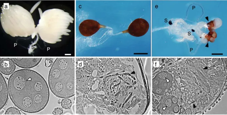
in Drosophila, F and Mdtra2 act cooperatively to set Mddsx into the female mode. To determine the level at which Mdtra2 acts relative to F, we silenced Mdtra2 in a strain carrying a gain-of-function allele of F, FD. Males of this strain are homozygous for F+and M (M/M; F+/F+). Zygotes that carry one copy of FDdevelop into fertile females even in the presence of two Ms (M/M; FD/F+). The FDallele in this strain is tightly linked to the dominant marker Ba (Bald abdomen). We injected the same dsRNA fragments of Mdtra2 into 500 embryos of which 71 survived to adulthood; 33 of these were Ba (M/M; FD/F+) and developed into normal-looking females; 38 were Ba+and were males (M/ M; F+/F+). Different from the previous silencing experi-ments, we could not observe any male structures in the dimorphic regions of the genotypically female class of survivors. Most of these females, however, were sterile and laid no eggs. Upon examination of their gonads, we observed that only 2 of 24 contained normal ovaries with mature oocytes, whereas the remaining had ovotestes (Fig.6e). These ovotestes were much smaller in size and typically contained an apical cap of pigmented testicular-like sheath. Shape and morphology of the ovotestes were similar to those observed in flies with compromised Mddsx activity (Hediger et al. 2004). We found sperma-togenic stages beneath the pigmented sheath (Fig. 6f). As the sexual fate of germ cells in Musca is solely dictated by the sex of the surrounding gonadal tissue, the occur-rence of male differentiated germ cells is a strong indi-cation that the overlying gonadal soma was transformed into testicular tissues (Hilfiker-Kleiner et al. 1994).
In contrast to the autosomal M and XX/XY strains used in the previous experiments, the FD strain appeared to be resistant to the masculinizing effect of Mdtra2 RNAi in non-gonadal soma. We infer from this result that, although the wild-type allele of F requires functional Mdtra2 to
maintain its female-promoting activity, the dominant FD allele does not depend on Mdtra2 to direct female dif-ferentiation in non-gonadal soma. This conclusion places Mdtra2 as a positive regulator upstream of F. On the other hand, the masculinized gonadal phenotypes in FDfemales with compromised Mdtra2 activity suggest that Mdtra2, at least in this tissue, acts downstream or in parallel to F.
Suppression of male courtship requires Mdtra2
The allele Fmanhas been described as a hypomorph of F (Schmidt et al.1997). Flies homozygous for this allele and with no M develop into males but 40% of these males produce no progeny. This sterility is not due to defects in spermatogenesis or abnormal gonadal or genital develop-ment, but to poor performance of these males to engage in courtship and copulation (S. Kaeppeli, unpublished results). This behavioral defect is aggravated when Fman homozy-gous males derive from a mother that carries a FDallele. All of her sons are behaviorally sterile as they make no attempts to copulate. A likely explanation is that courtship behavior of these males is suppressed by a residual female-promoting activity of Fman the level of which depends on maternal activity of F. Consistent with this presumption is the finding that the introduction of an M factor into Fmanhomozygous males renders them fertile.
If male courtship behavior is suppressed by F, it is conceivable that this process also requires functional Mdtra2. We thus injected Mdtra2 dsRNA into embryos derived from a cross between FD/Fmanmothers (strain 5) and Fman/Fmanfathers (strain 4). The surviving Fman/Fman males were tested for fertility in single matings with XX wild-type females. Twenty-one of 31 tested males (68%) sired progeny and thus were apparently fully rescued in Fig. 5 Mdtra2 is required for
female splicing of Mddsx. A Genomic organization of Mddsx (Hediger et al.2004); male-specific splice pattern is indicated in blue and the female-specific splice pattern in red. Arrows mark the positions of primers used below. B RT-PCR amplifications with male-specif-ic set (1/3) and female-specifmale-specif-ic set (1/2) of Mddsx primers. RNA preparations from a control adult female (non-injected bwb/bwb animal) produced only a product with the female-specific set (1/2). RNAi-treated bwb/bwb individuals: fly#14 had a male outer morphology, fly#20 dis-played a male genital region and a female interocular dis-tance. Preparations of RNA from both adult flies yielded a substantial amount of male-specific products
their capacity to copulate with females. This number is only mildly lower than the average 80% of successful matings of injected control wild-type males in single crosses. We conclude that repression of courtship behavior in Fman homozygous males requires the activity of Mdtra2. This result suggests that, besides an essential role in female differentiation, Mdtra2 is also engaged in the control of sex-specific behavior to fully implement the female program of development.
Discussion
Mdtra2 is essential for female development in Musca In Drosophila, the tra2 gene plays an important role in female development. It assists the tra gene in setting downstream regulators of the sex determination pathway into the female mode. Two direct targets, dsx and fru, are known, and their regulation by tra and tra2 has been studied in detail (Hedley and Maniatis1991; Heinrichs et al.1998; Hertel et al.1996; Lam et al.2003; Ryner and Baker1991; Tian and Maniatis1993). A structural and functional homo-logue of one of these targets in Musca, Mddsx, is sex-specifically regulated in a mode very similar to that of dsx in Drosophila. We examined whether the trans-acting factors involved in this process are conserved in the housefly. We describe the isolation and characterization of a tra2
homo-logue in Musca. Like its counterpart in Drosophila, it is needed for female-specific processing of Mddsx. Whether Mdtra2 participates directly or indirectly in this process remains to be examined, but the presence of well conserved TRA/TRA2 binding sites in the female-specific exon of Mddsx pre-mRNA favors a direct participation (Hediger et al. 2004). In Drosophila, these sequences serve as binding sites for the TRA/TRA2 splice-enhancer com-plex. It is thus conceivable that recognition of the female-specific 3′ splice in Mddsx also requires the assistance of a MdTRA/TRA2 splice-enhancer complex that binds to these sites. The structural features of MdTRA2 are con-sistent with such a mode of operation. It contains a well-conserved RRM which is likely to bind to the same type of ESE, as well as RS domains which provide a potential surface for interaction with other RS-containing proteins. Since Mdtra2 is equally expressed in females and males, this gene is an unlikely candidate for being the discrimina-tory component in the complex that determines the female fate. Rather its mode of action may correspond to that of Drosophila tra2, namely to act as an essential co-factor in the complex. A more likely candidate for the discriminatory factor in the complex is the product of the F gene. We have previously shown that F acts upstream of Mddsx (Hediger et al.2004), since Mddsx is spliced in the female mode only when F is active. If Mddsx is a direct target, F would functionally correspond to tra in Drosophila.
Fig. 6 Mdtra2 is required for female differentiation of the gonads. a Ovaries from a non-injected FD/F+female with fully differentiated eggs (P parovaria, S spermathecae). b Section through the ovary displays several cysts containing polyploid nuclei of nurse cells (white arrowhead). c Whole-mount of a pair of testes from a male of the same strain. Testes are pear-shaped and covered by a darkly pigmented epithelial sheath. d In a section through the testis, bundles of elongating spermatids can be recognized (arrowhead).
e Ovotestes of a FD/F+female treated with Mdtra2 RNAi. Some female structures (parovaria and spermathecae) are still present, but the gonads are greatly reduced in size and covered by a darkly pigmented male-like epithelial sheath. f Microscopic section through the ovotestis reveals the presence of both nurse cell-like nuclei (white arrowheads) and elongated bundles of spermatids (back arrowheads)
A dual role for Mdtra2 in female development
Activation of the F gene in the zygote depends on its own maternally provided activity (Dubendorfer and Hediger
1998). Once F is activated, it must be continuously active to keep the cells on the female pathway; removal of its activity at later stages of development leads to male development (Hilfiker-Kleiner et al.1993). Based on these findings it has been proposed that F relies on a feedback mechanism to maintain its female-promoting activity (Dubendorfer et al.
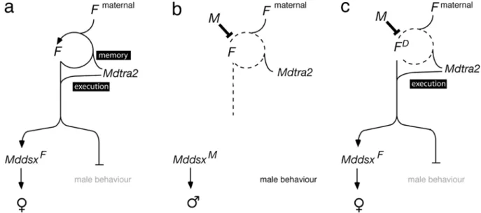
2002). As for the regulation of Mddsx, Mdtra2 may be an essential co-factor for the proposed auto-regulatory func-tion of F. A maternal contribufunc-tion of Mdtra2 to the activation of F in the zygote is suggested by the large amounts of Mdtra2 transcripts which are deposited into the egg chamber during oocyte maturation. In our model, this initial supply of Mdtra2 along with maternal F is needed to activate zygotic F (Fig.7a). The female-promoting activity by F is then sustained by a feedback loop which serves as a cellular memory for the proper execution of the female program. In the male zygote, establishment of this loop is prevented by the action of the paternally transmitted M factor and, as a result, male development follows (Fig.7b). Silencing of Mdtra2 by RNAi mimics the loop-breaking effect of M and results in complete masculinization. We explain this by the loss of Mdtra2 activity to form an active complex with maternal F that activates zygotic F. Conse-quently, the feedback loop of F cannot be engaged, and F remains functionally off.
The finding that the female-promoting activity of FD is neither repressed by M nor by Mdtra2 silencing suggests
that the FDallele does not depend on a feedback mechanism to produce active F products (Fig.7c). Rather, the gain-of-function nature of this allele seems to be based on con-stitutive expression of F activity independently of Mdtra2 or its own maternal activity (Dübendorfer and Hediger
1998). The gonadal soma in these FDfemales, however, is affected and becomes transformed into testes. The same phenotype can also be observed in standard XX individuals when Mddsx is downregulated by RNAi, suggesting that male transformation of gonadal soma in FD females with compromised Mdtra2 activity is caused by misregulation of Mddsx. This leads to the conclusion that Mdtra2 has two genetically separable functions: (1) upstream of F as a co-factor of the autocatalytic activity of F, and (2) parallel to F as a co-factor for the regulation of downstream targets such as Mddsx (Fig.7).
A very similar situation was described in the Mediterra-nean fruitfly, Ceratitis capitata. Pane et al. (2002) reported that the tra homologue in the Medfly plays a key role in sex determination based on an autoregulatory function that safeguards the propagation of the female determined state. The Ceratitis tra gene is regulated at the level of differential splicing and contains multiple TRA/TRA2 binding sites. The authors propose that the SR protein produced by Cctra imposes the female splicing mode on its own pre-mRNA and that of its target, the dsx homologue Ccdsx. Though the contribution of a putative Ceratitis homologue of tra2 in this process has not yet been studied, this modus operandi would be consistent with our model. It predicts that F, which functionally corresponds to Cctra, is directly reg-ulated at the post-transcriptional level by its own product
Fig. 7 Model for Mdtra2 in female development. a In the female zygote, Mdtra2 is required for autoregulation of the female-determining factor F. This autocatalytic loop of F is established early by maternally provided F product and by a maternal deposit of Mdtra2, and it is required to maintain the active female-determining state of F. Additionally, Mdtra2 is involved in the execution of the female pathway by assisting F in female-specific splicing of Mddsx and repressing male courtship behavior via a yet unknown target. b In the male zygote, the presence of a paternally transmitted male-determining factor M factor prevents the establishment of
the loop and F will remain in an inactive state. As a result, Mddsx will produce a male-specific activity that directs male differentiation and the branch that instates male behavior is derepressed. c The gain-of-function FD allele does not depend on the feedback loop to produce a female-promoting activity. Therefore, F and its co-factor Mdtra2 are dispensable in the activating process. M cannot prevent the execution of the female pathway in FD females, because it exclusively interferes with the activation process of F, but not with the downstream functions of F
and by Mdtra2. In this regard the sex-determining cascades of these two dipteran species appear strikingly similar. It supports the idea that Drosophila may be the exception in involving another level of control upstream of tra to memorize and execute the instruction given by the primary signal (Saccone et al. 2002). Though well-conserved ho-mologues of Sxl are present and expressed in the housefly and in the Mediterranean fruitfly, they appear not to play a role in sexual development (Meise et al.1998; Saccone et al. 1998).
Mdtra2 and courtship behavior
Besides the regulation of Mddsx and F, our study implicates Mdtra2 in the control of male courtship behavior. Our data suggest that Mdtra2, together with F, represses male courtship behavior. This type of control does not seem to be mediated by Mddsx, but by a different branch of the cascade. First, the hypomorphic activity of the Fman mutation impairs courtship behavior, but does not affect cuticular differentiation of homozygous males and, second, the splicing pattern of Mddsx is male-specific in Fmanmales (Hediger et al. 2004). In Drosophila, male courtship be-havior is under the control of tra but not of dsx. Instead, TRA in concert with TRA2 regulates splicing of a different target, namely of fru, a gene that is expressed in about 0.5% of the neurons of the central nervous system (Hall 1994; Ryner et al.1996; Taylor et al.1994). By a splice enhancer-dependent mechanism, the TRA/TRA2 complex activates a female-specific fru 5′ splice site and thereby prevents the production of male-specific fru transcripts which are essential for male courtship behavior (Lam et al. 2003). It is conceivable that a functional counterpart to fru exists in Musca that is regulated by F and Mdtra2. Likewise, this target may produce a male-specific activity that is essential for courtship behavior. The disturbed sexual behavior of Fman/Fmanmales may be caused by some residual activity of F which is still sufficient to repress the male mode of this CNS-specific branch of the cascade.
Acknowledgements We are deeply indebted to Dr. Rolf Nöthiger for his continuing support and for many stimulating discussions. We also thank him and Dr. Mary Bownes for helpful comments on this manuscript. Mark Robertson and Dr. Peter Atkinson (Department of Entomology, University of California, Riverside, USA) are gratefully acknowledged for providing degenerate primers and cloning strate-gies. We thank Simone Kaeppeli for sharing unpublished results. We thank Claudia Brunner for technical assistance and Johanna Nägeli and Raymond Grunder for Musca stock keeping. This work was supported by a grant of the Swiss National Foundation (31-67993.02).
References
Amrein H, Gorman M, Nöthiger R (1988) The sex-determining gene tra-2 of Drosophila encodes a putative RNA binding protein (published erratum appears in Cell 1989 Jul 28; 58(2): fol-lowing 419). Cell 55:1025–1035
Amrein H, Maniatis T, Nöthiger R (1990) Alternatively spliced transcripts of the sex-determining gene tra-2 of Drosophila encode functional proteins of different size. EMBO J 9:3619– 3629
Amrein H, Hedley ML, Maniatis T (1994) The role of specific protein-RNA and protein-protein interactions in positive and negative control of pre-mRNA splicing by Transformer 2. Cell 76:735–746
Baker BS, Wolfner MF (1988) A molecular analysis of doublesex, a bifunctional gene that controls both male and female sexual differentiation in Drosophila melanogaster. Genes Dev 2:477– 489
Beil B, Screaton G, Stamm S (1997) Molecular cloning of htra2-beta-1 and htra2-beta-2, two human homologs of tra-2 gen-erated by alternative splicing. DNA Cell Biol 16:679–690 Bell LR, Horabin JI, Schedl P, Cline TW (1991) Positive
auto-regulation of Sex-lethal by alternative splicing maintains the female determined state in Drosophila. Cell 65:229–239 Burtis KC, Baker BS (1989) Drosophila doublesex gene controls
somatic sexual differentiation by producing alternatively spliced mRNAs encoding related sex-specific polypeptides. Cell 56: 997–1010
Chandler D, McGuffin ME, Piskur J, Yao J, Baker BS, Mattox W (1997) Evolutionary conservation of regulatory strategies for the sex determination factor transformer-2. Mol Cell Biol 17:2908–2919
Cline TW, Meyer BJ (1996) Vive la différence: males vs females in flies vs worms. Annu Rev Genet 30:637–702
Dauwalder B, Amaya-Manzanares F, Mattox W (1996) A human homologue of the Drosophila sex determination factor trans-former-2 has conserved splicing regulatory functions. Proc Natl Acad Sci USA 93:9004–9009
Dubendorfer A, Hediger M (1998) The female-determining gene F of the housefly, Musca domestica, acts maternally to regulate its own zygotic activity. Genetics 150:221–226
Dubendorfer A, Hediger M, Burghardt G, Bopp D (2002) Musca domestica, a window on the evolution of sex-determining mechanisms in insects. Int J Dev Biol 46:75–79
Fortier E, Belote JM (2000)Temperature-dependent gene silencing by an expressed inverted repeat in Drosophila. Genesis 26:240– 244
Goralski TJ, Edstrom JE, Baker BS (1989) The sex determination locus transformer-2 of Drosophila encodes a polypeptide with similarity to RNA binding proteins. Cell 56:1011–1018 Hall JC (1994) The mating of a fly. Science 264:1702–1714 Hazelrigg T, Tu C (1994) Sex-specific processing of the Drosophila
exuperantia transcript is regulated in male germ cells by the tra-2 gene. Proc Natl Acad Sci USA 91:10752–10756 Hediger M, Niessen M, Wimmer EA, Dubendorfer A, Bopp D
(2001) Genetic transformation of the housefly Musca domestica with the lepidopteran derived transposon piggyBac. Insect Mol Biol 10:113–119
Hediger M, Burghardt G, Siegenthaler C, Buser N, Hilfiker-Kleiner D, Dubendorfer A, Bopp D (2004) Sex determination in Drosophila melanogaster and Musca domestica converges at the level of the terminal regulator doublesex. Dev Genes Evol 214:29–42
Hedley ML, Maniatis T (1991) Sex-specific splicing and poly-adenylation of dsx pre-mRNA requires a sequence that binds specifically to tra-2 protein in vivo. Cell 65:579–586
Heinrichs V, Ryner LC, Baker BS (1998) Regulation of sex-specific selection of fruitless 5′ splice sites by transformer and transformer-2. Mol Cell Biol 18:450–458
Hertel KJ, Lynch KW, Hsiao EC, Liu EH, Maniatis T (1996) Structural and functional conservation of the Drosophila doublesex splicing enhancer repeat elements. RNA 2:969–981 Hilfiker-Kleiner D, Dübendorfer A, Hilfiker A, Nöthiger R (1993) Developmental analysis of two sex-determining genes, M and F, in the housefly, Musca domestica. Genetics 134:1189–1194
Hilfiker-Kleiner D, Dubendorfer A, Hilfiker A, Nothiger R (1994) Genetic control of sex determination in the germ line and soma of the housefly, Musca domestica. Development 120:2531– 2538
Hoshijima K, Inoue K, Higuchi I, Sakamoto H, Shimura Y (1991) Control of doublesex alternative splicing by transformer and transformer-2 in Drosophila. Science 252:833–836
Inoue K, Hoshijima K, Sakamoto H, Shimura Y (1990) Binding of the Drosophila sex-lethal gene product to the alternative splice site of transformer primary transcript. Nature 344:461–463 Ito H, Fujitani K, Usui K, Shimizu-Nishikawa K, Tanaka S,
Yamamoto D (1996) Sexual orientation in Drosophila is altered by the satori mutation in the sex-determination gene fruitless that encodes a zinc finger protein with a BTB domain. Proc Natl Acad Sci USA 93:9687–9692
Kohtz JD, Jamison SF, Will CL, Zuo P, Luhrmann R, Garcia-Blanco MA, Manley JL (1994) Protein-protein interactions and 5 ′-splice-site recognition in mammalian mRNA precursors. Nature 368:119–124
Lam BJ, Bakshi A, Ekinci FY, Webb J, Graveley BR, Hertel KJ (2003) Enhancer-dependent 5′-splice site control of fruitless pre-mRNA splicing. J Biol Chem 278:22740–22747
Lynch K, Maniatis T (1995) Synergistic interactions between two distinct elements of a regulated splicing enhancer. Genes Dev 9:284–293
Lynch KW, Maniatis T (1996) Assembly of specific SR protein complexes on distinct regulatory elements of the Drosophila doublesex splicing enhancer. Genes Dev 10:2089–2101 Madigan SJ, Edeen P, Esnayra J, McKeown M (1996) att, a target
for regulation by tra2 in the testes of Drosophila melanogaster, encodes alternative RNAs and alternative proteins. Mol Cell Biol 16:4222–4230
Manley JL, Tacke R (1996) SR proteins and splicing control. Genes Dev 10:1569–1579
Mattox W, Baker BS (1991) Autoregulation of the splicing of transcripts from the transformer-2 gene of Drosophila. Genes Dev 5:786–796
Mattox W, Palmer MJ, Baker BS (1990) Alternative splicing of the sex determination gene transformer-2 is sex-specific in the germ line but not in the soma. Genes Dev 4:789–805 Mattox W, McGuffin ME, Baker BS (1996) A negative feedback
mechanism revealed by functional analysis of the alternative isoforms of the Drosophila splicing regulator transformer-2. Genetics 143:303–314
McDonald IC, Evenson P, Nickel CA, Johnson OA (1978) House fly genetics: isolation of a female determining factor on chromosome 4. Ann Entomol Soc Am 71:692–694
McGregor AP, Shaw PJ, Hancock JM, Bopp D, Hediger M, Wratten NS, Dover GA (2001) Rapid restructuring of bicoid-dependent hunchback promoters within and between Dipteran species: implications for molecular coevolution. Evol Dev 3:397–407 McGuffin ME, Chandler D, Somaiya D, Dauwalder B, Mattox W
(1998) Autoregulation of transformer-2 alternative splicing is necessary for normal male fertility in Drosophila. Genetics 149:1477–1486
Meise M, Hilfiker-Kleiner D, Dubendorfer A, Brunner C, Nothiger R, Bopp D (1998) Sex-lethal, the master sex-determining gene in Drosophila, is not sex-specifically regulated in Musca domestica. Development 125:1487–1494
Nöthiger R, Steinmann-Zwicky M (1985) A single principle for sex determination in insects, vol 50. Cold Spring Harbor Sympo-sium, Cold Spring Harbor, N.Y., pp 615–621
Pane A, Salvemini M, Delli Bovi P, Polito C, Saccone G (2002) The transformer gene in Ceratitis capitata provides a genetic basis for selecting and remembering the sexual fate. Development 129:3715–3725
Parkhurst SM, Meneeley PM (1994) Sex determination and dosage compensation: lessons from flies and worms. Science 264:924– 932
Perje A-M (1948) Studies on the spermatogenesis in Musca domestica. Hereditas 34:209–232
Rubini PG, Franco MG, Vanossi Este S (1972) Polymorphisms for heterochromosomes and autosomal sex-determinants in Musca domestica L. Atti IX Congr Naz It Entomol 9:341–352 Ryner LC, Baker BS (1991) Regulation of doublesex pre-mRNA
processing occurs by 3′ splice site activation. Genes Dev 5:2071–2085
Ryner LC, Goodwin SF, Castrillon DH, Anand A, Villella A, Baker BS, Hall JC, Taylor BJ, Wasserman SA (1996) Control of male sexual behavior and sexual orientation in Drosophila by the fruitless gene. Cell 87:1079–1089
Saccone G, Peluso I, Artiaco D, Giordano E, Bopp D, Polito LC (1998) The Ceratitis capitata homologue of the Drosophila sex-determining gene sex-lethal is structurally conserved, but not sex-specifically regulated. Development 125:1495–1500 Saccone G, Pane A, Polito LC (2002) Sex determination in flies,
fruitflies and butterflies. Genetica 116:15–23
Schmidt R, Hediger M, Nöthiger R, Dübendorfer A (1997) The mutation masculinizer (man) defines a sex determining gene with maternal and zygotic functions in Musca domestica L. Genetics 145:173–183
Schutt C, Nothiger R (2000) Structure, function and evolution of sex-determining systems in dipteran insects. Development 127:667–677
Sosnowski BA, Belote JM, McKeown M (1989) Sex-specific alternative splicing of RNA from the transformer gene results from sequence-dependent splice site blockage. Cell 58:449–459 Tautz D, Pfeifle C (1989) A non-radioactive in situ hybridization method for the localization of specific RNAs in Drosophila embryos reveals translational control of the segmentation gene hunchback. Chromosoma 98:81–85
Taylor BJ, Villella A, Ryner LC, Baker BS, Hall JC (1994) Behavioral and neurobiological implications of sex-determin-ing factors in Drosophila. Dev Genet 15:275–296
Tian M, Maniatis T (1993) A splicing enhancer complex controls alternative splicing of doublesex pre-mRNA. Cell 74:105–114 Wu JY, Maniatis T (1993) Specific interactions between proteins implicated in splice site selection and regulated alternative splicing. Cell 75:1061–1070