Cornputatlonal Modeling of Blast-Induced
Traumatic
Brainr
Injury
l
iyMicl le K. Nvein
S.B.. ('Cimstry. Maslmisetts
Instituteof 'ecniology (2004)
J.D..
H arvard Lniversity (20)7)
Sulnanit t"d
m the)Depart;w
of
Avrorautitxs and AsT ronamiCsill pnjrrl Fiillnenti of the reqluirLTlernts for the dcgree of
\wster
of
Sciute
at
r.hc
MASSA( "ILUSETTS [NSTIT'UTE OF TECH NO
XY
JImI
20
IMO
ARCGHVES
MASSACHUSETTS INSTMT-OF TECHNOLOYyCJUN
2 3 2010
LIBRARIES
Massah~use~ts
hittite
of TchnIh>y 2104. All rights rserved.A
i
T
... ...---AeTolni
1leNUS w1i1d AstmlUt itSMty
"1. 2010
Certified hv...
...
Rail A.
ILaZlvitzkv'
Assoriat e P'rofesor f
Aeroma
its amd A stronai uticsLILSiS Supervixor
/
/If
L iAccepteii 1). ...
-Associate Profes-sor of
Eltan H. Mruiijo
t/Croniaut
l-s and Ast-ronamicshair.
C4jmmitte
OTn radteStIudnts
Computational Modeling of Blast-Induced Traumatic Brain
Injury
by
Michelle K. Nyein
Submitted to the Department of Aeronautics and Astronautics on May 21, 2010, in partial fulfillment of the
requirements for the degree of Master of Science
Abstract
Blast-induced TBI has gained prominence in recent years due to the conflicts in Iraq and Afghanistan, yet little is known about the mechanical effects of blasts on the human head; no injury thresholds have been established for blast effects on the head, and even direct transmission of the shock wave to the intracranial cavity is disputed. Still less is known about how personal protective equipment such as the Advanced Combat Helmet (ACH) affect the brain's response to blasts. The goal of this thesis is to investigate the mechanical response of the human brain to blasts and to study the effect of the ACH on the blast response of the head. To that end, a biofidelic computational model of the human head consisting of 11 distinct structures was developed from high-resolution medical imaging data. The model, known as the DVBIC/MIT Full Head Model (FHM), was subjected to blasts with incident overpressures of 6 atm and 30 atm and to a 5 m/s lateral impact. Results from the simulations demonstrate that blasts can penetrate the intracranial cavity and generate intracranial pressures that exceed the pressures produced during impact; the results suggest that blasts can plausibly directly cause traumatic brain injury. Subsequent investigation of the effect of the ACH on the blast response of the head found that the ACH provided minimal mitigation of blast effects. Results from the simulations conducted with the FHM extended to include the ACH suggest that the ACH can slightly reduce peak pressure magnitudes and delay peak pressure arrival times, but the benefits are minimal because the ACH does not protect the main pathways of load transmission from the blast to brain tissue. A more effective blast mitigation strategy might involve altering the helmet design to more completely surround the head in order to protect it from direct exposure to blast waves.
Thesis Supervisor: Ra6l A. Radovitzky
Acknowledgments
I would like to thank my advisor, Prof. Radovitzky, for providing invaluable guidance and support throughout the years and for continually encouraging me to grow as a graduate student.
I am thankful for the financial support of the Joint Improvised Explosive Device Defeat Organization (JIEDDO) through the Army Research Office.
I would also like to thank Dr. David Moore for providing the geometry for the DVBIC/MIT Full Head Model, supplying material properties for the head structures, and generally serving as a font of medical knowledge.
I would like to express my deep appreciation for the members, past and present, of the RR Group. Dr. Antoine Jerusalem, in particular, patiently answered questions and provided much needed guidance when I first started working on this project. Other postdoctoral researchers in the group, including Dr. Ludovic Noels, Dr. Tan Bui, Dr. Claudio Pita, and Dr. Julian Rimoli, have been more than generous with their time, knowledge, and advice. And I am truly grateful to the other students in the RR Group, including Michael Tupek, Lei Qiao, Andrew Seagraves, Srikanti Rupa Avasarala, Brandon Talamini, Li Yu, Amanda Jason, Piotr Fidkowski, Riley Schutt, and Ganesh Gurumurthy, for their friendship, support, and encouragement.
Last, but certainly not least, I would like to extend my deepest, most heartfelt gratitude to David Carpenter, who has been by my side through every step of my journey through grad school. During even the most difficult times, he provided
Contents
1 Introduction
1.1 Blast-Induced TBI . . . .
1.1.1 Blast Injury Mechanisms . . .
1.1.2 Definition . . . .
1.1.3 Symptoms . . . .
1.1.4 Pathophysiology ... . . . .
1.2 Summary . . . .
2 Existing TBI Models
2.1 Impact-Related TBI . . . .
2.1.1 Cadaver Experiments . . . . .
2.1.2 Finite Element Head Models .
2.2 Blast-Related TBI . . . . 2.3 Summary . . . . 3 Modeling Framework 3.1 Computational Framework . . . . 3.1.1 Adlib . . . . 3.1.2 AMROC . . . . 3.1.3 Fluid-Structure Interaction . . 3.2 Mesh Generation . . . .
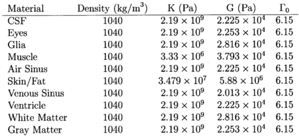
3.3 Material Models and Properties . . .
3.3.1 Hugoniot Equation of State .
13 . . . . . 17 . . . . 17 . . . - 19 . . . . 2 1 . . . . 23 . . . . 28 31 . . . . 3 1 . . . . 3 1 . . . . 33 . . . . 42 . . . . 46 49 . . . . 49 . . . .- 49 . . . . 54 . . . . 55 . . . . 55 . . . . 58 . . . . 59
3.3.2 Tait Equation of State . . . . 59
3.3.3 Deviatoric Elasticity . . . . 60
3.3.4 Material Properties . . . . 60
3.4 Summary . . . . 68
4 Full Head Simulations 69 4.1 Overview . . . . 69
4.2 6 atm Blast Simulation . . . . 70
4.3 30 atm Blast Simulation . . . . 73
4.4 5 m/s Impact Simulation . . . . 77
4.5 Comparison of Blast and Impact Simulations . . . . 81
4.6 Discussion . . . . 85
5 Helmet Simulations 87 5.1 Overview . . . . 87
5.2 Head-Helmet Model . . . . 88
5.2.1 Material Model and Properties . . . . 88
5.3 30 atm Blast Simulation . . . . 89
5.4 5 m/s Impact Simulation . . . . 98
5.5 Discussion . . . . 99
6 Conclusions 101 6.1 Future Work. . . . . 103
List of Figures
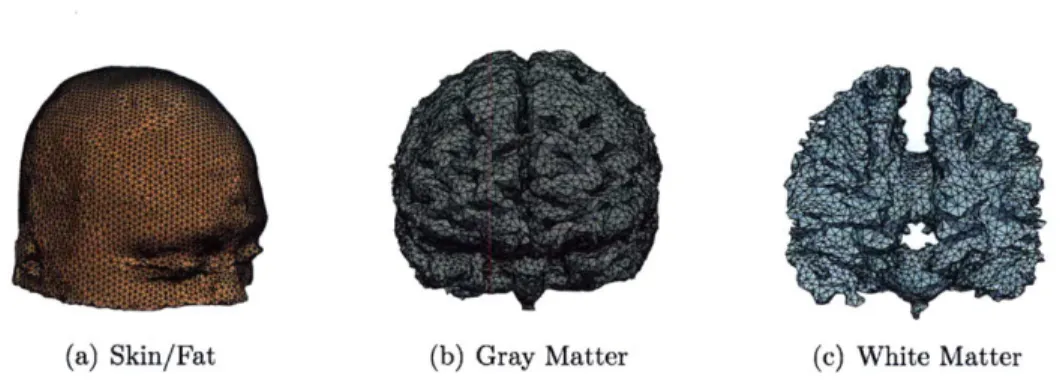
3-1 Detailed views of the skin/fat, gray matter, and white matter
struc-tures in the FHM . . . . 56
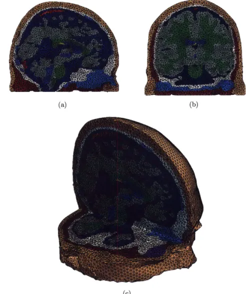
3-2 Full head model: sagittal cut 3-2(a), coronal cut 3-2(b), and combined sagittal and axial cut showing detail of the full mesh 3-2(c) . . . . 57
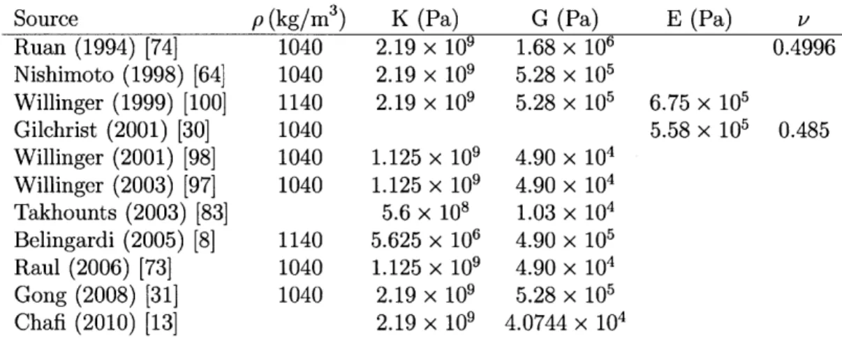
4-1 Snapshots of pressure from the 6 atm head blast simulation. The scale for the color bar is from -1.0 to 1.0 MPa . . . . 71 4-2 Pressure and Von Mises stress envelopes from the 6 atm head blast
simulation. The scale is from 0 to 17 MPa. . . . . 72 4-3 Snapshots of pressure from the 30 atm head blast simulation. The
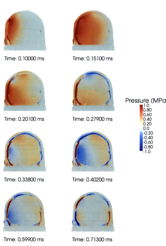
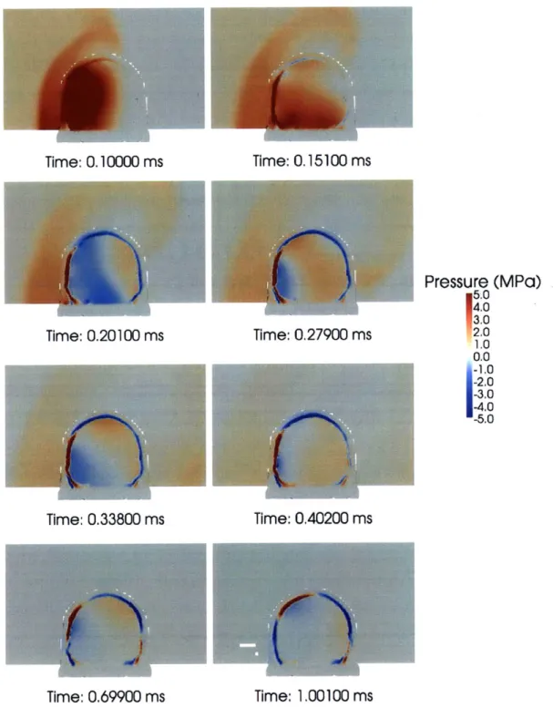
scale for the color bar is from -5.0 to 5.0 MPa . . . . 74 4-4 Pressure and Von Mises stress envelopes from the 30 atm head blast
simulation. The scale is from 0 to 120 MPa. . . . . 75 4-5 Snapshots of pressure from the 5 m/s head impact simulation. The
scale for the color bar is from -1.0 to 1.0 MPa. . . . . 78 4-6 Pressure and Von Mises stress envelopes from the 5 m/s head impact
simulation. The scale is from 0 to 50 MPa. . . . . 79 4-7 Comparison of pressure envelopes from the 6 atm, 30 atm, and 5 m/s
impact head simulations for gray matter, white matter, muscle, and
sku ll. . . . . 80
4-8 Pressure histories from the 6 atm, 30 atm, and 5 m/s impact head
4-9 Pressure histories from the 6 atm, 30 atm, and 5 m/s impact head
simulations at points located in the mid-sagittal plane. . . . . 84
5-1 Computational head-helmet mesh. . . . . 88
5-2 Snapshots of pressure from the 30 atm helmet blast simulation. The scale for the color bar is from -5.0 to 5.0 MPa. . . . . 90 5-3 Pressure contours from the 30 atm helmet blast simulation in
mid-coronal sections of the head. The scale for the color bar is from -5.0 to 5.0 M P a. . . . . 91 5-4 Pressure envelopes from the head and helmet 30 atm blast simulations.
The scale is from -1 to 41 MPa. . . . . 92 5-5 Von Mises stress envelopes from the head and helmet 30 atm blast
simulations. The scale is from 0 to 120 MPa. . . . . 93 5-6 Pressure histories from the head and helmet 30 atm blast simulations
at points in the mid-coronal plane. The scale is from -5 to 15 MPa. . 94 5-7 Pressure histories from the head and helmet 30 atm blast simulations
at points in the mid-sagittal plane. The scale is from -2.5 to 6 MPa. . 96 5-8 Snapshots of pressure from the helmet impact simulation. The scale
for the color bar is from -1.0 to 1.0 MPa. . . . . 97 5-9 Pressure envelopes from the head and helmet impact simulations. The
scale is from 0 to 22 MPa . . . . 98
5-10 Von Mises stress envelopes from the head and helmet impact simula-tions. The scale is from 0 to 50 MPa. . . . . 99
List of Tables
3.1 Hugoniot Equation of State and Deviatoric Elasticity Parameters . 60
3.2 Tait Equation of State and Deviatoric Elasticity Parameters . . . . . 61
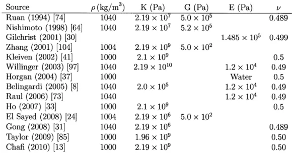
3.3 CSF Material Properties from the Literature . . . . 62
3.4 Brain Material Properties from the Literature . . . . 64
3.5 Gray Matter Material Properties from the Literature . . . . 64
3.6 White Matter Material Properties from the Literature . . . . 65
3.7 Skin-Related Material Properties from the Literature . . . . 66
3.8 Skull (Cortical and Trabecular Bone) Material Properties from the Literature . . . . 67
3.9 Cortical Bone Material Properties from the Literature . . . ... . . . . 67
3.10 Trabecular Bone Material Properties from the Literature . . . . 67
Chapter 1
Introduction
Traumatic brain injury (TBI) has been a major public health concern in the United States for decades; from 1995 to 2001, an average of 1.4 million Americans a year sustained a TBI, leading to 50,000 deaths, 235,000 hospitalizations, and 1.1 million emergency department visits [45]. Of these, an estimated 80-90,000 experienced the onset of long-term disability [45, 88]. These TBIs were largely caused by falls, motor vehicle crashes, sports collisions, and firearm assaults [45]. Even with an average of 1.4 million Americans sustaining a TBI each year, though, the average annual incidence of TBI was only 0.5% [45]. Since Operation Enduring Freedom (OEF) began in Afghanistan in October 2001 and Operation Iraqi Freedom (OIF) began in Iraq in May 2003, the incidence of TBI among U.S. service members has been significantly higher. Indeed, TBI has been labeled as the signature injury of OEF and OIF [34].
The U.S. Department of Defense has estimated that 10-20% of OEF/OIF Army and Marine Corps service members have sustained a mild TBI, based on data from 2004 to 2006 at selected military installations [65]. Other studies have found that any-where from 12 to 22.8% of OEF/OIF veterans have experienced TBIs. For example, a recent study found that 22.8% of soldiers in an Army Brigade Combat Team return-ing from Iraq had clinician-confirmed TBI [86]. A 2008 RAND survey of OEF/OIF veterans found that 19.5% had experienced a probable TBI during deployment; given that prevalence of TBI, it was estimated that approximately 320,000 OEF/OIF vet-erans could have experienced a TBI [84]. A 2006 study of U.S. Army infantry soldiers
3-4 months after their return from a year-long deployment to Iraq found that 15% reported an injury during deployment that involved loss of consciousness or altered mental status [34], and a 2005 survey of OEF/OIF veterans who had left combat theaters by September 2004 found that about 12% of the 2,235 respondents reported a history consistent with mild TBI [77]. Among those who have been medically evac-uated from theater, the proportion who have suffered a TBI is predictably higher; screening at Walter Reed Army Medical Center (WRAMC), for example, found that 30% of service members medically evacuated there from the OEF/OIF combat the-aters from January 2003 to June 2007 had sustained a TBI [65]. And of 50 OEF/OIF veterans treated at the Tampa Veterans Affairs Polytrauma Rehabilitation Center, 80% had incurred combat-related TBI, with 70% of the injuries caused by impro-vised explosive devices (IEDs) [14]. Given the high prevalence of TBI among U.S. service members, the costs of military-related TBI are enormous. The 2008 RAND study conservatively estimated the total cost for deployment-related TBI from 2001 to 2007 to be $554-854 million [84]. It was noted that the estimate likely understates the costs of deployment-related TBI, because while it includes the cost of treatment and reduction in productivity in the first year following injury and costs associated with mortality and suicide, it does not include cost of treatment or reduction in pro-ductivity beyond the first year, caregiver burden, TBI-related health problems, or downstream costs stemming from substance abuse, domestic violence, homelessness, and other factors [84]. While the precise cost of military-related TBI over the last decade remains unclear, it is apparent that a significant fraction of U.S. service mem-bers have sustained a TBI during OEF/OIF, and the costs associated with those deployment-related TBIs have been massive.
For active duty military personnel in war zones, blasts are the primary cause of TBI [84]. 68% of the OEF/OIF soldiers at WRAMC who screened positive for TBI had been injured by a blast [95], and data collected from March to September 2004 from Navy-Marine Corps medical facilities in Iraq found that IEDs were responsible for 62% of combat-related TBIs, even though 69% of the TBI patients had worn a helmet [27]. One reason blasts have become such a large cause of military-related TBI
is because the current conflicts in Iraq and Afghanistan involve battling insurgents who favor cheap, easily concealed explosive weapons capable of killing large numbers of people. As a result, approximately 60% of total combat casualties [49] and 67% of Army war zone evacuations [94] have been attributed to explosive blasts. The IED in particular has become the most common explosive weapon [49], accounting for about 40% of all casualties [84]. IEDs can be manufactured from materials ranging from 155 mm artillery shells to plastic explosives to barrels of gasoline [3]; they have evolved from relatively crude devices detonated by simple mechanisms to sophisticated devices capable of penetrating the armor of an M-1 Abrams tank [84]. Exposure to these blasts often leads to head and neck injuries. For example, 97% of the injuries in one Marine unit in Iraq were due to explosions (65% to IEDs), with 53% involving the head and neck [82], and a study of 4,831 patients at a U.S. Army echelon II medical facility in Iraq between October 1, 2003, and June 30, 2004, found that 88% of U.S military personnel treated had been injured by IEDs or mortars, with up to 47% of those injuries involving the head [60]. According to the Joint Theater Trauma Registry compiled by the U.S. Army Institute of Surgical Research, 22% of wounded OEF/OIF service members who passed through Landstuhl Regional Medical Center in Germany had injuries to the head, face, or neck [67]. It is likely that a significant number of these head injuries caused by explosions included blast-related TBIs (bTBIs).
Blast-related TBI has also gained visibility due to advances in military medicine and personal protective equipment (PPE) that allow U.S. service members to survive blasts that previously would have been fatal. In the current conflicts, the survival rates are dramatically higher than in previous conflicts. While 30% of wounded U.S. soldiers died in World War II and 24% of wounded U.S. soldiers died in the Viet-nam conflict, only about 10% of wounded U.S. soldiers have died in Afghanistan and Iraq [28]. The high survival rate can be attributed in part to significant advances in military medicine, including deployment of leaner, more mobile surgical teams far
forward on the battlefield and rapid medical evacuation from combat zones - the
average time from battlefield to arrival in the United States is now less than 4 days, compared to 45 days during the Vietnam conflict [28]. In addition, important
ad-vances in TBI treatment have been made, including early decompressive craniectomy, neurocritical care, cerebral angiography, transcranial Doppler, hypertonic saline, and TBI clinical management guidelines [49]. The high survival rate can also be attributed in part to advances in PPE, with improvements in body armor allowing soldiers to survive injuries that previously would have been fatal [67]. However, the improved protective gear do not necessarily prevent blast-related TBI; PPE have historically been designed to protect against ballistic impact, not blasts. Until recently, blast protection has not been a primary objective in the design of PPE, and any secondary blast protection provided has been fortuitous; the low incidence of blast lung nowa-days, for example, suggests that Kevlar vests are effective in preventing those types of injuries. The influence of other PPE, such as the Advanced Combat Helmet (ACH), on the brain's response to blast waves remains largely unknown.
TBI, and blast-related TBI in particular, has thus emerged as the leading injury among OEF/OIF service members [65]. Despite the importance of understanding blast-related TBI, however, little is known about the mechanical effect of blasts on the human head; no injury thresholds have been established for blast effects on the head, and even direct transmission of the shock wave to the intracranial cavity is disputed. Alternative mechanisms have been proposed, including acceleration of the head and transmission of the blast wave to the brain through a thoracic mechanism [17, 12]. Some have questioned the link between mild TBI and adverse physical health outcomes at all [34]. Additionally, it is not understood how the ACH affects the brain's response to blasts.
This thesis aims to investigate the mechanical response of the human brain to blast waves and to examine the effect of the ACH on that response through numerical simulation. The remainder of Chapter 1 will provide background on blast-related TBI, and Chapter 2 will discuss existing computational TBI models. Chapter 3 will present the modeling framework used in the blast-related TBI simulations; the computational code, full head model, material models, and material properties will be detailed. In Chapter 4, the results from the simulations will be presented and discussed, and it will be demonstrated that a blast wave can propagate through the cranial cavity
and result in stresses that match or exceed those observed in impact-related TBI. In Chapter 5, the results from simulations conducted with the full head model extended to include the ACH will be presented; it will be shown that the addition of the ACH minimally delays and reduces the magnitudes of stresses transmitted to the head by the blast wave. Chapter 6 will contain conclusions and comments on future work.
1.1
Blast-Induced TBI
1.1.1
Blast Injury Mechanisms
When an explosive device detonates, a chemical reaction occurs that rapidly releases gas and heat. The gas radially expands as a supersonic, high-pressure blast wave, and the leading edge of the blast wave compresses the host medium to create a shock front. For an ideal free-field explosion in air, the blast wave can be modeled as a Friedlander waveform, which is characterized by a rapid rise to peak pressure immediately followed by an exponential decay of the overpressure and a relatively prolonged underpressure [6]. If the blast wave encounters a solid object, such as a wall or other structure, it is significantly modified as it reflects from the object and diffracts around it; assuming that air behaves as an ideal gas, the overpressure of the reflected wave can be up to 8 times greater than the overpressure of the incident wave, and taking real gas effects into account, the reflected overpressure can be even larger, perhaps 20 or more times greater than the incident overpressure [6]. The three-dimensional fluid flow field characterizing an explosion can thus be very complex, and there is considerable room for variability in military-associated blast exposure.
Blasts can result in four types of injury: 1) primary blast injury, which results from the impact of the blast wave with body tissue; 2) secondary blast injury, which results from debris and shrapnel; 3) tertiary injury, which results from individuals being thrown by blast wind and impacting stationary objects; and 4) quaternary blast injury, which encompasses all other explosion-related injuries, including burns, inhalation injuries, crush injuries, and asthma [11]. While secondary, tertiary, and
quaternary blast injuries are not unique to blasts and have been studied extensively for decades, primary blast injury is not as well understood, particularly as it relates to TBI. The organs most susceptible to primary blast injury are the air-filled organs such as the ears, lungs, and gastrointestinal (GI) tract [25], with the most common injury being rupture of the tympanic membranes [63]. Although research efforts in prior decades focused on blast lung injuries, such injuries have occurred only infre-quently during OEF/OIF thanks to advances in body armor, and blast-related GI tract injuries have been even rarer [49]. Now, with soldiers surviving higher intensity blasts that previously would have been fatal due to lung or GI tract injury, focus has shifted to blast-related brain injury. The brain, residing in a fluid-filled cavity, is vulnerable to blasts [25], but little is known about how blasts affect the brain; no injury thresholds have been established, and direct transmission of the non-linear shock wave into the intracranial cavity has been disputed. Here, focusing solely on primary blast injury, we investigate the effects of blast waves on the human brain.
There is little question that exposure to blasts can harm the brain, as numerous animal studies have linked blasts to various manifestations of brain damage. One study of rats, for example, found that application of 12.5 MPa shock waves resulted in cerebral contusional hemorrhage associated with neuronal apoptosis, while appli-cation of 1.0 MPa shock waves resulted in mild morphological changes in neurons [39]. Another study found that rats subjected to 147 kPa air blasts suffered from prominent areas of cortical loss, gliosis, and infiltration, as well as hemorrhage and extensive necrosis [50], and a different study found that subjecting rats to 20 kPa blasts resulted in cortical neuron degeneration and significant impairment of perfor-mance on tests of coordination, strength, and startle response [57]. A study of pigs found that exposure to free air explosions with peak overpressure of 237 kPa resulted in transient flattening of the electroencephalogram (EEG) and short-lasting apnea, indicating a blast wave-induced effect on the brainstem or higher controlling center [80]. From these studies, it is evident that blasts can have a detrimental effect on the brain.
dis-pute. One proposed mechanism suggests that shock waves could directly propagate through the cranial cavity as stress waves, which travel around the speed of sound with high amplitude and can injure tissue through spalling, implosion, and pressure differentials [47]. A 1961 study exposing rhesus monkeys to air blasts found a large fraction of the pressure was transmitted into the brain through the skull, while little was transmitted from the torso to the brain [47]. Studies of rats and rabbits exposed to blasts also demonstrated that pressure waves could be transmitted directly to the brain with only slight change in amplitude [47]. Alternatively, it has been suggested that the central nervous system (CNS) could be injured by blasts through the cerebral vasculature via a thoracic mechanism [17]. Studies have shown that ballistic pressure waves, which are generated when a projectile enters a viscous medium, can cause remote injuries; for example, studies have found that shooting pigs in the thigh can result in apnea, EEG suppression, microscopic neuronal damage, and elevated pres-sures within the brain [17]. Similarly, studies have shown that behind armor trauma, which results when impacts to body armor transmit sufficient force to tissue behind the armor, can result in EEG suppression and death [17]. These studies demonstrate that localized trauma can lead to remote injuries, and they provide some support for the hypothesis that blast waves can be transmitted from the torso to the brain through the vasculature. Finally, it has been suggested that acceleration of the head can lead to coup-contrecoup injury akin to that observed in impact-related TBI [21]. Peak intracranial pressures due to acceleration have been found to be in the same range as pressure magnitudes that result in mTBI in lateral fluid percussion (LFP) models [17]. Still, it remains unknown which mechanism, or combination of mech-anisms, accounts for blast-related TBI. To develop a more complete understanding of TBI, we now examine its medical and biological aspects, considering its clinical definition, symptoms, and pathophysiology.
1.1.2
Definition
In 1995, the Centers for Disease Control and Prevention (CDC) published Guidelines for Surveillance of Central Nervous System Injury, which defined TBI in terms of
ICD-9-CM (International Classification of Diseases, Ninth Revision, Clinical Modification) diagnostic codes [89]. To summarize, TBI was defined as an occurrence of head injury that is associated with decreased level of consciousness, amnesia, other neurological or neuropsychological abnormalities, skull fracture, diagnosed intracranial lesions, or death [88]. TBIs may be classified by severity as mild, moderate, or severe. The American Congress of Rehabilitation Medicine has defined mild TBI (mTBI) as a head injury resulting in at least one of the following: (1) loss of consciousness (LOC) for approximately 30 minutes or less; (2) post-traumatic amnesia (PTA) for less than 24 hours; (3) any alteration in mental state at the time of the accident; and (4) focal neurological deficit(s) that may or may not be transient, with a Glasgow Coma Scale (GCS) score of 13 or greater 30 minutes after injury [53, 62]. Moderate TBI is typically associated with a presenting GCS score of 9-13, and severe TBI with a GCS score of 8 or lower [49]. A new classification specific to blast-related TBI has been proposed, in which a mild bTBI would be characterized by LOC for less than 1 hour and PTA for less than 24 hours following exposure to an explosive blast, a moderate bTBI would be characterized by LOC for 1-24 hours and PTA for 1-7 days, and severe bTBI would be characterized by LOC for more than 24 hours and PTA for more than 7 days [49].
The vast majority of TBIs in the civilian population are mild; the CDC has reported that up to 75% of TBIs that occur each year are mild [11], and a World Health Organization task force reported that 70-90% of all treated TBI were mild [44]. Mild TBI is typically not associated with abnormalities in brain imaging [67], and most patients with mild TBI recover fully in 4-12 weeks [40, 2]. However, mTBI patients with more severe injuries, such as those who experienced LOC lasting more than 10 minutes or PTA lasting more than 4-6 hours, may require months to years to recuperate [2]. In addition, some mTBI patients develop postconcussive syndrome (PCS), experiencing persistent cognitive, behavioral, and/or somatic symptoms [77, 2, 32]. Studies have shown that 15-35% of patients with mild TBI experience this onset of long-term disability [2, 32, 88]. It is not known why some patients develop PCS, although some explanations have included high level of preexisting emotional
stress, severe pain, and genetic predisposition to poor TBI outcome [2, 40]. Repeated exposure to blasts has also been found to lead to PCS and generally worsen TBI symptoms [1, 82]. One study of 126 veterans with a history of mild blast-related TBI found that of the 63% of veterans who had residual impairments on neurological or neuropsychological examinations, 91% had reported multiple episodes of loss of consciousness [75].
In the military context, a significant proportion of TBIs sustained in combat are moderate or severe. One study of patients at Navy-Marine Corps medical facilities in Iraq found that 54% of TBI patients who had been wounded in action were medically evacuated [27]. Many TBI patients who are medically evacuated from combat zones have moderate or severe TBIs; 56% of TBI patients seen at Walter Reed Army Med-ical Center from January 2003 to February 2005, for example, suffered a moderate or severe TBI [94]. Combat-injured patients with moderate bTBI generally require prompt medical evacuation, while combat-injured patients with severe TBI gener-ally require advanced medical care on the battlefield, then evacuation to the nearest combat hospital with neurosurgical capability [49]. Fortunately, a number of neuro-surgical techniques have been developed to mitigate the effects of severe TBI, such as intravenous infusions of hypertonic saline, mild hypothermia, and early decompressive craniectomy [49]. Nevertheless, 30-50% of severe TBI patients die, with 90% of the deaths occurring within 48 hours of injury [69]. The deaths are typically caused by uncontrolled raised intracranial pressure, which results in brainstem herniation [69].
1.1.3
Symptoms
Common symptoms of TBI include a variety of cognitive, behavioral, and physi-cal/somatic changes. Cognitive changes may include disturbances in attention, mem-ory, language, or executive functioning, such as poor planning, organizing, or sequenc-ing, and/or impaired judgment and impulse control [67, 44, 72]. Behavioral changes may include mood changes, depression, anxiety, impulsiveness, emotional outbursts, irritability, or inappropriate laughter [67, 40]. Physical or somatic symptoms may include headaches, fatigue, sleep disturbances, dizziness, problems with motor skills,
and sensitivity to light and noise [67, 40]. In addition, following TBI, some patients experience neuropsychiatric problems such as major depression, anxiety disorders, and psychosis [72], and up to 50% of patients with TBI suffer from impaired neuroen-docrine function, particularly growth hormone deficiency [40]. TBI may also cause patients to be more sensitive to many medications [40]. For blast-injured patients in particular, common symptoms include subdural hematoma, headache, blurring of vi-sion, transient deafness, and psychoneuroses [47]. Blast-related TBI patients are also more likely than other TBI patients to report neurological disorders such as insomnia, impaired concentration, memory loss, and hypervigilance [47, 94].
Many mild TBI symptoms overlap with post-traumatic stress disorder (PTSD) symptoms, and a number of patients suffer from both disorders [67]. Overlapping symptoms include cognitive problems such as impaired learning, forgetfulness, at-tention and concentration difficulties, slower processing speed, and a sense of being overwhelmed with once simple tasks, and changes in personality such as impulsive-ness, reduced insight, rigid thinking, and reduced motivation [40]. Studies have shown that PTSD is strongly associated with mild TBI; one survey of soldiers from two U.S. Army combat infantry brigades 3-4 months after returning from a yearlong deploy-ment in Iraq found that 32.6% of soldiers with mild TBI met the criteria for PTSD [34], and another survey of OIF/OEF veterans from all branches of the military found that 39.6% of respondents with mild TBI had PTSD [77]. It has been suggested that the strong association between mild TBI and PTSD can be explained by the fact that the life-threatening, traumatic events that lead to combat-related mTBI are also likely to result in PTSD, or by the possibility that symptoms associated with PTSD may be a manifestation of brain injury [34, 77]. In addition, it has been suggested that PTSD can modify the effects of mTBI [34, 77], and in particular, exacerbate cognitive symptoms of mTBI [40]. Studies have also found that PTSD is strongly
associated with persistent postconcussive symptoms following mTBI. In the Hoge et al. study, it was found that while soldiers with mild TBI were significantly more likely to report poor general health, missed workdays, medical visits, and a high number of somatic and postconcussive symptoms, after adjustment for PTSD and depression,
the association between mTBI and poor physical health outcomes disappeared, ex-cept for headache; the poor physical health outcomes occurred almost exclusively in soldiers with PTSD, and among those soldiers with PTSD, the proportion with poor physical health symptoms did not significantly differ according to type of injury [341.
In the Schneiderman et al. study, it was found that 35% of respondents with mild TBI reported persistent postconcussive symptoms, but 66% of respondents with both mild
TBI and PTSD reported persistent postconcussive symptoms [771. The association
between mTBI, PTSD, and physical health problems remains under investigation, but one thing is clear soldiers who reported mTBI events were at very high risk for physical and mental health problems [34, 77].
Many of the symptoms of TBI can be treated by pharmaceuticals. For example, stimulants such as methylphenidate or dextroamphetamine are commonly used to treat problems with attention or information processing, selective serotonin-reuptake inhibitor antidepressants are sometimes prescribed for irritability or angry outbursts, and valproate is often prescribed for migraines and behavioral symptoms [67]. How-ever, there is still no broad-based cure for TBI, in large part due to the heterogeneous nature of TBI. While a number of pharmacological agents, including calcium channel blockers, corticosteroids, magnesium, N-methyl-D-aspartate (NMDA) receptor antag-onists, and progesterone, have shown promise in preclinical studies, and some have even had encouraging results in phase II clinical trials, none have demonstrated effi-cacy in phase III clinical trials thus far [101, 7]. Similarly, no single biomarker of TBI has yet been identified, although it has been suggested that a combination of markers, such as S-1000, neuron specific enolase, glial fibrillary acid protein, myelin basic pro-tein, cl-spectrin breakdown products, NMDA-R fragments, and anti-inflammatory cytokines could effectively detect TBI and predict outcomes [81].
1.1.4
Pathophysiology
TBI comprises two phases of injury: primary and secondary. Primary injury en-compasses direct, unavoidable injury that occurs during the initial insult, including contusions, lacerations, hemorrhages, and axonal shearing. Secondary injury follows
from the primary injury and occurs through biochemical processes in the hours and days following the initial insult.
Primary Injury
A mechanical insult to the head may cause blood vessels to rupture, leading to con-tusions, hemorrhages, and hematomas. Cerebral concon-tusions, or bruises, are caused when capillaries bleed into the brain tissue. They are commonly located within the gray matter or at the gray-white matter interface, particularly in the frontal and tem-poral lobes due to their proximity to bony protuberances on the inside surface of the skull [44, 82, 70, 40]. The contusions can directly disrupt function in cortical and sub-cortical regions [44], contributing to local necrotic and apoptotic neuronal deaths [70]. Some studies have linked focal cortical contusions in the anterior frontal and temporal lobes to impairments in executive functioning, working memory, memory encoding and retrieval, higher order attenuation, and behavior modulation, and contusions in the posterior temporal lobe to language disorders [70]. Rupture of blood vessels can also lead to hemorrhages, such as subarachnoid hemorrhage, which involves bleeding in the space between the arachnoid membrane and the pia mater. Presence of sub-arachnoid hemorrhage typically indicates a more severe injury and often heralds acute severe cerebral edema and hyperemia, as well as delayed vasospasm [49]. In addition, contusions and hemorrhages can lead to hematomas, or collections of blood outside blood vessels. Subdural hematoma, for example, is caused by bleeding from the veins that run between the dura mater and the brain, and epidural hematoma is caused by bleeding between the dura mater and the skull. Hematomas can be dangerous be-cause they can compress the brain and raise intracranial pressure, leading to cerebral ischemia, which contributes to about 90% of deaths after closed head injuries [91].
A mechanical insult to the head can also cause diffuse axonal injury (DAI), in which axons are damaged by shearing forces, leading to degeneration of some axons' distal projections and diffuse loss of synaptic terminals [69, 67]. Occurring in all severities of TBI, it has been increasingly recognized as central to a patient's outcome, causing at least one-third of the poor outcomes of TBI [69]. It can commonly be
found in the gray-white matter junction, internal capsule, deep gray matter, rostral brainstem, and corpus callosum [82, 64, 44]. Damage to these regions is associated with high mortality because the sites serve as neural relay stations and as centers for vital functions; for example, trauma to the brainstem occurs in 70% of head injuries with survival times of less than 48 hours [4]. Loss of neural connections may lead to many of the symptoms associated with brain injuries [67]. Deeper lesions correspond to more severe injuries and greater disability, and involvement of the corpus callosum or upper brainstem corresponds to more severe levels of DAI [70]. One study demonstrated that moderate and severe TBI subjects had reduced white matter integrity in 13 distinct regions, including the corpus callosum and corona radiata, and mild TBI subjects had reduced white matter integrity in 3 regions -the superior longitudinal fasciculus, sagittal stratum, and corticospinal tract [44].
Secondary Injury
Secondary injury mechanisms are the physiological responses to primary injury [49]. Following the initial insult, primary injuries can trigger secondary injury through a number of mechanisms, including excitotoxicity, oxidative stress, and inflammation.
Excitotoxicity, for example, can lead to neuronal and axonal death. Primary injury typically leads to excessive extracellular concentrations of the excitatory neurotrans-mitters (and excitotoxins) glutamate and aspartate [69, 91, 40]. Animal studies have demonstrated significant increases in extracellular glutamate and aspartate adjacent to the trauma site, with the increases being proportional to the severity of the in-jury [26]. The glutamate and aspartate act on NMDA receptors to alter cell wall permeability, allowing increases in intracellular calcium and sodium [91, 69]. The excess calcium and sodium activate cysteine proteases such as calpains and caspases that can degrade a variety of proteins, including cytoskeletal proteins, membrane re-ceptors, and signal transduction enzymes [93]; this leads to necrotic and apoptotic cell death [69, 70]. In axons, for example, where mechanical forces associated with injury can also trigger focal alterations in axolemmal permeability that allow influx of calcium [70], the calpains and caspases degrade proteins responsible for shape and
transport, ultimately leading to axonal disconnection [69, 91]. In humans, this pro-cess can take several hours or days postinjury [70]. Following axonal damage and disconnection, the axon undergoes Wallerian degeneration, a several month process in which the portion of the axon separated from the neuron's nucleus disintegrates [70]. Some data suggest that this sets the stage for intact nerve fibers to sprout, leading to recovery of synaptic input [70], while other data suggest that glial scars may form in the spaces left by degenerating axons, obliterating pathways for axonal growth or actively inhibiting axonal growth. Preliminary studies have shown that the neuronal cell bodies linked to traumatically damaged axons do not progress to rapid cell death, but rather undergo perturbation of neuronal protein translation that persists for several days and is followed by cellular recovery [70].
Excitotoxicity can also lead to oxidative stress, a secondary injury mechanism that involves reactive oxygen species inducing oxidative damage [69, 21]. For example, ex-cessive NMDA receptor activity can increase production of nitric oxide, while excess intracellular calcium can increase production of superoxide; the nitric oxide and su-peroxide react to produce peroxynitrite, a highly reactive oxidant that can produce nitration of amino acid aromatic rings, lipid peroxidation, and DNA fragmentation, all of which rapidly lead to cell death [69]. In general, reactive oxygen species can cause considerable damage to proteins, lipids, and DNA through peroxidation of cellu-lar and vascucellu-lar structures, DNA damage, protein oxidation, lipid/protein nitration, and inhibition of the mitochondrial electron transport chain, leading to initiation of necrotic and apoptotic cell death cascades [21, 96]. One rat study found levels of oxidative stress to be increased within a few hours of insult, with a return to normal levels by 5 days postinjury [21].
Additional secondary responses include neuroinflammation, reduced cerebral metabolic and energetic states, and cholinergic deficits. For example, primary brain injury can trigger cellular and humoral inflammatory responses that can last hours to days [91, 1]. Following primary injury, concentrations of pro-inflammatory cytokines such as interleukin-6 are increased [91, 1], and microglia, the resident immune cells of the central nervous system, are activated, releasing various chemokines and cytokines that
can act as pro-inflammatory factors [1]. These processes act to eliminate injured and adjacent tissue, replacing them with newly synthesized scar tissue [96]. In addition to inflammation, primary insults can lead to a reduction in cerebral metabolic and energetic states. TBI has been found to lead to a sharp reduction in intracellular free magnesium [26]. Since intracellular magnesium regulates many processes responsible for cellular metabolism and bioenergetics, cerebral metabolic and energetic states are frequently reduced after TBI, with degree of metabolic failure related to severity of the primary insult [96]. Some researchers have found a correlation between reduced cerebral metabolic rate in the brainstem, thalamus, and cerebellum and level of con-sciousness in patients recovering from TBI [70]. Primary insults can also lead to a decrease in acetylcholine, a neurotransmitter considered critical for arousal and at-tention, declarative memory, and executive function; cholinergic deficit is thought to be the neurochemical basis for cognitive deficits following TBI [40].
A number of secondary outcomes are particularly common sequelae of blast-related TBI. For example, blast-related TBI frequently results in vasogenic or cytotoxic cere-bral edema [1]. Cerecere-bral edema, or swelling of the brain due to water imbalance, is dangerous because it can rapidly develop and lead to intracranial hypertension, hy-poxia, ischemia, and necrotic and apoptotic neuronal cell death [1, 50, 3]. Vasogenic edema, which is seen primarily in cerebral white matter, occurs rapidly as the inflam-matory response breaks down the blood-brain barrier and allows for transfer of ions and proteins from the intravascular to the extracellular brain compartments [3, 1, 96]. In contrast, cytotoxic edema is seen primarily in gray matter and develops slowly as excitotoxins cause accumulation of intracellular calcium and sodium, leading to an osmotic gradient that draws in water [3, 1]. Military neurosurgeons have noted the common presence of diffuse cerebral edema among severe bTBI patients [49].
Another particularly common secondary outcome of blast-induced TBI is va-sospasm, a condition in which blood vessels spasm, leading to vasoconstriction and possible ischemia and necrosis [96]. Vasospasm indicates severe damage to the brain, and it is worse with higher injury severity [96, 49]. Vasospasm can be caused by chronic depolarization of vascular smooth muscle due to reduced potassium channel
activity, release of endothelin along with reduced availability of nitric oxide, cyclic GMP depletion of vascular smooth muscle, potentiation of prostaglandin-induced vasoconstriction, or free radical formation; onset varies from the 2nd to 15th day post-injury, and hypoperfusion occurs in 50% of patients developing vasospasm [96, 49]. Often it is the cause of delayed neurological deterioration [49]. It has been reported that vasospasm occurs in more than 1/3 of patients with TBI [96], and one study found that 47% of blast-related TBI patients developed cerebral vasospasm [49]. Va-sospasm is more prevalent when traumatic subarachnoid hemorrhage is also present acutely [49, 76].
Cerebral edema and vasospasm can both lead to cerebral ischemia, or
restric-tion in blood supply, which is associated with poor neurological outcome - death
or vegetative state [96]. Cerebral ischemia may occur through morphological injury as a result of mechanical displacement, hypotension in the presence of autoregula-tory failure, inadequate availability of nitric oxide or cholinergic neurotransmitters, or vasoconstriction [96]. Ischemia can lead to brain tissue hypoxia, or deprivation of oxygen supply, which can cause infarction of neuronal tissue and thus poor outcome [96, 21, 91]. Additionally, hyperemia, or increase in blood flow, may follow post-traumatic ischemia, leading to increases in cerebral blood volume and intracranial pressure [96]. Severe bTBI patients have been known to develop delayed increased intracranial pressure 14-21 days after the initial insult [49]. Both ischemia and hy-peremia refer to mismatch between cerebral blood flow and cerebral metabolism; following TBI, cerebral blood flow autoregulation is impaired in most patients [96]. It has been claimed that the presence of hypotension is the single most important predictor of mortality [21].
1.2
Summary
Blast-related TBI is a complex, heterogeneous, multiphasic injury, and in the military context, all levels of severity are well-represented. Although studies have demon-strated that exposure to blasts can lead to brain damage, the mechanism through
which that damage occurs is not yet known. What is known is that mechanical forces are imparted from the blast wave to the brain, leading to primary injuries such as contusions, hemorrhages, hematomas, and diffuse axonal injury. The primary injuries then trigger secondary injuries through a series of diverse biochemical and physiolog-ical mechanisms, including excitotoxicity, oxidative stress, and inflammation. The heterogeneity of the mechanisms leads to a wide range of cognitive, behavioral, and physical/somatic symptoms, and results in difficulty developing a broad-based bTBI cure. Although many of the symptoms can be treated by pharmacological agents, no cure yet exists for blast-related TBI.
Chapter 2
Existing TBI Models
For decades, researchers have studied impact-related TBI, seeking to understand trauma arising from automobile accidents and sports collisions; through animal and cadaver experiments and computational models of varying complexity, researchers have identified injury mechanisms and developed injury tolerance criteria. Much more recently, researchers have also begun investigating blast-related TBI. In the fol-lowing sections, we describe examples of the previous work done on impact-related and blast-related TBI.
2.1
Impact-Related TBI
2.1.1
Cadaver Experiments
Over the years, a number of researchers have conducted impact-related TBI experi-ments. Two sets of experimental cadaver data in particular have been widely used to validate computational impact-related TBI models. The first set is from Nahum, who in 1977 conducted two series of frontal head impact experiments on cadavers [61]. In the experiments, seated, stationary cadavers were impacted by a rigid mass traveling at a constant velocity; the skull was rotated so that the anatomical plane was inclined 45 degrees to the horizontal, and the blow was delivered to the frontal bone in the mid-sagittal plane in an anteroposterior direction. The cadavers were repressurized
to in vivo pressure levels and were instrumented with pressure transducers at five locations: in the frontal region adjacent to the impact site, immediately posterior and superior to the coronal and squamosal sutures respectively in the parietal bone, inferior to the lambdoidal suture in the occipital bone, and at the posterior fossa in the occipital bone. The first series consisted of 8 single impact experiments in which impactors with masses ranging from 5.23 to 23.09 kg were launched with velocities ranging from 8.41 to 12.95 m/s. The experiments found high positive peak pres-sures at the impact site, or coup site, decreasing pressure magnitudes that eventually became negative as the contrecoup site, opposite the impact site, was approached, and high negative pressures at the contrecoup site. Data from experiment 37, which involved a 5.59 kg impactor traveling at 9.94 m/s with a peak input force of 7.9 kN, are often used for validation of finite element (FE) models, since intracranial pressure histories were published for that experiment only. In experiment 37, peak positive pressure was reported to be 141.2 kPa in the frontal region, and peak negative pressure was reported to be -60.3 kPa in the posterior fossa region; no discernible hemorrhage was reported. The second series of experiments focused on repeated impacts on a single specimen, varying the impact velocity and impact interface material. A linear relationship was found between pressure and head acceleration. These cadaver exper-iments conducted by Nahum demonstrated the coup-contrecoup intracranial pressure response to direct head impacts and provided useful data against which simulation data can be compared.
More recently, in 1992, Trosseille conducted a series of head impact experiments in which repressurized cadavers were hung in a sitting position with a harness and impacted in the anteroposterior direction [90]. In the tests, the impactor had a mass of 23.4 kg, the initial velocity ranged'from 5 to 7 m/s, and the point of impact varied. The cadavers were instrumented to measure intracranial pressures in the frontal, parietal, and occipital lobes, and in certain tests, to measure ventricular pressures. Data from three of the tests indicated that the occipital region pulled away from the skull, while data from two of the tests indicated that the brain and skull remained well attached. Data from these cadaver tests have also been used for validation of
computational head models.
2.1.2
Finite Element Head Models
Given the difficulties associated with conducting TBI experiments on cadavers or live humans, many researchers have turned to increasingly sophisticated computational head models. The following are examples of finite element models that have been developed for use in simulations of impact-related TBI.
Wayne State University Brain Injury Model
One three-dimensional (3D) computational head model that has been developed is the Wayne State University Brain Injury Model (WSUBIM). One of the earliest ver-sions of the WSUBIM had 1,110 elements and consisted of a 3-layer skull (outer table, diploe, inner table), CSF, and brain; all the materials were assumed to be homoge-neous, isotropic, and linearly elastic [74]. The model was validated using data from Nahum's frontal impact experiments and was then used in simulations of side and rear impacts. Under all impact conditions, positive pressure was observed at the coup site and negative pressure was observed at the contrecoup site, with localized skull deformation at the impact site. In both the frontal and rear impact simulations, coup pressure was larger than contrecoup pressure, with coup pressure higher in the frontal simulation and contrecoup pressure higher in the rear simulation. In the side impact simulation, there was a more symmetric pressure response, with almost iden-tical values for the coup and contrecoup pressure. It was also found that variations in the Young's modulus of the skull and bulk modulus of the brain and CSF affected intracranial pressure response.
A number of other versions of the WSUBIM were subsequently developed. Com-parison of frontal and lateral impacts using one 22,995-element version found that lateral impacts result in larger skull deformation, higher intracranial pressure, and larger shear deformation than frontal impacts [104]. A 41,354-element version, which differentiated gray matter from white matter, was used to predict the location of DAI
in the brain and to predict the directional sensitivity of the brain to impacts from varying directions; that version was later revised and upgraded to include a sliding interface between the skull and brain surface [103].
In 2001, an entirely new version of the WSUBIM was developed in order to bet-ter model direct and indirect impacts over a wide range of impact severities [103]. The geometry of the head was taken from a previous model, and boundaries between anatomical structures were refined and improved based on published anatomical draw-ings. The revised model, consisting of over 314,500 elements with an average charac-teristic length of around 2 mm, simulated all essential anatomical features of a 50th percentile male head, including the scalp, 3-layer skull, dura, falx and tentorium, pia, sagittal and transverse sinuses, CSF, white and gray matter, cerebellum, brainstem, lateral ventricles, third ventricles, and bridging veins. The model also included an anatomically detailed facial model based on Magnetic Resonance Imaging (MRI) and Computed Tomography (CT) scans from the Visible Human Project (VHP) database. The CSF was modeled using solid elements with the same bulk modulus as water and a very low shear modulus. The brain, consisting of gray matter, white matter, brain-stem, and cerebellum, was modeled as an isotropic material with a shear viscoelastic and volumetric elastic response. To account for the fibrous nature of white matter, the shear modulus of the white matter was assigned to be 25% higher than that of the gray matter, and to capture regional differences, the short-term shear modulus used for the brainstem was 80% higher than that for the cerebral white matter. An elastic-plastic model was used for the facial bones, and an elastic model was used for the facial skin and muscle. This new version of the WSUBIM was then validated us-ing data from Nahum and Trosseille. Coup and contrecoup pressures predicted usus-ing the model correlated reasonably well with peak pressure data from six of Nahum's tests, and intracranial and ventricular pressures predicted using the model matched data from one of Trosseille's experiments well up to 14 ms, after which there was some discrepancy, possibly due to the CSF not being represented by fluid elements. In addition, the model was found to be stable up to a peak translational acceleration of 200 g with a duration of 20 ms and a peak rotational acceleration of 12,000 rad/s2
with a duration of 30 ms.
The new WSUBIM was later modified to improve mesh quality and material definitions, and it was used in simulations of 24 head-to-head collisions that occurred
during National Football League (NFL) games [105]. The simulations, which were
conducted using PAM-CRASH, an explicit finite element analysis code, predicted pressure gradients stretching uniformly across the brain. Peak positive pressures were found to occur at the coup site, in the front temporal lobe, and peak negative pressures were found to occur at the contrecoup site, in the right occipital lobe near the posterior cerebellum. The mean values of coup and contrecoup pressure were found to be 90 ± 24 kPa and 76 ± 26 kPa respectively in injury cases. The simulations predicted a different pattern of response for the shear stresses in the brain; while high shear stresses appeared on the cortical surface of the brain initially, they gradually concentrated on the central core region of the brain, particularly the midbrain and thalamus. The mean values of shear stress in the injury cases were found to be 4.5 ± 1.2 kPa in the thalamus and 8.4 ± 2.2 kPa in the midbrain. It was found that the maximum shear stress response at the brainstem provided the strongest correlation with occurrence of mTBI, and a shear stress of 7.8 kPa was proposed as the tolerance level for a 50% probability of sustaining an mTBI.
Universite Louis Pasteur Model
Another 3D human head FE model that was developed was the Universit6 Louis
Pasteur (ULP) finite element head model [38]. The skull geometry was obtained
by digitizing the internal and external surfaces of an adult human skull, and the remaining head geometry was obtained using data from an anatomical atlas. The model included a skull, falx and tentorium, subarachnoid space, scalp, cerebrum, cerebellum, and brainstem. A 13,208-element mesh was generated using HyperMesh; the falx and tentorium were modeled by a layer of shell elements, the skull was modeled by a 3-layered composite shell, and the remaining features, including the CSF, were modeled with brick elements. The CSF, scalp, facial bones, and falx and tentorium were modeled as elastic materials, the skull was modeled as an elastic brittle
material capable of fracture, and the brain was modeled as a viscoelastic material. Using the RADIOSS finite element code, the ULP model was validated using data from Nahum's experiment 37; good agreement was found between simulation results and experimental data, with a maximum difference in the main pressure peaks of less than 7%. The validated ULP model was then used to replicate a head impact caused by a motorcycle accident; high shear stress regions predicted by the model appeared in good correspondence with actual observed sites of contusion in the right temporal lobe. Later, the ULP model was coupled to a helmet FE model consisting of a linear elastic helmet shell and elastic-plastic padding [99]. The helmeted ULP model was subjected to a frontal impact with an initial velocity of 7.5 m/s, resulting in a maximum compressive stress of 169 kPa in the frontal region and a maximum tensile stress of -90 kPa in the occipital region. The highest Von Mises stresses occurred in the occipital region, in the base of the brain and the brainstem. The pressures experienced by the helmeted ULP model were largely similar to the pressures experienced by the unhelmeted ULP model during validation, although the helmeted model experienced compressive pressures that were slightly lower and tensile pressures that were slightly higher than those experienced by the unhelmeted model.
The ULP model was subsequently further validated using data from Trosseille [97]. To improve the simulation of the CSF during long duration impacts, the model was altered to represent the CSF with 3 layers of brick elements in Arbitrary Lagrangian Euler (ALE) formulation. It was found that the ALE formulation led to pressure-time histories that better matched experimental results; remaining discrepancies were attributed to lack of accurate material properties. It was noted that pressure predic-tions became less accurate as the distance from site of impact increased, especially for long duration impacts. The ULP model was then used to simulate 64 real world
accidents - 35 helmeted and 29 unhelmeted direct head impacts. The helmeted
impacts were from accidents involving motorcyclists and football players, and the unhelmeted impacts were from pedestrians impacting a car's windshield. Intracranial responses were computed using the RADIOSS code, and histograms of several me-chanical parameters were correlated with injury types to derive tolerance limits for