HAL Id: inserm-00508037
https://www.hal.inserm.fr/inserm-00508037
Submitted on 28 Dec 2010
HAL is a multi-disciplinary open access
archive for the deposit and dissemination of
sci-entific research documents, whether they are
pub-lished or not. The documents may come from
teaching and research institutions in France or
abroad, or from public or private research centers.
L’archive ouverte pluridisciplinaire HAL, est
destinée au dépôt et à la diffusion de documents
scientifiques de niveau recherche, publiés ou non,
émanant des établissements d’enseignement et de
recherche français ou étrangers, des laboratoires
publics ou privés.
Evaluation of the (R)VAD+C regimen for the treatment
of newly diagnosed mantle cell lymphoma. Combined
results of two prospective phase II trials from the French
GOELAMS group.
Rémy Gressin, Sylvie Caulet-Maugendre, Eric Deconinck, Olivier Tournilhac,
Emmanuel Gyan, Marie Pierre Moles, Abderrazak El Yamani, Jerome
Cornillon, Jean François Rossi, Steven Le Gouill, et al.
To cite this version:
Rémy Gressin, Sylvie Caulet-Maugendre, Eric Deconinck, Olivier Tournilhac, Emmanuel Gyan, et
al.. Evaluation of the (R)VAD+C regimen for the treatment of newly diagnosed mantle cell
lym-phoma. Combined results of two prospective phase II trials from the French GOELAMS group..
Haematologica, Ferrata Storti Foundation, 2010, 95 (8), pp.1350-7. �10.3324/haematol.2009.011759�.
�inserm-00508037�
Manuscript received on June 10, 2009. Revised version arrived on February 15, 2010. Manuscript accepted on February 15, 2010. Correspondence: Rémy Gressin, Département d’Onco-Hématologie, CHU Michallon, BP217, 38043 Grenoble cedex 09 France/Team 7, INSERM-Université Joseph Fourier Research Centre Unité 823, Institut Albert Bonniot, Grenoble, France. E-mail: RGressin@chu-grenoble.fr Background
There is currently no international consensus for first-line treatment (prior to autologous stem cell transplantation) in mantle cell lymphoma patients. Here, we investigated the efficacy and tolerance of VAD associated with chlorambucil (VAD+C) and rituximab or not before autolo-gous stem cell transplantation.
Design and Methods
Between 1996 and 2005, 113 previously untreated mantle cell lymphoma patients were enrolled in two consecutive prospective phase II studies. Responses and response factors to the (R)VAD+C regimen were evaluated. The survival prognostic value of the MIPI score and Ki67 were also analyzed.
Results
The induction phase of 4 courses of (R)VAD+C showed very low hematologic and extra-hema-tologic toxicity (grade 3-4 thrombopenia and neutropenia, 9% and 2.7%, respectively and grade 3-4 extra-hematologic toxicities, 1.6%). Overall and complete response rates were 73% and 46%, respectively, and rose to 83% and 51% for the 70% of patients with less than two independent response factors (LDH, B symptoms and lymphocytosis). At the end of treatment, 65% of patients were in complete remission. Progression free and overall survival were signif-icantly better in the transplanted population. The MIPI score was confirmed as a predictor of survival. Ki67, serum LDH, Performance Status (PS) and B symptoms were identified as inde-pendent prognostic factors of survival. A prognostic scoring system could stratify patients into three risk groups with markedly different median overall survival of 112, 44 and 11 months, respectively.
Conclusions
The (R)VAD+C is an effective regimen with very low toxicity. In addition to the MIPI score, Ki67 expression provides additional independent prognostic information for the prediction of overall survival (ClinicalTrials.gov Identifier: NCT00285389).
Key words: autologous stem cell transplantation, mantle cell lymphoma, chlorambucil.
Citation: Gressin R, Caulet-Maugendre S, Deconinck E, Tournilhac O, Gyan E, Moles MP, El Yamani A, Cornillon J, Rossi JF, Le Gouill S, Lepeu G, Damaj G, Solal Celigny P, Maisonneuve H, Corront B, Vilque JP, Casassus P, Lamy T, Colonna M, and Colombat P for the French GOE-LAMS group. Evaluation of the (R)VAD+C regimen for the treatment of newly diagnosed mantle cell lymphoma. Combined results of two prospective phase II trials from the French GOELAMS group. Haematologica 2010;95(8):1350-1357: doi:10.3324/haematol.2009.011759
©2010 Ferrata Storti Foundation. This is an open-access paper.
Evaluation of the (R)VAD+C regimen for the treatment of newly diagnosed
mantle cell lymphoma. Combined results of two prospective phase II trials
from the French GOELAMS group
Rémy Gressin,1,2Sylvie Caulet-Maugendre,3Eric Deconinck,4Olivier Tournilhac,5Emmanuel Gyan,6
Marie Pierre Moles,7Abderrazak El Yamani,8Jerome Cornillon,9Jean François Rossi,10Steven Le Gouill,11
Gérard Lepeu,12Ghandi Damaj,13Philippe Solal Celigny,14Hervé Maisonneuve,15Bernadette Corront,16
Jean Pierre Vilque,17Philippe Casassus,18Thierry Lamy,3Marc Colonna,19and Philippe Colombat6
for the French GOELAMS group
1Centre Hospitalo-Universitaire (CHU) de Grenoble; 2INSERM, U 823, Institut Albert Bonniot, Grenoble; 3CHU de Rennes; 4CHU de Besançon; 5CHU de Clermont-Ferrand; 6CHU de Tours; 7CHU d’Angers; 8Centre Hospitalier (CH) de Le Mans; 9Institut de
cancérologie de la Loire, Saint Etienne; 10CHU Montpellier; 11CHU Nantes; 12CH d’Avignon; 13CHU d’Amiens; 14Clinique Victor Hugo
de Le Mans; 15CH La Roche sur Yon; 16CH d’Annecy; 17Centre anticancéreux Baclesse de Caen; 18CHU de Bobigny; 19Isere’s Cancer
Registry, Grenoble, France
Introduction
Mantle cell lymphoma (MCL) is an aggressive form of lymphoma described in the international WHO classifica-tion.1 Over the last decade the median overall survival
(OS) for MCL patients has risen from 3-4 years to five years.2 A much shorter survival is, however, associated
with some histological subtypes, such as blastic variants where median OS is 14 months compared to 53 months for the common forms.3
There is currently no consensual front-line therapy for MCL. Prior to the introduction of rituximab, and despite the non-superiority of regimens containing anthracy-clines,4,5the CHOP regimen was initially widely used as
the reference treatment in view of its efficacy in aggressive large B-cell lymphoma. Reported overall response (OR) and complete remission rates (CR) range from 57 to 92%, and 7 to 30%, respectively.6-8High-dose cytarabine-based
regimens, such as DHAP8 or Hyper-CVAD,9 have been
shown to achieve better responses; around 95% OR and between 38% and 84% CR, respectively.9,10 However,
higher toxicities make these regimens difficult to complete in elderly patients.
Several reports have demonstrated that high-dose con-solidation therapy including autologous stem cell trans-plantation (auto-SCT) improves PFS8,9,11-15 This was
con-firmed by the European MCL network in a randomized prospective phase III study.16Auto-SCT associated to
rit-uximab treatment of molecular relapse has been shown to extend median OS to five years.17
Here we report the long-term follow-up results of two French GOELAMS studies which evaluated the tolerance and efficacy of the VAD+C regimen with or without rit-uximab in a total of 113 newly diagnosed MCL patients.18
The prognostic value of the recently-devised MIPI score19
and Ki67 expression status were also evaluated.
Design and Methods
Study design
The designs of the LM 1996 and LM2001 trials are summarized in Figure 1. The goal of LM 1996 was to evaluate the efficacy and tolerance of the VAD+C regimen.18 This trial was opened for
patients aged between 18 and 75 years old. The primary objective of LM2001 was to evaluate the association of rituximab to the VAD+C, in terms of efficacy and toxicity in patients under 65 years of age.
In LM1996, all patients underwent an induction phase of treat-ment including 4 cycles of the VAD+C regimen. The VAD regi-men (vincristine 0.4 mg/d, doxorubicine 9 mg/m2/d infused intra-venously over four days associated with dexamethasone 20 mg/12h intravenous (IV) or oral (PO) administration on days 1 to 4) was applied every five weeks (d1 to d1) and associated with ten days of chlorambucil 12 mg/d PO from day 20 to day 29 of each cycle.18Responses were evaluated after four cycles. All patients
showing more than a partial response (over 50%) were eligible for the second phase. During this second phase, patients under 61 years of age received 2 additional cycles of the VAD+C regimen followed four weeks later by high-dose melphalan (140 mg/m2) and a fractioned total body irradiation (TBI) (8 grays, 4 fractions) with peripheral blood stem cell (PBSC) support. The fourth VAD cycle did not contain chlorambucil to limit the risk of harvest fail-ure. Patients over 61 years not eligible for high-dose therapy received 4 additional cycles of VAD+C.
In the LM2001 trial, all 39 patients received the VAD+C induc-tion associated to rituximab, (375 mg/m2, at day 1 of each cycle). PBSC collection was performed as for LM1996 with, in addition, high-dose cyclophosphamide (4 g/m2), mobilization (one course) and in vivo purging with one injection of rituximab (375 mg/m2). Prior to autologous transplantation, patients received one cycle of RVAD+C and one cycle of VAD+C. The preparative regimen for transplant was identical to that used in the LM1996 trial.
Patients’ selection
From 1996 to 2005, 113 newly diagnosed, previously untreated mantle cell lymphoma patients (according to the WHO classifica-tion1) were enrolled in the two consecutive phase II trials by the
French GOELAMS group described above. Ninety patients were included in LM1996 (inclusions proceeded from September 1996 through December 2000) and 39 patients in the LM2001 trial (from September 2003 through December 2005). Additional inclu-sion criteria were an Ann Arbor (AA) stage II-IV and a perform-ance status (PS) between 0 and 2 according to the ECOG scale. Ann Arbor staging was based on clinical examination, CT scan, bone marrow biopsy and gastric endoscopy. Peripheral blood infil-tration was assessed by lymphocyte count. Patients were required to have normal renal (creatinine clearance > 50 mL/mn), cardiac (ventricular ejection fraction > 50%) and hepatic (ASAT/ALAT < 3 times the upper limit) functions. Patients with positivity for HIV, HCV or HBV, or reporting a previous malignancy, were not includ-ed. These phase II studies were approved by the ethics committee of Grenoble University Hospital and by the GOELAMS institu-tional review board (IRB). All recruited patients provided written informed consent.
Tumor analysis
The initial pathological examination prior to inclusion was per-formed locally and included morphological analysis and immuno-histochemical detection of at least CD20 expression. All diagnoses were reviewed centrally by 3 pathologists from the GOELAMS pathology panel. MCL were classified, according to the criteria of the WHO 2001 classification of Lymphoma, in two groups: the common group with two variants (small cells and marginal zone-like cells) and the blastoid group with the lymphoblastic-zone-like and the pleomorphic variants.
A total of 127 tumors were reviewed (83 lymph nodes and 44 extranodal tissues as follows: bone marrow n=18, spleen n=11, gastrointestinal (GI) tract n=8, tonsils n=3, skin n=1, orbital tumor n=2, salivary gland n=1).
Immunohistochemistry was performed using a labeled strepta-vidin-biotin-peroxydase system with diamino-benzidin as chro-mogen (Ultra-tech, Beckman Coulter, Miami, FL, USA). The fol-lowing monoclonal antibodies were used: anti-CD5 (Novocastra, Newcastle upon Tyne, UK), anti-CD23 (Novocastra), anti-IgD (Dako, Glostrup, Denmark) anti-cyclin D1 CCND1 (Novocastra) and anti-Ki67 (Novocastra). In the LM1996 study, Ki67 immunos-taining and quantification were performed by counting a total of one thousand cells in two areas of high Ki67 expression (2x500). The mean Ki67 count (26%) was used as the cut-off that could dis-tinguish two different prognostic groups. In the 2001 trial, Ki67 was quantified by counting 2x250 cells also showing high CCND1 expression. If this first count yielded 20-30% Ki67 positivity (approaching the cut off of 26%), an additional observation of 2x250 cells was performed to confirm the initial count.
Evaluation and response criteria
Physical examination and complete blood cell count (CBC) pre-ceded each course of (R)VAD. A CT scan was performed after the induction phase and after completion of the treatment plan. A (R)VAD and chlorambucil for first line treatment of MCL
bone marrow aspiration was also performed at these check-points. Subsequently, follow up was performed every three months during the first year and then every six months, including physical examination and CBC counts each time. CT scans were repeated every six months and bone marrow biopsies once year-ly. Response was defined according to the International Working Group criteria.20
Statistical and prognostic factor analyses
Sixteen parameters were analyzed as potential adverse prognos-tic factors for response rate: age at diagnosis (<61 years vs. ≥61 years), sex, pathology subtype (common form vs. blastoid vari-ants), lymphocytosis at diagnosis (<5¥109/L vs. ≥5¥109/L), PS (ECOG 0-1 vs. 2), B symptoms, LDH serum level (Normal vs. > N), bulky tumor (maximal diameter <10 cm vs. ≥10cm), number of extranodal sites (<2 vs. ≥2), bone marrow infiltration, GI tract localization, spleen localization, rituximab administration or not, and Ki67 status. Response rate after the induction phase (<50% vs. ≥50%) and auto-SCT (yes vs. no) were also studied as prognostic factors of survival.
A logistic regression was applied with the SPSS (Chicago, IL, USA) software to identify which factors impacted on the response rate. According to the international response criteria,21the
proba-bility of overall survival (OS) was calculated for all patients from day one of the first cycle, until death, and the probability of pro-gression free survival (PFS) until death or propro-gression. OS and PFS were plotted with the StatView software (1998 SAS institute Inc.). Cox’s regression analysis with the forward stepwise method was applied with all factors, except the response after induction phase, to establish which clinical or biological factors at diagnosis impact-ed independently on OS and/or PFS. The log rank test was usimpact-ed to validate an index, determined by independent factors, which was compared with the MIPI score. P values lower than 0.05 were con-sidered significant.
Results
Patients’ characteristics
Patients’ characteristics are summarized in Table 1. After pathology review and control of the inclusion/exclu-sion criteria, 74 patients were included in the LM1996 and all 39 patients of the LM2001 trial were considered eligi-ble for the study. Fourteen patients of the 129 initially selected (15%) were excluded because of initial misdiag-nosis (7 patients diagnosed as B chronic lymphocytic leukemia, 4 follicular and 3 marginal zone lymphoma). Two other patients presented exclusion criteria.
Eighty-five patients (75%) were diagnosed as common MCL and 27 (24%) with a blastoid variant. One patient could not be classified because only a bone marrow biop-sy was available.
There were 84 males and 29 females (ratio 2.9). All but 4 patients had stage III-IV disease (n=109, 96%). There was bone marrow involvement in 88% of the patients (99/113) and 60% (68/113) showed spleen involvement, 16% (18/113) head/neck or orbit involvement and 18% (20/113) a GI localization (3 with polyposis). Fifty-nine percent of the patients had more than one extranodal site localization. Bulky tumor, defined by a diameter larger than 10 cm, was present in 23% of the cases and 39% of the cases displayed B symptoms. A PS over 1 was observed for 9% of the patients; 38% had increased LDH serum levels. Lymphocytosis over 5¥109/L was observed
in 31% of the patients and Ki67 over the defined
thresh-old in 43%. According to the MIPI score, 47% (n=52/110) of the patients were in the low-risk group, 31% (n=34/110) in the intermediate group and 22% (n=24/110) in the high-risk group. There was no statistically signifi-cant difference in clinical and biological characteristics between the patients receiving the VAD+C regimen with-out rituximab and those who received rituximab (Table 1). Seventy-eight patients (70%, 78/113) were considered, at diagnosis, to be eligible for intensive therapy, i.e. 39 patients in each trial.
(R)VAD+C toxicity
Of the 414 VAD+C cycles performed during the induc-tion phase (including rituximab in 150 cycles), 18 (4%) were delayed. WHO grade 3-4 neutropenia and throm-bopenia were seen after 35 cycles (8%) and 11 cycles (3%), respectively. Fifteen patients required red blood cell (n=13) or platelet (n=9) transfusion during this phase. All presented with anemia or thrombopenia at diagnosis. The most serious extra-hematologic side effects consisted of WHO grade 3-4 infections (n=8), grade 3 cardiac side-effects (n=2) and grade 3 transaminase alterations (n=1). Fifty-eight patients had at least one WHO Grade 1-2 gas-trointestinal, infectious, neurological, or cardiac side-effects. No renal complications were reported.
Response rates after 4 (R)VAD+C (induction phase)
Overall (ORR) and complete (CR/CRu) response rates after 4 cycles of (R)VAD+C were 73% (n=82) and 46% (n=52), respectively. There were no differences in ORR or CR/CRu between the VAD+C and R-VAD+C regimen (73% vs. 72% for ORR and 49% vs. 44% for CR/Cru, respectively). ORR and CR/CRu did not differ between young (n=78) and elderly patients (n=35): ORR was 70.5% (55/78) and 77% (27/35) (P=0.46) and CR/CRu was 44% (34/78) and 48% (17/35) (P=0.62). Thirty-one patients (27%) were not eligible for the second phase: one patient died, 12 did not achieve partial response (PR), and 18 pro-gressed while on therapy. The median OS of these patients was ten months.Figure 1.Study design of the LM 1996 and LM 2001 trials and num-ber of responders at the intermediate and final step of evaluation. N= number of patients recruited in each arm. VAD+C: vincristine 0.4 mg/D, D1 to D4; doxorubicin 9 mg/m2/D, D1 to D4; dexamethasone 40 mg/D, D1 to D4; chlorambucil 12 mg/D, D20 to D29 ; 2 cycle delay 35 days. RVAD+C: rituximab 375 mg/m2, D1 of each VAD cycle. H1 and H2: stem cell harvest; H1, steady state manner; H2, cyclophosphamide mobilization 4 g/m2and in vitro purge with ritux-imab 375 mg/m2. 2(R)VAD+C*, one RVAD+C followed by one VAD+C. §Melphalan 140 mg/m2and TBI 8 gy/4 fractions.
Induction phase LM 1996 N=74 LM 2001 N=39 > 60 years N=35 4 VAD+C 4 VAD+C n=31 2VAD+C auto-SCT§ n=23 2R-VAD+C* auto-SCT§ n=26 2R-VAD+C n=2 4 VAD+C 4R-VAD+C Yes n=27 Yes n=27 Yes n=28 No n=8 No n=12 No n=11 n=4 n=2 H1 H2 < 60 years N=39 < 65 years N=39 Response>50% n=82 Second phase
Stem cell collection was performed in 52 young patients. A median of 2 cytaphereses were necessary to collect an average 3.46¥106 CD34+ cells/kg (0.9-11.5):
3.34¥106 CD34+ cells/kg (0.9-10.7) in LM1996 and
5.94¥106 CD34+ cells/kg (2.02-11.5) in LM2001. Harvest
failed in 3 patients.
Response rate at the end of the treatment plan
Out of the 82 responding patients, 33 were not trans-planted. Among them, 31 received a total of 8 cycles of VAD+C without rituximab (27 elderly and 4 young patients of LM 1996) and 2 young patients of LM 2001 received a total of 6 cycles of R-VAD+C.
Forty-nine young patients were transplanted: 23 of LM1996 patients received 6 cycles of VAD+C and 26 of LM2001 patients received 6 cycles of R-VAD+C before SCT. Six patients did not receive the scheduled auto-SCT because of stem cell mobilization failure (n=3), pro-tocol violation (n=1), coronary thrombosis (n=1) or metastatic carcinoma (n=1).
For the 33 patients treated with (R)VAD+C alone, dis-ease status at the end of treatment was CR/CRu in 29 cases (88%), PR in one case and three progressions. For the 49 transplanted patients, the disease status at the end of the treatment was CR/CRu in 44 cases (89%), PR in 3 cases, one progression and one death. In summary, 73 of the 113 patients (66%) were in CR/CRu after the end of treatment.
Overall survival and progression free survival
The final analyses were performed in December 2007 and September 2009 for LM 1996 and LM 2001, respec-tively. The median follow up (FU) for the 39 living patients was 62 months (104 and 53 months, respectively, for LM1996 and LM2001 patients). The 3-year OS rate of the 113 patients was 62% (IC 95%, 46-68%) and median OS was 52 months. For the 78 young patients, the 3-year OS rate was 66.5% with a median OS of 63 months while for the 35 elderly patients treated with 8 VAD+C without rit-uximab these values were 51% and 36 months, respec-tively (Figure 2A). OS was significantly better for the 49 transplanted patients compared to 64 non-transplanted patients: 3-year OS=81% (95%CI, 50-90%) vs. 47% (95%CI, 37 to 56%) P<0.0001 (Figure 2B). The PFS was also significantly better for patients who received auto-SCT: 3-year FS = 62% (95%IC, 28-68%) vs. 6% (95% IC, 2-12%) P<0.0001 (Figure 2C).
For the 35 elderly patients, 3-year PFS and median PFS were 11% and 16 months, respectively. For the 49 trans-planted patients there was a trend to a better PFS for those receiving rituximab prior to auto-SCT (P=0.054) (Figure 3A) which did not translate into a better OS (P=0.49) (Figure 3B).
Response factors to the induction phase
and prognostic factors for survival
Predictive factors of response to the 4 cycles of induc-tion were analyzed. Logistic regression analyses identified three independent factors: LDH (P=0.009), B symptoms (P=0.007) and lymphocytosis (P=0.05). The ORR and CR/CRu between 78 patients (70%) with few adverse response factors (RF) (0 or 1) and 35 patients (30%) with high RF (2 or 3) were statistically different: ORR 83% with few RF vs. 47% with high RF (P=0.001) and CR/CRu 51% for few RF vs. 31% (P=0.047).
We next analyzed prognostic factors (PF) influencing OS. In a monoparametric analysis, eight PF had a signifi-cant impact: response to the induction phase, auto-SCT, PS, lymphocytosis more than 5¥109/L, presence of B
symptoms, elevated LDH, spleen involvement and Ki67 expression. In a multiparametric Cox’s regression model, four independent PF influenced OS: LDH level, Ki67 pro-liferation index, PS and B symptoms. It is, therefore, pos-sible to propose a “GOELAMS index” of MCL prognostic factors related to the number of pejorative PF observed at diagnosis within these four criteria: Ki67>26%, ECOG>1, B symptoms and LDH>N. This allowed an OS prognostic index to be defined that stratified patients into three sta-tistically significant distinct risk groups (Figure 4A, P<0.0001): group 1 (16 patients with 3-4 PF) with a medi-an OS of 11 months, group 2 (64 patients with 1-2 PF) with a median OS of 44 months and group 3 (33 patients without any PF) with a median OS of 112 months.
The MIPI score, applied to 110 patients of our cohort, was efficient in distinguishing three independent prognos-tic groups: the high-risk group (n=24) had a median OS of 20 months, the intermediate group (n=34) had a median OS of 42 months and the low-risk group (n=52) had a median OS of 75 months (Figure 4B).
Discussion
In this study, 113 previously untreated mantle cell lym-phoma patients were enrolled into two consecutive phase II prospective studies in order to explore the efficacy and tolerance of the VAD+C regimen, previously tested in (R)VAD and chlorambucil for first line treatment of MCL
Table 1. Patients’ and tumor characteristics.
Clinical presentation n/N (%) n/N (%) P
Trial LM2001 LM1996
N=39 N=74
Sex, male 27/39 (70) 57/74 (77) ns Ann Arbor Stage III-IV 36/39 (92) 73/74 (98) ns Bone marrow infiltration 35/39 (89) 64/74 (86) ns Spleen localization 24/39 (61) 44/72 (60) ns GI tract localization 8/39 (20) 12/74 (16) ns Head and neck localization 3/39 (8) 12/74 (16) ns Extra nodal sites >1 23/39 (59) 43/73 (59) ns Bulky > 10 cm 8/39 (20) 18/74 (24) ns ECOG status > 1 2/39 (5) 8/73 (11) ns B symptoms 15/39 (39) 29/74 (39) ns LDH level > N 16/39 (42) 27/72 (37) ns Lymphocytes > 5¥109/L 9/39 (23) 26/74 (35) ns
MIPI low ; int ; high (%) 58;29;13 42;32;26 ns Tumor characteristics Blastoid subtype 12/39 (31) 15/73 (21) ns CD5 positive 35/39 (90) 72/74 (97) ns CD10 negative 25/25 (100) 66/66 (100) ns CD23 negative 36/37 (97) 65/69 (94) ns BCL1 positive 28/29 (97) 68/74 (92) ns Ki67 > 26% 14/33 (42) 30/70 (43) ns
n, number of patients with informative characteristics; N, number of patients studied. GI, gastro-intestinal; ECOG, Eastern Cooperative Oncology Group; LDH,
relapsed patients,18with or without addition of rituximab.
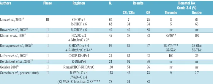
With 73% and 46% ORR and CR rates, respectively, 4 cycles of the (R)VAD+C compared favorably with 6 cycles of CHOP.8,16 However 70% of our patients (n=78) with
less than 2 independent response factors integrating LDH level, B symptoms and lymphocyte count had response rates close to 6 cycles of RCHOP22,23, with a more
favor-able toxicity profile (Tfavor-able 2). Moreover, the median OS (36 months) and PFS (16 months) of our 35 elderly patients treated with 8 cycles of VAD+C without rituximab was similar to that reported in the literature with 6 CHOP or 6 RCHOP cycles.22,23 Rituximab did not improve the
response rate of the VAD+C regimen in this study. One reason could be a trend to a higher MIPI score for patients treated with rituximab (Table 1). Indeed, rituximab has been reported to improve all other regimens in first line (CHOP, DHAP or HyperCVAD) (Table 2).22,24,26As
expect-Figure 2. (A) Overall Survival (OS) as intent to treat of 78 young and 35 elderly patients receiving first-line (R)VAD+C regimen followed or not by auto-SCT. (B) Overall survival. (C) Progression free survival, of the 49 transplanted patients (Tr) versus 64 non-transplanted patients (no Tr).
Figure 3. (A) Progression free survival and (B) overall survival, of the transplanted patients after RVAD+C or VAD+C regimen.
Figure 4. (A) Overall survival according to the GOELAMS index relat-ed to the number of criteria observrelat-ed at diagnosis among 4 pejora-tive prognostic factors (PF): Ki67>26%, ECOG>1, B symptoms and LDH>N. g1 = group with 3 or 4 PF, g2 = group with 1 or 2 PF and g3 = group without PF. (B) Overall survival according to the MIPI, Low=low-risk, Int = intermediate-risk and High = high-risk. 0 20 40 60 80 100 120 140 Time in months young Tr no Tr no Tr Tr g1 g2 g3 VAD+C VAD+C RVAD+C RVAD+C Low Int High Tr 1 0.8 0.6 0.4 0.2 0.0 1 0.8 0.6 0.4 0.2 0.0 1 0.8 0.6 0.4 0.2 0.0 1 0.8 0.6 0.4 0.2 0.0 1 0.8 0.6 0.4 0.2 0.0 1 0.8 0.6 0.4 0.2 0.0 1 0.8 0.6 0.4 0.2 0.0 elderly S u rv iv a l S u rv iv a l S u rv iv a l S u rv iv a l S u rv iv a l S u rv iv a l S u rv iv a l P=0.016 P<0.0001 P<0.0001 P<0.0001 P<0.0001 P=0.49 P=0.054 0 20 40 60 80 100 120 140 Time in months 0 20 40 60 80 100 120 140 Time in months 0 20 40 60 80 100 120 140 Time in months 0 20 40 60 80 100 120 140 Time in months 0 20 40 60 80 100 120 140 Time in months 0 20 40 60 80 100 120 140 Time in months
ed, the efficacy was lower than that observed for high-dose cytarabine-containing regimens R-Hyper-CVAD or R-DHAP.8,24,25However, the low toxicity profile observed
means the (R)VAD+C regimen offers a good efficacy/tol-erance ratio particularly recommended for elderly patients. RVAD+C has to be compared with other combi-nation regimens which have shown a good efficacy in first line for elderly patients. The RFC regimen is currently compared to RCHOP in a phase III study of the EU MCL network. The British national cancer research network is completing a phase II trial that compares FC with or with-out rituximab in order to evaluate the impact of rituximab in non-transplanted MCL patients. Of note, the ben-damustin-rituximab (BR) regimen has shown a good effi-cacy/toxicity profile on indolent lymphoma including MCL in relapse and was recently reported as effective as RCHOP in first line.27,28
This study confirms that intensive strategies including auto-SCT prolong PFS, as previously demonstrated in a randomized study conducted by the European MCL net-work.16We also demonstrated a better OS in transplanted
patients; but these were non-randomized trials and the populations of transplanted and non-transplanted patients differed by age. The 3-year OS and PFS rates (81% and 62%, respectively) of the 49 transplanted patients were comparable to those previously published.8,16,17,26 Unlike
the recent study previously reported by the Nordic group, no survival plateau was observed in our study.17The use of
rituximab before auto-SCT seemed to give a PFS advan-tage as recently reported.29,30 We also observed, as
pub-lished by Murali et al., long-term relapses with up to ten years of follow up in patients who did not benefit from auto-SCT.31In this setting, maintenance therapy strategies
or treatment of molecular relapses as recently published are of particular interest.32
In addition to confirming the prognostic value of the MIPI score, this work shows that additional important prognostic information can be obtained from quantifying
Ki67 expression, in agreement with recent studies from the EU MCL network and German low-grade lymphoma study group (GLSG).19Recent recommendations to assess
the Ki67 index have been published by the MCL net-work.33 We used very similar screening procedures for
Ki67 evaluation. Ki67 expression, together with three additional parameters available at diagnosis (PS > 1, pres-ence of B symptoms, elevated serum LDH), was combined into a new scoring system that proved particularly power-ful for the identification of very high- and low-risk patients, respectively. Of note, the 26% threshold for Ki67 expression is very close to that (30%) reported by the EU MCL network and the GLSG.34The very high-risk group
could benefit from more intensive treatment strategies such as allogeneic stem cell transplantation which has shown promise in relapsed MCL.35-37Conversely, low-risk
patients could benefit from less intensive (continuous) treatment regimens, and the (R)VAD+C could be a good option in this setting.38
In conclusion, this study demonstrates the low toxicity and efficacy of the (R)VAD+C regimen as first-line thera-py in mantle cell lymphoma patients. This regimen can be considered an efficient therapeutic option especially for elderly patients and for low-risk younger patients sched-uled to undergo auto-SCT. However, it is reasonable to propose that younger patients would benefit from more intensive treatment programs such as R-DHAP or RmaxiCHOP-RHDAraC followed by auto-SCT.17,25
Refinement of the current prognostic scoring systems for MCL could be very useful to accurately assign MCL patients to appropriate treatment plans as early as possi-ble, particularly in the younger patient group.
Acknowledgments
The GOELAMS group acknowledges the following clinicians for their signifi-cant contributions to this study: Dr. Dutel JL; Dr. de Jaureguiberry JP; Dr. Maloisel F; Dr.Rodon P; Dr. Thyss A; Pr. Feugier P; Dr. Le Maignan C; Dr. Caillères S; Dr. Lucas V; Dr. Grulois I; Dr.Bellange A and Dr. Ghandour.C ; The
GOE-(R)VAD and chlorambucil for first line treatment of MCL
Table 2.Selected trials of the main different (R)-Chemotherapy protocols used in first line for mantle cell lymphoma comparing efficacy and hematologic toxicity.
Authors Phase Regimen N. Results Hematol Tox
Grade 3-4 (%)
CR/CRu OR Thrombo Neutro
Lenz et al., 200516 III CHOP x 6 60 7 75 8 62 R-CHOP x 6 62 34 94 5 63 Howard et al., 200223 II R-CHOP x 6 40 40 88 nr nr Khouri er al., 19989 II HCVAD x 2 45 38 93 85/46** 100
+ MtxAraC x 2*
Romaguera et al., 200524 II R-HCVAD x 3-4 97 87 97 28-37/c*** 35-61/c + R-MtxAraC x 3-4* 37-57/c 59-71/c Lefrere et al., 200210 II CHOP-DHAPx4 21 84 92 100 29 De Guibert et al., 200625 II R-DHAPx4 24 92 96 nr nr Geisler 200817 II RmaxiCHOP-RHDAraC 160 54 96 nr nr Gressin et al., present study II R-VAD+C x 4 113 46 73 9 2.7
/ VAD+C x 4
(R) VAD+C less than 2 RF**** 78 51 83
Hematol Tox, Hematologic toxicities (NCI criteria). %, percentage of patients showing a grade 3-4 toxicity. nr, not reported. *alternated regimen. **85% after HCVAD/ 43% after MtxAraC . ***/c, % percentage of patients showing a grade 3-4 toxicity after each cycle of R-HCVAD and R-MtxAraC. ****3 independent Response Factors (RF) were defined LDH>N,
LAMS acknowledges the following pathologists for their participation sending slides or blocks for the histological review and study: Angonin R, Peoc’h M, Déchelotte P, Kemeny JL, Rousselet MC, Lefrancq T, De Muret A, Boucheron S, Gentil-Perret A, Mosnier JF, Fabiani B, Michenet P, Patey M, Eric Keddari, Fonck A, Memeteau F, Kambouchner M, Lagorce , Marck MF, Raymond-Gellé MF, Tas P, Chenard-Neu MP, Eloit S, Corvasier P, Julié C, Lhuintre P, Petit J, Fabre-Bocquentin B, Cazal-Hatem D, Rousset T, Lemaire JC, Akpo-Alavo B, Douvin D. We thank particularly the colleagues of the pathological review board: Dr Fanny Gaillard, Dr Josette Brière, Dr Martine Raphael (for the first trial) and the assistance of Dr S Saïkali. We are very grateful to the data managers of the GOE-LAMS group, in particular Valérie Rolland. We thank Dr. Mary Callanan, Prof. Marie-Christine Béné, and Prof. Jean-Yves Cahn for critically reviewing the man-uscript and for English editing. We express sincere thanks to Prof. Jean-Jacques Sotto for his constant support and encouragement of this work.
Authorship and Disclosures
RG designed the trial; SC-M reviewed all the patholog-ic samples; RG, ED, OT, EG, MP, AE-Y, JC, JFR, SLG, GL, GD, PSC, HM, BC, JPV, PC, TL and PC were involved in the care of patients, the collections and the validation of the clinical data with the clinical research associates of the Goelams group; MC has provided statistical analysis; RG, SLG, TL and PC wrote the manuscript; all the authors have checked the final version of the manuscript.
JFR received honooraria from HOSPIRA; RG and TLdLC received honoraria from CELGENE.
The other authors reported no potential conflicts of interest.
References
1. Harris NL, Jaffe ES, Diebold J, Flandrin G, Muller-Hermelink HK, Vardiman J, et al. The World Health Organization classifica-tion of neoplasms of the hematopoietic and lymphoid tissues: report of the Clinical Advisory Committee meeting--Airlie House, Virginia, November, 1997. Hematol J. 2000;1(1):53-66.
2. Herrmann A, Hoster E, Zwingers T, Brittinger G, Engelhard M, Meusers P, et al. Improvement of overall survival in advanced stage mantle cell lymphoma. J Clin Oncol. 2009;27(4):511-8.
3. Bernard M, Gressin R, Lefrere F, Drenou B, Branger B, Caulet-Maugendre S, et al. Blastic variant of mantle cell lymphoma: a rare but highly aggressive subtype. Leukemia. 2001;15(11):1785-91.
4. Meusers P, Engelhard M, Bartels H, Binder T, Fulle HH, Gorg K, et al. Multicentre ran-domized therapeutic trial for advanced cen-trocytic lymphoma: anthracycline does not improve the prognosis. Hematol Oncol. 1989;7(5):365-80.
5. Zucca E, Roggero E, Pinotti G, Pedrinis E, Cappella C, Venco A, et al. Patterns of sur-vival in mantle cell lymphoma. Ann Oncol. 1995;6(3):257-62.
6. Majlis A, Pugh WC, Rodriguez MA, Benedict WF and Cabanillas F. Mantle cell lymphoma: correlation of clinical outcome and biologic features with three histologic variants. J Clin Oncol. 1997;15(4):1664-71. 7. Hiddemann W, Unterhalt M, Herrmann R,
Woltjen HH, Kreuser ED, Trumper L, et al. Mantle-cell lymphomas have more wide-spread disease and a slower response to chemotherapy compared with follicle-cen-ter lymphomas: results of a prospective comparative analysis of the German Low-Grade Lymphoma Study Group. J Clin Oncol. 1998;16(5):1922-30.
8. Lefrere F, Delmer A, Levy V, Delarue R, Varet B and Hermine O. Sequential chemotherapy regimens followed by high-dose therapy with stem cell transplantation in mantle cell lymphoma: an update of a prospective study. Haematologica. 2004;89(10):1275-6.
9. Khouri IF, Romaguera J, Kantarjian H, Palmer JL, Pugh WC, Korbling M, et al. Hyper-CVAD and high-dose methotrex-ate/cytarabine followed by stem-cell trans-plantation: an active regimen for aggressive
mantle-cell lymphoma. J Clin Oncol. 1998;16(12):3803-9.
10. Lefrere F, Delmer A, Suzan F, Levy V, Belanger C, Djabarri M, et al. Sequential chemotherapy by CHOP and DHAP regi-mens followed by high-dose therapy with stem cell transplantation induces a high rate of complete response and improves event-free survival in mantle cell lym-phoma: a prospective study. Leukemia. 2002;16(4):587-93.
11. Milpied N, Gaillard F, Moreau P, Mahe B, Souchet J, Rapp MJ, et al. High-dose thera-py with stem cell transplantation for man-tle cell lymphoma: results and prognostic factors, a single center experience. Bone Marrow Transplant. 1998;22(7):645-50. 12. Freedman AS, Neuberg D, Gribben JG,
Mauch P, Soiffer RJ, Fisher DC, et al. High-dose chemoradiotherapy and anti-B-cell monoclonal antibody-purged autologous bone marrow transplantation in mantle-cell lymphoma: no evidence for long-term remission. J Clin Oncol. 1998;16(1):13-8. 13. Andersen NS, Pedersen L, Elonen E,
Johnson A, Kolstad A, Franssila K, et al. Primary treatment with autologous stem cell transplantation in mantle cell lym-phoma: outcome related to remission pre-transplant. Eur J Haematol. 2003;71(2):73-80.
14. Vandenberghe E, Ruiz de Elvira C, Loberiza FR, Conde E, Lopez-Guillermo A, Gisselbrecht C, et al. Outcome of autolo-gous transplantation for mantle cell lym-phoma: a study by the European Blood and Bone Marrow Transplant and Autologous Blood and Marrow Transplant Registries. Br J Haematol. 2003;120(5):793-800. 15. Mangel J, Leitch HA, Connors JM,
Buckstein R, Imrie K, Spaner D, et al. Intensive chemotherapy and autologous stem-cell transplantation plus rituximab is superior to conventional chemotherapy for newly diagnosed advanced stage mantle-cell lymphoma: a matched pair analysis. Ann Oncol. 2004;15(2):283-90.
16. Dreyling M, Lenz G, Hoster E, Van Hoof A, Gisselbrecht C, Schmits R, et al. Early con-solidation by myeloablative radiochemotherapy followed by autolo-gous stem cell transplantation in first remis-sion significantly prolongs progresremis-sion-free survival in mantle-cell lymphoma: results of a prospective randomized trial of the European MCL Network. Blood. 2005;105(7):2677-84.
17. Geisler CH, Kolstad A, Laurell A, Andersen NS, Pedersen LB, Jerkeman M, et al. Long-term progression-free survival of mantle cell lymphoma after intensive front-line immunochemotherapy with in vivo-purged stem cell rescue: a nonrandomized phase 2 multicenter study by the Nordic Lymphoma Group. Blood. 2008;112(7): 2687-93.
18. Gressin R, Legouffe E, Leroux D, Jacob MC, Swiercz P, Peoch M, et al. Treatment of mantle-cell lymphomas with the VAD +/-chlorambucil regimen with or without sub-sequent high-dose therapy and peripheral blood stem-cell transplantation. Ann Oncol. 1997;8 (Suppl 1):103-6.
19. Hoster E, Dreyling M, Klapper W, Gisselbrecht C, van Hoof A, Kluin-Nelemans HC, et al. A new prognostic index (MIPI) for patients with advanced-stage mantle cell lymphoma. Blood. 2008;111(2):558-65.
20. Cheson BD, Horning SJ, Coiffier B, Shipp MA, Fisher RI, Connors JM, et al. Report of an international workshop to standardize response criteria for non-Hodgkin's lym-phomas. NCI Sponsored International Working Group. J Clin Oncol. 1999;17(4): 1244.
21. Cheson BD, Pfistner B, Juweid ME, Gascoyne RD, Specht L, Horning SJ, et al. Revised response criteria for malignant lym-phoma. J Clin Oncol. 2007;25(5):579-86. 22. Lenz G, Dreyling M, Hoster E, Wormann B,
Duhrsen U, Metzner B, et al. Immunochemotherapy with rituximab and cyclophosphamide, doxorubicin, vin-cristine, and prednisone significantly improves response and time to treatment failure, but not long-term outcome in patients with previously untreated mantle cell lymphoma: results of a prospective ran-domized trial of the German Low Grade Lymphoma Study Group (GLSG). J Clin Oncol. 2005;23(9):1984-92.
23. Howard OM, Gribben JG, Neuberg DS, Grossbard M, Poor C, Janicek MJ, et al. Rituximab and CHOP induction therapy for newly diagnosed mantle-cell lym-phoma: molecular complete responses are not predictive of progression-free survival. J Clin Oncol. 2002;20(5):1288-94.
24. Romaguera JE, Fayad L, Rodriguez MA, Broglio KR, Hagemeister FB, Pro B, et al. High rate of durable remissions after treat-ment of newly diagnosed aggressive man-tle-cell lymphoma with rituximab plus
hyper-CVAD alternating with rituximab plus high-dose methotrexate and cytara-bine. J Clin Oncol. 2005;23(28):7013-23. 25. de Guibert S, Jaccard A, Bernard M, Turlure
P, Bordessoule D and Lamy T. Rituximab and DHAP followed by intensive therapy with autologous stem-cell transplantation as first-line therapy for mantle cell lym-phoma. Haematologica. 2006;91(3):425-6. 26. Gianni AM, Magni M, Martelli M, Di
Nicola M, Carlo-Stella C, Pilotti S, et al. Long-term remission in mantle cell lym-phoma following high-dose sequential chemotherapy and in vivo rituximab-purged stem cell autografting (R-HDS regi-men). Blood. 2003;102(2):749-55. 27. Rummel MJ, Al-Batran SE, Kim SZ, Welslau
M, Hecker R, Kofahl-Krause D, et al. Bendamustine plus rituximab is effective and has a favorable toxicity profile in the treatment of mantle cell and low-grade non-Hodgkin's lymphoma. J Clin Oncol. 2005;23(15):3383-9.
28. Rummel MJ, Niederle N, Maschmayer G, Banat A, von Grunhagen U, Losem C, et al. Bendamustine Plus Rituximab Is Superior in Respect of Progression Free Survival and CR Rate When Compared to CHOP Plus Rituximab as First-Line Treatment of Patients with Advanced Follicular, Indolent, and Mantle Cell Lymphomas: Final Results of a Randomized Phase III Study of the StiL
(Study Group Indolent Lymphomas, Germany). Blood. 2009;114:168.
29. Tam CS and Khouri IF. Autologous and allogeneic stem cell transplantation: rising therapeutic promise for mantle cell lym-phoma. Leuk Lymlym-phoma. 2009:1-10. 30. Dreger P, Rieger M, Seyfarth B, Hensel M,
Kneba M, Ho AD, et al. Rituximab-aug-mented myeloablation for first-line autolo-gous stem cell transplantation for mantle cell lymphoma: effects on molecular response and clinical outcome. Haematologica. 2007;92(1):42-9.
31. Murali S, Winton E, Waller EK, Heffner LT, Lonial S, Flowers C, et al. Long-term progres-sion-free survival after early autologous transplantation for mantle-cell lymphoma. Bone Marrow Transplant. 2008;42(8):529-34. 32. Andersen NS, Pedersen LB, Laurell A, Elonen E, Kolstad A, Boesen AM, et al. Pre-emptive treatment with rituximab of molecular relapse after autologous stem cell transplantation in mantle cell lymphoma. J Clin Oncol. 2009;27(26):4365-70. 33. Klapper W, Hoster E, Determann O,
Oschlies I, van der Laak J, Berger F, et al. Ki-67 as a prognostic marker in mantle cell lymphoma-consensus guidelines of the pathology panel of the European MCL Network. J Hematop. 2009 Jun 16. [Epub ahead of print]
34. Determann O, Hoster E, Ott G, Wolfram
Bernd H, Loddenkemper C, Leo Hansmann M, et al. Ki-67 predicts outcome in advanced-stage mantle cell lymphoma patients treated with anti-CD20 immunochemotherapy: results from ran-domized trials of the European MCL Network and the German Low Grade Lymphoma Study Group. Blood. 2008; 111(4):2385-7.
35. Khouri IF, Lee MS, Saliba RM, Jun G, Fayad L, Younes A, et al. Nonablative allogeneic stem-cell transplantation for advanced/recurrent mantle-cell lymphoma. J Clin Oncol. 2003;21(23):4407-12. 36. Maris MB, Sandmaier BM, Storer BE,
Chauncey T, Stuart MJ, Maziarz RT, et al. Allogeneic hematopoietic cell transplanta-tion after fludarabine and 2 Gy total body irradiation for relapsed and refractory man-tle cell lymphoma. Blood. 2004;104(12): 3535-42.
37. Ganti AK, Bierman PJ, Lynch JC, Bociek RG, Vose JM and Armitage JO. Hematopoietic stem cell transplantation in mantle cell lymphoma. Ann Oncol. 2005;16(4):618-24.
38. Coleman M, Martin P, Ruan J, Furman R, Niesvizky R, Elstrom R, et al. Low-dose metronomic, multidrug therapy with the PEP-C oral combination chemotherapy reg-imen for mantle cell lymphoma. Leuk Lymphoma. 2008;49(3):447-50.