HAL Id: hal-01744606
https://hal.sorbonne-universite.fr/hal-01744606
Submitted on 27 Mar 2018HAL is a multi-disciplinary open access archive for the deposit and dissemination of sci-entific research documents, whether they are pub-lished or not. The documents may come from teaching and research institutions in France or abroad, or from public or private research centers.
L’archive ouverte pluridisciplinaire HAL, est destinée au dépôt et à la diffusion de documents scientifiques de niveau recherche, publiés ou non, émanant des établissements d’enseignement et de recherche français ou étrangers, des laboratoires publics ou privés.
ATP binding cassette (ABC) transporters: expression
and clinical value in glioblastoma
Antonin Dréan, Shai Rosenberg, François-Xavier Lejeune, Larissa Goli,
Aravindan Arun Nadaradjane, Jeremy Guehennec, Charlotte Schmitt, Maïté
Verreault, Franck Bielle, Karima Mokhtari, et al.
To cite this version:
Antonin Dréan, Shai Rosenberg, François-Xavier Lejeune, Larissa Goli, Aravindan Arun Nadaradjane, et al.. ATP binding cassette (ABC) transporters: expression and clinical value in glioblastoma. Journal of Neuro-Oncology, Springer Verlag, In press, �10.1007/s11060-018-2819-3�. �hal-01744606�
1
ATP Binding Cassette (ABC) transporters: expression and clinical value in
glioblastoma
Dréan Antonin1,2, Rosenberg Shai1,3, Lejeune François-Xavier1, Goli Larissa1,
Nadaradjane Aravindan Arun1, Guehennec Jérémy1, Schmitt Charlotte1, Verreault
Maïté1, Bielle Franck1,4,7, Mokhtari Karima1,4,7, Sanson Marc1,5,7, Carpentier
Alexandre6, Delattre Jean-Yves1,5,7, Idbaih Ahmed1,5
1 Inserm U 1127, CNRS UMR 7225, Sorbonne Universités, UPMC Univ Paris 04
UMR S 1127, Institut du Cerveau et de la Moelle épinière, ICM, F-75013, Paris,
France
2 Equipe de recherche CarThera, Institut du Cerveau et de la Moelle épinière, Ipeps
ICM, F-75013, Paris, France.
3 Hadassah - Hebrew University Medical Center, Israel (S.R.)
4 AP-HP, Hôpitaux Universitaires La Pitié Salpêtrière - Charles Foix, Service de
Neuropathologie, F-75013, Paris, France.
5 AP-HP, Hôpitaux Universitaires La Pitié Salpêtrière - Charles Foix, Service de
Neurologie 2-Mazarin, F-75013, Paris, France.
6 AP-HP, Hôpitaux Universitaires La Pitié Salpêtrière - Charles Foix, Service de
Neurochirurgie, F-75013, Paris, France.
2
Corresponding author
Ahmed Idbaih. Hôpitaux Universitaires La Pitié Salpêtrière - Charles Foix, Service de
Neurologie 2-Mazarin, F-75013, Paris, France. Tél: 00 33 1 42140385. Fax 00 33 1
3
Abstract
ATP-binding cassette transporters (ABC transporters) regulate traffic of multiple
compounds, including chemotherapeutic agents, through biological membranes.
They are expressed by multiple cell types and have been implicated in the drug
resistance of some cancer cells. Despite significant research in ABC transporters in
the context of many diseases, little is known about their expression and clinical value
in glioblastoma (GBM).
We analyzed expression of 49 ABC transporters in both commercial and
patient-derived GBM cell lines as well as from 51 human GBM tumor biopsies. Using The
Cancer Genome Atlas (TCGA) cohort as a training dataset and our cohort as a
validation dataset, we also investigated the prognostic value of these ABC
transporters in newly diagnosed GBM patients, treated with the standard of care.
In contrast to commercial GBM cell lines, GBM-patient derived cell lines (PDCL),
grown as neurospheres in a serum-free medium, express ABC transporters similarly
to parental tumors. Serum appeared to slightly increase resistance to temozolomide
correlating with a tendency for an increased expression of ABCB1. Some differences
were observed mainly due to expression of ABC transporters by microenvironmental
cells. Together, our data suggest that the efficacy of chemotherapeutic agents may
be misestimated in vitro if they are the targets of efflux pumps whose expression can
be modulated by serum. Interestingly, several ABC transporters have prognostic
value in the TCGA dataset. In our cohort of 51 GBM patients treated with radiation
therapy with concurrent and adjuvant temozolomide, ABCA13 overexpression is
associated with a decreased progression free survival in univariate (p<0.01) and
multivariate analyses including MGMT promoter methylation (p=0.05) suggesting
4
Expression of ABC transporters is: (i) detected in GBM and microenvironmental cells
and (ii) better reproduced in GBM-PDCL. ABCA13 expression is an independent
prognostic factor in newly diagnosed GBM patients.
Further prospective studies are warranted to investigate whether ABCA13 expression
can be used to further personalize treatments for GBM.
5
Introduction
Glioblastoma (GBM) is the most common and aggressive primary brain cancer in
adults, with a median overall survival below 20 months after the initial diagnosis [1].
Virtually, all GBM patients experience tumor relapse after first-line standard of care.
The blood-brain barrier (BBB), a biophysical barrier limiting penetration of
chemotherapeutic agents within the brain and the brain tumor, is one of the actors
involved in intrinsic resistance of drugs in GBM patients [2].
ATP binding cassette (ABC) transporters are a family of 49 proteins that actively
transport many substrates. This family contains the main efflux pumps that expel
drugs out of cells, therefore impacting chemosensitivity [3]. Efflux pumps are
expressed by many cells in the body. In the brain, they are expressed by microglial
cells, astrocytes, neurons, pericytes and, endothelial cells [4]. Endothelial cells, which
are involved in the normal functioning of the BBB, mainly express ABCB1 (P-gp) and
ABCG2 (BCRP) to efflux xenobiotics from the brain parenchyma to the bloodstream
[2]. In addition, many ABC transporters are involved in cell metabolism and/or
transport of biologic molecules [3].
ABC transporters have a large spectrum of substrates, including many
chemotherapeutic agents [5]. The efflux of these therapeutic agents from the brain to
the blood by the BBB hampers their access to tumor cells and their efficacy. Efflux
pumps, from the ABC transporter family, can also be expressed by tumor cells
themselves, leading to the development of intrinsic chemoresistance [6].
Here the expression of all ABC transporters in GBM cells and their microenvironment
was assessed and their potential prognostic impact in a homogeneous population of
6
Material and methods
Public dataset
Levels of mRNA expression of the 49 ABC transporters were downloaded from The
Cancer Genome Atlas (TCGA) GBM dataset [7]. The threshold of z-scores (RNA Seq
V2 RSEM) was set at ±2 of distinguishing over- and under-expressing samples.
Overall survival (OS; from diagnosis to death) and progression-free survival (PFS;
from diagnosis to first tumor progression) were compared using log-rank test.
P-values below 0.05 were considered significant. RNAseq data for the U251 cell line
were obtained from Lundberg et al. [8]. Expression levels of ABCB1, ABCG1 and
ABCG2 by normal CNS cells were obtained from
http://web.stanford.edu/group/barres_lab/brain_rnaseq.html [9].
OncoNeuroTek tissue and data as validation cohort
For the validation, patients and tumors from the OncoNeuroTek tissue bank were
selected according to the following inclusion criteria : (i) newly diagnosed and
histologically proven GBM, (ii) age below 70 years, (iii) Karnofsky performance status
above 70%, (iv) treated with radiation therapy with concurrent and adjuvant
temozolomide [10], (v) available MGMT promoter methylation status, (vi) available
clinical data, (vii) available tumor tissue for additional molecular analysis and, (viii)
signed informed consent form for molecular analysis.
GBM cell lines
Nine GBM-patient derived cell lines (PDCL) named 4339, 4371, 5706, 6190, 6240,
7
paired human tumor samples was available for further molecular analysis. These
samples and associated clinico-molecular annotations were previously reported [11].
Cell culture
Cell cultures were performed as previously described [11]. GBM-PDCL were
established (passage > 5) and cultured in DMEM-F12 (Life Technologies, 31331028)
supplemented with 1% penicillin/streptomycin, B27 supplement 50X (Life
Technologies, 17504-044), 20 ng/mL human bFGF (Peprotech, 100-18B) and 20
ng/ml human EGF (Peprotech, 100-15). Cells were cultured as gliomaspheres.
Dissociation was performed with Accutase StemPro (Life Technologies, A11105-11)
after washing with HBSS (Life Technologies, 1150029). For culture in serum, PDCL
were cultured in DMEM (Life Technologies,31885049) supplemented with 10% FBS
(Thermo Fisher Life) and 1% penicillin/streptomycin.
Viability test.
For drug sensitivity assays, 96-well plates were used. For viability assay on PDCL, wells were coated with 10 μg/mL laminin (cat no. L2020, Sigma-Aldrich) at 37°C for 1
hour for PDCL. Six thousand cells were plated per well. Temozolomide
(Sigma-Aldrich, T2577) was resuspended in DMSO as a 20 mg/ml stock solution and was
added 24 hours after plating. 150 hours after drug addition, WST-1 reagent (Roche)
was added according to the manufacturer's instructions. WST-1 salt is cleaved to a
soluble formazan dye by a NAD(P)H-dependent reaction in viable cells. Plates were
incubated for 3 hours and read by spectrophotometry at 450 nm and 620 nm
8
RNA extraction
RNA samples from the 9 GBM tumors and their 9 paired GBM-PDCL were provided
by OncoNeuroTek tissue bank. RNA extraction for tumor samples was performed as
previously described using the RNeasy Lipid Tissue Mini Kit (Qiagen) [12].
For GBM-PDCL, 1 x 10⁴ dissociated cells were cultured in a 75 cm2 flask for four
days, with culture medium changed at day 3, and cells were harvested the next day
as a dry pellet and stored at -80°C as described previously [11]. RNA extraction was
performed using the kit NucleoSpin® RNA (Macherey-Nagel, 740955) according the manufacturer’s guidelines.
Reverse Transcription and Real-Time Quantitative PCR (RT-qPCR)
Reverse transcription was performed using the kit Maxima First Strand cDNA
Synthesis Kit for RT-qPCR (ThermoScientific, K1442) according to the manufacturer’s guidelines starting with 750 ng of RNA. cDNAs were stored at -20°C.
qPCR was used to examine expression levels of ABCA13, ABCB1, ABCG1, and
ABCG2 in the GBM-PDCL cells. Paired tumor samples were analyzed using the
LightCycler Probe Master mix 2X (Roche, 04887301001) and the UPL detection
system (Roche, 04483433001; primers are listed in supplementary table 1) in a Light
Cycler 96 (Roche). For each qPCR, we made a duplicate measure in each
experiment and we ran three distinct experiments at three different passages. PPIA
expression was used for normalization. qPCR was used to investigate expression
levels of ABCA1, ABCA4, ABCA13, ABCC1, ABBCC12, ABCC13, and ABCG1 in the
validation cohort of GBM patients were performed in a 1534 wells plate using a Sybr
green detection system in a Light Cycler 1534 (Roche). PPIA and PPIB expression
9
Immunohistochemistry
Formalin-fixed, paraffin-embedded (FFPE) tumor tissue sections were deparaffinized,
rehydrated, and incubated in citrate buffer at pH 4.0 for epitope retrieval in a
microwave at 400W for 15 minutes. Blockade solution was made using PBS 0.1M
supplemented with 0.4% triton, 3% BSA and 10% donkey serum. Anti-human ABCG2
antibody (BXP-21; Euromedex) and anti-human CD31 antibody (SP38; Thermo
Fischer Scientific) were diluted at 1:100 in the blockade solution and incubated on
the tissue slices overnight at 4°C. Secondary antibodies conjugated with Alexa 488
and 447 were incubated on the slices for 2h at room temperature. The slices were
then mounted using fluoromount, supplemented with DAPI (0100-20; Clinisciences)
and visualized using a microscope (Apotome Axio M2, Zeiss).
Statistical analysis
Comparison between ABC transporter expression in GBM-PDCL and tumors was
performed by a paired t-test with a Bonferroni correction using R.
RT-qPCR data bar charts were plotted with GraphPad PRISM 4, and analyzed by a
two-way ANOVA test.
For analysis of OS and PFS, patients were divided in two groups from each side of
the median. Kaplan Meier survival curves were made on GraphPad PRISM 4. Survivals of groups with “low” versus “high” expression were compared using
Log-rank (Mantel-Cox) test. Additional Cox test for survival in a multivariate modality was
performed with R to assess the impact of MGMT promoter methylation status after a
10 Results
Expression of ABC transporters in the commercial U251 GBM cell line, in GBM-PDCL and paired-GBM tumors
Out of the 49 ABC transporters, 19 are measurably expressed in U251 GBM cell
lines versus 42/49 in the GBM-PDCL line [8]. This highlights that GBM cell lines may
lose ABC transporter expression over time in adherent culture conditions including
bovine fetal serum (Fig. 1, Panel A). When cultured in a differentiating medium
containing fetal calf serum, the N13-1520 PDCL shows a slight non-significant trend
for increased expression of ABCB1 in RT-qPCR (Fig. 1C) associated with higher
resistance to temozolomide in an in vitro viability assay (p<0.0001) (Fig. 1D). These
results suggest that the expression loss of most of the ABC transporters may be due
to the high passages after cell line establishment rather than exposure to fetal calf
serum. It also highlights the importance of using a non-differentiating serum-free
medium.
We already showed that, overall, GBM-PDCL are molecularly very close to their
parental human tumors in a multi-omics analysis when cultured in a
non-differentiating serum-free medium [11]. We analyzed the same data set (i.e. 9
GBM-PDCL and their tumor of origin) with a specific focus on expression levels of the 49
ABC transporters acquired using Affymetrix® expression profiling microarray and
RNA Seq platforms. Three ABC transporters were differentially expressed between
the GBM-PDCL and their parental tumor in both transcriptomes (i.e. Affymetrix and
RNA Seq): ABCB1, ABCG1 and ABCG2 (Figures 1B, 2A and 2B, and Table 1).
RT-qPCR validated the difference of expression for the three transporters (Figs. 2C,
11
ABC transporters differentially expressed in GBM-PDCL compared to their parental tumors are expressed in the microenvironnement
We hypothesized that ABC transporters are differentially expressed between
GBM-PDCL and their parental tumors (i.e. underexpression in GBM-GBM-PDCL versus paired
human tumors) due to the expression by microenvironmental cells present in bulk
tumor. We questioned http://web.stanford.edu/group/barres_lab/brain_rnaseq.html for
expression of ABCB1, ABCG1 and ABCG2 in normal murine cells [13]. ABCB1,
ABCG1 and ABCG2 are expressed by endothelial cells supporting our hypothesis.
ABCG1 and ABCG2 are also expressed in additional cell types (i.e. astrocytes,
neurons, oligodendrocytes) (Figs. 2E, 2F and 2G). Indeed, ABCG2 and CD31
expression are colocalized using immunohistochemistry in a human GBM (Fig. 2H).
Clinical value of ABC transporter expression in GBM patients
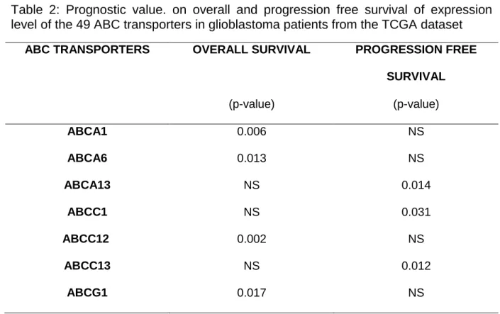
We then investigated the prognostic value of the 49 ABC transporters in the 2013
TCGA GBM cohort. An overview of the expression of these transporters is presented
in supplementary Fig. 2. Kaplan-Meier analysis highlighted an unfavorable prognostic
impact of: (i) over-expression of ABCA1, ABCA4, ABCC12 and ABCG1 on OS, and
(ii) over-expression of ABCA13, ABCC1 and ABCC13 on PFS (Table 2).
To validate this finding, expression of ABCA1, ABCA4, ABCA13, ABCC1, ABCC12,
ABCC13 and ABCG1 was assessed in a homogeneously treated cohort of 51 newly
diagnosed GBM patients (Fig. 3A). Only overexpression of ABCA13 was validated as
negative prognostic factor in terms of PFS (p=0.004, Figure 3B) and OS in univariate
analysis (p=0.047, Figure 3C). In multivariate analysis, ABCA13 overexpression was
12
1.12) when MGMT promoter methylation status (p=0.002 for MGMT, HR=3.13) is
13
Discussion
GBM is a challenging cancer to treat. Indeed, GBM cells are highly resistant to
treatments. The blood-brain and blood-tumor barriers limit the efficacy of drug
treatments in GBM patients and are a major factor in primary resistance of GBM cells
to chemotherapy.
The blood-brain and blood-tumor barriers are physical and biological barriers. ABC
transporters play a pivotal role in the biology of both barriers. Indeed, they can expel
chemotherapeutic agents from the brain tissue to the bloodstream [2]. Expression of
ABC transporters by the tumor cells themselves can also be responsible for tumor
resistance to chemotherapies. Therefore, in the current work we have investigated
their expression in GBM cell lines used for preclinical investigations of new
therapeutic compounds and in human tumors.
Commercial GBM cell lines lack expression of most ABC transporters. Additional in
vitro models that better recapitulate ABC transporter expression patterns of human
tumors must be also used before moving drugs to in vivo investigations. GBM-PDCL
is an important tool expressing a larger number of ABC transporters expressed in
human GBM. However, addition of serum in the culture medium appeared to change
expression patterns of ABC transporters. A trend to the increased expression of
ABCB1 and an increased resistance to temozolomide in a serum-complemented
medium is consistent with the fact that temozolomide is a substrate of ABCB1 [14].
Together, our data suggest that the efficacy of chemotherapeutic agents might be
changed in vitro, if they are substrates of efflux pumps not expressed in the cell line
used and whose expression can be modulated by fetal calf serum. Using GBM-PDCL
14
To identify ABC transporters impacting response to the standard of care of newly
diagnosed GBM patients, we have correlated prognosis of GBM patients to ABC
transporter expression in a training dataset (i.e. TCGA) and a validation dataset (i.e.
personal cohort). ABCA13 expression was found to be a prognostic factor for PFS
and OS in univariate analyses. Interestingly, based on multivariate analysis, ABCA13
expression provides additional prognostic information, in terms of PFS, to MGMT
promoter methylation in young patients (<70 years), in good clinical condition
(KPS>70%) suffering from newly diagnosed GBM and treated with the standard of
care. This supports for the first time ABCA13 expression as an independent
prognostic biomarker in newly diagnosed GBM patients. Additional prospective and
functional studies are warranted to investigate our findings further.
Little is known about ABCA13. ABCA13 is a large protein of 5058 amino acids
encoded by ABCA13 located on chromosome region 7p12.3 that is often polysomic
in GBM. ABCA13 is highly expressed in the bone marrow, but not in normal brain [9].
Therefore, in GBM, ABCA13 is likely to be primarily expressed by tumor cells rather
than microenvironmental cells. The molecular function of ABCA13 remains largely
unknown [15]. However, ABCA13 has been linked to multiple human diseases,
including cancer. ABCA13 genetic polymorphisms have been associated with psychiatric disorders [16–21] and success of coronary artery bypass surgery [22].
ABCA13 expression has also been linked to outcome in various cancers. In line with
our study, ABCA13 overexpression is associated with poor prognosis in renal cell
carcinoma [23], in gastric adenocarcinoma [24], and in ovarian serous carcinoma
[25]. In contrast, ABCA13 overexpression is associated with improved outcomes in
breast cancer [26] and in colorectal cancer [27]. Yun et al. reported an association of ABCA13 and stem-like phenotype in the “side population” of metastatic renal cell
15
carcinoma. Overexpression of ABCA13 was also correlated with drug resistance,
suggesting a potential role in drug efflux [23].
Overall our study suggests that ABC binding cassette transporters, particularly
ABCA13, are important in both preclinical and clinical research in GBM. We have
found that ABCA13 is a potential prognostic factor in GBM patients and a putative
actor of therapy resistance.Further studies are warranted to support our findings and
to better understand the role of ABCA13.
Acknowledgments
Institut Universitaire de Cancérologie
Fondation ARC pour la recherche sur le cancer
Association pour la Recherche sur les Tumeurs Cérébrales
Dr Michael Canney.
OncoNeuroTek tissue bank, Paris.
Compliance with Ethical Standards
This study was funded by the Fondation ARC pour la recherche sur le cancer and the
Association pour la Recherche sur les Tumeurs Cérébrales.
All authors declare they have no conflict of interest with the present study.
All procedures performed in studies involving human participants were in accordance
with the ethical standards of the institutional and/or national research committee and
with the 1964 Helsinki declaration and its later amendments or comparable ethical
16
References
1. Ostrom QT, Bauchet L, Davis FG, et al (2014) The epidemiology of glioma in adults: a “state of the science” review. Neuro-Oncol 16:896–913 . doi: 10.1093/neuonc/nou087
2. Dréan A, Goldwirt L, Verreault M, et al (2016) Blood-brain barrier, cytotoxic chemotherapies and glioblastoma. Expert Rev Neurother 1–16 . doi: 10.1080/14737175.2016.1202761
3. Vasiliou V, Vasiliou K, Nebert DW (2009) Human ATP-binding cassette (ABC) transporter family. Hum Genomics 3:281–290
4. Hartz AMS, Bauer B (2011) ABC transporters in the CNS - an inventory. Curr Pharm Biotechnol 12:656–673
5. Sparreboom A, Danesi R, Ando Y, et al (2003) Pharmacogenomics of ABC transporters and its role in cancer chemotherapy. Drug Resist Updat Rev Comment Antimicrob Anticancer Chemother 6:71–84
6. Khamisipour G, Jadidi-Niaragh F, Jahromi AS, et al (2016) Mechanisms of tumor cell resistance to the current targeted-therapy agents. Tumour Biol J Int Soc Oncodevelopmental Biol Med 37:10021–10039 . doi: 10.1007/s13277-016-5059-1
7. Brennan CW, Verhaak RGW, McKenna A, et al (2013) The somatic genomic landscape of glioblastoma. Cell 155:462–477 . doi: 10.1016/j.cell.2013.09.034
8. Lundberg E, Fagerberg L, Klevebring D, et al (2010) Defining the transcriptome and proteome in three functionally different human cell lines. Mol Syst Biol 6:450 . doi: 10.1038/msb.2010.106
9. Uhlén M, Hallström BM, Lindskog C, et al (2016) Transcriptomics resources of human tissues and organs. Mol Syst Biol 12:862
10. Stupp R, Mason WP, van den Bent MJ, et al (2005) Radiotherapy plus concomitant and adjuvant temozolomide for glioblastoma. N Engl J Med 352:987–996 . doi: 10.1056/NEJMoa043330
11. Rosenberg S, Verreault M, Schmitt C, et al (2016) Multi-omics analysis of primary glioblastoma cell lines shows recapitulation of pivotal molecular features of parental tumors. Neuro-Oncol. doi: 10.1093/neuonc/now160
12. Kamoun A, Idbaih A, Dehais C, et al (2016) Integrated multi-omics analysis of oligodendroglial tumours identifies three subgroups of 1p/19q co-deleted gliomas. Nat Commun 7:11263 . doi: 10.1038/ncomms11263
13. Zhang Y, Chen K, Sloan SA, et al (2014) An RNA-sequencing transcriptome and splicing database of glia, neurons, and vascular cells of the cerebral cortex. J Neurosci Off J Soc Neurosci 34:11929–11947 . doi: 10.1523/JNEUROSCI.1860-14.2014
17
14. Goldwirt L, Beccaria K, Carpentier A, et al (2014) Irinotecan and temozolomide brain distribution: a focus on ABCB1. Cancer Chemother Pharmacol 74:185–193 . doi: 10.1007/s00280-014-2490-0
15. Tomioka M, Toda Y, Kurisu J, et al (2012) The effects of neurological disorder-related codon variations of ABCA13 on the function of the ABC protein. Biosci Biotechnol Biochem 76:2289–2293 . doi: 10.1271/bbb.120563
16. Knight HM, Pickard BS, Maclean A, et al (2009) A cytogenetic abnormality and rare coding variants identify ABCA13 as a candidate gene in schizophrenia, bipolar disorder, and depression. Am J Hum Genet 85:833–846 . doi: 10.1016/j.ajhg.2009.11.003
17. Dwyer S, Williams H, Jones I, et al (2011) Investigation of rare non-synonymous variants at ABCA13 in schizophrenia and bipolar disorder. Mol Psychiatry 16:790–791 . doi: 10.1038/mp.2011.2
18. Pickard BS, Van Den Bossche MJA, Malloy MP, et al (2012) Multiplex amplicon quantification screening the ABCA13 gene for copy number variation in schizophrenia and bipolar disorder. Psychiatr Genet 22:269–270 . doi: 10.1097/YPG.0b013e32835185b3
19. Degenhardt F, Priebe L, Strohmaier J, et al (2013) No evidence for an involvement of copy number variation in ABCA13 in schizophrenia, bipolar disorder, or major depressive disorder. Psychiatr Genet 23:45–46 . doi: 10.1097/YPG.0b013e328358645b
20. Ma J, Lan X, Gao N, et al (2013) A genetic association study between common variants in the ABCA13 gene and schizophrenia in a Han Chinese population. Psychiatry Res 209:748–749 . doi: 10.1016/j.psychres.2013.07.013
21. Chen J, Khan RAW, Wang M, et al (2016) Association between the variability of the ABCA13 gene and the risk of major depressive disorder and schizophrenia in the Han Chinese population. World J Biol Psychiatry Off J World Fed Soc Biol Psychiatry 1–7 . doi: 10.1080/15622975.2016.1245442
22. Shah AA, Haynes C, Craig DM, et al (2015) Genetic variants associated with vein graft stenosis after coronary artery bypass grafting. Heart Surg Forum 18:E1-5
23. Yun E-J, Zhou J, Lin C-J, et al (2017) The network of DAB2IP-miR-138 in regulating drug resistance of renal cell carcinoma associated with stem-like phenotypes. Oncotarget. doi: 10.18632/oncotarget.17756
24. Araújo TM, Seabra AD, Lima EM, et al (2016) Recurrent amplification of RTEL1 and ABCA13 and its synergistic effect associated with clinicopathological data of gastric adenocarcinoma. Mol Cytogenet 9:52 . doi: 10.1186/s13039-016-0260-x 25. Nymoen DA, Holth A, Hetland Falkenthal TE, et al (2015) CIAPIN1 and ABCA13
are markers of poor survival in metastatic ovarian serous carcinoma. Mol Cancer 14:44 . doi: 10.1186/s12943-015-0317-1
18
26. Hlaváč V, Brynychová V, Václavíková R, et al (2013) The expression profile of ATP-binding cassette transporter genes in breast carcinoma. Pharmacogenomics 14:515–529 . doi: 10.2217/pgs.13.26
27. Hlavata I, Mohelnikova-Duchonova B, Vaclavikova R, et al (2012) The role of ABC transporters in progression and clinical outcome of colorectal cancer. Mutagenesis 27:187–196 . doi: 10.1093/mutage/ger075
19
20
Figure 1: mRNA expression levels of the 49 ABC transporters. Panel A: Expression of the ABC transporters in nine previously
characterized GBM-PDCL and the adherent, serum-cultured human GBM cell line U251 MG. U251 MG transcriptome was extracted
from published data [8]. Panel B: Expression of the ABC transporters in the nine GBM-PDCL and their parental tumors. Panel C:
Expression of ABCB1, ABCA13 and ABCG2 in the N13-1520 PDCL grown in its serum-free versus serum-complemented medium
measured by RT-qPCR (T-test: p=0.0620 for ABCB1; NS for each couples ; error bars are in SD). Panel D: viability test of the
N13-1520 PDCL grown in serum-free and serum-complemented medium after exposure to temozolomide (p<0.0001; error bars are in
22
Figure 2: Correlation of mRNA expression levels of the 49 ABC transporters in the nine GBM-PDCL versus their matched parental
tumors. Panel A: Using transcriptome from Affymetrix expression profiling micro-arrays. Panel B: Using transcriptome from RNA
sequencing. In both panels, red dots indicate the ABC transporters differentially expressed between the GBM-PDCL and their
paired parental tumor in the paired t-test. Panels C, D and E represent qPCR data validating the expression of the three ABC
transporters (i.e. ABCB1, ABCG1 and ABCG2 respectively) differentially, in both expression profiling platforms, expressed between
GBM-PDCL and paired parental tumors. Tumor samples for the 4190 and the 7142 PDCL were ran out and could not be used for
RT-qPCR. T-test PDCL vs tumor for ABCB1, ABCG1 and ABCG2 : p<0.05. Error bars are in SD. Bar charts in panels F, G and H
represent the expression of ABCB1, ABCG1 and ABCG2 respectively by the different cell types in the brain extracted from a
database [9]. E : endothelial cells ; M : Microglia ; MO : myelinating oligodendrocytes ; NFO : Newly formed oligodendrocytes ; OPC
: oligodendrocyte progenitor cells ; N : Neurons ; A : Astrocytes. Panel I shows colocalization of ABCG2 and CD31 in
24
Figure 3: impact of ABCA1, ABCA4, ABCA13, ABCC1, ABCC12, ABCC13 and ABCG1 in clinic. Panel A: Expression of ABCA1,
ABCA4, ABCA13, ABCC1, ABCC12, ABCC13 and ABCG1 measured by RT-qPCR in the 51 GBM from OncoNeuroTek. Panel B:
ABCA13 overexpression is significantly associated with shorter OS. Panel C: ABCA13 overexpression is significantly associated
26
Supplementary Figure 1: mRNA expression levels of ABCB1, ABCG1 and ABCG2 (from top to bottom) in the 9 GBM-PDCL versus
their paired parental tumors measured used RNAseq and Affymetrix microarray expression profiling and validated using qPCR
(from left to right). RNAseq levels are closer to qPCR level compared to Affymetrix microarray expression profiling. Error bars are in
27
Supplementary Figure 2: mRNA expression levels of the 49 ABC transporters using RNA sequencing for the TCGA set of patients
28
Table 1: Expression level of the ABC transporters in GBM-PDCL compared to their
parental tumors using two gene expression profiling platforms (i.e. RNA-seq and
Affymetrix gene expression profiling microarray)
ADJUSTED P-VALUES RNASEQ AFFYMETRIX
ABCB1 0.004 0.008
ABCB1/ABCB4 NA 0.004
ABCG1 0.004 <0.001
ABCG2 0.001 0.031
Legend : NA. not available in the design of the microarray. pval. p-value in the paired t-test. p_adjusted.
Table 2: Prognostic value. on overall and progression free survival of expression level of the 49 ABC transporters in glioblastoma patients from the TCGA dataset
ABC TRANSPORTERS OVERALL SURVIVAL PROGRESSION FREE SURVIVAL (p-value) (p-value) ABCA1 0.006 NS ABCA6 0.013 NS ABCA13 NS 0.014 ABCC1 NS 0.031 ABCC12 0.002 NS ABCC13 NS 0.012 ABCG1 0.017 NS
29
Supplementary Table 1: qPCR primers and UPL probes
GENE PRIMER LEFT PRIMER RIGHT UPL (IF
USED)
ABCA1 tgctgcatagtcttgggactc acctcctgtcgcatgtcact
ABCA6 aaccatctacgggcatagacc taacgactgcctggattgc
ABCA13 aagccctgctgtggaaga ggccagaagaattcagcaag Probe #21
ABCB1 gaaatttagaagatctgatgtcaaaca actgtaataataggcatacctggtca Probe #65
ABCC1 ccatgtgggaaaacacatctt ctgtgcgtgaccaagatcc
ABCC12 gaggaagatgctggtataatcgtt gagctggtgctcaggaactt
ABCC13 gcccactcataatgaagcaaa cagagtttgcaaaaagactacaaca
ABCG1 tcagggacctttcctattcg ttcctttcaggagggtcttgt Probe #22