HAL Id: inserm-01322505
https://www.hal.inserm.fr/inserm-01322505
Submitted on 27 May 2016
HAL is a multi-disciplinary open access
archive for the deposit and dissemination of
sci-entific research documents, whether they are
pub-lished or not. The documents may come from
teaching and research institutions in France or
abroad, or from public or private research centers.
L’archive ouverte pluridisciplinaire HAL, est
destinée au dépôt et à la diffusion de documents
scientifiques de niveau recherche, publiés ou non,
émanant des établissements d’enseignement et de
recherche français ou étrangers, des laboratoires
publics ou privés.
RECQ helicases are deregulated in hematological
malignancies in association with a prognostic value
Elena Viziteu, Alboukadel Kassambara, Philippe Pasero, Bernard Klein,
Jerome Moreaux
To cite this version:
Elena Viziteu, Alboukadel Kassambara, Philippe Pasero, Bernard Klein, Jerome Moreaux. RECQ
he-licases are deregulated in hematological malignancies in association with a prognostic value. Biomarker
Research, BioMed Central, 2016, 4, pp.3. �10.1186/s40364-016-0057-4�. �inserm-01322505�
R E S E A R C H
Open Access
RECQ helicases are deregulated in
hematological malignancies in association
with a prognostic value
Elena Viziteu
2, Alboukadel Kassambara
1,2, Philippe Pasero
2, Bernard Klein
1,2,3and Jerome Moreaux
1,2,3*Abstract
Background: RECQ helicase family members act as guardians of the genome to assure proper DNA metabolism in
response to genotoxic stress. Hematological malignancies are characterized by genomic instability that is possibly
related to underlying defects in DNA repair of genomic stability maintenance.
Methods: We have investigated the expression of
RECQ helicases in different hematological malignancies and in
their normal counterparts using publicly available gene expression data. Furthermore, we explored whether
RECQ
helicases expression could be associated with tumor progression and prognosis.
Results: Expression of at least one
RECQ helicase family member was found significantly deregulated in all hematological
malignancies investigated when compared to their normal counterparts. In addition,
RECQ helicase expression
was associated with a prognostic value in acute myeloid leukemia, chronic lymphocytic leukemia, lymphoma and
multiple myeloma.
Conclusion:
RECQ helicase expression is deregulated in hematological malignancies compared to their normal
counterparts in association with a prognostic value. Deregulation of RECQ helicases appears to play a role in
tumorigenesis and represent potent therapeutic targets for synthetic lethal approaches in hematological malignancies.
Keywords: RECQ helicases, Gene expression, Hematological malignancies, Prognostic markers, Therapeutic targets
Background
The RECQ family of DNA helicases is a family of
con-served enzymes that display highly-specialized and vital
roles in the maintenance of genome stability [1].
In humans, RECQ helicase family has five members
with similar catalytic core: RECQ1, BLM, WRN, RECQ4
and RECQ5 [1]. Mutations in three of the five human
RECQ helicases, BLM, WRN and RECQ4, lead to genetic
disorders as Bloom, Rothmund-Thompson and Werner
’s
syndromes that are associated with cancer predisposition,
premature ageing and developmental abnormalities [1, 2].
The Bloom
’s syndrome helicase, BLM has DNA
anneal-ing and unwindanneal-ing activities. Through its interaction with
TOPOIII
α, BLM unwinds the short stretches of naked
duplex DNA and processes homologous recombination
(HR) intermediates containing a double holiday junction
[1, 2]. This helicase appears to prefer specific structures
including D-loops and Holliday junctions and promotes
Holliday junction branch migration [3]. It may suppress
hyper-sister chromatid exchange (SCE) by disruption of
D-loop recombination intermediates and also might be
involved in the suppression of crossing over during
homology-mediated recombination [4]. BLM-mediated
crossover suppression may involve synthesis-dependent
strand annealing (SDSA) [3]. This helicase facilitates
telomere replication by resolving G4 structures [5].
De-fects in BLM are also associated with the cancer
pheno-type [4].
Unlike the other members of the RECQ helicase family,
the Werner
’s syndrome helicase, WRN contains both the
classical helicase activity and 3
’-to 5’ exonuclease activity
that target multiple DNA or RNA-DNA hybrid structures
[1, 2]. As BLM, WRN appears to have robust in vitro G4
* Correspondence:jerome.moreaux@igh.cnrs.fr 1
Laboratory for Monitoring Innovative Therapies, Department of Biological Hematology, Hôpital Saint-Eloi - CHRU de Montpellier, 80, av. Augustin Fliche, 34295 Montpellier, Cedex 5, France
2Institute of Human Genetics, CNRS-UPR1142, Montpellier F-34396, France
Full list of author information is available at the end of the article
© 2016 Viziteu et al. Open Access This article is distributed under the terms of the Creative Commons Attribution 4.0 International License (http://creativecommons.org/licenses/by/4.0/), which permits unrestricted use, distribution, and reproduction in any medium, provided you give appropriate credit to the original author(s) and the source, provide a link to the Creative Commons license, and indicate if changes were made. The Creative Commons Public Domain Dedication waiver (http://creativecommons.org/publicdomain/zero/1.0/) applies to the data made available in this article, unless otherwise stated.
unwinding activity [6] and plays a specialized role in
telo-mere replication by disruption of G-quadruplex stretches
[7]. Another specific substrate of WRN is D-loop. WRN
repress the inappropriate telomeric recombination
inter-mediates through its ability to resolve the D-loops [8].
This helicase was found to be involved in the repair of
double strand DNA breaks and studies on Werner’s
syn-drome fibroblasts have shown defects in recombination
intermediate resolution which suggests that WRN is
in-volved in HR [9]. WRN can bind to NBS1 [10], a member
of the MRN complex, and to the Ku70/Ku80 heterodimer
[11], a core non-homologous end joining DNA repair
(NHEJ) complex.
RECQ1 is the shortest of the human RECQ family
heli-cases. RECQ1 displays specific functions in branch
migra-tion and restart of reversed DNA replicamigra-tion forks upon
DNA topoisomerase I inhibition that is not shared by
other human RECQ helicases [12, 13]. Studies have also
shown that RECQ1 plays a role in DNA strand breaks
re-pair, mismatch repair and resistance to replication stress
[1, 2, 13, 14]. RECQ1 can also contribute to tumor
devel-opment and progression by regulating the expression of
key genes that promote cancer cell migration, invasion
and metastasis [15].
The gene encoding for RECQ5 helicase was found to
be localized on human chromosome 17q23–25, a region
associated with both breast and ovarian cancer [16].
RECQ5 was found to cause a significant increase in the
frequency of spontaneous SCE [1, 2]. As BLM, RECQ5
was shown to play an essential role in suppression of
crossovers [17]. RECQ5 was identified as a potential
proto-oncogene in mouse leukemia [18]. RECQ5 is the
only member of RECQ family associated with RNA
poly-merase II, maintaining genomic stability during
tran-scription [19].
RECQ helicase are at the crossroad between
replica-tion, recombinareplica-tion, DNA repair and transcription and
could represent potent therapeutic targets for cancer
therapy [20]. Hematological malignancies are
character-ized by genomic instability that is possibly related to
underlying defects in DNA repair of genomic stability
maintenance. Since these helicases play important roles
in the maintenance of chromosomal stability [21], we
fo-cused on
RECQ helicases expression in hematological
cancers compared to their normal counterparts and the
association with prognostic impact.
Results and discussion
RECQ helicase gene expression levels were analyzed in
different types of hematological malignancies and in
their normal counterparts using Oncomine Cancer
Microarray database [22] as indicated in Table 1.
Abnor-mal expression of at least one
RECQ helicase was
identi-fied in all analyzed hematological malignancies (Table 1).
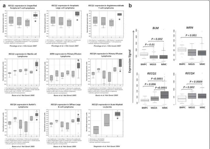
RECQ1 was found to be significantly overexpressed in
mantle cell lymphoma (P = 0.0025) [23], in unspecified
peripheral T-cell lymphoma (P = 0.0038) [24], anaplastic
large lymphoma (P = 0.0024) [24] and
angioimmunoblas-tic large cell lymphoma (P = 0.001) [24] (Fig. 1a).
WRN is significantly overexpressed in primary effusion
lymphoma compared to normal B cells (p = 0.003) [23]
(Fig. 1a).
RECQ4 expression was increased in Burkitt
Lymph-oma (P = 0.001) [23, 25], in diffuse large B cell
lymph-oma (P = 0.004) [23] and also in primary effusion
lymphoma (P = 0.005) [23] compared to normal
coun-terpart (Fig. 1a).
RECQ5 is significantly overexpressed in acute myeloid
leukemia (P = 0,001) [26] (Fig. 1a).
Comparing
RECQ helicases expression between normal
plasma cells (BMPC), premalignant cells from MGUS
pa-tients and multiple myeloma cells (MMC) [27],
BLM and
WRN were found to be significantly downregulated in
MMC compared to normal BMPC (P = 0.002 and P =
Table 1
RECQ family member expression in hematological malignancies compared to that of their normal tissue counterparts using
publicly available gene expression data, including the Oncomine Cancer Microarray and Genomicscape databases
Hematological Malignancies Datasets Gene overexpression compared to normal tissue counterpart RECQ1 BLM WRN RECQ4 RECQ5 Myeloid Neoplasms Acute Myeloid Leukemia 12 No No No No YES B-cell neoplasms Primary Effusion Lymphoma 8 No No YES YES No
Chronic Lymphocyte Leukemia 11 No No YES No No Diffuse Large B-cell Lymphoma 8 No No No YES No Burkitt's Lymphoma 8 No No No YES No Mantle Cell Lymphoma 11 YES No No No No Multiple Myeloma 1 YES YES YES YES No T-cell neoplasms Unspecified Peripheral T-cell Lymphoma 11 YES No No No No Anaplastic Large Cell Lymphoma 11 YES No No No No Angioimmunoblastic T-Cell Lymphoma 11 YES No No No No
0.001 respectively) (Fig. 1b). A decreased expression of
BLM was also observed in MGUS compared to BMPC
(P = 0.01). RECQ1 and RECQ4 are overexpressed in
MGUS (P = 0.006 and P = 0.002) and MMC (P < 0.0001
and
P = 0.0009) compared to BMPC. Furthermore, a
significant increased expression of
RECQ1 in MMC
compared to MGUS was identified (Fig. 1b).
Furthermore, using the human protein atlas database
[28–30], the expression of RECQ1, RECQ4 and RECQ5
could be confirmed at protein level in myeloid and
lymphoid cancer cell lines (Additional file 1: Figure S1).
We investigated whether
RECQ helicases expression
could be associated with tumor progression and
progno-sis in hematological malignancies (Table 2).
In AML patients with abnormal karyotype (Verhaak
cohort,
N = 521 patients) [31], a high expression of
BLM and RECQ4 is associated with a better overall
survival (OS) (P = 0.01 and P = 0.003). At the
oppos-ite, high
RECQ5 expression was linked with a poor
prognosis in the same cohort of patient (P = 0.008)
(Fig. 2a). In cox multivariate analysis, only
RECQ5
expression kept prognostic value (P = 0.01, hazard
ra-tio (HR) = 1.43).
In AML with normal karyotype, (Metzeler cohort,
N =
78) [32], gene expression of four
RECQ helicases out of
five were identified to predict for OS. High expression of
RECQ1 (P = 0.02), BLM (P = 0.01) and RECQ5 (P = 0.03)
were found to be associated with poor prognosis. In
con-trast, high
RECQ4 expression was linked with a better
out-come (P = 0.03) (Fig. 2b). When tested together in a cox
multivariate analysis, only
RECQ4 expression remained
significant (P = 0.009; HR = 0.4).
Interestingly,
RECQ5 overexpression was only
iden-tified in myeloid malignancies in association with an
adverse prognosis.
RECQ5 increased expression was
recently reported in JAK2V617F myeloproliferative
neoplasms [33]. RECQ5 depletion in
JAK2V617F-mutant cells impairs replication after hydroxyurea
treatment leading to a significant increased
double-stranded breaks and apoptosis [33]. RECQ5 represents
a potent regulator of genome stability in
myeloprolif-erative neoplasms in association with drug resistance
a
b
Fig. 1 Increased RECQ helicase gene expression in hematological malignancies compared to normal counterparts using Oncomine database (a) and Genomicscape database (b). Data sets in a given panel were from the same study. GEP data are log transformed (Oncomine) or not (Genomicscape) and normalized as previously described [64]
[33].
RECQ5 overexpression could also be involved in
AML pathophysiology and chemoresistance.
Even if RECQ1 mutations have been recently shown
to been associated with predisposition to breast cancer
[34, 35], no link between
RECQ1, BLM, WRN, and
RECQ4 deregulation and lymphoid malignancies were
previously reported.
In chronic lymphocytic leukemia (CLL), a poor
prog-nosis was linked with high
RECQ5 expression (P = 8E-8)
and a better outcome was associated with high
WRN
ex-pression (P = 0.0006) (Fig. 3a).
In a cohort of patients with follicular lymphoma (FL)
(Staudt cohort,
N = 180) [36], high RECQ1 and RECQ5
ex-pression represented adverse prognostic factors (P = 0.003
and
P = 0.0006 respectively) whereas RECQ4 expression
was found to be associated with a good prognosis (P =
0.009) (Fig. 3b). Interestingly,
RECQ1, RECQ4 and
RECQ5 expresison remained independent when tested
in cox multivariate analysis (P < 0.0001; HR = 2.8; P =
0.002; HR = 0.49 and
P < 0.01; HR = 1.78 respectively).
In diffuse large B cell lymphoma (DLBCL), only
RECQ5
expression was associated with a prognostic value. Low
RECQ5 expression was a poor prognostic marker in two
independent cohorts of patients (P = 0.02 in a cohort of
patients treated by combination of cyclophosphamide,
doxorubicin, vincristine and prednisone (CHOP)
ther-apy (N = 181) and P = 0.01 in a cohort of patients
treated by Rituximab combined with CHOP (R-CHOP)
regimen (N = 233)) (Fig. 3c) [37].
In MM, high
RECQ1, WRN and RECQ4 expression are
associated with an adverse prognosis in the UAMS
co-hort treated with total therapy 2 (Fig. 4) [27].
These data demonstrate a link between
RECQ
heli-case expression and a prognostic value in different
hematological malignancies.
Hematological malignancies are characterized by
gen-omic instability that could be related to defects in DNA
repair [21]. The RECQ family of DNA helicases is a
fam-ily of conserved enzymes that display highly specialized
and vital roles in the maintenance of genome stability.
Mutations in three of the five human RECQ helicases,
BLM, WRN and RECQL4, are associated with genetic
disorders characterized by chromosomal instability and
increased susceptibility to cancer including leukemia
[38, 39]. Mutations in
BLM result in a dramatic lowering
of
BLM mRNA levels and premature termination of
pro-tein translation owing to nonsense-mediated mRNA
decay [1, 40]. Patients with Bloom syndrome exhibit
cancer predisposition including most types of cancers
and particularly non-Hodgkin’s lymphoma, leukemias
and carcinomas of skin, breast and colon [1].
Interest-ingly, a low
BLM expression is associated with a poor
prognosis in AML with complex karyotypes (Fig. 2a).
The cancer spectrum observed in patients with Werner’s
syndrome is characterized mainly by cancers of
mesenchy-mal origin and some epithelial cancers [1, 41]. RECQ4
mutations are found in Rothmund-Thomson syndrome,
RAPADILINO syndrome and Baller-Gerold syndrome
[1, 41]. Rothmund-Thomson syndrome are
character-ized by predisposition to mainly osteosarcoma whereas
RAPADILINO syndrome are linked with lymphoma
and osteosarcoma predisposition [1].
Specific recurrent chromosomal translocations have
been associated with DNA repair deficiencies linked
with repression of DDR (DNA damage response) genes
in AML [42]. In PML-RARA, PML and BLM are
delo-calized from the nuclear bodies into microspeckled
nu-clear regions [43]. All trans retinoic acid (ATRA)
treatment of APL patients leads to degradation of
PML-RARA and relocalization of BLM to nuclear
bod-ies [43] suggesting that PML-RARA are involved in
genomic instability in APL through disruption of BLM
and PML localization and activity. Interestingly, we
re-ported that low
BLM and RECQ4 expression are
associ-ated with a poor prognosis in AML with abnormal
karyotype (Fig. 2a), suggesting that downregulation of
RECQ helicases could be involved in leukemogenesis
and genomic instability. At the opposite, in AML with
normal karyotype,
RECQ1, BLM and RECQ5 high
ex-pression are associated with a poor prognosis (Fig. 2b).
As reported in solid cancer,
RECQ helicase
overexpres-sion could be a marker of chemoresistance and higher
Table 2 Link between
RECQ helicase gene expression and prognostic value in hematological malignancies
RECQ helicase
Prognostic value
AML LLC FL DBLCL MM
normal karyotype abnormal karyotype
WRN - - GOOD GOOD - BAD
BLM BAD GOOD - - -
-RECQ1 BAD - - BAD - BAD
RECQ4 GOOD GOOD - GOOD - BAD
RECQ5 BAD - BAD BAD GOOD
-GOOD : A highRECQ helicase expression is associated with a better outcome BAD : A highRECQ helicase expression is associated with a poor prognosis
proliferation helping AML cells to deal with replication
stress [44, 45]. B lymphocytes are continuously
pro-duced during adult life and they undergo different
gen-etic alterations associated with DNA breaks, including
VDJ recombination, Ig class switch recombination and
somatic hypermutation. These mechanisms must be
tightly regulated to prevent tumorigenesis and ensure
efficient immune response [46]. Collapsed DNA
repli-cation forks occurring in rapidly dividing lymphocytes
leads to a restart failure and results in an interruption
of the normal developmental program [47]. HR is
re-quired for lymphoid development [47]. Aberrations
a
b
Fig. 2 Overall survival related to RECQ helicase gene expression in acute myeloid leukemia with abnormal karyotype (a) and normal karyotype (b). The prognostic value of gene expression was determined using the MaxStat R function in R software. The overall survival of subgroups of patients was compared with the log-rank test and survival curves computed with the Kaplan–Meier method (R software) [66]
affecting HR actors are correlated with genomic
in-stability in B cell cancers [48]. By their involvement in
HR and also by their ability to resolve and to continue
the normal fork replication after DNA damage or
rep-lication fork arrest, WRN [49], BLM [4], RECQ1 [12]
and RECQ5 [50] helicases might be crucial in
lymph-oid development and aberration in their expression or
function can lead to cancer genesis. Interestingly, low
expression of WRN in CLL and FL, low
RECQ4
ex-pression in FL and low
RECQ5 in DLBCL are
associ-ated with a poor prognosis (Fig. 3). Furthermore, high
RECQ5 expression in CLL and FL and high RECQ1
ex-pression in FL are associated with a poor prognosis
and could be involved in chemoresistance. In
lymph-oma, deregulation of DDR is associated with
tumori-genesis [51, 52], poor prognosis [53, 54] and could
represent a potent therapeutic target [53, 55, 56].
In MM, patients with extensive chromosomal
instabil-ity and replicative stress are associated with an adverse
outcome [27, 57–59]. Accordingly, high RECQ1, WRN
and
RECQ4 expression is associated with a significant
poor survival in MM patients (Fig. 4). Although
WRN
was found to be significantly downregulated in MMC
compared to normal BMPC (Fig. 1b), patients with high
expression display a poor prognosis.
WRN is located on
chromosome 8p deleted in 25 % of MM patients without
prognostic value [60]. These data could explain the
sig-nificant downregulation of
WRN expression in MM
compared to normal BMPC.
Recently, a set of molecule inhibitors of WRN and
BLM was characterized [61, 62]. These new molecules
could open up new therapeutic strategies for targeting
hematological malignancies characterized by RECQ
heli-case deregulation and a poor prognosis.
Conclusion
The analysis reported here demonstrates that
RECQ
helicase expression is deregulated in hematological
malignancies compared to their normal counterparts
in association with a prognostic value in AML, CLL,
lymphoma and MM. Deregulation of RECQ helicases
appears to play a role in tumorigenesis and could be
involved in genomic instability and chemoresistance
in hematological malignancies. RECQ helicases
repre-sent potent therapeutic targets for synthetic lethal
approaches.
Methods
Databases: We used Oncomine Cancer Microarray
database (http://www.oncomine.org) [22] and
Geno-micscape (http://genoGeno-micscape.com/) [63] to study
gene expression of RECQ family members in nine
dif-ferent human hematological malignancies and their
normal tissue counterpart as indicated in Table 1. To
compare the gene expression of a tumor type to its
normal counterpart, we used gene expression data
from a same study with the same methodology. All
data were log transformed, median centered per array,
Fig. 3 Overall survival related toRECQ helicase gene expression in chronic lymphocytic leukemia (a), follicular lymphoma (b) and diffuse large B cell lymphoma (c)
and the standard deviation was normalized to one per
array [22].
Statistical comparisons were done with Mann–Whitney
or Student
t-test as previously published [64].
Prognosis values of each member of RECQ family in
hematological malignancies were determined by using
Maxstat R package based on publicly available data
(Gene Expression Omnibus
(http://www.ncbi.nlm.nih.-gov/geo/); accession numbers GSE6891, GSE12417,
GSE22762, GSE16131, GSE10846 and GSE4581) analyzed
with Genomicscape [63] as previously reported [65].
Additional file
Additional file 1: Figure S1. RECQ helicase gene and protein expression in myeloid and lymphoid cell lines using using the human protein atlas database. RECQ1, RECQ4 and RECQ5 expression could be confirmed at protein level in myeloid and lymphoid cancer cell lines. (PDF 599 kb)
Competing interests
The authors declare that they have no competing interests.
Authors’ contributions
EV performed research, bioinformatics, and participated in the writing of the paper. AK participated in the bioinformatics. PP participated in the writing of the paper. BK participated in the research and in the writing of the paper. JM supervised the research, bioinformatics, and the writing of the paper. All authors read and approved the final manuscript.
Acknowledgements
This work was supported by grants from French INCA (Institut National du Cancer) Institute (2012-109/087437), Languedoc Roussillon CRLR (R14026FF), Fondation de France (201400047510), ITMO Cancer (MM&TT) and AXLR SATT (30041633). EV is supported by a grant from Guillaume Espoir association (Saint- Genis-Laval, France).
Author details
1Laboratory for Monitoring Innovative Therapies, Department of Biological
Hematology, Hôpital Saint-Eloi - CHRU de Montpellier, 80, av. Augustin Fliche, 34295 Montpellier, Cedex 5, France.2Institute of Human Genetics, CNRS-UPR1142, Montpellier F-34396, France.3University of Montpellier 1, UFR
de Médecine, Montpellier, France.
Received: 5 November 2015 Accepted: 8 February 2016
References
1. Chu WK, Hickson ID. RecQ helicases: multifunctional genome caretakers. Nat Rev Cancer. 2009;9(9):644–54. doi:10.1038/nrc2682.
2. Larsen NB, Hickson ID. RecQ Helicases: Conserved Guardians of Genomic Integrity. Adv Exp Med Biol. 2013;767:161–84. doi:10.1007/978-1-4614-5037-5_8. Fig. 4 Overall survival related toRECQ helicase gene expression in multiple myeloma
3. Weinert BT, Rio DC. DNA strand displacement, strand annealing and strand swapping by the Drosophila Bloom's syndrome helicase. Nucleic Acids Res. 2007;35(4):1367–76. doi:10.1093/nar/gkl831.
4. Sung P, Klein H. Mechanism of homologous recombination: mediators and helicases take on regulatory functions. Nat Rev Mol Cell Biol. 2006;7(10): 739–50. 10. 1038/nrm2008.
5. Drosopoulos WC, Kosiyatrakul ST, Schildkraut CL. BLM helicase facilitates telomere replication during leading strand synthesis of telomeres. J Cell Biol. 2015;210(2):191–208. doi:10.1083/jcb.201410061.
6. Kamath-Loeb A, Loeb LA, Fry M. The Werner syndrome protein is distinguished from the Bloom syndrome protein by its capacity to tightly bind diverse DNA structures. PLoS One. 2012;7(1):e30189. doi:10.1371/ journal.pone.0030189.
7. Damerla RR, Knickelbein KE, Strutt S, Liu FJ, Wang H, Opresko PL. Werner syndrome protein suppresses the formation of large deletions during the replication of human telomeric sequences. Cell Cycle. 2012;11(16):3036–44. doi:10.4161/cc.21399.
8. Opresko PL, Mason PA, Podell ER, Lei M, Hickson ID, Cech TR, et al. POT1 stimulates RecQ helicases WRN and BLM to unwind telomeric DNA substrates. J Biol Chem. 2005;280(37):32069–80. doi:10.1074/jbc.M505211200. 9. Chen L, Huang S, Lee L, Davalos A, Schiestl RH, Campisi J, et al. WRN, the
protein deficient in Werner syndrome, plays a critical structural role in optimizing DNA repair. Aging Cell. 2003;2(4):191–9.
10. Kobayashi J, Okui M, Asaithamby A, Burma S, Chen BP, Tanimoto K, et al. WRN participates in translesion synthesis pathway through interaction with NBS1. Mech Ageing Dev. 2010;131(6):436–44. doi:10.1016/j.mad.2010.06.005. 11. Karmakar P, Snowden CM, Ramsden DA, Bohr VA. Ku heterodimer binds to
both ends of the Werner protein and functional interaction occurs at the Werner N-terminus. Nucleic Acids Res. 2002;30(16):3583–91.
12. Berti M, Ray Chaudhuri A, Thangavel S, Gomathinayagam S, Kenig S, Vujanovic M, et al. Human RECQ1 promotes restart of replication forks reversed by DNA topoisomerase I inhibition. Nat Struct Mol Biol. 2013;20(3): 347–54. doi:10.1038/nsmb.2501.
13. Lu X, Parvathaneni S, Hara T, Lal A, Sharma S. Replication stress induces specific enrichment of RECQ1 at common fragile sites FRA3B and FRA16D. Mol Cancer. 2013;12(1):29. doi:10.1186/1476-4598-12-29.
14. Popuri V, Croteau DL, Brosh Jr RM, Bohr VA. RECQ1 is required for cellular resistance to replication stress and catalyzes strand exchange on stalled replication fork structures. Cell Cycle. 2012;11(22):4252–65. doi:10.4161/ cc.22581.
15. Li XL, Lu X, Parvathaneni S, Bilke S, Zhang H, Thangavel S, et al. Identification of RECQ1-regulated transcriptome uncovers a role of RECQ1 in regulation of cancer cell migration and invasion. Cell Cycle. 2014;13(15):2431–45. 16. Sekelsky JJ, Brodsky MH, Rubin GM, Hawley RS. Drosophila and human
RecQ5 exist in different isoforms generated by alternative splicing. Nucleic Acids Res. 1999;27(18):3762–9.
17. Hu Y, Lu X, Barnes E, Yan M, Lou H, Luo G. Recql5 and Blm RecQ DNA helicases have nonredundant roles in suppressing crossovers. Mol Cell Biol. 2005;25(9):3431–42. doi:10.1128/MCB.25.9.3431-3442.2005.
18. Hansen GM, Skapura D, Justice MJ. Genetic profile of insertion mutations in mouse leukemias and lymphomas. Genome Res. 2000;10(2):237–43. 19. Saponaro M, Kantidakis T, Mitter R, Kelly GP, Heron M, Williams H, et al.
RECQL5 controls transcript elongation and suppresses genome instability associated with transcription stress. Cell. 2014;157(5):1037–49. doi:10.1016/j. cell.2014.03.048.
20. Rezazadeh S. RecQ helicases; at the crossroad of genome replication, repair, and recombination. Mol Biol Rep. 2011. doi:10.1007/s11033-011-1243-y 21. Economopoulou P, Pappa V, Papageorgiou S, Dervenoulas J,
Economopoulos T. Abnormalities of DNA repair mechanisms in common hematological malignancies. Leuk Lymphoma. 2011;52(4):567–82. doi:10. 3109/10428194.2010.551155.
22. Rhodes DR, Yu J, Shanker K, Deshpande N, Varambally R, Ghosh D, et al. ONCOMINE: a cancer microarray database and integrated data-mining platform. Neoplasia. 2004;6(1):1–6.
23. Basso K, Margolin AA, Stolovitzky G, Klein U, Dalla-Favera R, Califano A. Reverse engineering of regulatory networks in human B cells. Nat Genet. 2005;37(4):382–90. doi:10.1038/ng1532.
24. Piccaluga PP, Agostinelli C, Califano A, Rossi M, Basso K, Zupo S, et al. Gene expression analysis of peripheral T cell lymphoma, unspecified, reveals distinct profiles and new potential therapeutic targets. J Clin Invest. 2007; 117(3):823–34. doi:10.1172/JCI26833.
25. Brune V, Tiacci E, Pfeil I, Doring C, Eckerle S, van Noesel CJ, et al. Origin and pathogenesis of nodular lymphocyte-predominant Hodgkin lymphoma as revealed by global gene expression analysis. J Exp Med. 2008;205(10):2251–68. doi:10.1084/jem.20080809.
26. Stegmaier K, Ross KN, Colavito SA, O'Malley S, Stockwell BR, Golub TR. Gene expression-based high-throughput screening(GE-HTS) and application to leukemia differentiation. Nat Genet. 2004;36(3):257–63. doi:10.1038/ng1305. 27. Zhan F, Huang Y, Colla S, Stewart JP, Hanamura I, Gupta S, et al. The
molecular classification of multiple myeloma. Blood. 2006;108(6):2020–8. 28. Uhlen M, Fagerberg L, Hallstrom BM, Lindskog C, Oksvold P, Mardinoglu A,
et al. Proteomics. Tissue-based map of the human proteome. Science. 2015; 347(6220):1260419. doi:10.1126/science.1260419.
29. Ponten F, Jirstrom K, Uhlen M. The Human Protein Atlas–a tool for pathology. J Pathol. 2008;216(4):387–93. doi:10.1002/path.2440.
30. Uhlen M, Bjorling E, Agaton C, Szigyarto CA, Amini B, Andersen E, et al. A human protein atlas for normal and cancer tissues based on antibody proteomics. Mol Cell Proteomics. 2005;4(12):1920–32. doi:10.1074/mcp.M500279-MCP200. 31. Verhaak RG, Wouters BJ, Erpelinck CA, Abbas S, Beverloo HB, Lugthart S, et
al. Prediction of molecular subtypes in acute myeloid leukemia based on gene expression profiling. Haematologica. 2009;94(1):131–4. doi:10.3324/ haematol.13299.
32. Metzeler KH, Maharry K, Radmacher MD, Mrozek K, Margeson D, Becker H, et al. TET2 mutations improve the new European LeukemiaNet risk
classification of acute myeloid leukemia: a Cancer and Leukemia Group B study. J Clin Oncol. 2011;29(10):1373–81. doi:10.1200/JCO.2010.32.7742. 33. Chen E, Ahn JS, Sykes DB, Breyfogle LJ, Godfrey AL, Nangalia J, et al. RECQL5
Suppresses Oncogenic JAK2-Induced Replication Stress and Genomic Instability. Cell reports. 2015;13(11):2345–52. doi:10.1016/j.celrep.2015.11.037. 34. Sun J, Wang Y, Xia Y, Xu Y, Ouyang T, Li J, et al. Mutations in RECQL Gene
Are Associated with Predisposition to Breast Cancer. PLoS Genet. 2015;11(5): e1005228. doi:10.1371/journal.pgen.1005228.
35. Cybulski C, Carrot-Zhang J, Kluzniak W, Rivera B, Kashyap A, Wokolorczyk D, et al. Germline RECQL mutations are associated with breast cancer susceptibility. Nat Genet. 2015;47(6):643–6. doi:10.1038/ng.3284. 36. Leich E, Salaverria I, Bea S, Zettl A, Wright G, Moreno V, et al. Follicular
lymphomas with and without translocation t(14;18) differ in gene expression profiles and genetic alterations. Blood. 2009;114(4):826–34. doi: 10.1182/blood-2009-01-198580.
37. Lenz G, Wright G, Dave SS, Xiao W, Powell J, Zhao H, et al. Stromal gene signatures in large-B-cell lymphomas. N Engl J Med. 2008;359(22):2313–23. doi:10.1056/NEJMoa0802885.
38. Suhasini AN, Brosh Jr RM. Fanconi anemia and Bloom's syndrome crosstalk through FANCJ-BLM helicase interaction. Trends Genet. 2012;28(1):7–13. doi: 10.1016/j.tig.2011.09.003.
39. Poppe B, Van Limbergen H, Van Roy N, Vandecruys E, De Paepe A, Benoit Y, et al. Chromosomal aberrations in Bloom syndrome patients with myeloid malignancies. Cancer Genet Cytogenet. 2001;128(1):39–42.
40. German J, Sanz MM, Ciocci S, Ye TZ, Ellis NA. Syndrome-causing mutations of the BLM gene in persons in the Bloom's Syndrome Registry. Hum Mutat. 2007;28(8):743–53. doi:10.1002/humu.20501.
41. Goto M, Miller RW, Ishikawa Y, Sugano H. Excess of rare cancers in Werner syndrome (adult progeria). Cancer Epidemiol Biomarkers Prev. 1996;5(4):239–46. 42. Esposito MT, So CW. DNA damage accumulation and repair defects in acute
myeloid leukemia: implications for pathogenesis, disease progression, and chemotherapy resistance. Chromosoma. 2014;123(6):545–61. doi:10.1007/ s00412-014-0482-9.
43. Zhong S, Hu P, Ye TZ, Stan R, Ellis NA, Pandolfi PP. A role for PML and the nuclear body in genomic stability. Oncogene. 1999;18(56):7941–7. doi:10.1038/sj.onc.1203367.
44. Sanada S, Futami K, Terada A, Yonemoto K, Ogasawara S, Akiba J, et al. RECQL1 DNA repair helicase: a potential therapeutic target and a proliferative marker against ovarian cancer. PLoS One. 2013;8(8):e72820. doi: 10.1371/journal.pone.0072820.
45. Matsushita Y, Yokoyama Y, Yoshida H, Osawa Y, Mizunuma M, Shigeto T, et al. The level of RECQL1 expression is a prognostic factor for epithelial ovarian cancer. J Ovarian Res. 2014;7:107. doi:10.1186/s13048-014-0107-1. 46. Gennery AR, Cant AJ, Jeggo PA. Immunodeficiency associated with DNA
repair defects. Clin Exp Immunol. 2000;121(1):1–7.
47. Caddle LB, Hasham MG, Schott WH, Shirley BJ, Mills KD. Homologous recombination is necessary for normal lymphocyte development. Mol Cell Biol. 2008;28(7):2295–303. doi:10.1128/MCB.02139-07.
48. Hasham MG, Donghia NM, Coffey E, Maynard J, Snow KJ, Ames J, et al. Widespread genomic breaks generated by activation-induced cytidine deaminase are prevented by homologous recombination. Nat Immunol. 2010;11(9):820–6. doi:10.1038/ni.1909.
49. Sidorova JM, Li N, Folch A, Monnat Jr RJ. The RecQ helicase WRN is required for normal replication fork progression after DNA damage or replication fork arrest. Cell Cycle. 2008;7(6):796–807.
50. Yin L. Chondroitin synthase 1 is a key molecule in myeloma cell-osteoclast interactions. J Biol Chem. 2005;280(16):15666–72.
51. Gu X, Booth CJ, Liu Z, Strout MP. AID-associated DNA repair pathways regulate malignant transformation in a murine model of BCL6-driven diffuse large B cell lymphoma. Blood. 2015. doi:10.1182/blood-2015-02-628164 52. Hathcock KS, Padilla-Nash HM, Camps J, Shin DM, Triner D, Shaffer AL, 3rd
et al. ATM deficiency in absence of T cells promotes development of NF-kB-dependent murine B cell lymphomas that resemble human ABC DLBCL. Blood. 2015. doi:10.1182/blood-2015-06-654749
53. Bret C, Klein B, Cartron G, Schved JF, Constantinou A, Pasero P et al. DNA repair in diffuse large B-cell lymphoma: a molecular portrait. Br J Haematol. 2014. doi:10.1111/bjh.13206
54. Bret C, Klein B, Moreaux J. Gene expression-based risk score in diffuse large B-cell lymphoma. Oncotarget. 2012;3(12):1700–10.
55. Kwok M, Davies N, Agathanggelou A, Smith E, Petermann E, Yates E, et al. Synthetic lethality in chronic lymphocytic leukaemia with DNA damage response defects by targeting the ATR pathway. Lancet. 2015;385 Suppl 1: S58. doi:10.1016/S0140-6736(15)60373-7.
56. Bret C, Klein B, Moreaux J. Nucleotide excision DNA repair pathway as a therapeutic target in patients with high-risk diffuse large B cell lymphoma. Cell Cycle. 2013;12(12):1811–2. doi:10.4161/cc.25115.
57. Cottini F, Hideshima T, Suzuki R, Tai YT, Bianchini G, Richardson PG, et al. Synthetic Lethal Approaches Exploiting DNA Damage in Aggressive Myeloma. Cancer Discov. 2015;5(9):972–87. doi:10.1158/2159-8290.CD-14-0943. 58. Hose D, Reme T, Hielscher T, Moreaux J, Messner T, Seckinger A, et al.
Proliferation is a central independent prognostic factor and target for personalized and risk-adapted treatment in multiple myeloma. Haematologica. 2011;96(1):87–95. doi:10.3324/haematol.2010.030296. 59. Kassambara A, Gourzones-Dmitriev C, Sahota S, Reme T, Moreaux J,
Goldschmidt H, et al. A DNA repair pathway score predicts survival in human multiple myeloma: the potential for therapeutic strategy. Oncotarget. 2014;5(9):2487–98.
60. Walker BA, Leone PE, Chiecchio L, Dickens NJ, Jenner MW, Boyd KD, et al. A compendium of myeloma-associated chromosomal copy number abnormalities and their prognostic value. Blood. 2010;116(15):e56–65. doi:10. 1182/blood-2010-04-279596.
61. Aggarwal M, Banerjee T, Sommers JA, Brosh Jr RM. Targeting an Achilles' heel of cancer with a WRN helicase inhibitor. Cell Cycle. 2013;12(20):3329–35. doi:10.4161/cc.26320.
62. Nguyen GH, Dexheimer TS, Rosenthal AS, Chu WK, Singh DK, Mosedale G, et al. A small molecule inhibitor of the BLM helicase modulates chromosome stability in human cells. Chem Biol. 2013;20(1):55–62. doi:10. 1016/j.chembiol.2012.10.016.
63. Kassambara A, Reme T, Jourdan M, Fest T, Hose D, Tarte K, et al. GenomicScape: an easy-to-use web tool for gene expression data analysis. Application to investigate the molecular events in the differentiation of B cells into plasma cells. PLoS Comput Biol. 2015;11(1):e1004077. doi:10.1371/ journal.pcbi.1004077.
64. Kassambara A, Klein B, Moreaux J. MMSET is overexpressed in cancers: link with tumor aggressiveness. Biochem Biophys Res Commun. 2009;379(4): 840–5. doi:10.1016/j.bbrc.2008.12.093.
65. Kassambara A, Hose D, Moreaux J, Walker BA, Protopopov A, Reme T, et al. Genes with a spike expression are clustered in chromosome (sub)bands and spike (sub)bands have a powerful prognostic value in patients with multiple myeloma. Haematologica. 2012;97(4):622–30. doi:10.3324/haematol.2011.046821. 66. Moreaux J, Kassambara A, Hose D, Klein B. STEAP1 is overexpressed in
cancers: A promising therapeutic target. Biochem Biophys Res Commun. 2012;429(3-4):148–55. doi:10.1016/j.bbrc.2012.10.123.
• We accept pre-submission inquiries
• Our selector tool helps you to find the most relevant journal • We provide round the clock customer support
• Convenient online submission • Thorough peer review
• Inclusion in PubMed and all major indexing services • Maximum visibility for your research
Submit your manuscript at www.biomedcentral.com/submit