Bimodal rheotactic behavior reflects flagellar
beat asymmetry in human sperm cells
The MIT Faculty has made this article openly available.
Please share
how this access benefits you. Your story matters.
Citation
Bukatin, Anton et al. “Bimodal Rheotactic Behavior Reflects
Flagellar Beat Asymmetry in Human Sperm Cells.” Proceedings of
the National Academy of Sciences 112.52 (2015): 15904–15909. ©
2017 National Academy of Sciences
As Published
http://dx.doi.org/10.1073/pnas.1515159112
Publisher
National Academy of Sciences (U.S.)
Version
Final published version
Citable link
http://hdl.handle.net/1721.1/110648
Terms of Use
Article is made available in accordance with the publisher's
policy and may be subject to US copyright law. Please refer to the
publisher's site for terms of use.
Bimodal rheotactic behavior reflects flagellar beat
asymmetry in human sperm cells
Anton Bukatina,b,1, Igor Kukhtevichb,c,1, Norbert Stoopd,1, Jörn Dunkeld,2, and Vasily Kantslere
aSt. Petersburg Academic University, St. Petersburg 194021, Russia;bInstitute for Analytical Instrumentation of the Russian Academy of Sciences,
St. Petersburg 198095, Russia;cITMO University, St. Petersburg 197101, Russia;dDepartment of Mathematics, Massachusetts Institute of Technology,
Cambridge, MA 02139-4307; andeDepartment of Physics, University of Warwick, Coventry CV4 7AL, United Kingdom
Edited by Charles S. Peskin, New York University, New York, NY, and approved November 9, 2015 (received for review July 30, 2015)
Rheotaxis, the directed response to fluid velocity gradients, has been shown to facilitate stable upstream swimming of mamma-lian sperm cells along solid surfaces, suggesting a robust physical mechanism for long-distance navigation during fertilization. How-ever, the dynamics by which a human sperm orients itself relative to an ambient flow is poorly understood. Here, we combine micro-fluidic experiments with mathematical modeling and 3D flagellar beat reconstruction to quantify the response of individual sperm cells in time-varying flow fields. Single-cell tracking reveals two kinematically distinct swimming states that entail opposite turning behaviors under flow reversal. We constrain an effective 2D model for the turning dynamics through systematic large-scale parameter scans, and find good quantitative agreement with experiments at different shear rates and viscosities. Using a 3D reconstruction algorithm to identify the flagellar beat patterns causing left or right turning, we present comprehensive 3D data demonstrating the rolling dynamics of freely swimming sperm cells around their longitudinal axis. Contrary to cur-rent beliefs, this 3D analysis uncovers ambidextrous flagellar wave-forms and shows that the cell’s turning direction is not defined by the rolling direction. Instead, the different rheotactic turning behaviors are linked to a broken mirror symmetry in the midpiece section, likely arising from a buckling instability. These results challenge current theoretical models of sperm locomotion.
sperm swimming
|
rheotaxis|
fluid dynamics|
microfluidics|
simulationsT
axis, the directed kinematic response to external signals, is a defining feature of living things that affects their reproduc-tion, foraging, migrareproduc-tion, and survival strategies (1–4). Higher organisms rely on sophisticated networks of finely tuned sensory mechanisms to move efficiently in the presence of chemical or physical stimuli. However, various fundamental forms of taxis are already manifest at the unicellular level, ranging from che-motaxis in bacteria (5) and phototaxis in unicellular green algae (2) to the mechanical response (durotaxis) of fibroblasts (6) and rheotaxis (7, 8) in spermatozoa (3, 9–12). Over the last few de-cades, much progress has been made in deciphering chemotactic, phototactic, and durotactic pathways in prokaryotic and eukaryotic model systems. In contrast, comparatively little is known about the physical mechanisms that enable flow gradient sensing in sperm cells (3, 9–13). Recent studies (3, 12) suggest that mamma-lian sperm use rheotaxis for long-distance navigation, but it remains unclear how shear flows alter flagellar beat patterns in the vicinity of surfaces and, in particular, how such changes in the beat dynamics affect the steering process. Answering these questions will be es-sential for evaluating the importance of chemical (14) and physical (4) signals during mammalian fertilization (15–17).A necessary requirement for any form of directed kinematic response is the ability to change the direction of locomotion. Multiflagellate bacteria achieve this feat by varying their motor activity, resulting in alternating phases of entangled and disen-tangled flagellar dynamics that give rise to run-and-tumble motion (5). A similar mechanism was recently discovered in the biflagellate eukaryote Chlamydomonas reinhardtii (18). This unicellular green alga actively redirects its swimming motion through occasional desynchronization of its two cilia (19), although it is still debated
whether this effect is of mechanical (20) or hydrodynamic (21, 22) origin. Experiments (23) show that the alga’s reorientation dynamics can lead to localization in shear flow (24, 25), with potentially profound implications in marine ecology. In contrast to taxis in multiflagellate organisms (2, 5, 18, 26, 27), the navi-gation strategies of uniflagellate cells are less well understood. For instance, it was discovered only recently that uniflagellate marine bacteria, such as Vibrio alginolyticus and Pseudoalteromonas haloplanktis, use a buckling instability in their lone flagellum to change their swimming direction (28). However, as passive pro-karyotic flagella differ fundamentally from their active eupro-karyotic counterparts, it is unclear to what extent such insights translate to spermatozoa.
Earlier studies of human sperm locomotion have identified several potential steering and transport mechanisms, including thermotaxis (4), uterine peristalsis (29, 30), and chemotaxis (14, 16, 31), but their relative importance has yet to be quantified. Recent experiments (3, 32, 33) demonstrate that rheotaxis, combined with steric surface alignment (12, 34), enables robust long-distance navigation by turning sperm cells preferentially against an externally imposed flow direction (9, 10), but how exactly this realignment process happens is unknown. It has been suggested (32, 35, 36) that the intrinsic curvature or chiral beat dynamics (37, 38) of the flagellum could play an essential role in rheotactic steering, but this remains to be confirmed in experi-ments. Similarly, an increasing number of theoretical models (36, 39–47) still await empirical validation, because 3D data for the beat pattern of sperm swimming close to surfaces has been lacking. To examine the dynamics of human sperm rheotaxis quantita-tively, we here combine microfluidic experiments with mathematical modeling and 3D flagellar beat reconstruction. Single-cell tracking
Significance
Successful sperm navigation is essential for sexual reproduc-tion, yet we still understand relatively little about how sperm cells are able to adapt their swimming motion in response to chemical and physical cues. This lack of knowledge is owed to the fact that it has been difficult to observe directly the full 3D dynamics of the whip-like flagellum that propels the cell through the fluid. To overcome this deficiency, we apply a new algorithm to reconstruct the 3D beat patterns of human sperm cells in experiments under varying flow conditions. Our anal-ysis reveals that the swimming strokes of human sperm are considerably more complex than previously thought, and that sperm may use their heads as rudders to turn right or left. Author contributions: A.B., I.K., N.S., J.D., and V.K. designed research; A.B., I.K., N.S., J.D., and V.K. performed research; A.B., I.K., N.S., J.D., and V.K. analyzed data; and N.S., J.D., and V.K. wrote the paper.
The authors declare no conflict of interest. This article is a PNAS Direct Submission.
Freely available online through the PNAS open access option. 1A.B., I.K., and N.S. contributed equally.
2To whom correspondence should be addressed. Email: [email protected]. This article contains supporting information online atwww.pnas.org/lookup/suppl/doi:10. 1073/pnas.1515159112/-/DCSupplemental. BIOPHYSICS AND COMPUTATION AL BIOLOGY
reveals the existence of two kinematically distinct swimming states that result in opposite turning behaviors under flow reversal. We quantify this effect for a range of viscosities and shear rates, and use these comprehensive data to constrain an effective 2D model through a systematic large-scale scan (>6,000 parameter com-binations). To identify the details of the flagellar beat dynamics during rheotaxis, we developed an algorithm that translates 2D intensity profiles into 3D positional data. Our 3D analysis con-firms that human sperm perform a rolling motion (48), charac-terized by weakly nonplanar beat patterns and a rotating beat plane. However, contrary to current beliefs, we find that neither the rolling direction nor beat helicity determine the turning direction after flow reversal. Instead, the rheotactic turning behavior corre-lates with a previously unrecognized asymmetry in the midpiece, likely caused by a buckling instability. These findings call for a re-vision and extension of current models (36, 39–44, 46).
Results
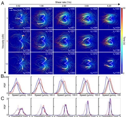
Reorientation Dynamics After Flow Reversal. Understanding how efficiently a sperm cell can react to directional changes in ambient fluid flows is a first step toward evaluating the importance of uterine peristalsis on sperm transport during reproduction (29, 30). To examine the response of human sperm after a sudden flow reversal, we tracked individual cells in microfluidic channels (Experimental Details) at three different kinematic viscositiesν = 1 centi-Stokes (cSt), 3 cSt, and 12 cSt, and five different shear rates, _γ = 0.52 s−1, 1.56 s−1, 2.60 s−1, 3.64 s−1, and 5.20 s−1(Fig. 1). Hydrodynamic and steric forces cause sperm to accumulate near solid boundaries (12), where they remain trapped for several minutes while being exposed to a locally linear normal flow gradient. In our experiments, cells
generally accumulated at distances of<10 μm from the wall. We therefore fixed the focal plane parallel to the upper wall of the microfluidic chamber, using a large depth of field to track all cells within distance 10μm from the wall. The results presented below are thus integrated measurements over this accumulation layer. At time t< 0, a constant external flow field was applied in negative x direction, causing the cells to align preferentially in positive x di-rection (3, 32). At t= 0, the flow direction was rapidly reversed (switching time K 1 s), and the motions of 300 to 1,000 randomly selected sperm cells were tracked for a period of>30 s for each parameter pair (ν, _γ).
Trajectory analysis shows that approximately half of the tracked cells respond to flow reversal by making a right turn whereas the other half pursue a left turn (Fig. 1A andMovies S1–S6). In both cases, the majority of cells perform a complete U-turn, provided the shear rate is sufficiently large _γ > 1.56 s−1. As the value of _γ is increased, the characteristic curvature of the U-turns also in-creases, and the spread around the mean trajectories, obtained by averaging positions at equal time t> 0, is reduced (thick white-shaded lines in Fig. 1A).
The initial speed distributions, measured at the moment of the flow switch t= 0, show little variation between left-turning and right-turning cells (Fig. 1B). As expected, the maximum of the speed distribution is shifted to a lower value at high viscosity (blue curves in Fig. 1B). Strikingly, the initial offset anglesφð0Þ of left-turning and right-turning individuals are bimodally distrib-uted, suggesting that exposure to constant flow for t< 0 separates two different alignment modes that become magnified during a flow reversal (Fig. 1C).
Fig. 1. Turning behavior of human sperm under flow reversal reveals two kinematically distinct swimming states. (A) Trajectories of individual sperm cells swimming close to the channel boundary in theðx, yÞ plane, with initial positions superimposed at time t = 0 (viewed from inside the channel). Equal-time trajectory averages for left- and right-turning cells are shown as thick white-shaded lines. Flow was reversed at t= 0, pointing in positive x direction for t > 0 (white arrow). The shear velocity increases linearly in z direction. Color encodes time. (Scale bar, 200μm.) (B) Normalized speed distributions before flow reversal at time t = 0. Faint lines indicate left-turning cells, and the other lines indicate right-turning cells. (C) Distribution of the orientation anglesφð0Þ, measured relative to the x axis before flow reversal at time t= 0, signals two kinematically distinct cell populations. Colors in B and C indicate different viscosities (black, 1 cSt; red, 3 cSt; blue, 12 cSt).
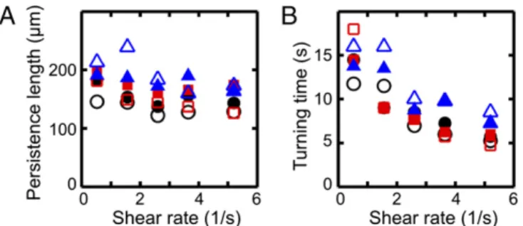
To characterize the typical distance scale associated with the turning process, we consider the downstream persistence length Λ, defined as the maximum of the x component of a given mean trajectory in Fig. 1A. In our experiments,Λ is found to increase slightly with viscosityν while showing a weak systematic decrease with the shear rate_γ (Fig. 2A). The downstream persistence length Λ defines a characteristic turning time T > 0, corresponding to the time after which a given average trajectory in Fig. 1A begins to point against the flow. Assuming a passive response to the flow reversal, basic dimensional arguments suggest T∝ 1=_γ. This trend is con-firmed in our experiments (Fig. 2B).
Effective 2D Model Captures Turning Dynamics. To capture the experimentally observed turning dynamics quantitatively in a mathematical model, we assume that the quasi-2D locomotion of a sperm cell near the boundary can be effectively described in terms of a position vectorXðtÞ ∈ R2and a unit orientation vector NðtÞ = ðcos φðtÞ, sin φðtÞÞ ∈ S1. Considering flow along the x axis,
the translation dynamics is governed by dX
dt= VN + σUex [1] where V> 0 is the self-swimming speed, σðtÞ ∈ f±1g is the flow direction, and U> 0 is the mean advective flow speed experi-enced by the cell. The reorientation dynamics dN=dt = ð−sin φðtÞ, cosφðtÞÞ dφ=dt is determined by an Adler-type equation
dφ dt= σ τR sinφ +χ τC [2]
whereτR> 0 is the rheotacic realignment time scale and τC> 0 is
an intrinsic turning time, withχ = ±1 accounting effectively for a preferential turning behavior. As in the experiments, we assume that the cells are viewed from inside the microchannel so that, in the absence of external flow (corresponding toσ = 0), the param-eter choiceχ = +1 corresponds to a left-turning cell. A micro-scopic physical mechanism underlyingχ will be identified below. For the values of the shear-rate_γ probed in our experiments, the rheotactic response is faster than the intrinsic circling period, τR< τC. Instead of adding rotational noise (32) in Eq. 2, we
sampled the turning times from Gaussian distributions ( Param-eter Scans), which seems more realistic because sperms cells ex-perience only weak rotational diffusion due to their large size but may exhibit systematically different beat patterns across a range of individuals.
To determine how the model parameters (U,τC± ΔτC,τR± ΔτR)
depend on the experimental control parametersðν, _γÞ, we simulated Eqs.1 and 2 for >6,000 parameter combinations (Parameter Scans). Distribution parameters of the self-swimming speed V were directly estimated from experiments performed at _γ = 0 (Fig. S1 and
Parameter Scans). For each parameter pairðν, _γÞ, a best-fit model was determined by identifying the simulation run that most ac-curately reproduced the experimentally measured ensemble mean trajectories (Parameter Scans).
The best-fit simulation runs and the experimentally measured mean trajectories are compared in Fig. S1. Although the un-derlying mathematical model is relatively basic, the numerically obtained mean trajectories generally agree well with their ex-perimental counterparts (thick white-shaded lines inFig. S1A). Note that the satisfactory quantitative agreement holds not only for the mean trajectory shape but also for the color-coded tem-poral progression. One can therefore conclude that the effective model defined by Eqs.1 and 2 provides an adequate quantitative minimal representation of the rheotactic alignment process.
The dependencies between the best-fit model parameters and experimental control parametersðν, _γÞ are summarized in Fig. S1B: At high viscosity, the typical free-swimming speed V is significantly reduced. The mean advection speed U grows line-arly with the shear rate_γ. The rheotacic realignment time scale
τRdecays with_γ, and so does the intrinsic circling time scale τC,
consistent with the experimental results in Fig. 2B.
The model defined in Eqs.1 and 2 postulates an intrinsic pref-erence for left/right turning, encoded by the parameterχ = ±1 and required to reproduce the experimentally observed turning dy-namics. To understand how the flagellar beat dynamics determines the sign ofχ, we next reconstructed the 3D flagellar pattern of in-dividual sperm cells swimming freely near a surface in shear flow.
Three-Dimensional Beat Reconstruction.To identify the microscopic origin of the two distinct rheotactic responses, we developed a two-step algorithm that reconstructs the vertical beat component from 2D bright-field high-speed [450−600 frames per second (fps)] microscopy images (Fig. 3 A and B andMovie S7). The algorithm first identifies the projected 2D shape of the flagellum, based on the pixel intensity levels. Subsequently, the z coordinate is estimated by analyzing the intensity profile along cross sections normal to the flagellum (Beat ReconstructionandFig. S2). As in
Rayleigh−Som-merfeld back-propagation (49, 50), this reconstruction method ex-ploits that an object located behind the focal plane appears bright with a dark halo, whereas a point source in front of the focal plane appears dark with a bright halo (51). The algorithm robustly re-covers the 3D beat dynamics of the anterior∼70% of the flagellum (Movie S7). The tail resolution is limited by the frame rate. The necessity of a full 3D analysis becomes evident when one compares the dynamics of the 2D projected (52) tangent angle αðs*Þ at s* ≈ 15 μm (Fig. 3) with the corresponding 3D angle (Fig. S3A). Althoughαðs*Þ is periodic in time (Fig. S3B), it exhibits a spurious mode (36) that vanishes in the 3D signal (Fig. S3 B and C).
Three-Dimensional Rolling Motion and Beat Planarity. At low vis-cosities, human sperm cells swim in a rolling mode, characterized by a rotation of the flagellum around its longitudinal axis (35, 53). In our experiments, almost all cells perform rolling, as evi-dent from the rotation of the sperm head (Movie S7). The 3D conical rolling beat pattern, thought to be linked to a calcium signaling pathway, leads to strong rheotactic alignment (3). Hy-drodynamic models predict that the rolling motion and rheotaxis are caused by an out-of-plane component in the flagellar beat (40, 42, 46), but dynamics and geometry of this beat mode have yet to be confirmed through 3D measurements on freely swimming cells. To test the robustness of our reconstruction algorithm, we first analyzed the 3D rolling motion. When viewed from head on, we found that the flagellum beats almost always in a counterclockwise rolling motion as indicated in Fig. 3C, whereas the normal vector angleθ of the corresponding beat plane rotates in the opposite direction (Fig. 3 C and D; details and additional examples in
Figs. S4 and S5). The rotation of the beat plane is synchro-nized with the beat dynamics such that the flagellum typically performs half a beat in a plane of fixed orientation, but then
Fig. 2. Characteristics of the turning process for the cell trajectories in Fig. 1, using the same color coding for viscosities, with filled (unfilled) symbols in-dicating right-turning (left-turning) trajectories. (A) The persistence lengthΛ, defined as the maximum of the x component of each mean trajectory in Fig. 1A, shows weak variation with shear rate_γ. (B) The mean turning time T, defined as the time after which the x component of a mean trajectory reaches its maximum, is approximately inversely proportional to the shear rate, T∝ 1=_γ.
BIOPHYSICS
AND
COMPUTATION
AL
completes the second half in another plane. The beat envelope is not symmetric about the vertical axis, due to the presence of the wall, which is located below the cell in the body-centered frame (Fig. 3C). A large number of beats are performed parallel to the wall, reflecting inhibition of rolling by hydrodynamic and steric interactions between wall and flagellum, as also observed in previous experiments (48) and simulations (42).
We quantify the beat planarity through the length ratio P= jr−j=jr+j of the two minor axis vectorsr±of the flagellar inertial
ellipsoid (Fig. S6). The shortest axisr−is normal to the best-fit plane through the flagellum, and P= 0 for planar curves. We find that the flagellum remains mostly planar (Fig. S7), with a sample mean of hPi ≈ 0.2, in excellent agreement with estimates from previous 2D orthogonal measurements (35). Tracking a single point at arc length s* ≈ 15 μm from the head, we obtain flagelloid curves similar to those observed in head-fixated mouse sperma-tozoa (Fig. S5) (48) and recent hydrodynamic simulations (40), corroborating the accuracy of the 3D reconstruction.
Turning Behavior Is Independent of Rolling.To test if the rolling motion causes different turning directions, we track the normal vectorn = ðenx,eny,enzÞ of the best-fit plane through the flagellum
in the head-centered frame (Fig. 3C). The projected orientation angleθðtÞ = tan−1ðenz=enyÞ is found to undergo persistent clockwise
rotation, interrupted by short periods of counterclockwise rota-tion (Fig. 3D). These results reconcile seemingly contradicting earlier reports of purely unidirectional (54) and bidirectional (35) rolling motion in human sperm. Importantly, however, our data show no correlation between rolling and rheotactic turning direction of the sperm cells (Fig. 3D).
Turning Behavior Is Independent of Beat Chirality.It has been sug-gested that the beat patterns of human sperm flagella resemble spirals of well-defined helicity (32, 35, 36). If true, then the dif-ferent turning behaviors could be caused by a chiral mechanism. Even though helicoidal models of human sperm swimming are widely used in theoretical studies (32, 40, 46), the helicity of the beat patterns has never been measured directly in experiments. Using our 3D data, we can determine the local helicity of the flagellum shapeΓðsÞ at time t from the binormal vector bðt, sÞ = Γ′ðsÞ × Γ″ðsÞ=jΓ′ðsÞ × Γ″ðsÞj. In the head-centered frame, a heli-coidal flagellum winding in counterclockwise direction when viewed from the front has local helicity hðt, sÞ = bðt, sÞ · ex> 0, whereas
hðt, sÞ < 0 if the winding is clockwise. Plotting hðt, sÞ along the flagellum as a function of time, we find no persistent helicity (Fig. 3F). Instead, the flagellar dynamics is dominated by helicity waves of either handedness (Fig. 3F). The mean helicity HðtÞ =
1 L
RL
0 hðt, sÞ ds fluctuates around zero, showing no discernible
dif-ference between left-turning and right-turning sperm (Figs. S8−S10).
Midpiece Asymmetry Determines Turning Direction.The midpiece connecting head and flagellar tail of a human sperm cell is∼5 μm long, and its microstructure differs from that of the remaining flagellum (55). Our 3D data reveal that, unexpectedly, left-turning and right-turning sperm exhibit a notably different midpiece curvature. To quantify this effect, we measured the bend angleδ between the tangent at s= 0 and the secant through sc≈ 4 μm, and
found that the bend angle distributions of left-turning cells are centered near zero, whereas right-turning cells exhibit a mean bend angle of δ ≈ 0.04 rad for ν = 1 cSt and _γ = 2.56 s−1 (Fig. 3E and
Fig. S11). Discussion
Structure of the 2D Model.The 2D trajectory data reveal two ki-netically distinct swimming states, corresponding to χ = ±1 in Eqs.1 and 2. More precisely, it is necessary to postulate an in-trinsic preference for left turning (χ = +1) or right turning (χ = −1) in the effective 2D model because of the experimental ob-servation that, after reversal of the flow direction, the majority of cells perform a complete U-turn (Fig. 1 andMovies S1–S6). To clarify this important detail, we may consider a hypothetical collection of cells without intrinsic turning preference, corre-sponding toχ = 0. If the flow is along the negative x direction (σ = −1), the only stable fixpoint of Eq. 2 is φ = 0, corresponding to exact alignment against the flow direction. If we further as-sume that the cell orientations are approximately symmetrically distributed around this fixed point, then, after a flow reversal fromσ = −1 to σ = +1, about 50% of the cells would turn left and right, respectively. However, each of those subpopulations would stop turning once they reach the new stable orientation fixed pointφ = π. Thus, the resulting trajectory ensemble would trace out an open W shape instead of the experimentally observed “closed heart” shape (Fig. 1A).
Ambidextrous Beat Helicity.Our 3D analysis implies that neither rolling direction nor helicity controls the rheotactic turning
A
B
C
D
E
F
Fig. 3. A 3D flagellar beat reconstruction reveals that a mirror symmetry breaking in the midpiece curvature separates left-turning from right-turning sperm. (A) A 2D bright-field image and tracked flagellum in the head-centered comoving frame, with arc-length s and normal line n. (Scale bar, 10μm.) (B) A 3D beat reconstruction in the head-centered frame (Beat ReconstructionandMovie S7). (C) Typical 3D beat plane rotation for a single sperm, seen from head-on, with beat period of∼0.08 s. The circular arrow indicates rolling direction of the flagellum. (D) The cumulative beat plane rotation θ, shown for 10 typical samples of left-turning (L) and right-turning (R) cells, implies that the rolling and turning direction are not correlated. (E) Midpiece curvature, quantified by the bend angleδ between the tangent at s = 0 and the secant through sc≈ 4 μm, correlates strongly with the turning direction (three different donors, sample size in
brackets). (F) A 3D reconstruction reveals ambidextrous helicity in the first∼70% of the flagellum.
behavior of sperm cells, challenging the current paradigm of human sperm rheotaxis. Although it is, in principle, possible that helicity becomes biased near the posterior tip, which could not be reliably tracked due to the experimental frame rate limita-tions, such a scenario seems rather implausible, as the chiral waves should propagate through the whole flagellum. More-over, a recent 3D analysis of the malaria parasite Plasmodium berghei, which has a 9+2 axoneme structure similar to human sperm, also challenges the picture of persistent helicity in flagellar propulsion (50).
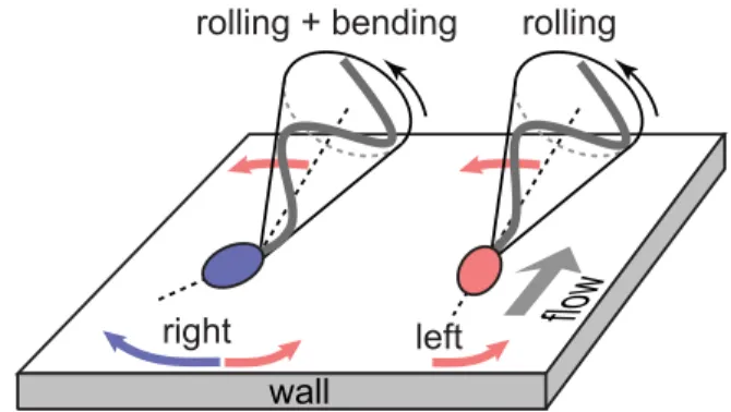
Intrinsic Midpiece Curvature vs. Buckling. Our data demonstrate that the rheotactic turning behavior correlates with an asym-metry in the midpiece curvature distributions (Fig. 3E andFig. S11). Recent hydrodynamic simulations show that midpiece curvature asymmetries strongly affect swimming trajectories, thus providing an effective long-range steering mechanism (42). Possible explanations for a curved midpiece are intrinsic curvature or symmetry breaking caused by a dynamic buckling instability. Intrinsic midpiece curvature is a known feature of rodent sperm (56) and has been linked to strongly asymmetric beat patterns (57). A less pronounced intrinsic curvature has been reported for human sperm, requiring artificial elevation of the intracellular calcium levels (58) absent in our experiments. An intrinsically curved midpiece per se does not explain the observed asymmetry in the distribution of the projected 2D bend angles (Fig. 3E), as the rolling motion would effectively symmetrize the distributions, and both left-turning and right-turning cells exhibit nearly identical rolling statistics (Fig. 3D). Therefore, a more plausible explana-tion is dynamic buckling, which is common in thin structures such as flagellar axonemes and cross-linked filament bundles (59). For sea urchin sperm, a buckling instability was suggested as an explanation for asymmetric compressed beat patterns at high vis-cosities (60). Nonlinear analysis and hydrodynamic simulations revealed that flagellar buckling arises generically from the interplay between elasticity and fluid forces (61), resulting in spontaneously broken symmetries of beat patterns and curved swimming trajec-tories. Although these buckling instabilities were predicted to affect the entire flagellum, localized buckling as observed in our ex-periments can be caused by inhomogeneities in the flagellar elastic stiffness. In human sperm cells, the midpiece carries the mitochondrial gyres and is therefore thicker than the tail of the flagellum (55); ergo, it is plausible that buckling occurs primarily at the transition point between midpiece and tail, triggered by perturbations that arise from the inhomogeneous material proper-ties. In this picture, left-turning cells fluctuate symmetrically around the unbuckled stateδ = 0 whereas right-turning cells predominately occupy a buckled configuration with positive bend angleδ (Fig. 3E). The two distinct turning behaviors can then be inferred from basic force balance considerations (32, 33, 62): Approximating the fla-gellar beat envelope by a cone rotating counterclockwise around its symmetry axis, hydrodynamic interactions with the wall effectively result in a left-turning torque (Fig. 4). This rheotactic turning mechanism depends only on the flagellar rolling motion but not on the head and its geometry, in agreement with recent experi-ments demonstrating rheotaxis for headless mouse sperm (3). For right-turning cells trapped in a buckled state, the head acts as a tilted hydrofoil (“front rudder”) that overcomes the rolling force. Thus, mirror symmetry is not just broken by the rolling motion but also by the midpiece bend.
Conclusions
Our joint experimental and theoretical study focused on the dynamics of human sperm cells swimming under the influence of a time-dependent linear flow gradient near a solid surface, a situation that is common in a wide range of external and internal fertilization processes. We identified two kinematically distinct rheotactic turning behaviors and showed that an effective 2D
mathematical model suffices to reproduce quantitatively the ex-perimentally observed trajectory statistics. Building on, to our knowledge, the first systematic 3D beat reconstruction for freely swimming sperm cells, we found that human sperm flagella perform nearly planar beats in a stepwise-rotating plane. However, the rheotactic turning behaviors are independent of this rolling motion. Contrary to current opinion, the 3D beat patterns exhibit no persistent helicity but instead are composed of helical waves of either handedness. Interestingly, similar beating modes were reported recently for Trypanosoma brucei (63) and malaria par-asites P. berghei (50). Taken together, these results suggest that ambidextrous beat patterns may be a common feature of eukaryotic uniflagellates that share the canonical 9+2 axoneme structure. Furthermore, our 3D beat pattern analysis reveals that rheotactic separation into left-turning and right-turning cells is related to a curved midpiece section. In the absence of evidence for intrinsic midpiece curvature in human sperm, a buckling in-stability combined with the wall-induced partial suppression of rolling can provide a plausible explanation for the observed bend angle asymmetry in right-turning cells. Recent experiments showed (28) that uniflagellate marine bacteria use buckling to change their swimming direction. Although passive prokaryotic and active eukaryotic flagella are built differently, buckling could provide a general physical mechanism for controlling reor-ientation in single-cell uniflagellates.
More generally, the above 3D observations call for a revision and extension of the currently prevailing helical models of fla-gellar propulsion. Future theoretical studies should focus on the interplay between rolling dynamics, wall interactions, and structural and elastic inhomogeneities in midpiece and flagellum. The basic ingredients for a bimodal rheotactic response—cell rolling and beat curvature asymmetry—are generic features of many sperm species (42, 48, 60, 61). It will thus be important to investigate experi-mentally if bimodal rheotactic response occurs in other species, in particular those featuring strong head asymmetries that may criti-cally affect the proposed hydrofoil effect. Moreover, it will be in-teresting to study how calcium concentration (3), increased viscous load (36), and viscoelastic fluid environments affect the flagellar dynamics. The 3D beat reconstruction approach implemented here provides a promising starting point for such future studies.
ACKNOWLEDGMENTS. The authors thank Jeff Guasto, Andreas Hilfinger, and Ingmar Riedel-Kruse for helpful discussions and the Bourn Hall Clinic and Martyn Blayney for fruitful collaboration. This work was supported by the Skolkovo Foundation (Grant agreement for Russian educational and scientific organiza-tion no. 4dd.25.12.2014 to A.B.), the Swiss Naorganiza-tional Science Foundaorganiza-tion Grant 148743 (to N.S.), an MIT Solomon Buchsbaum Fund Award (to J.D.), and an Alfred P. Sloan Research Fellowship (to J.D.).
1. Häder D-P (1987) Polarotaxis, gravitaxis and vertical phototaxis in the green flagel-late, Euglena gracilis. Arch Microbiol 147(2):179–183.
2. Witman GB (1993) Chlamydomonas phototaxis. Trends Cell Biol 3(11):403–408. 3. Miki K, Clapham DE (2013) Rheotaxis guides mammalian sperm. Curr Biol 23(6):443–452.
Fig. 4. Turning mechanisms implied by 3D data. Left-turning (red) and right-turning (blue) sperm cells roll their flagellum counterclockwise with a conical beat envelope, resulting in a left-turning torque. For right-turning sperm, this effect is counteracted by a larger opposing force due to the tilted head acting as a hydrofoil or“rudder.”
BIOPHYSICS
AND
COMPUTATION
AL
4. Bahat A, et al. (2003) Thermotaxis of mammalian sperm cells: A potential navigation mechanism in the female genital tract. Nat Med 9(2):149–150.
5. Berg HC, Brown DA (1972) Chemotaxis in Escherichia coli analysed by three-dimensional tracking. Nature 239(5374):500–504.
6. Lo CM, Wang HB, Dembo M, Wang YL (2000) Cell movement is guided by the rigidity of the substrate. Biophys J 79(1):144–152.
7. Pedley TJ, Kessler JO (1992) Hydrodynamic phenomena in suspensions of swimming microorganisms. Annu Rev Fluid Mech 24:313–358.
8. Marcos F, Fu HC, Powers TR, Stocker R (2012) Bacterial rheotaxis. Proc Natl Acad Sci USA 109(13):4780–4785.
9. Adolphi H (1905) Die Spermatozoen der Säugetiere schwimmen gegen den Strom. Anat Anz 26(20-21):549–559.
10. Rothschild L (1963) Non-random distribution of bull spermatozoa in a drop of sperm suspension. Nature 198(4886):1221–1222.
11. Zimmer RK, Riffell JA (2011) Sperm chemotaxis, fluid shear, and the evolution of sexual reproduction. Proc Natl Acad Sci USA 108(32):13200–13205.
12. Kantsler V, Dunkel J, Polin M, Goldstein RE (2013) Ciliary contact interactions domi-nate surface scattering of swimming eukaryotes. Proc Natl Acad Sci USA 110(4): 1187–1192.
13. Elgeti J, Winkler RG, Gompper G (2015) Physics of microswimmers—Single particle motion and collective behavior: A review. Rep Prog Phys 78(5):056601.
14. Brenker C, et al. (2012) The CatSper channel: A polymodal chemosensor in human sperm. EMBO J 31(7):1654–1665.
15. Eisenbach M, Giojalas LC (2006) Sperm guidance in mammals—An unpaved road to the egg. Nat Rev Mol Cell Biol 7(4):276–285.
16. Kaupp UB, Kashikar ND, Weyand I (2008) Mechanisms of sperm chemotaxis. Annu Rev Physiol 70:93–117.
17. Alvarez L, Friedrich BM, Gompper G, Kaupp UB (2014) The computational sperm cell. Trends Cell Biol 24(3):198–207.
18. Polin M, Tuval I, Drescher K, Gollub JP, Goldstein RE (2009) Chlamydomonas swims with two“gears” in a eukaryotic version of run-and-tumble locomotion. Science 325(5939):487–490.
19. Goldstein RE (2015) Green algae as model organisms for biological fluid dynamics. Annu Rev Fluid Mech 47:343–375.
20. Friedrich BM, Jülicher F (2012) Flagellar synchronization independent of hydrody-namic interactions. Phys Rev Lett 109(13):138102.
21. Goldstein RE, Polin M, Tuval I (2011) Emergence of synchronized beating during the regrowth of eukaryotic flagella. Phys Rev Lett 107(14):148103.
22. Brumley DR, Wan KY, Polin M, Goldstein RE (2014) Flagellar synchronization through direct hydrodynamic interactions. eLife 3:e02750.
23. Durham WM, Kessler JO, Roman S (2009) Disruption of vertical motility by phyto-plankton layers. Science 323:1067–1070.
24. Zöttl A, Stark H (2013) Periodic and quasiperiodic motion of an elongated micro-swimmer in Poiseuille flow. Eur Phys J E Soft Matter 36(1):4.
25. Rusconi R, Guasto JS, Stocker R (2014) Bacterial transport suppressed by fluid shear. Nat Phys 10:212–217.
26. Leptos KC, et al. (2013) Antiphase synchronization in a flagellar-dominance mutant of Chlamydomonas. Phys Rev Lett 111(15):158101.
27. Bennett RR, Golestanian R (2015) A steering mechanism for phototaxis in Chlamy-domonas. J R Soc Interface 12(104):20141164.
28. Son K, Guasto JS, Stocker R (2013) Bacteria can exploit a flagellar buckling instability to change direction. Nat Phys 9:1–5.
29. Kunz G, Beil D, Deininger H, Wildt L, Leyendecker G (1996) The dynamics of rapid sperm transport through the female genital tract: Evidence from vaginal sonography of uterine peristalsis and hysterosalpingoscintigraphy. Hum Reprod 11(3):627–632. 30. Fauci LJ, Dillon R (2006) Biofluidmechanics of reproduction. Annu Rev Fluid Mech 38:
371–394.
31. Spehr M, et al. (2003) Identification of a testicular odorant receptor mediating human sperm chemotaxis. Science 299(5615):2054–2058.
32. Kantsler V, Dunkel J, Blayney M, Goldstein RE (2014) Rheotaxis facilitates upstream navigation of mammalian sperm cells. eLife 3:02403.
33. Tung CK, et al. (2015) Emergence of upstream swimming via a hydrodynamic tran-sition. Phys Rev Lett 114(10):108102.
34. Denissenko P, Kantsler V, Smith DJ, Kirkman-Brown J (2012) Human spermatozoa migration in microchannels reveals boundary-following navigation. Proc Natl Acad Sci USA 109(21):8007–8010.
35. Ishijima S, Hamaguchi MS, Naruse M, Ishijima SA, Hamaguchi Y (1992) Rotational movement of a spermatozoon around its long axis. J Exp Biol 163:15–31. 36. Smith DJ, Gaffney EA, Gadêlha H, Kapur N, Kirkman-Brown JC (2009) Bend
propa-gation in the flagella of migrating human sperm, and its modulation by viscosity. Cell Motil Cytoskeleton 66(4):220–236.
37. Hilfinger A, Jülicher F (2008) The chirality of ciliary beats. Phys Biol 5(1):016003.
38. Friedrich BM, Riedel-Kruse IH, Howard J, Jülicher F (2010) High-precision tracking of sperm swimming fine structure provides strong test of resistive force theory. J Exp Biol 213(Pt 8):1226–1234.
39. Fauci LJ, McDonald A (1995) Sperm motility in the presence of boundaries. Bull Math Biol 57(5):679–699.
40. Smith DJ, Gaffney EA, Blake JR, Kirkman-Brown JC (2009) Human sperm accumulation near surfaces: A numerical study. J Fluid Mech 621:289–320.
41. Evans AA, Lauga E (2010) Propulsion by passive filaments and active flagella near boundaries. Phys Rev E Stat Nonlin Soft Matter Phys 82(4 Pt 1):041915.
42. Elgeti J, Kaupp UB, Gompper G (2010) Hydrodynamics of sperm cells near surfaces. Biophys J 99(4):1018–1026.
43. Gaffney EA, Gadelha H, Smith DJ, Blake JR, Kirkman-Brown JC (2011) Mammalian sperm motility: Observation and theory. Annu Rev Fluid Mech 43:501–528. 44. Montenegro-Johnson TD, Smith AA, Smith DJ, Loghin D, Blake JR (2012) Modelling
the fluid mechanics of cilia and flagella in reproduction and development. Eur Phys J E Soft Matter 35(10):111.
45. Lauga E, Eloy C (2013) Shape of optimal active flagella. J Fluid Mech 730:R1. 46. Ishimoto K, Gaffney EA (2015) Fluid flow and sperm guidance: A simulation study of
hydrodynamic sperm rheotaxis. J R Soc Interface 12(106):20150172.
47. Lauga E, Powers TR (2009) The hydrodynamics of swimming microorganisms. Rep Prog Phys 72:096601.
48. Woolley DM (2003) Motility of spermatozoa at surfaces. Reproduction 126(2):259–270. 49. Lee S-H, Grier DG (2007) Holographic microscopy of holographically trapped
three-dimensional structures. Opt Express 15(4):1505–1512.
50. Wilson LG, Carter LM, Reece SE (2013) High-speed holographic microscopy of malaria parasites reveals ambidextrous flagellar waveforms. Proc Natl Acad Sci USA 110(47): 18769–18774.
51. Wilson L, Zhang R (2012) 3D localization of weak scatterers in digital holographic mi-croscopy using Rayleigh-Sommerfeld back-propagation. Opt Express 20(15):16735–16744. 52. Riedel-Kruse IH, Hilfinger A, Howard J, Jülicher F (2007) How molecular motors shape
the flagellar beat. HFSP J 1(3):192–208.
53. Phillips DM (1972) Comparative analysis of mammalian sperm motility. J Cell Biol 53(2):561–573.
54. Linnet L (1979) Human spermatozoa: Unidirectional rotation of the tail as indicated by head-to-head agglutinates. Arch Androl 2(2):157–161.
55. Mundy AJ, Ryder TA, Edmonds DK (1995) Asthenozoospermia and the human sperm mid-piece. Hum Reprod 10(1):116–119.
56. Lindemann CB, Goltz JS (1988) Calcium regulation of flagellar curvature and swim-ming pattern in triton X-100−extracted rat sperm. Cell Motil Cytoskeleton 10(3): 420–431.
57. Chang H, Suarez SS (2011) Two distinct Ca2+signaling pathways modulate sperm flagellar beating patterns in mice. Biol Reprod 85(2):296–305.
58. Bedu-Addo K, et al. (2008) Mobilisation of stored calcium in the neck region of hu-man sperm—A mechanism for regulation of flagellar activity. Int J Dev Biol 52(5-6): 615–626.
59. Gadêlha H, Gaffney EA, Goriely A (2013) The counterbend phenomenon in fla-gellar axonemes and cross-linked filament bundles. Proc Natl Acad Sci USA 110(30): 12180–12185.
60. Woolley DM, Vernon GG (2001) A study of helical and planar waves on sea urchin sperm flagella, with a theory of how they are generated. J Exp Biol 204(Pt 7):1333–1345. 61. Gadêlha H, Gaffney EA, Smith DJ, Kirkman-Brown JC (2010) Nonlinear instability
in flagellar dynamics: A novel modulation mechanism in sperm migration? J R Soc Interface 7(53):1689–1697.
62. Lauga E, DiLuzio WR, Whitesides GM, Stone HA (2006) Swimming in circles: Motion of bacteria near solid boundaries. Biophys J 90(2):400–412.
63. Rodríguez JA, et al. (2009) Propulsion of African trypanosomes is driven by bihelical waves with alternating chirality separated by kinks. Proc Natl Acad Sci USA 106(46): 19322–19327.
64. Herráez-Domínguez JV, Gil Garcia de León F, Diez-Sales O, Herraez-Dominguez M (2005) Rheological characterization of two viscosity grades of methylcellulose: An approach to the modeling of the thixotropic behaviour. Colloid Polym Sci 284(1): 86–91.
65. Aziz N, Fear S, Taylor C, Kingsland CR, Lewis-Jones DI (1998) Human sperm head morphometric distribution and its influence on human fertility. Fertil Steril 70(5): 883–891.
66. Grubbs FE (1969) Procedures for detecting outlying observations in samples. Technometrics 11(1):1–21.
67. Carlson AE, et al. (2003) CatSper1 required for evoked Ca2+entry and control of flagellar function in sperm. Proc Natl Acad Sci USA 100(25):14864–14868. 68. Werner S, Rink JC, Riedel-Kruse IH, Friedrich BM (2014) Shape mode analysis exposes
movement patterns in biology: Flagella and flatworms as case studies. PLoS One 9(11): e113083.
69. Bergou M, Wardetzky M, Robinson S, Audoly B, Grinspun E (2008) Discrete elastic rods. ACM Trans Graph 27(3):63.