HAL Id: hal-02325921
https://hal.archives-ouvertes.fr/hal-02325921
Submitted on 25 Oct 2019
HAL is a multi-disciplinary open access archive for the deposit and dissemination of sci-entific research documents, whether they are pub-lished or not. The documents may come from teaching and research institutions in France or abroad, or from public or private research centers.
L’archive ouverte pluridisciplinaire HAL, est destinée au dépôt et à la diffusion de documents scientifiques de niveau recherche, publiés ou non, émanant des établissements d’enseignement et de recherche français ou étrangers, des laboratoires publics ou privés.
Histone variants: critical determinants in tumour
heterogeneity
Tao Wang, Florent Chuffart, Ekaterina Bourova-Flin, Jin Wang, Jianqing Mi,
Sophie Rousseaux, Saadi Khochbin
To cite this version:
Tao Wang, Florent Chuffart, Ekaterina Bourova-Flin, Jin Wang, Jianqing Mi, et al.. Histone variants: critical determinants in tumour heterogeneity. Frontiers of Medicine in China, Springer Verlag, 2019, 13 (3), pp.289-297. �10.1007/s11684-018-0667-3�. �hal-02325921�
Histone variants: critical determinants in tumour heterogeneity
Tao Wang1,2, Florent Chuffart1, Ekaterina Bourova-Flin1, Jin Wang2, Jianqing Mi2, Sophie Rousseaux1, Saadi Khochbin (
✉
)11
CNRS UMR 5309, Inserm, U1209, University of Grenoble Alpes, Institute for Advanced Biosciences, 38706, Grenoble, France;2State Key Laboratory for Medical Genomics and Department of Hematology, Shanghai Institute of Hematology, Collaborative Innovation Center of Systems Biomedicine, Pôle Sino-Français des Sciences du Vivant et Genomique, Ruijin Hospital Affiliated to Shanghai Jiao Tong University School of Medicine, Shanghai 200025, China
© The Author(s) 2018. This article is published with open access at link.springer.com and journal.hep.com.cn 2018
Abstract Malignant cell transformation could be considered as a series of cell reprogramming events driven by oncogenic transcription factors and upstream signalling pathways. Chromatin plasticity and dynamics are critical determinants in the control of cell reprograming. An increase in chromatin dynamics could therefore constitute an essential step in driving oncogenesis and in generating tumour cell heterogeneity, which is indispensable for the selection of aggressive properties, including the ability of cells to disseminate and acquire resistance to treatments. Histone supply and dosage, as well as histone variants, are the best-known regulators of chromatin dynamics. By facilitating cell reprogramming, histone under-dosage and histone variants should also be crucial in cell transformation and tumour metastasis. Here we summarize and discuss our knowledge of the role of histone supply and histone variants in chromatin dynamics and their ability to enhance oncogenic cell reprogramming and tumour heterogeneity.
Keywords cancer-testis; TH2B; TH2A; H1T; H1.0; H1F0; linker histones
Introduction
Malignant transformation of normal somatic cells is a multistep process eventually leading to the selection of deadly metastasis-prone cancer cells. Within this context, the molecular mechanisms that generate genetic and epigenetic heterogeneity are essential to provide the indispensable ground for the selection of such cells [1]. The selective pressure here operates on genetic/epigenetic elements [2] to stably support the acquisition of the so-called cancer hallmarks [3]. The transformation of a normal cell into a cell with malignant characteristics could therefore be qualified as an oncogenic reprogramming of cells.
This process could be compared to the induced cell reprogramming, first defined by Yamanaka [4], with a major difference: malignant reprogramming relies on the aberrant activation of intrinsic reprogramming events [5]. Successful malignant transformation should therefore obey
to the same laws as those underlying the induced reprogramming of differentiated somatic cells [6].
Indeed, not only there is a need to activate driver transcription factors and appropriate cell signalling, but there is also a requirement to break reprogramming barriers, mostly of epigenetic nature [7]. Among these reprogramming barriers important ones are the Suv39H1 enzyme, H3K9 tri-methylation and other factors respon-sible for heterochromatin formation [8,9].
In addition to the erasure of repressive epigenetic marks, a critical factor for successful cell reprogramming is increased chromatin dynamics [10]. Factors capable of sustaining enhanced chromatin dynamics seem to also improve cell reprogramming [11]. Chromatin dynamics could be directly modulated by canonical replication-dependent histones through the control of their assembly during DNA replication, as well as through induced histone degradation and histone under-dosage or indepen-dently, through the assembly of a specific class of histone variants [12–16].
Regulated replication-dependent nucleosome assembly is in fact an important element in defining the dynamic states of chromatin. Accordingly, two studies based on interference with CAF1 activity, a histone chaperone
REVIEW
Received April 4, 2018; accepted July 2, 2018 Correspondence: Saadi Khochbin,
required for the replication-dependent chromatin assembly [17], highlighted the critical role of this factor in modulating cell reprogramming. Down-regulation of CAF1 leads to an increased chromatin accessibility, a shift in the cell gene expression programs and the acquisition of new characters [18,19].
Histone variants that are tissue-specific or whose expression depends on specific regulatory signals, also play their role by conferring specific states to chromatin, either globally in a particular cell type, or locally on defined genomic regions. Some of these histones are associated with more stable and transcriptionally repressed chromatin such as the H2A variant, macroH2A [20], while others, in contrast, are associated with unstable nucleo-somes such as H2A.B.3 and H2A.L.2 in spermatogenic cells [21,22]. Interestingly, somatic cell reprogramming assays showed that macroH2A expression is associated with a resistance to reprogramming [23], while the ectopic expression of testis/oocyte-specific histone variants of the H2A and H2B types (THA, TH2B) infibroblasts, greatly stimulates the process of reprogramming [24].
All these data highlight the importance of histone metabolism, histone dosage and histone type synthesis in the capacity of cells to be reprogrammed. Mechanisms controlling histone turnover and the expression of histone variants are largely affected in cancer [25]. More specifically, cancer cells express almost systematically tissue-restricted genes [26], among which, genes encoding histone variants [27] as well as mutated histones known as “oncohistones” [28].
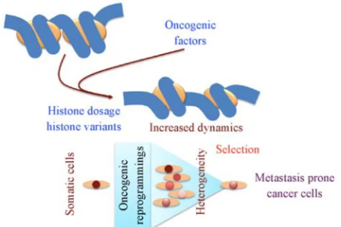
Here we develop a discussion on the roles of specific types of histone variants and of histone dosage in cell epigenetic reprogramming that could be essential for the establishment of malignant transformation and tumour heterogeneity (Fig. 1).
Male germ cells express the largest set of
histone variants
In addition to canonical histones that are the building blocks of nucleosomes, the human genome encodes a number of histone variants of the H3, H2A and H2B types. Many of these variants are predominantly or exclusively expressed in spermatogenic cells [14]. The reason is that thefinal stages of male germ cell differentiation involve one of the most dramatic chromatin remodelling events, characterized by an almost genome-wide eviction of histones and their replacement by protamines [29]. Functional, biochemical and structural analyses of these histone variants showed that many of them present the ability to generate unstable nucleosomes. This is true for H3, H2B and H2A variants [14]. Taking into account the final dismantlement of chromatin in post-meiotic sperma-togenic cells, before the generation of mature spermatozoa, one can easily understand why most of these variants
confer nucleosome instability. Indeed, a more open and dynamic chromatin should lower the energy supply required for the genome wide removal of histones.
There are data strongly supporting the idea that most of these variants are in fact nucleosome-destabilizing ele-ments. Structural studies of the testis-specific histone H3, H3T/t, showed that both the human and the mouse members create unstable nucleosomes [30,31]. More particularly, in H3t, a single amino acid, H42, is critical to generate a flexible linker DNA at the entry and exit of the nucleosome [30]. This ability to open the nucleosome with flexible DNA ends is also shared by several H2A variants that are expressed at different stages of sperma-togenesis, namely H2A.B.3 [21] and H2A.L.2 in mouse [22], as well as H2A.Bbd (H2.B.1 and H2A.B.2) in human [32]. Finally, the major testis-specific H2B variant, TH2B, also induces nucleosome instability, especially when it is paired with TH2A [24,33].
Since nucleosome instability underlies chromatin dynamics, and that the latter is essential for efficient cell reprogramming, the question arises on the role of these histone variants in enhancing cell reprograming. A positive answer then invites to consider if this histone driven chromatin dynamics could also induce malignant cell reprogramming and thereby contribute to tumour hetero-geneity.
TH2B/TH2A enhances somatic cell
reprogramming
Among the male germ cell histone variants, TH2B and
Fig. 1 Histone-based malignant transformation, tumour heterogeneity and selection of aggressive characters. Pro-oncogenic events could lead to aberrant activation of silenced histone variants-encoding genes or histone assembly defects or histone under-dosage, leading to increased chromatin dynamics and enhanced genome reprogramming by onco-genic factors. The resulting heterogeneity would create a window of opportunity for the selection of newly reprogrammed oncogenic cells capable of surviving and disseminating.
TH2A show very particular characteristics, since they act during two unique periods of the male genome life. At the time of commitment of male germ cells into meiotic divisions, these variants accumulate and gradually become the major nucleosomal H2A and H2B histone types until their genome-wide eviction and replacement, prior to the generation of mature spermatozoa [33,34]. Interestingly, these histone variants also accumulate in maturing oocytes and are stored until fertilization. Upon removal of protamines from the male genome, maternal TH2A/ TH2B become again associated with the male genome [35], and spread over the zygote’s genome at a period critical for epigenetic programming in pre-implantation embryonic cells. During later embryonic cell divisions, TH2A/TH2B are gradually replaced by somatic type H2A/ H2B histones [33,34]. Their presence at two critical periods of general genome programming,first during the male germ cell differentiation in preparation of histone-to-protamine exchange and again during histone-to- protamine-to-histone replacement and embryonic cell genome program-ing, strongly suggests that the properties conferred by these histone variants to chromatin are of critical importance to make the genome programmable.
This hypothesis has been actually elegantly confirmed by Ishii’s group, who showed that the ectopic expression of TH2B/TH2A in somatic cells strongly enhances induced pluripotent stem cells (iPS) formation by Yama-naka factors. Additionally, in the absence of both TH2B and TH2A (double KO mice), fertilized eggs do not develop properly, again indicating the importance of these histone variants in early development [24].
Taking into account the essential role of these two histone variants in genome reprogramming, we also hypothesized that their ectopic activation could help oncogenic cell transformation or create a heterogeneity among tumour cell populations, which would facilitate the evolution of cancer cells toward increased aggressiveness. To test this hypothesis the expression of TH2A and TH2B was monitored in transcriptomic data available from several cohorts of cancer patients. Fig.2A shows that in human both TH2A and TH2B encoding genes are indeed tissue-restricted genes specifically expressed in testis. The analysis of a cohort of breast cancers as well as of two series of lung tumour samples shows that both genes could become aberrantly active in a subset of tumours (Fig. 2B). However, these data do not allow us to conclude on a
Fig. 2 Aberrant activation of testis-oocyte specific TH2A/TH2B in various cancers. (A) Expression of TH2A and TH2B genes in normal human tissues samples from RNA-seq data, provided by the Genotype-Tissue Expression (GTEx) project [56]. (B) Expression of TH2A and TH2B genes in breast and lung cancer samples. Breast cancer RNA-seq data are provided by the TCGA-BRCA project [57]. Lung cancer RNA-seq data are provided by the NCBI GEO GSE81089 [58], TCGA-LUAD and TCGA-LUSC projects [57]. For all plots, the expression level of genes is represented as a distribution of log-transformed RPKM values, after addition of a pseudo count of 1 (log2 (1+ RPKM)). Breast cancer: NT Breast = non tumoral breast; Breast K = breast cancer. Lung cancer: NT Lung = non tumoral lung; L. ADC = Lung adenocarcinoma; L. SQC = Lung squamous cell carcinoma; other LK = lung tumours of other histological subtypes.
relationship between the activation of these genes and the process of oncogenesis. Indeed, in most cases, the tumours analysed here were harvested long after the initial pro-oncogenic crisis and the malignant transformation process. It is therefore likely that a counter-selection against the expression of both or either of these genes is required for tumour cells to survive beyond the initial events, with a stable gene expression program (Fig. 1). In support of this hypothesis, in the normal frame of TH2A/TH2B expres-sion, it has been shown that embryonic development is associated with a sharp decrease in both TH2A and TH2B in embryonic cells, indicating a requirement for the silencing of these genes after the initial reprogramming period. The sporadic expression of these genes in established cancer cells in tumours would merely be part of the vestiges of their full activation at the time of oncogenic transformation.
Short H2A variants
One of the most remarkable characteristics of the canonical histone H2A is the presence of an acidic patch, which locates at the surface of nucleosomes and was shown to mediate the compaction of the chromatinfibre [21,32,36]. The other important contribution of H2A to the nucleo-some structure is through its C-terminal region, known as the“docking domain,” involved in the stabilization of H3 a-N helix. The H2A docking domain also contributes to the stability of the H2A-H2B dimer–H3-H4 tetramer interac-tion [37]. Indeed, H2A variants with a short docking domain [38] are unable to form stable histone octamers. Within a nucleosome, a H2A with a short docking domain disrupts the ability of the H3 a-N helix to stabilize nucleosomal DNA and leads to the release of unwrapped DNA ends [39,40].
Taking into account the ability of the H2A variants to open and destabilise nucleosomes, it is expected that their incorporation into nucleosomes should increase chromatin dynamics and genome reprogramming ability. Unfortu-nately, however, the role of this category of short H2A variants in cell reprogramming has not yet been investi-gated.
Interestingly, although most of the short H2A variants, including human H2A.Bbd, are testis-specific [38], several investigations pointed to their contribution to oncogenic cell programming in specific sets of cancers. Indeed, the H2A.Bbd-encoding gene was found de-repressed in Hodgkin lymphoma [41]. The analyses of various Hodgkin lymphoma cell lines showed that the expression of this histone variant is remarkably variable [42], suggesting that the ectopic activation of this gene could have been important in the process of malignant transformation and that a counter-selection against its expression operates after the establishment of the transformed state.
Ectopic testis-specific linker histone
expression as a measure of tumour
heterogeneity
Similar to core histones, linker histones are also encoded by a variety of canonical replication-dependent as well as replication-independent and tissue-specific genes [43].
There are testis-specific linker histones named H1T and HILS1 in human. Here we first verified their tissue-restricted pattern of expression in human adult tissues. Fig.3 shows that while, as expected, H1T shows a strict testis-specific expression, HILS1 is also expressed in other tissues such as in muscle (not shown). The questions are whether the H1T gene could be aberrantly activated in cancers, whether its activation would reflect tumour heterogeneity and whether its expression could be associated with tumour types, sub-types and prognosis.
To answer these questions the expression of H1T was monitored in breast and lung tumours where the ectopic activation of genes was observed in a significant number of tumours (Fig. 3). Since HILS1 did not show the expected testis-specific expression pattern, its expression in cancer was not considered. The ectopic activation of H1T, as in the case of TH2A/TH2B, could be an important event during malignant transformation and the presence of H1T in some of the analysed tumours could be a vestige of this initial oncogenic reprogramming process (and the subse-quent counter-selection required to stabilize the epigenome of the established malignant cells).
However, in the case of H1T, this hypothesis remains highly speculative since, in contrast to the TH2A/TH2B situation, the role of H1T in genome reprogramming has not been shown. Therefore additional experimental data on the reprogramming capacity of H1T and its expression in oocytes and early development are required to support the hypothesis.
Linker histone variants and tumour
heterogeneity
Among linker histone variants genes, H1F0 encodes histone H1.0, which presents a differentiation-dependent expression.
H1.0 is a conserved linker histone present in all vertebrates, except in birds. The corresponding gene is also present in some invertebrates such as sea urchin [43]. Interestingly, not only the H1.0 protein shows conserved features, but also the regulatory circuits that control the expression of its gene is conserved in different species [44]. These data highlight the fact that the differentiation-dependent nature of H1.0 expression is also an evolu-tionary conserved characteristic of cell differentiation and hence should probably contribute to the epigenetic stability of differentiated cells.
The gene is repressed during early embryonic develop-ment and in non-differentiated stem cells as well as in various cancer cell lines but is induced upon the commitment of cells into differentiation [43]. The study of established cancer cell lines such as Friend murine erythroleukemia [45] and murine melanoma cells [46] showed that the induced differentiation of these cells is associated with the activation of H1.0-encoding gene.
Additionally, in contrast to other linker histone-encoding genes, H1F0 expression is potently induced by histone-deacetylase inhibitors in cultured cells in all vertebrates [43] and in developing embryos in a stage-specific manner [47–50].
A recent study showed that heterogeneous H1.0 expression in tumour cells directly reflects tumour heterogeneity, with more differentiated cells expressing higher levels of H1.0. An absence of H1.0 in tumour cells correlates with the stem type nature of the cells [51]. The
degree of H1.0 expression could therefore be considered as a measure of the level of differentiation of tumour cells and hence reflect tumour aggressiveness. This work also shows that H1.0 restricts self-renewal and favours differentiation. These data are in agreement with data we previously published on H1.0 expression in hepatocytes after partial hepatectomy. Indeed, the H1.0 content dramatically decreases after partial hepatectomy corresponding to the natural reprogramming of hepatocytes and the induction of cell proliferation [52]. Altogether, these data show that reprogramming of adult differentiated cells should be associated with a decrease in H1.0 content, either in a physiological setting, after partial hepatectomy, or in the pathological condition of malignant cell transformation [51]. In addition, we do not expect the oncogenic transformation of stem types of cells to be associated with any change in H1.0 gene expression, since non-transformed adult stem cells do not express high levels of
Fig. 3 Aberrant activation of testis-specific H1T in various cancers. Expression of H1T gene in normal (left panel) and tumour (right panels) samples from RNA-seq data, provided by the GTEx [56], TCGA-BRCA [57] and NCBI GEO GSE81089 [58] datasets. The expression level of genes is represented as a distribution of log-transformed RPKM values, after addition of a pseudo count of 1 (log2 (1+ RPKM)). Breast cancer: NT Breast = non tumoral breast; Breast K = breast cancer. Lung cancer: NT Lung = non tumoral lung; L. ADC = Lung adenocarcinoma; L. SQC = Lung squamous cell carcinoma; other LK = lung tumours of other histological subtypes.
H1.0. However, the induced expression of H1.0 in cancer cells could reflect the proliferation and differentiation of malignant stem cells.
Following these data, we analysed the relationship between H1F0 gene expression and patient survival data taking into account published cancer data used by Torres and colleagues [51], as well as some other cancer transcriptome series not considered by these authors (Fig. 4).
Unexpectedly, we observe that, in all the considered cases, H1F0 expression was lower in normal control tissues compared to the corresponding cancer series (Fig. 4). Two additional observations could help explain-ing this observation. First, the H1F0 gene promoter bears regulatory elements which are shared with the replication-dependent H4 encoding genes [53]. Second, in cells in culture, H1F0 shows an increased expression in S phasis [54]. Therefore, the higher expression of H1F0 genes in cancers compared to their non-transformed counter-parts could actually reflect the presence of proliferative sub-populations in the considered tumours.
The capacity of the H1F0 gene promoter to be responsive to both cell differentiation and cell proliferation signals could appear contradictory. The higher levels of H1F0 gene expression in tumour cells compared to their non-transformed counterparts could be explained by the higher proportion of proliferative cells in tumours.
However, taking into account the heterogeneity of H1.0 in a given tumour [51], it can be proposed that, within the context of proliferative cancer cells, a higher level of differentiation would lead to an even higher expression of this gene therefore allowing the identification of less aggressive more differentiated cancer cells.
However, in contrast to the reported data [51], when we looked for a correlation between the level of H1F0 expression and survival in a cohort of breast cancer patients and in three cohorts of patients with lung cancer and we found no significant association between expres-sion of H1F0 and survival probability (Fig. 4).
In conclusion, although a variable expression of H1.0 might be used as a measure of tumour heterogeneity [51], its level of expression cannot be reliably used as an indicator of prognosis.
In addition to H1.0, other tissue-restricted linker histones are also expressed in vertebrates. Birds express a linker histone known as H5 only in erythrocytes, a gene, which is probably related to an ancestral H1.0-encoding gene, uniquely expressed in amphibian erythrocytes [55]. The specific/high expression of H1.0/H5 in amphibian/ birds erythrocytes is certainly linked to the functional inactivation of the nucleus in these cells, which in contrast to mammals, remain nucleated. There are however no data on the expression of H5 and cancer in avian.
Fig. 4 H1F0 gene expression is activated in different cancers. Expression of H1F0 gene in breast and lung tumour samples with corresponding Kaplan–Meyer survival curves. Breast cancer RNA-seq data are provided by the TCGA-BRCA project [57]. Lung cancer RNA-seq data are provided by the NCBI GEO GSE81089 [58], TCGA-LUAD and TCGA-LUSC projects [57]. For all plots, the expression level of genes is represented as a distribution of log-transformed RPKM values, after addition of a pseudo count of 1 (log2 (1+ RPKM)). Breast cancer: NT Breast = non tumoral breast; Breast K = breast cancer. Lung cancer: NT Lung = non tumoral lung; L. ADC = Lung adenocarcinoma; L. SQC = Lung squamous cell carcinoma; L. LCNE = Lung large cell neuroendocrine tumours; L. BAS = Lung basaloid tumours; L. SCC = Lung small cell carcinoma; L. CARCI = Lung carcinoid tumours; other LK = lung tumours of other histological subtypes.
Discussion
Epigenetic stability is a critical barrier to physiological and pathological cell reprogramming. The establishment of specific strategies to induce reprograming in adult differentiated cells also enabled to highlight the epigenetic determinants that prevent cell reprogramming. Histone dosage seems to be an essential barrier in hindering reprogramming factors’ action. Further investigations suggest that increased chromatin dynamics could explain the effect of histone under-dosage and under-assembly in facilitating reprogramming. Indeed, we know that histone loss increases chromatin dynamics [13] and increased chromatin dynamics is associated with enhanced repro-gramming capacity of the cells [11]. Therefore, histone assembly defects or histone under-dosage as well as the expression of specific classes of histone variants should break the adult cell resistance to reprogramming and facilitate malignant transformation and tumour cell heterogeneity.
Here by analysing cancer transcriptomic data we show that malignant cells express histones that are normally either expressed in a tissue-restricted manner (H1T, TH2A, TH2B) or expressed during a particular physiological state such as differentiation (H1.0). We also show that the aberrant activation of these genes could give a clear measure of tumour heterogeneity. Additionally, they may also functionally impact the cells and facilitate oncogenic cell reprogramming as they do in reprogramming assays (TH2A/TH2B).
Taken together we can conclude that, beyond the specific emerging role of oncohistones [28], the general concept of histone-driven oncogenesis should be consid-ered and promises to increase our understanding of malignant transformation and the molecular basis of tumour cell heterogeneity.
Acknowledgements
This review was prepared in frame of“Pitcher” research program on tumour heterogeneity (Plan Cancer: No. C16012CS). WT is a post-doctoral fellow on this program. Research in SK and JM is supported by Cai Yuanpei program and by the “Pôle Sino-Français des Sciences du Vivant et Genomique.”
SK laboratory is also supported by a grant from“Foundation pour la Recherche Medicale (FRM)” “analyse bio-informatique pour la recherche en biologie” program, as well as by ANR Episperm3 program. Additional support is from: the “Université Grenoble Alpes” ANR-15-IDEX-02 LIFE and SYMER programs as well as from Fondation ARC “Canc’air” project (No. RAC16042CLA), Plan Cancer (No. CH7-INS15B66).
Compliance with ethics guidelines
Tao Wang, Florent Chuffart, Ekaterina Bourova-Flin, Jin Wang,
Jianqing Mi, Sophie Rousseaux, and Saadi Khochbin declare that they have no conflict of interest. This manuscript is a review article and does not involve a research protocol requiring approval by the relevant institutional review board or ethics committee.
Open Access This article is distributed under the terms of the Creative Commons Attribution 4.0 International License (http:// creativecommons.org/licenses/by/4.0/), which permits unrestricted use, distribution, and reproduction in any medium, provided the appropriate credit is given to the original author(s) and the source, and a link is provided to the Creative Commons license, which indicates if changes are made.
References
1. Assenov Y, Brocks D, Gerhäuser C. Intratumor heterogeneity in epigenetic patterns. Semin Cancer Biol 2018; 51: 12–21
2. Mazor T, Pankov A, Song JS, Costello JF. Intratumoral hetero-geneity of the epigenome. Cancer Cell 2016; 29(4): 440–451 3. Hanahan D, Weinberg RA. Hallmarks of cancer: the next
generation. Cell 2011; 144(5): 646–674
4. Takahashi K, Yamanaka S. Induction of pluripotent stem cells from mouse embryonic and adultfibroblast cultures by defined factors. Cell 2006; 126(4): 663–676
5. Ecker S, Pancaldi V, Valencia A, Beck S, Paul DS. Epigenetic and transcriptional variability shape phenotypic plasticity. BioEssays 2018; 40(2): 1700148
6. Puisieux A, Pommier RM, Morel AP, Lavial F. Cellular pliancy and the multistep process of tumorigenesis. Cancer Cell 2018; 33(2): 164–172
7. Decottignies A, d’Adda di Fagagna F. Epigenetic alterations associated with cellular senescence: a barrier against tumorigenesis or a red carpet for cancer? Semin Cancer Biol 2011; 21(6): 360–366 8. De Carvalho DD, You JS, Jones PA. DNA methylation and cellular
reprogramming. Trends Cell Biol 2010; 20(10): 609–617 9. Becker JS, Nicetto D, Zaret KS. H3K9me3-dependent
heterochro-matin: barrier to cell fate changes. Trends Genet 2016; 32(1): 29–41 10. Burton A, Torres-Padilla ME. Chromatin dynamics in the regulation of cell fate allocation during early embryogenesis. Nat Rev Mol Cell Biol 2014; 15(11): 723–734
11. Apostolou E, Hochedlinger K. Chromatin dynamics during cellular reprogramming. Nature 2013; 502(7472): 462–471
12. Cheloufi S, Hochedlinger K. Emerging roles of the histone chaperone CAF-1 in cellular plasticity. Curr Opin Genet Dev 2017; 46: 83–94
13. Hauer MH, Gasser SM. Chromatin and nucleosome dynamics in DNA damage and repair. Genes Dev 2017; 31(22): 2204–2221 14. Hoghoughi N, Barral S, Vargas A, Rousseaux S, Khochbin S.
Histone variants: essential actors in male genome programming. J Biochem 2018; 163(2): 97–103
15. Gaume X, Torres-Padilla ME. Regulation of reprogramming and cellular plasticity through histone exchange and histone variant incorporation. Cold Spring Harb Symp Quant Biol 2015; 80: 165– 175
16. Yang P, Wu W, Macfarlan TS. Maternal histone variants and their chaperones promote paternal genome activation and boost somatic
cell reprogramming. BioEssays 2015; 37(1): 52–59
17. Gurard-Levin ZA, Quivy JP, Almouzni G. Histone chaperones: assisting histone traffic and nucleosome dynamics. Annu Rev Biochem 2014; 83(1): 487–517
18. Cheloufi S, Elling U, Hopfgartner B, Jung YL, Murn J, Ninova M, Hubmann M, Badeaux AI, Euong Ang C, Tenen D, Wesche DJ, Abazova N, Hogue M, Tasdemir N, Brumbaugh J, Rathert P, Jude J, Ferrari F, Blanco A, Fellner M, Wenzel D, Zinner M, Vidal SE, Bell O, Stadtfeld M, Chang HY, Almouzni G, Lowe SW, Rinn J, Wernig M, Aravin A, Shi Y, Park PJ, Penninger JM, Zuber J, Hochedlinger K. The histone chaperone CAF-1 safeguards somatic cell identity. Nature 2015; 528(7581): 218–224
19. Ishiuchi T, Enriquez-Gasca R, Mizutani E, Bošković A, Ziegler-Birling C, Rodriguez-Terrones D, Wakayama T, Vaquerizas JM, Torres-Padilla ME. Early embryonic-like cells are induced by downregulating replication-dependent chromatin assembly. Nat Struct Mol Biol 2015; 22(9): 662–671
20. Rivera-Casas C, Gonzalez-Romero R, Cheema MS, Ausió J, Eirín-López JM. The characterization of macroH2A beyond vertebrates supports an ancestral origin and conserved role for histone variants in chromatin. Epigenetics 2016; 11(6): 415–425
21. Soboleva TA, Nekrasov M, Pahwa A, Williams R, Huttley GA, Tremethick DJ. A unique H2A histone variant occupies the transcriptional start site of active genes. Nat Struct Mol Biol 2011; 19(1): 25–30
22. Barral S, Morozumi Y, Tanaka H, Montellier E, Govin J, de Dieuleveult M, Charbonnier G, Couté Y, Puthier D, Buchou T, Boussouar F, Urahama T, Fenaille F, Curtet S, Héry P, Fernandez-Nunez N, Shiota H, Gérard M, Rousseaux S, Kurumizaka H, Khochbin S. Histone variant H2A.L.2 guides transition protein-dependent protamine assembly in male germ cells. Mol Cell 2017; 66(1): 89–101.e8
23. Pasque V, Gillich A, Garrett N, Gurdon JB. Histone variant macroH2A confers resistance to nuclear reprogramming. EMBO J 2011; 30(12): 2373–2387
24. Shinagawa T, Takagi T, Tsukamoto D, Tomaru C, Huynh LM, Sivaraman P, Kumarevel T, Inoue K, Nakato R, Katou Y, Sado T, Takahashi S, Ogura A, Shirahige K, Ishii S. Histone variants enriched in oocytes enhance reprogramming to induced pluripotent stem cells. Cell Stem Cell 2014; 14(2): 217–227
25. Quénet D. Histone variants and disease. Int Rev Cell Mol Biol 2018; 335: 1–39
26. Rousseaux S, Debernardi A, Jacquiau B, Vitte AL, Vesin A, Nagy-Mignotte H, Moro-Sibilot D, Brichon PY, Lantuejoul S, Hainaut P, Laffaire J, de Reyniès A, Beer DG, Timsit JF, Brambilla C, Brambilla E, Khochbin S. Ectopic activation of germline and placental genes identifies aggressive metastasis-prone lung cancers. Sci Transl Med 2013; 5(186): 186ra66
27. Govin J, Caron C, Rousseaux S, Khochbin S. Testis-specific histone H3 expression in somatic cells. Trends Biochem Sci 2005; 30(7): 357–359
28. Mohammad F, Helin K. Oncohistones: drivers of pediatric cancers. Genes Dev 2017; 31(23-24): 2313–2324
29. Gaucher J, Reynoird N, Montellier E, Boussouar F, Rousseaux S, Khochbin S. From meiosis to postmeiotic events: the secrets of histone disappearance. FEBS J 2010; 277(3): 599–604
30. Ueda J, Harada A, Urahama T, Machida S, Maehara K, Hada M,
Makino Y, Nogami J, Horikoshi N, Osakabe A, Taguchi H, Tanaka H, Tachiwana H, Yao T, Yamada M, Iwamoto T, Isotani A, Ikawa M, Tachibana T, Okada Y, Kimura H, Ohkawa Y, Kurumizaka H, Yamagata K. Testis-specific histone variant H3t gene is essential for entry into spermatogenesis. Cell Reports 2017; 18(3): 593–600 31. Tachiwana H, Kagawa W, Osakabe A, Kawaguchi K, Shiga T,
Hayashi-Takanaka Y, Kimura H, Kurumizaka H. Structural basis of instability of the nucleosome containing a testis-specific histone variant, human H3T. Proc Natl Acad Sci USA 2010; 107(23): 10454–10459
32. Zhou J, Fan JY, Rangasamy D, Tremethick DJ. The nucleosome surface regulates chromatin compaction and couples it with transcriptional repression. Nat Struct Mol Biol 2007; 14(11): 1070–1076
33. Montellier E, Boussouar F, Rousseaux S, Zhang K, Buchou T, Fenaille F, Shiota H, Debernardi A, Héry P, Curtet S, Jamshidikia M, Barral S, Holota H, Bergon A, Lopez F, Guardiola P, Pernet K, Imbert J, Petosa C, Tan M, Zhao Y, Gérard M, Khochbin S. Chromatin-to-nucleoprotamine transition is controlled by the histone H2B variant TH2B. Genes Dev 2013; 27(15): 1680– 1692
34. Shinagawa T, Huynh LM, Takagi T, Tsukamoto D, Tomaru C, Kwak HG, Dohmae N, Noguchi J, Ishii S. Disruption of Th2a and Th2b genes causes defects in spermatogenesis. Development 2015; 142(7): 1287–1292
35. Iuso D, Czernik M, Toschi P, Fidanza A, Zacchini F, Feil R, Curtet S, Buchou T, Shiota H, Khochbin S, Ptak GE, Loi P. Exogenous expression of human protamine 1 (hPrm1) remodels fibroblast nuclei into spermatid-like structures. Cell Reports 2015; 13(9): 1765–1771
36. Chodaparambil JV, Barbera AJ, Lu X, Kaye KM, Hansen JC, Luger K. A charged and contoured surface on the nucleosome regulates chromatin compaction. Nat Struct Mol Biol 2007; 14(11): 1105– 1107
37. Luger K, Mäder AW, Richmond RK, Sargent DF, Richmond TJ. Crystal structure of the nucleosome core particle at 2.8 Å resolution. Nature 1997; 389(6648): 251–260
38. Molaro A, Young JM, Malik HS. Evolutionary origins and diversification of testis-specific short histone H2A variants in mammals. Genome Res 2018; 28(4): 460–473
39. Bao Y, Konesky K, Park YJ, Rosu S, Dyer PN, Rangasamy D, Tremethick DJ, Laybourn PJ, Luger K. Nucleosomes containing the histone variant H2A.Bbd organize only 118 base pairs of DNA. EMBO J 2004; 23(16): 3314–3324
40. Syed SH, Boulard M, Shukla MS, Gautier T, Travers A, Bednar J, Faivre-Moskalenko C, Dimitrov S, Angelov D. The incorporation of the novel histone variant H2AL2 confers unusual structural and functional properties of the nucleosome. Nucleic Acids Res 2009; 37(14): 4684–4695
41. Winkler C, Steingrube DS, Altermann W, Schlaf G, Max D, Kewitz S, Emmer A, Kornhuber M, Banning-Eichenseer U, Staege MS. Hodgkin’s lymphoma RNA-transfected dendritic cells induce cancer/testis antigen-specific immune responses. Cancer Immunol Immunother 2012; 61(10): 1769–1779
42. Sansoni V, Casas-Delucchi CS, Rajan M, Schmidt A, Bönisch C, Thomae AW, Staege MS, Hake SB, Cardoso MC, Imhof A. The histone variant H2A.Bbd is enriched at sites of DNA synthesis.
Nucleic Acids Res 2014; 42(10): 6405–6420
43. Khochbin S. Histone H1 diversity: bridging regulatory signals to linker histone function. Gene 2001; 271(1): 1–12
44. Peretti M, Khochbin S. The evolution of the differentiation-specific histone H1 gene basal promoter. J Mol Evol 1997; 44(2): 128–134 45. Rousseau D, Khochbin S, Gorka C, Lawrence JJ. Regulation of histone H1(0) accumulation during induced differentiation of murine erythroleukemia cells. J Mol Biol 1991; 217(1): 85–92 46. Rousseau D, Khochbin S, Gorka C, Lawrence JJ. Induction of H1
(0)-gene expression in B16 murine melanoma cells. Eur J Biochem 1992; 208(3): 775–779
47. Khochbin S, Wolffe AP. Developmental regulation and butyrate-inducible transcription of the Xenopus histone H1(0) promoter. Gene 1993; 128(2): 173–180
48. Seigneurin D, Grunwald D, Lawrence JJ, Khochbin S. Devel-opmentally regulated chromatin acetylation and histone H1(0) accumulation. Int J Dev Biol 1995; 39(4): 597–603
49. Grunwald D, Lawrence JJ, Khochbin S. Accumulation of histone H1(0) during early Xenopus laevis development. Exp Cell Res 1995; 218(2): 586–595
50. Izzo A, Ziegler-Birling C, Hill PWS, Brondani L, Hajkova P, Torres-Padilla ME, Schneider R. Dynamic changes in H1 subtype composition during epigenetic reprogramming. J Cell Biol 2017; jcb.201611012
51. Torres CM, Biran A, Burney MJ, Patel H, Henser-Brownhill T, Cohen AS, Li Y, Ben-Hamo R, Nye E, Spencer-Dene B, Chakravarty P, Efroni S, Matthews N, Misteli T, Meshorer E, Scaffidi P. The linker histone H1.0 generates epigenetic and functional intratumor heterogeneity. Science 2016; 353(6307): aaf1644
52. Gorka C, Lawrence JJ, Khochbin S. Variation of H1(0) content throughout the cell cycle in regenerating rat liver. Exp Cell Res 1995; 217(2): 528–533
53. Khochbin S, Wolffe AP. Developmentally regulated expression of linker-histone variants in vertebrates. Eur J Biochem 1994; 225(2): 501–510
54. Grunwald D, Khochbin S, Lawrence JJ. Cell cycle-related accumulation of H1(0) mRNA: induction in murine
erythroleuke-mia cells. Exp Cell Res 1991; 194(2): 174–179
55. Brocard MP, Triebe S, Peretti M, Doenecke D, Khochbin S. Characterization of the two H1(zero)-encoding genes from Xenopus laevis. Gene 1997; 189(1): 127–134
56. Lonsdale J, Thomas J, Salvatore M, Phillips R, Lo E, Shad S, Hasz R, Walters G, Garcia F, Young N, Foster B, Moser M, Karasik E, Gillard B, Ramsey K, Sullivan S, Bridge J, Magazine H, Syron J, Fleming J, Siminoff L, Traino H, Mosavel M, Barker L, Jewell S, Rohrer D, Maxim D, Filkins D, Harbach P, Cortadillo E, Berghuis B, Turner L, Hudson E, Feenstra K, Sobin L, Robb J, Branton P, Korzeniewski G, Shive C, Tabor D, Qi L, Groch K, Nampally S, Buia S, Zimmerman A, Smith A, Burges R, Robinson K, Valentino K, Bradbury D, Cosentino M, Diaz-Mayoral N, Kennedy M, Engel T, Williams P, Erickson K, Ardlie K, Winckler W, Getz G, DeLuca D, MacArthur D, Kellis M, Thomson A, Young T, Gelfand E, Donovan M, Meng Y, Grant G, Mash D, Marcus Y, Basile M, Liu J, Zhu J, Tu Z, Cox NJ, Nicolae DL, Gamazon ER, Im HK, Konkashbaev A, Pritchard J, Stevens M, Flutre T, Wen X, Dermitzakis ET, Lappalainen T, Guigo R, Monlong J, Sammeth M, Koller D, Battle A, Mostafavi S, McCarthy M, Rivas M, Maller J, Rusyn I, Nobel A, Wright F, Shabalin A, Feolo M, Sharopova N, Sturcke A, Paschal J, Anderson JM, Wilder EL, Derr LK, Green ED, Struewing JP, Temple G, Volpi S, Boyer JT, Thomson EJ, Guyer MS, Ng C, Abdallah A, Colantuoni D, Insel TR, Koester SE, Little AR, Bender PK, Lehner T, Yao Y, Compton CC, Vaught JB, Sawyer S, Lockhart NC, Demchok J, Moore HF; GTEx Consortium. The Genotype-Tissue Expression (GTEx) project. Nat Genet 2013; 45 (6): 580–585
57. Peng L, Bian XW, Li DK, Xu C, Wang GM, Xia QY, Xiong Q. Large-scale RNA-seq transcriptome analysis of 4043 cancers and 548 normal tissue controls across 12 TCGA cancer types. Sci Rep 2015; 5(1): 13413
58. Djureinovic D, Hallström BM, Horie M, Mattsson JSM, La Fleur L, Fagerberg L, Brunnström H, Lindskog C, Madjar K, Rahnenführer J, Ekman S, Ståhle E, Koyi H, Brandén E, Edlund K, Hengstler JG, Lambe M, Saito A, Botling J, Pontén F, Uhlén M, Micke P. Profiling cancer testis antigens in non-small-cell lung cancer. JCI Insight 2016; 1(10): e86837
![Fig. 2 Aberrant activation of testis-oocyte speci fi c TH2A/TH2B in various cancers. (A) Expression of TH2A and TH2B genes in normal human tissues samples from RNA-seq data, provided by the Genotype-Tissue Expression (GTEx) project [56]](https://thumb-eu.123doks.com/thumbv2/123doknet/14701389.564914/4.892.113.814.587.978/aberrant-activation-various-cancers-expression-provided-genotype-expression.webp)
![Fig. 3 Aberrant activation of testis-specific H1T in various cancers. Expression of H1T gene in normal (left panel) and tumour (right panels) samples from RNA-seq data, provided by the GTEx [56], TCGA-BRCA [57] and NCBI GEO GSE81089 [58] datasets](https://thumb-eu.123doks.com/thumbv2/123doknet/14701389.564914/6.892.109.815.121.637/aberrant-activation-specific-various-cancers-expression-provided-datasets.webp)
