HAL Id: hal-01910310
https://hal.archives-ouvertes.fr/hal-01910310
Submitted on 9 Apr 2019
HAL is a multi-disciplinary open access
archive for the deposit and dissemination of
sci-entific research documents, whether they are
pub-lished or not. The documents may come from
teaching and research institutions in France or
abroad, or from public or private research centers.
L’archive ouverte pluridisciplinaire HAL, est
destinée au dépôt et à la diffusion de documents
scientifiques de niveau recherche, publiés ou non,
émanant des établissements d’enseignement et de
recherche français ou étrangers, des laboratoires
publics ou privés.
Using MALDI-TOF MS to identify mosquitoes collected
in Mali and their blood meals
Fatalmoudou Tandina, Sirama Niare, Maureen Laroche, Abdoulaye K. Kone,
Adama Z. Diarra, Abdoulaye Ongoiba, Jean Michel Berenger, Ogobara K.
Doumbo, Didier Raoult, Philippe Parola
To cite this version:
Fatalmoudou Tandina, Sirama Niare, Maureen Laroche, Abdoulaye K. Kone, Adama Z. Diarra, et al..
Using MALDI-TOF MS to identify mosquitoes collected in Mali and their blood meals. Parasitology,
Cambridge University Press (CUP), 2018, 145 (9), pp.1170-1182. �10.1017/S0031182018000070�.
�hal-01910310�
Using MALDI-TOF MS to identify mosquitoes collected in Mali and their blood
meals
Article in Parasitology · February 2018
DOI: 10.1017/S0031182018000070 CITATIONS 3 READS 284 10 authors, including:
Some of the authors of this publication are also working on these related projects:
Identification of arthropod vectors by MALDI-TOF MS View project
cimex population morphologyView project Fatalmoudou Tandina Aix-Marseille Université 9PUBLICATIONS 24CITATIONS SEE PROFILE Niare Sirama Aix-Marseille Université 6PUBLICATIONS 28CITATIONS SEE PROFILE Maureen Laroche
IHU Méditerranée Infection 17PUBLICATIONS 62CITATIONS SEE PROFILE Abdoulaye Kone University of Bamako 76PUBLICATIONS 2,081CITATIONS SEE PROFILE
Parasitology
cambridge.org/par
Research Article
Cite this article:Tandina F et al. Using MALDI-TOF MS to identify mosquitoes collected in Mali and their blood meals. Parasitology https://doi.org/10.1017/S0031182018000070 Received: 15 July 2017
Revised: 19 December 2017 Accepted: 21 December 2017 Key words:
MALDI-TOF MS; mosquito; field; blood meals; Whatman; Mali
Author for correspondence:
Philippe Parola, Email: [email protected]
© Cambridge University Press 2018
Using MALDI-TOF MS to identify mosquitoes
collected in Mali and their blood meals
Fatalmoudou Tandina1,2, Sirama Niaré1,2, Maureen Laroche1,
Abdoulaye K Koné2, Adama Z Diarra1,2, Abdoulaye Ongoiba2,
Jean Michel Berenger1, Ogobara K Doumbo2, Didier Raoult1and Philippe Parola1
1Aix Marseille Univ, IRD, AP-HM, IHU-Méditerranée Infection, SSA, VITROME, Marseille, France and2Department
of Epidemiology of Parasitic Diseases, Malaria Research and Training Center, University of Science, Techniques and Technologies of Bamako, Mali
Abstract
Matrix-assisted laser desorption/ionization time-of-flight mass spectrometry (MALDI-TOF MS) has been recently described as an innovative and effective tool for identifying arthropods and mosquito blood meal sources. To test this approach in the context of an entomological survey in the field, mosquitoes were collected from five ecologically distinct areas of Mali. We successfully analysed the blood meals from 651 mosquito abdomens crushed on Whatman filter paper (WFPs) in the field using MALDI-TOF MS. The legs of 826 mosquitoes were then submitted for MALDI-TOF MS analysis in order to identify the different mosquito species. Eight mosquito species were identified, including Anopheles gambiae Giles, Anopheles coluzzii, Anopheles arabiensis, Culex quinquefasciatus, Culex neavei, Culex perexiguus, Aedes aegypti and Aedes fowleri in Mali. The field mosquitoes for which MALDI-TOF MS did not provide successful identification were not previously available in our database. These spe-cimens were subsequently molecularly identified. The WFP blood meal sources found in this study were matched against human blood (n = 619), chicken blood (n = 9), cow blood (n = 9), donkey blood (n = 6), dog blood (n = 5) and sheep blood (n = 3). This study reinforces the fact that MALDI-TOF MS is a promising tool for entomological surveys.
Introduction
Mosquito-borne infectious diseases are a public health concern in tropical countries, and an
emerging problem in temperate areas (Becker et al. 2010). The main mosquito vectors,
which may transmit pathogens during their blood meals, belong to three main genera, namely
Aedes, Culex and Anopheles (Becker et al.2010). Aedes spp. mosquitoes are vectors for several
arboviruses including the Yellow Fever, Dengue, Chikungunya and Zika viruses, which have
come to the world’s attention in recent years (Gardner and Ryman, 2010; Vasilakis et al.
2011; Caglioti et al. 2013). Culex mosquitoes are responsible for West Nile Virus (WNV)
and Japanese encephalitis virus transmission around the world (Komar,2003; Anosike et al.
2005; de Wispelaere et al. 2017). Anopheles mosquitoes are the primary vectors of malaria.
Female Anopheles mosquitoes are able to transmit six species of Plasmodium to humans: P. falciparum, P. vivax, P. malariae, P. ovale wallikeri, P. ovale curtisi, P. knowlesi and P. simium
(WHO,2016; Brasil et al.2017). Malarial transmission remains high in Africa, with 117 886
deaths in 2015 (WHO,2016). In Mali, West Africa, 1544 deaths were recorded as being
attrib-utable to malaria in 2015 (WHO, 2016). It is reasonable to assume that the number of
malaria-associated deaths remains underestimated.
The precise identification of mosquito fauna is essential in entomological surveys, and in
order to plan control measures and monitor their impact (Bass et al. 2007). Furthermore,
the identification of mosquito blood meal sources is essential to understanding the biting
behaviour of mosquito vectors (anthropophilic or zoophilic) (Muturi et al.2013).
Mosquitoes are most frequently identified at the genus and species levels by morphological characteristics and using molecular tools. Morphological identification requires well-trained
entomologists using dichotomous identification keys (Gillies MT1987). Morphological
iden-tification continues to be the standard approach for arthropod studies. However, it presents some limits in terms of discriminating cryptic or sibling species. In recent years, molecular tools have emerged and can identify mosquitoes by amplifying different target genes. The tar-get gene, such as the cytochrome c oxidase (COI), internal transcribed spacer 2, IGS regions of rDNA, has been used to satisfactorily identify mosquitoes up to sibling species with great
specificity and sensitivity (Folmer et al.1994).
Several approaches have been developed to identify the host vertebrate blood source of mosquito meals. The main tools include a serological approach which involves precipitin
tests and enzyme-linked immunosorbent assays (ELISA) (Fyodorova et al. 2006; Gomes
et al.2013). However, these techniques present several limitations, including the availability
of specific antisera against a broad diversity of host species and the cross-reactivity of anti-bodies for close species. To this end, molecular methods have also been developed to identify
mosquito blood meal sources, such as mammalian blood and
avian blood from Culex pipiens complex (Gomes et al. 2013).
However, molecular methods also present several constraints, such as their cost, the time they take and the need for bulky equipment.
Matrix-assisted laser desorption/ionization time-of-flight mass spectrometry (MALDI-TOF MS) has recently been used as an alternative tool for rapid arthropod identification. The mass spec-trum from a new sample, generated using MALDI-TOF MS, is compared with a library of spectra from a reference database. In our laboratory, the MALDI-TOF MS approach has been rou-tinely used to identify arthropods such as ticks (using their legs)
(Yssouf et al. 2013, 2015; Kumsa et al. 2016), fleas (bodies of
fleas without the abdomen) (Yssouf et al. 2014b), sand flies
(using their thoraces, wings and legs) (Lafri et al. 2016), adult
mosquitoes (using their legs) (Yssouf et al. 2013, 2014a) and
mosquito larvae (using whole mosquitoes) (Dieme et al.2014).
Preliminary studies have also reported that MALDI-TOF MS may be used for mosquito blood meal identification. When the MS spectra obtained from the abdomen of mosquitoes which had been experimentally engorged on different blood meals source were tested, the MS protein profiles were clearly distinct according to the origin of the mosquito blood meals, up to
24 h post-feeding (Niare et al.2016). During entomological
sur-veys, it may be difficult to preserve samples, and entomologists frequently use Whatman filter papers (WFPs) to preserve mos-quito blood meals in the field by crushing the engorged abdo-mens onto WFPs.
In this study, the goal was to use the proteomic MALDI-TOF MS approach to identify mosquitoes collected in Mali and deter-mine the sources of their blood meals. For this purpose, mosqui-toes were collected in different ecological areas of Mali and tested by MALDI-TOF MS in Marseille, France. The abdomens of engorged female mosquitoes were crushed onto WFP to deter-mine the blood meal sources using MALDI-TOF MS.
Materials and methods Ethics statement
Consent was obtained from the heads of families where the quitoes were collected. Ethical approval for the collection of mos-quito was granted by authorities from the National Malaria Control Program (NMCP) and approved by the Faculty of Medicine Ethical Committee, Bamako, Mali (N°2016/113/CE/ FMPOS). The mosquito samples were processed and stored in line with the World Health Organization (WHO) Good Laboratory Practices guidance and documents on mosquito sam-pling handling procedures.
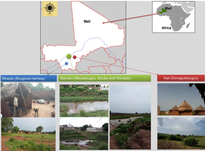
Collection sites
This study was conducted in three different localities in Mali, namely Donéguébougou, Bougoula-hameau and Bamako. In Bamako, the collection was performed in the three semi-urban areas of Sotuba, Yirimadio and Missabougou. The geo-positions of each collection site are as follows: Bougoula-hameau (−5°
66′13.1′′, 11°30′95.2′′E); Donéguébougou (−7°98′39.8′′N, 12°
80′44.9′′E) and the semi-urban areas of Bamako, Sotuba (−9°
18′65.7′N, 8°23′07.4′E), Yirimadio (−9°18′56.5′N, 6°23′01.8′′E)
and Missabougou (−9°18′77.5′′N, 8°23′03.9′′E).
Mosquito collection
Mosquitoes were collected from the various sites during the mid-dle of the rainy season between July and August 2016 (WHO,
1992). The peak densities and consequentially of anopheline
mos-quitoes in Mali occur in August (Sogoba et al.2007). Mosquitoes
were collected over three consecutive days per week. On each day, mosquitoes were aspirated from 10 houses using a mouth aspir-ator (Model 612, John W Hock, Gainesville, Florida, USA). All mosquitoes were collected indoors in the morning between 8 am and noon. The mosquito specimens were identified using
morphological criteria (Gillies MT 1987). After being collected,
mosquito specimens were kept at room temperature (RT) between 2 and 4 h during the female abdomens crushed process and then
were stored at −20 °C. Each mosquito specimen was then
indi-vidually transferred to a 1.5 mL Eppendorf tube labelled with a reference number, the gender of the specimen, the date and site of collection.
Mosquito abdomens with visible blood meals were crushed on WFPs (Whatman International Ltd., Maidstone, England, approved by BSI). Following the entomological stage, all samples were transported to Aix-Marseille University for mosquito and blood meal identification using MALDI-TOF MS in September and October 2016.
Preparation of samples for MALDI-TOF MS analysis Mosquito identification
The legs of the specimens were cleaned in 70% (v/v) ethanol for between one to two minutes, then rinsed in high performance liquid chromatography (HPLC) grade water. The legs from each mosquito were individually placed in 1.5 mL Eppendorf tubes with glass powder (Sigma, Lyon, France), 15 µL of 70% (v/v) for-mic acid (Sigma, Lyon, France), and 15 µL of 50% (v/v) aceto-nitrile (Fluka, Buchs, Switzerland). The samples were crushed using a TissueLyser device (Qiagen, Hilden, Germany) over
three cycles of 30 m s−1for 60 s (Nebbak et al.2016). The samples
were centrifuged at 200 g for one minute, and 1.5 µL of super-natant of each homogenate was deposited on the MALDI-TOF target plate in quadruplicate (Bruker Daltonics, Wissembourg, France) and covered with 1.5 µL of CHCA matrix solution
com-posed of saturated α-cyano-4-hydroxycynnamic acid (Sigma,
Lyon, France), 50% acetonitrile (v/v), 2.5% trifluoroacetic acid (v/v) (Aldrich, Dorset, UK), and HPLC grade water (Yssouf
et al.2013; Nebbak et al.2016). The target plate was dried for
sev-eral minutes at RT and placed in the Microflex LT MALDI-TOF Mass Spectrometer (Bruker Daltonics, Wissembourg, France) for
analysis (Yssouf et al.2013,2016; Nebbak et al.2016).
Bloody Whatman filter papers (BWFPs)
A piece of the WFPs (i.e. about 1 mm2) containing crushed
abdo-mens from engorged mosquitoes was individually cut using a ster-ile scalpel and transferred to a new 1.5 mL Eppendorf tube (Niare
et al.2017). For each piece of WFPs, 20 µL of formic acid (70%, v/v)
plus 20 µL of acetonitrile (50% v/v) (Fluka, Buchs, Switzerland) was added and incubated for 10 min at RT. After a fast spin (i.e. 10 000 rpm for 20 s), 1 µL of the supernatant of each sample was loaded onto the MALDI-TOF target plate in quadruplicate
and covered with 1 µL of CHCA matrix (Niare et al. 2016).
After drying for several minutes at RT, the MALDI-TOF target plate was placed in the Microflex LT MALDI-TOF Mass Spectrometer (Bruker Daltonics, Bremen, Germany) for analysis. To control loading on mass spectra steel, matrix quality and MALDI-TOF apparatus performance, the matrix solution was loaded in duplicate onto each MALDI-TOF plate with or without a bacterial test standard (Bruker protein Calibration Standard I)
Spectral analysis
Protein mass profiles were acquired using a Microflex LT MALDI-TOF Mass Spectrometer, with detection in the linear positive-ion mode at a laser frequency of 50 Hz within a mass range of 2-20 kDa. The acceleration voltage was 20 kV, and the extraction delay time was 200 ns. Each spectrum corresponded to ions obtained from 240 laser shots performed in six regions of the same spot and automatically acquired using the AutoXecute of the Flex Control v.2.4 software (Bruker Daltonics, Bremen, Germany). The spectrum profiles obtained from mosquito legs and bloody WFPs were visualized with Flex analysis v.3.3 software and were exported to ClinProTools version v.2.2 (Bruker Daltonics, Bremen, Germany) and MALDI-Biotyper v.3.0. (Bruker Daltonics, Bremen, Germany) for data processing (smoothing, baseline sub-traction, peak picking) and evaluated using cluster analysis. Spectra of low quality were excluded from the study.
MALDI-TOF identification of mosquitoes
We used our in-lab arthropod MALDI-TOF database, which includes spectra obtained from various arthropods listed in
Table 1. The database was upgraded with the spectra of three Culex quinquefasciatus mosquitoes and one Culex neavei mos-quito collected and molecularly identified during this study. A comparison of the spectrum of each specimen of mosquito legs from Mali was evaluated against the home-made MS reference spectra database using the MALDI-Biotyper software v3.0. tool (Bruker Daltonics, Bremen, Germany). The level of significance was determined using the log score values (LSVs) provided by the MALDI-Biotyper software v.3.3. corresponding to a matched degree of signal intensities of mass spectra of the query and the reference spectra. LSVs ranged from zero to three. To determine the origin of blood meals, MALDI-TOF MS spectra from the
abdominal proteins of engorged mosquitoes crushed on WFPs were also blindly queried against the database. A sample was con-sidered to be correctly and significantly identified at the species level when the queried spectrum had a log score value (LSV)
⩾1.8 (Niare et al.2016).
Cluster analysis
Cluster analysis on MSP (MSP, Main Spectrum Profile) spectra was performed and the comparison of the main spectra given by the MALDI-Biotyper software was clustered according to pro-tein mass profile (i.e. their mass signals and intensities). We per-formed hierarchical clustering of the mass spectra of two specimens per mosquito species using the MSP dendrogram func-tion. The clustering analyses were performed to visualize the homogeneity level of MS spectra from specimens belonging to the same species level. The resulting MSP dendrogram shows how samples are related to one another.
Molecular identification
A molecular tool was used to confirm MALDI-TOF MS identifi-cation in randomly selected mosquitoes. Molecular identifiidentifi-cation was also conducted for specimens whose spectra did not match with any mosquito spectrum in our database. When it was demonstrated that a high quality spectrum had been obtained from a mosquito species missing from our database, this new spectrum was added to the database. DNA extractions from individual mosquito heads and thorax samples were performed using the EZ1 DNA Tissue Kit (Qiagen, Hilden, Germany) according to the
manufac-turer’s recommendations. A set of primers specifically amplifying
a fragment of 710 bp of the mosquito’s cytochrome c oxidase I
gene (mCOI) was used (LCO1490 (forward): 5’-GGTCAAC
Table 1.List of the arthropod species present in our home-made MALDI-TOF MSadatabase.
Mosquitoes Imago: Aedes albopictus, Ae. excrucians, Ae. vexans, Ae. rusticus, Ae. dufouri, Ae. cinereus, Ae. fowleri, Ae. aegypti, Ae. caspius, Anopheles gambiae Giles, An. coluzzii, An. funestus, An. ziemanni, An. arabiensis, An. wellcomei, An. rufipes, An. pharoensis, An. coustani, An. claviger, An. hyrcanus, An. maculipennis, Culex quinquefasciatus, Cx. pipiens, Cx. modestus, Cx. insignis, Cx. neavei, Mansonia uniformis, Culiseta longiareolata, Orthopodomyia reunionensis, Coquillettidia richiardii and Lutzia tigripes.
Larvae: Aedes aegypti, Ae. albopictus, Anopheles gambiae Giles, An. coluzzii, Cx. pipiens, Cx. molestus, Culiseta sp.
Sand flies Phlebotomus papatasi, P. (Larrousius) longicuspis, P. (Larrousius) perfiliewi, P. (Larrousius) perniciosus, P. (Paraphlebotomus) sergenti and Sergentomyia minuta
Triatomines Triatoma infestans, Rhodnius prolixus, Rh. pictipes, Rh. robustus, Eratyrus mucronatus and Panstrongylus geniculatus
Ticks Legs: Amblyomma cohaerens, Am. gemma, Am. variegatum, Dermacentor marginatus, D. reticulatus,
Haemaphysalis leachi, Hae. concinna, Hae. spinulosa, Hyalomma marginatum rufipes, H. truncatum, H. detritum, Rhipicephalus decoloratus, Ixodes hexagonus, I. ricinus, Rh. bergeoni, Rh. e. evertsi,
Rh. praetextatus, Rh. pulchellus, Rh. sanguineus, Rh. sulcatus, Rh. microplus, Rh. annulatus, Rh. turanicus and Rh. bursa.
Hemolymph: Am. variegatum, D. marginatus, H. marginatum rufipes, Rh. bursa and Rh. sanguineus.
Mites Leptotrombidium chiangraiensis, L. imphalum and L. deliense
Bedbugs Cimex lectularius
Lice Pediculus humanus, Damalinia bovis, D. caprae, D. ovis, Haematopinus eurysternus, Linognatus vituli and L.
africanus
Fleas Ctenocephalides felis, Ct. canis, Archaeopsylla erinacei, Xenopsylla cheopis and Stenoponia tripectinata
Abdomen of mosquitoes engorged Anopheles gambiae Giles fed on: Homo sapiens, Equus caballus, Ovis aries, rabbit, Balb/C mouse, Rattus norvegicus, Canis familiaris, Bos taurus, Capra hircus, Gallus gallus, Equus asinus, Tapirus indicus, Tapirus terrestris, Carollia perspicillata, Thraupis episcopus, Erythrocebus patas and Callithrix pygmaea blood Aedes albopictus fed on: Homo sapiens blood
Anopheles gambiae Giles blood meals from Whatman Filter paper
Ovis aries and Homo sapiens blood
aMALDI-TOF MS, matrix-assisted laser desorption/ionization time-of-flight mass spectrometry.
AAATCATAAGATATTGG-3’; HC02198 (reverse): 5’-TAAAC
TTCAGGGTGACCAAAAAATCA-3’ (Folmer et al. 1994). We
used gene Acetylcholinesterase-2 to amplify a fragment of 610 bp of Culex pipiens and a fragment of 274 bp of Cx.
quinque-fasciatus. The primers set were ACEquin (forward): 5′-CCTT
CTTGAATGG CTGTGGCA-3′, ACEpip (forward): 5′-GGAAA
CAACGACGTATGTACT-3′, B1246s (reverse): 5′-TGGAGCC
TCCTCTTCACGGC-3′(Smith and Fonseca,2004).
A set of primers specifically amplifying a fragment of 310 bp of the Anopheles gambiae mosquito complex Acomplex_28S_MBF
5′-AGCKCGTCTTGGTCTGGGG-3′ and Acomplex_28S_MBR
5′-GCCGACAAGCTCAYTAGTGT-3′was designed in our
labora-tory based on the work by Fanello et al. and PCR reactions were
processed as described (Fanello et al.2002). Molecular
identifica-tion of the blood was carried out on the bloody WFPs from 41 specimens randomly selected from the Malian samples, as
previ-ously described (Niare et al.2016). Positive PCR products were
then purified and sequenced using the same primers with the BigDye version 1-1 Cycle Sequencing Ready Reaction Mix (Applied Biosystems, Foster City, CA) and an ABI 3100 automated sequencer (Applied Biosystems, Foster City, CA). The sequences were assembled and analyzed using the ChromasPro software (ver-sion 1.34) (Technelysium Pty. Ltd., Tewantin, Australia) and the
NCBI BLAST website (http://blast.ncbi.nlm.nih.gov).
Results
Identification of the mosquitoes by MALDI-TOF MS
A total of 865 mosquitoes were captured by aspiration in Mali from various collection sites, including 257 in Bougoula-hameau,
168 in Donéguébougou, 230 in Sotuba, 125 in Missabougou and
85 in the Yirimadio semi-urban zones of Bamako (Fig. 1). All
spe-cimens collected were morphologically identified to genus level as Anopheles spp. (287/865; 33.18%), Culex spp. (573/865; 66.24%) and Aedes spp. (5/865; 0.58%).
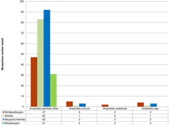
For MALDI-TOF analysis, MS spectra of good quality were obtained from 272 legs of Anopheles spp. Of these 272 Anopheles spp. tested against the arthropod MS database, 97% (n = 264/272) were identified with a log score value (LSV) ranging between 1.70 and 2.575. These 264 Anopheles specimens were identified as Anopheles gambiae Giles (95.80%, n = 253/264), Anopheles coluzzii (3.40%, n = 9/264) and Anopheles arabiensis
(0.80%, n = 2/264) (Fig. 2) by MALDI TOF MS. The remaining
eight Anopheles spp. were subjected to molecular identification. We tested the MS spectra from the legs of 549 Culex spp. against our arthropod database.
Of these 549 Culex spp. high-quality spectra, 98% (n = 537/549) were identified as species contained in our database. The 537 Culex specimens were identified by MALDI-TOF MS as Cx. quinquefas-ciatus (98%, n = 527/537) and Cx. neavei (2%, n = 10/537) from
Mali (Fig. 3). These 537 Culex obtained LSVs ranging from
1.713 to 2.611. The remaining twelve Culex spp. were subjected to molecular identification.
The five Aedes specimens were identified by MALDI-TOF MS as Aedes fowleri (n = 4) and Aedes aegypti (n = 1), with log score values ranging between 2.128 and 2.418.
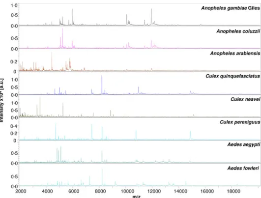
The MS spectra comparison from different mosquito species with Flex analysis software revealed an intra-species
reproducibil-ity and an inter-species specificreproducibil-ity (Fig. 4). Visually, the signals
and intensity of mosquito species’ protein profiles (Fig. 4) were
consistent for MALDI-TOF identification and revealed eight
Fig. 1.Ecological patterns and geographic distribution of mosquito collection in Mali. Sikasso: Bougoula-hameau (rural area), Bamako: Sotuba (peri-urban area), Missabougou, Yirimadio (urban areas) and Kati: Doneguebougou (rural area).
different species, namely Anopheles gambiae Giles, An. coluzzii, An. arabiensis, Cx. quinquefasciatus, Cx. neavei, Culex perexiguus, Ae. fowleri and Ae. aegypti. Clustering analysis of MSP spectra
from two specimens per mosquito species was used to generate a dendrogram. Clustering analysis revealed a gathering on distinct branches, following the eight species which were loaded
Fig. 2.MALDI-TOF MS Identification of 272 Anopheles spp. collected in Mali.
Fig. 3.MALDI-TOF MS Identification of 549 Culex spp. captured in Mali.
(Anopheles gambiae Giles, An. coluzzii, An. arabiensis, Cx. quin-quefasciatus, Cx. neavei, Cx. perexiguus, Ae. fowleri and Ae. aegypti) (Fig. 5). The clusters formed were consistent with the intra-species reproducibility and inter-species specificity visually observed on protein profiles.
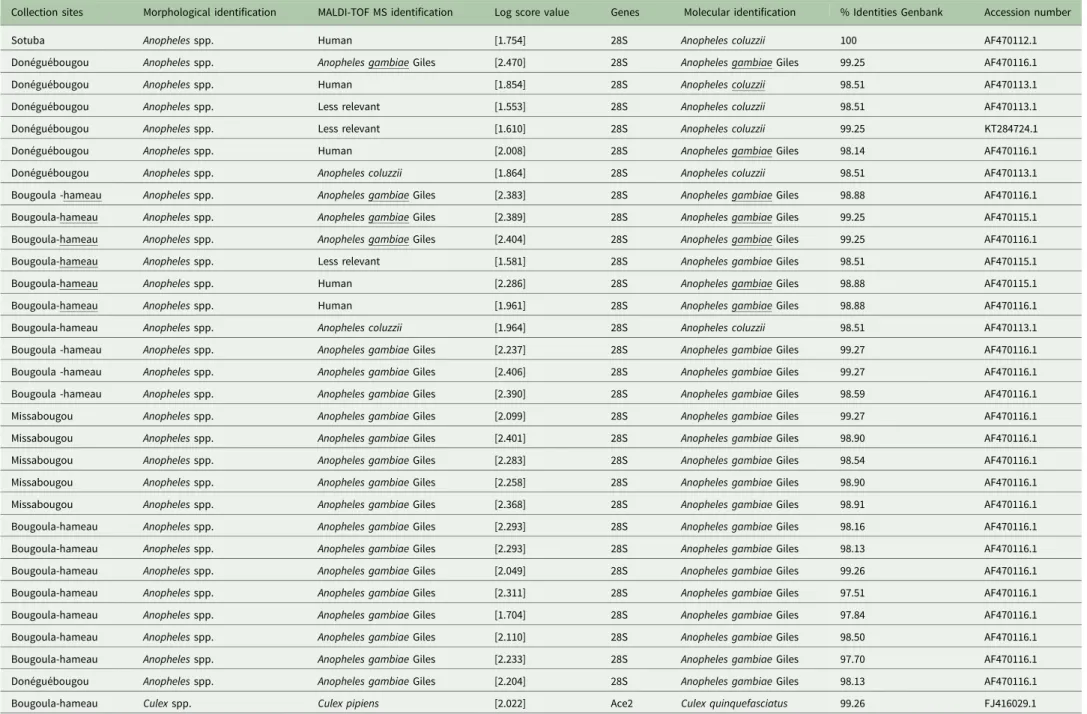
Molecular identification of mosquitoes collected in Mali Molecular biology was performed to confirm the mosquito iden-tification resulting from the MALDI-TOF MS analyses. For this purpose, we randomly selected 20/253 An. gambiae Giles, 2/9 An. coluzzii, 15/527 Cx. quinquefasciatus, 1/10 Cx. neavei for sequencing. The 28S gene sequencing of Anopheles corroborated the MALDI-TOF MS identification in all cases, with between
97.51 and 99.27% identity with Genbank sequences (Table 2).
The acetylcholinesterase-2 and COI genes were used to identify the Culex species. Sixteen specimens of Cx. quinquefasciatus (n = 15) and Cx. neavei (n = 1) were randomly selected for sequencing.
The molecular results were found to be highly consistent with the MALDI-TOF MS identification. Sequences obtained from Cx. quinquefasciatus and Cx. neavei were shown to share between
98.90 and 100% identity with Genbank (Table 2).
Molecular biology was also carried out on the mosquitoes that were not identified by MALDI-TOF MS (low scores), including the eight Anopheles spp. and 12 Culex spp. Sequencing of the 28S gene was performed to identify the eight Anopheles spp. (3%, n = 8/264). The matching sequences corresponded to Anopheles gambiae Giles (n = 4) and An. coluzzii (n = 4), which were shown to share between 98.52 and 100% identity with
Genbank (Table 2).
The acetylcholinesterase-2 and COI genes were amplified to identify the 12 Culex spp. which were misidentified (2%, n = 12/ 549) by MALDI-TOF MS. The sequences obtained correspond to the Cx. quinquefasciatus (n = 11) which were shown to share between 98.90 and 100% identity with Genbank and 100%
iden-tity with Cx. perexiguus (n = 1) (Table 2).
Fig. 4.Comparison of MALDI-TOF MS profiles of eight mosquito species collected in Mali. Spectra analysis was performed using Flex analysis 3.3 software. Abbreviations: a.u., arbitrary units; m/z, mass-to-charge ratio.
Fig. 5.MSP (Main Spectrum Profile) dendrograms of MALDI-TOF MS spectra of eight mosquito species collected in Mali. Clustering analysis was performed using MALDI Biotyper software. Distance unit corre-sponds to the relative similarity calculated from the distance matrix.
Table 2.Molecular identification of mosquitoes collected in Mali
Collection sites Morphological identification MALDI-TOF MS identification Log score value Genes Molecular identification % Identities Genbank Accession number
Sotuba Anopheles spp. Human [1.754] 28S Anopheles coluzzii 100 AF470112.1
Donéguébougou Anopheles spp. Anopheles gambiae Giles [2.470] 28S Anopheles gambiae Giles 99.25 AF470116.1
Donéguébougou Anopheles spp. Human [1.854] 28S Anopheles coluzzii 98.51 AF470113.1
Donéguébougou Anopheles spp. Less relevant [1.553] 28S Anopheles coluzzii 98.51 AF470113.1
Donéguébougou Anopheles spp. Less relevant [1.610] 28S Anopheles coluzzii 99.25 KT284724.1
Donéguébougou Anopheles spp. Human [2.008] 28S Anopheles gambiae Giles 98.14 AF470116.1
Donéguébougou Anopheles spp. Anopheles coluzzii [1.864] 28S Anopheles coluzzii 98.51 AF470113.1
Bougoula -hameau Anopheles spp. Anopheles gambiae Giles [2.383] 28S Anopheles gambiae Giles 98.88 AF470116.1
Bougoula-hameau Anopheles spp. Anopheles gambiae Giles [2.389] 28S Anopheles gambiae Giles 99.25 AF470115.1
Bougoula-hameau Anopheles spp. Anopheles gambiae Giles [2.404] 28S Anopheles gambiae Giles 99.25 AF470116.1
Bougoula-hameau Anopheles spp. Less relevant [1.581] 28S Anopheles gambiae Giles 98.51 AF470115.1
Bougoula-hameau Anopheles spp. Human [2.286] 28S Anopheles gambiae Giles 98.88 AF470115.1
Bougoula-hameau Anopheles spp. Human [1.961] 28S Anopheles gambiae Giles 98.88 AF470116.1
Bougoula-hameau Anopheles spp. Anopheles coluzzii [1.964] 28S Anopheles coluzzii 98.51 AF470113.1
Bougoula -hameau Anopheles spp. Anopheles gambiae Giles [2.237] 28S Anopheles gambiae Giles 99.27 AF470116.1
Bougoula -hameau Anopheles spp. Anopheles gambiae Giles [2.406] 28S Anopheles gambiae Giles 99.27 AF470116.1
Bougoula -hameau Anopheles spp. Anopheles gambiae Giles [2.390] 28S Anopheles gambiae Giles 98.59 AF470116.1
Missabougou Anopheles spp. Anopheles gambiae Giles [2.099] 28S Anopheles gambiae Giles 99.27 AF470116.1
Missabougou Anopheles spp. Anopheles gambiae Giles [2.401] 28S Anopheles gambiae Giles 98.90 AF470116.1
Missabougou Anopheles spp. Anopheles gambiae Giles [2.283] 28S Anopheles gambiae Giles 98.54 AF470116.1
Missabougou Anopheles spp. Anopheles gambiae Giles [2.258] 28S Anopheles gambiae Giles 98.90 AF470116.1
Missabougou Anopheles spp. Anopheles gambiae Giles [2.368] 28S Anopheles gambiae Giles 98.91 AF470116.1
Bougoula-hameau Anopheles spp. Anopheles gambiae Giles [2.293] 28S Anopheles gambiae Giles 98.16 AF470116.1
Bougoula-hameau Anopheles spp. Anopheles gambiae Giles [2.293] 28S Anopheles gambiae Giles 98.13 AF470116.1
Bougoula-hameau Anopheles spp. Anopheles gambiae Giles [2.049] 28S Anopheles gambiae Giles 99.26 AF470116.1
Bougoula-hameau Anopheles spp. Anopheles gambiae Giles [2.311] 28S Anopheles gambiae Giles 97.51 AF470116.1
Bougoula-hameau Anopheles spp. Anopheles gambiae Giles [1.704] 28S Anopheles gambiae Giles 97.84 AF470116.1
Bougoula-hameau Anopheles spp. Anopheles gambiae Giles [2.110] 28S Anopheles gambiae Giles 98.50 AF470116.1
Bougoula-hameau Anopheles spp. Anopheles gambiae Giles [2.233] 28S Anopheles gambiae Giles 97.70 AF470116.1
Donéguébougou Anopheles spp. Anopheles gambiae Giles [2.204] 28S Anopheles gambiae Giles 98.13 AF470116.1
Bougoula-hameau Culex spp. Culex pipiens [2.022] Ace2 Culex quinquefasciatus 99.26 FJ416029.1
(Continued ) P a rasitology 7 https://www.cambridge.org/core/terms . https://doi.org/10.1017/S0031182018000070 Downloaded from https://www.cambridge.org/core . Universite Mediterranee , on 27 Feb 2018 at 10:26:04
Table 2.(Continued.)
Collection sites Morphological identification MALDI-TOF MS identification Log score value Genes Molecular identification % Identities Genbank Accession number
Bougoula-hameau Culex spp. Culex pipiens [2.229] Ace2 Culex quinquefasciatus 98.90 FJ416029.1
Donéguébougou Culex spp. Culex quinquefasciatus [1.791] Ace2 Culex quinquefasciatus 99.26 FJ416029.1
Missabougou Culex spp. Human [2.182] COI Culex quinquefasciatus 99.38 KU920694.1
Missabougou Culex spp. Less relevant [1.610] COI Culex perexiguus 100 KU380476.1
Missabougou Culex spp. Culex pipiens [1.761] Ace2 Culex quinquefasciatus 99.63 FJ416025.1
Missabougou Culex spp. Human [2.042] COI Culex quinquefasciatus 99.08 KU920694.1
Missabougou Culex spp. Culex pipiens [2.139] Ace2 Culex quinquefasciatus 98.90 FJ416025.1
Missabougou Culex spp. Culex pipiens [2.231] Ace2 Culex quinquefasciatus 98.90 FJ416025.1
Missabougou Culex spp. Human [2.514] COI Culex quinquefasciatus 99.84 KU920694.1
Missabougou Culex spp. Culex pipiens [2.088] Ace2 Culex quinquefasciatus 98.90 FJ416029.1
Missabougou Culex spp. Human [2.294] COI Culex quinquefasciatus 99.69 KU920694.1
Yirimadio Culex spp. Culex pipiens [2.032] Ace2 Culex quinquefasciatus 98.90 FJ416029.1
Donéguébougou Culex spp. Culex quinquefasciatus [2.000] Ace2 Culex quinquefasciatus 99.26 FJ416029.1
Donéguébougou Culex spp. Culex quinquefasciatus [2.009] Ace2 Culex quinquefasciatus 99.27 FJ416025.1
Donéguébougou Culex spp. Culex quinquefasciatus [2.207] Ace2 Culex quinquefasciatus 98.90 FJ416025.1
Bougoula-hameau Culex spp. Culex quinquefasciatus [2.406] Ace2 Culex quinquefasciatus 98.90 FJ416019.1
Bougoula-hameau Culex spp Culex neavei [2.839] COI Culex neavei 99.16 KU380473.1
Bougoula-hameau Culex spp Culex quinquefasciatus [2.037] Ace2 Culex quinquefasciatus 99.11 FJ416029.1
Bougoula-hameau Culex spp Culex quinquefasciatus [2.197] Ace2 Culex quinquefasciatus 99.55 FJ416029.1
Bougoula-hameau Culex spp Culex quinquefasciatus [2.146] Ace2 Culex quinquefasciatus 99.56 FJ416029.1
Bougoula-hameau Culex spp Culex quinquefasciatus [2.383] Ace2 Culex quinquefasciatus 99.56 FJ416029.1
Bougoula-hameau Culex spp Culex quinquefasciatus [2.322] Ace2 Culex quinquefasciatus 99.13 FJ416029.1
Donéguébougou Culex spp. Culex quinquefasciatus [2.445] Ace2 Culex quinquefasciatus 99.57 FJ416029.1
Donéguébougou Culex spp. Culex quinquefasciatus [2.067] Ace2 Culex quinquefasciatus 98.71 FJ416029.1
Donéguébougou Culex spp. Culex quinquefasciatus [2.151] Ace2 Culex quinquefasciatus 100 FJ416029.1
Missabougou Culex spp. Culex quinquefasciatus [2.244] Ace2 Culex quinquefasciatus 98.68 FJ416029.1
Missabougou Culex spp. Culex quinquefasciatus [2.159] Ace2 Culex quinquefasciatus 99.56 FJ416029.1
MALDI-TOF MS, matrix-assisted laser desorption/ionization time-of-flight mass spectrometry; Ace 2: acetylcholinesterase-2; COI: the cytochrome c oxidase; %, per cent.
8 F a talmoudou T andina et al. . https://doi.org/10.1017/S0031182018000070 https://www.cambridge.org/core . Universite Mediterranee , on 27 Feb 2018 at 10:26:04
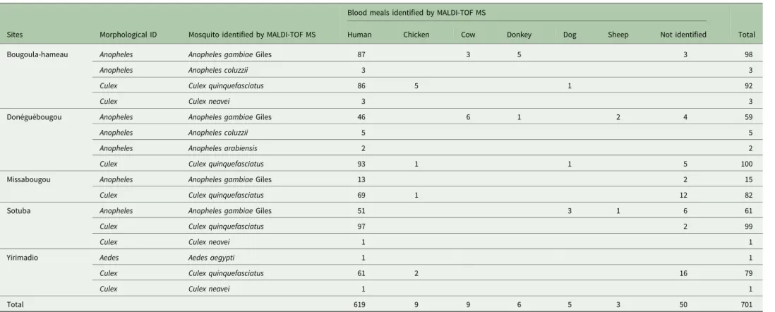
Table 3.Identification of the blood meals of mosquitoes collected in distinct ecological areas in Mali
Sites Morphological ID Mosquito identified by MALDI-TOF MS
Blood meals identified by MALDI-TOF MS
Total
Human Chicken Cow Donkey Dog Sheep Not identified
Bougoula-hameau Anopheles Anopheles gambiae Giles 87 3 5 3 98
Anopheles Anopheles coluzzii 3 3
Culex Culex quinquefasciatus 86 5 1 92
Culex Culex neavei 3 3
Donéguébougou Anopheles Anopheles gambiae Giles 46 6 1 2 4 59
Anopheles Anopheles coluzzii 5 5
Anopheles Anopheles arabiensis 2 2
Culex Culex quinquefasciatus 93 1 1 5 100
Missabougou Anopheles Anopheles gambiae Giles 13 2 15
Culex Culex quinquefasciatus 69 1 12 82
Sotuba Anopheles Anopheles gambiae Giles 51 3 1 6 61
Culex Culex quinquefasciatus 97 2 99
Culex Culex neavei 1 1
Yirimadio Aedes Aedes aegypti 1 1
Culex Culex quinquefasciatus 61 2 16 79
Culex Culex neavei 1 1
Total 619 9 9 6 5 3 50 701
ID, Identification; MALDI-TOF MS, matrix-assisted laser desorption/ionization time-of-flight mass spectrometry.
P a rasitology 9 https://www.cambridge.org/core/terms . https://doi.org/10.1017/S0031182018000070 Downloaded from https://www.cambridge.org/core . Universite Mediterranee , on 27 Feb 2018 at 10:26:04
Identification of the bloody WFPs sources by MALDI-TOF MS A total 701 abdomens of engorged mosquitoes were crushed in WFPs in the field in Mali. The 701 bloody BWFPs were submitted for MALDI-TOF MS analysis in Marseille one month after sam-pling. Of the 701 BWFPs, 651 (93%) high-quality spectra were obtained. The 651 BWFPs MS high-quality spectra were queried against our blood source MALDI-TOF MS database for identifi-cation. They matched with spectra from our database, including those of mosquito abdomens engorged with human blood (n = 619), chicken blood (n = 9), cow blood (n = 9), donkey blood (n = 6), dog blood (n = 5) and sheep blood (n = 3) (Table 3). These blood meals were identified using MALDI-TOF MS with log score values (LSVs) ranging from 1.707 to 2.731. The MS spectra comparison of different host blood revealed an intra-species reproducibility and an
inter-species specificity by Flex analysis (Fig. 6).
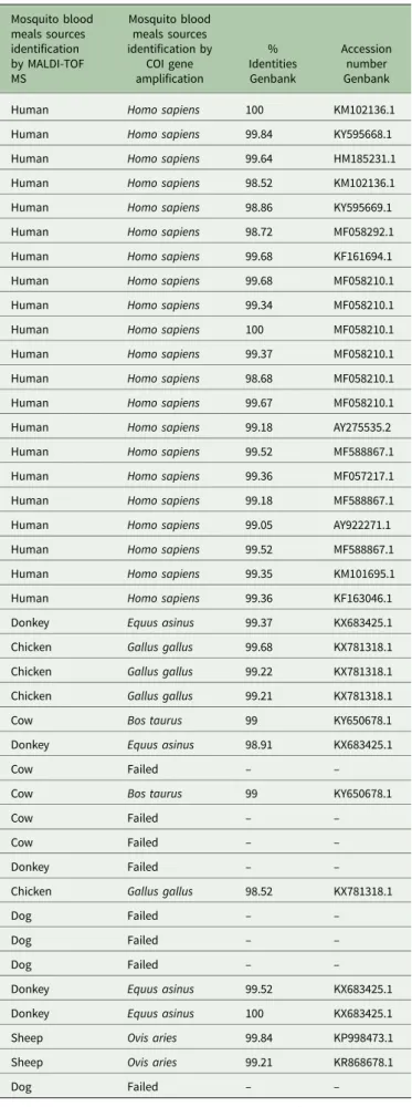
Molecular identification of the bloody mosquito WFPs A total of 41 bloody WFPs identified by MALDI-TOF MS as mosquito abdomens engorged with human blood (n = 21), don-key blood (n = 5), chicken blood (n = 4), cow blood (n = 5), dog blood (n = 4) and sheep blood (n = 2) were randomly selected for sequencing by COI gene amplification. Thirty-three bloody WFPs sequences were obtained which confirmed the accuracy of the MS identification. However, for eight bloody WFPs, no quality sequences could be obtained. The results of the PCR based on bloody WFP sequencing highly correlated with the
results of MALDI TOF MS identification (Table 4). The
sequences obtained from seventeen bloody WFPs had identities
between 98.52 and 100% against Genbank NCBI (Table 4).
Discussion
The goal of this work was not to provide precise data on the pres-ence and abundance of various mosquito species in specific areas in Mali. Indeed, these data vary according to the type of climate and the seasons. However, we did want to test the usefulness of MALDI-TOF MS using mosquitoes collected in the field, as most preliminary studies have used laboratory specimens.
The use of MALDI-TOF MS has recently emerged in medical entomology, including for the identification of arthropods, their blood meals and the detection of potential microorganisms
(Schaffner et al. 2014; Yssouf et al. 2016). The choice of the
arthropod body part is critical for specimen identification by
MALDI-TOF MS (Yssouf et al. 2016). For example, the legs
from adult mosquitoes have been shown to be sufficient for iden-tification, whereas whole specimens have been used for aquatic
stages (larvae) (Nebbak et al.2017).
Here, the MS spectra from mosquito legs collected in Mali, including 264 Anopheles, 549 Culex and five Aedes, permitted MALDI-TOF MS identification. The MS spectrum analyses from the mosquito legs revealed an intra-species reproducibility and inter-species specificity consistent with molecular validation (Fig. 5). Accurate identification of mosquitoes queried against the home-made MS database corresponded to 100% concordance
with molecular identification results (Table 2). The consistent
Fig. 6.The MS spectrum alignment from mosquito abdomen engorged on vertebrate host bloods and then crushed on Whatman filters. All bloody WFPs (BWFPs) were obtained from the field mosquitoes collected in Mali and crushed on WFPs. The MS spectrum alignment was performed by Flex analysis 3.3 software. The WFP blood free corresponds to the MS profiles of WFPs where no mosquito blood meal was released. The representative MS spectra from abdominal protein corre-sponds to Anopheles gambiae Giles abdomens BWFPs feed on human, donkey, cow and sheep blood, and Culex quinquefasciatus abdomens feed on chicken and dog blood. a.u. arbitrary units; m/z mass-to-charge ratio.
identification between molecular biology and MALDI-TOF MS was validated by the choice of the 28S gene for Anopheles species identification, and the acetylcholinesterase-2 and COI genes for Culex species identification. As shown in previous studies, the choice of these genes was highly relevant to discriminate and assess the phylogenetic relation between different mosquito
spe-cies (Folmer et al.1994; Fanello et al.2002; Smith and Fonseca,
2004). Here, the quality of spectra was a very important element
for identification, as more than 98% of the good quality spectra were identified with LSVs >1.8. The MALDI-TOF MS reference database has been updated with other mosquito species. It is necessary to create a reference database, which could subsequently be shared, and open access could be provided for routine arthro-pod identification. In this study, Aedes mosquitoes collected in Mali were correctly identified as Ae. fowleri using a database con-taining reference spectra of this species collected from La Reunion Island only, which is located in the Pacific Ocean. Therefore MALDI-TOF MS appears as an efficient tool for the identification of arthropods collected from distant geographical areas.
For Raharimalala et al. (2017), the usefulness and accuracy of
MALDI-TOF MS as a tool to identify vector mosquito species requires the creation of an international database (Raharimalala
et al. 2017). In this study, 2.40% of inconsistent MS leg results
were attributed to low-quality MS spectra for identification. The
MS spectra of some legs (n = 9) (Table 2) that matched with
ref-erence spectra of mosquito abdomens engorged with human blood were attributed to traces of blood on the legs during the abdomen crushing process onto WFPs. This phenomenon of low-quality spectra, leading to lower identification rates have been reported in arthropod identification such as at the aquatic
mos-quito stage (Dieme et al.2014). According to the reproducibility
of MS spectra, the hierarchical clustering showed that all speci-mens from the same species were grouped in the same branch. These results are similar to previous studies supporting inter-species reproducibility for mosquito identification (Yssouf et al.
2013). Additionally, we stress that MS cannot yet be considered
a reliable tool for the phylogenetic study of mosquito species
(Yssouf et al.2013).
Our results showed that 95% of the collected mosquitoes had fed on human blood. This result is not surprising because all mos-quitoes were collected inside homes. The advantage of our MALDI-TOF approach is its rapidity, effectiveness and reliability in determining bloody WFPs, since more than 100 bloody WFPs specimens were processed per day. Previously, the authors had demonstrated that the profiles of abdominal spectra of mosquito females engorged on human blood are the same, regardless of whether they were crushed or not crushed on WFPs (Niare
et al. 2017). Indeed, the home-made database contains filter
papers with Anopheles gambiae engorged blood such as human blood and sheep blood. These authors tested WFP either with the crushed abdomen of a non-engorged mosquito or simply as
a control (Niare et al.2017). These results suggest that
MALDI-TOF MS is not time-consuming in comparison with molecular tools and serological techniques. The eight bloody WFPs which failed molecular biology identification may be attributed to blood meal digestion. As previously reported, the time of the host blood digestion in the mosquito has an impact upon blood meal identification by MALDI-TOF MS and molecular biology
(Niare et al. 2016). Moreover, the molecular biology results of
the seventeen BWFPs sequences obtained by COI gene
amplifica-tion corroborated the MALDI-TOF MS identificaamplifica-tion (Table 4).
Interestingly, as we have recently found that MALDI-TOF may also recognize mixed blood meals (unpublished), we did not find any mixed blood meals either by molecular tools nor by MALDI-TOF. The authors experimentally engorged An. gambiae Giles mosquitoes with a mixture of blood from distinct vertebrate
Table 4. Molecular identification of the blood from mosquito’s abdomens crushed on Whatman filter papers
Mosquito blood meals sources identification by MALDI-TOF MS Mosquito blood meals sources identification by COI gene amplification % Identities Genbank Accession number Genbank
Human Homo sapiens 100 KM102136.1
Human Homo sapiens 99.84 KY595668.1
Human Homo sapiens 99.64 HM185231.1
Human Homo sapiens 98.52 KM102136.1
Human Homo sapiens 98.86 KY595669.1
Human Homo sapiens 98.72 MF058292.1
Human Homo sapiens 99.68 KF161694.1
Human Homo sapiens 99.68 MF058210.1
Human Homo sapiens 99.34 MF058210.1
Human Homo sapiens 100 MF058210.1
Human Homo sapiens 99.37 MF058210.1
Human Homo sapiens 98.68 MF058210.1
Human Homo sapiens 99.67 MF058210.1
Human Homo sapiens 99.18 AY275535.2
Human Homo sapiens 99.52 MF588867.1
Human Homo sapiens 99.36 MF057217.1
Human Homo sapiens 99.18 MF588867.1
Human Homo sapiens 99.05 AY922271.1
Human Homo sapiens 99.52 MF588867.1
Human Homo sapiens 99.35 KM101695.1
Human Homo sapiens 99.36 KF163046.1
Donkey Equus asinus 99.37 KX683425.1
Chicken Gallus gallus 99.68 KX781318.1
Chicken Gallus gallus 99.22 KX781318.1
Chicken Gallus gallus 99.21 KX781318.1
Cow Bos taurus 99 KY650678.1
Donkey Equus asinus 98.91 KX683425.1
Cow Failed – –
Cow Bos taurus 99 KY650678.1
Cow Failed – –
Cow Failed – –
Donkey Failed – –
Chicken Gallus gallus 98.52 KX781318.1
Dog Failed – –
Dog Failed – –
Dog Failed – –
Donkey Equus asinus 99.52 KX683425.1
Donkey Equus asinus 100 KX683425.1
Sheep Ovis aries 99.84 KP998473.1
Sheep Ovis aries 99.21 KR868678.1
Dog Failed – –
MALDI-TOF MS, matrix-assisted laser desorption/ionization time-of-flight mass spectrometry; COI, the cytochrome c oxidase; %, per cent.
hosts, such as human, sheep and dogs. Their results demon-strate that mixed mosquito blood meals can be successfully identified, depending on the concentration ratio (unpublished). Recently, some authors have also used the proteomic approach to identify the sources of tick mixed blood meals (Onder et al.
2013).
Of the mosquitoes identified by MALDI-TOF MS, A. gambiae Giles and Cx. quinquefasciatus were widely distributed across all collection sites. Our work enabled Cx. neavei and Cx. perexiguus to be detected for the first time in Mali. Currently, few studies have been carried out on the Culex species in Mali, particularly on their abundance, ecology and the infectious pathogens trans-mitted by these vectors. Culex species are widely distributed in West Africa and are found in any type of breeding sites (clear and polluted water), whereas the Anopheles species colonizes
sunny, fresh water (Becker et al.2010). There is an abundant
lit-erature on these mosquitoes, the well-known distribution of Cx. neavei and Cx. perexiguus in sub-Saharan Africa and their
impli-cation in the transmission of many arboviruses (Jupp et al.1986;
Fyodorova et al.2006; Nikolay et al.2012; Fall et al.2014; Gould
et al.2017). The presence of these potential vectors in Mali might
be of epidemiological importance.
Our study is the first to use MALDI-TOF MS as a tool for monitoring field mosquitoes in Africa, particularly in Mali, an endemic malarial area. Moreover, when the MALDI-TOF MS device is bought for clinical microbiology purposes, it can also be used for medical entomology at no additional cost. For example, at the Dakar hospital in Senegal, the MALDI-TOF MS equipment that was initially bought for clinical microbiology has been used for field entomology surveys and has successfully
identified Culicoides (Sambou et al.2015). In Senegal, the
acqui-sition of MALDI-TOF MS equipment has revolutionized bacteri-ology laboratories and clinical microbibacteri-ology domains, suggesting that this technique can be used as a front-line tool in tropical
countries (Lo et al.2015).
Although the time period for blood meal source determin-ation by MALDI-TOF MS was shorter than that of molecular biology or ELISA, the rapidity and low cost of the reagents made this proteomic method a financial and reliable competi-tive strategy. However, the relacompeti-tively high cost of the machine could be an impediment to implementation of this innovative tool in laboratories. The cost of purchasing the MALDI-TOF MS equipment in under-developed countries such as Mali (sub-Saharan Africa) could be a limitation to estimating the local vector-borne risk. However, when the device is bought by a microbiology lab it can be used in medical entomology at no additional cost.
Concluding remarks
The present study successfully identified field mosquitoes and the sources of their blood meals using MALDI-TOF MS. The mos-quitoes collected in Mali were correctly identified based on repro-ducibility and specificity from the protein profiles of leg extracts. The innovative MALDI-TOF MS tool enabled the rapid identifi-cation of eight mosquito species in Mali during entomological surveys. The challenge is to maintain and develop collaboration between north and south to facilitate the acquisition of the MALDI-TOF MS equipment.
Acknowledgements. We thank all members of the various MRTC sites where mosquitoes were collected. We would also like to acknowledge all the residents of the various concessions where we captured mosquitoes. Competing interests. The authors declare that they have no competing interests.
Author contributions. PP, TF and NS designed and developed the protocol. TF and NS performed the protocol. PP, TF, NS and ML analysed the data. KKA, DZA, OA, BMJ, OD and RD contributed reagents/materials/analysis tools. PP, TF and NS wrote the paper. OD and RD contributed to editing the paper. All authors agreed to publication.
Financial support. The project has received funding from the Excellence Initiative of Aix-Marseille University– A*MIDEX, a French ‘Investissements d’Avenir’ program (No. ANR-11-IDEX-0001-02) and grants from the Malian Minister of Health, the Foundation Mérieux, UMI3189, and the Malian Research and Training Center (MRTC) for the field specimen collection.
References
Anosike JC, Nwoke BE, Ajayi EG, Onwuliri CO, Okoro OU, Oku EE, Asor JE, Amajuoyi OU, Ikpeama CA, Ogbusu FI and Meribe CO (2005) Lymphatic filariasis among the Ezza people of Ebonyi state, Eastern Nigeria. Annals of Agricultural and Environnemental Medicine 12, 181–186.
Bass C, Williamson MS, Wilding CS, Donnelly MJ and Field LM(2007) Identification of the main malaria vectors in the Anopheles gambiae species complex using a TaqMan real-time PCR assay. Malaria Journal 6, 155. Becker N, Petriæ D, Zgomba M, Boase C, Dahl C, Madon M and Kaiser A
(2010) Mosquitoes and Their Control, 2nd Edn. Heidelberg, Germany: Springer.
Brasil P, Zalis MG, de Pina-Costa A, Siqueira AM, Junior CB, Silva S, Areas ALL, Pelajo-Machado M, de Alvarenga DAM, da Silva Santelli ACF, Albuquerque HG, Cravo P, Santos de Abreu FV,
Peterka CL, Zanini GM, Suarez Mutis MC, Pissinatti A,
Lourenco-de-Oliveira R, de Brito CFA, de Fatima Ferreira-da-Cruz, Culleton R and Daniel-Ribeiro CT (2017) Outbreak of human malaria caused by Plasmodium simium in the Atlantic forest in Rio de Janeiro: a molecular epidemiological investigation. Lancet Global Health 5, 1038–1046. Caglioti C, Lalle E, Castilletti C, Carletti F, Capobianchi MR and Bordi L (2013) Chikungunya virus infection: an overview. New Microbiology 36, 211–227.
de Wispelaere M, Despres P and Choumet V(2017) European Aedes albopic-tus and Culex pipiens Are competent vectors for Japanese encephalitis virus. PLoS Neglected Tropical Diseases 11, e0005294.
Dieme C, Yssouf A, Vega-Rua A, Berenger JM, Failloux AB, Raoult D, Parola P and Almeras L (2014) Accurate identification of Culicidae at aquatic developmental stages by MALDI-TOF MS profiling. Parasites &Vectors 7, 544.
Fall G, Diallo M, Loucoubar C, Faye O and Sall AA(2014) Vector compe-tence of Culex neavei and Culex quinquefasciatus (Diptera: Culicidae) from Senegal for lineages 1, 2, koutango and a putative new lineage of west Nile virus. American Journal of Tropical Medicine and Hygiene 90, 747–754. Fanello C, Santolamazza F and della TA, (2002) Simultaneous identification
of species and molecular forms of the Anopheles gambiae complex by PCR-RFLP. Medical and Veterinary Entomology 16, 461–464.
Folmer O, Black M, Hoeh W, Lutz R and Vrijenhoek R(1994) DNA primers for amplification of mitochondrial cytochrome c oxidase subunit I from diverse metazoan invertebrates. Molecular Marine Biology and Biotechnology 3, 294–299.
Fyodorova MV, Savage HM, Lopatina JV, Bulgakova TA, Ivanitsky AV, Platonova OV and Platonov AE(2006) Evaluation of potential west Nile virus vectors in volgograd region, Russia, 2003 (Diptera: Culicidae): species composition, bloodmeal host utilization, and virus infection rates of mos-quitoes. Journal of Medical Entomology 43, 552–563.
Gardner CL and Ryman KD(2010) Yellow fever: a reemerging threat. Clinics in Laboratory Medicine 30, 237–260.
Gillies MT and Coetzee M(1987) A supplement to the Anophelinae of Africa south of the Sahara. South African Institute for Medical Research 55, 143p. Gomes B, Sousa CA, Vicente JL, Pinho L, Calderon I, Arez E, Almeida AP, Donnelly MJ and Pinto J(2013) Feeding patterns of molestus and pipiens forms of Culex pipiens (Diptera: Culicidae) in a region of high hybridiza-tion. Parasites & Vectors 6, 93.
Gould E, Pettersson J, Higgs S, Charrel R and de Lamballerie X(2017) Emerging arboviruses: why today? One Health 4, 1–13.
Jupp PG, McIntosh BM and Blackburn NK(1986) Experimental assessment of the vector competence of Culex (Culex) neavei theobald with west Nile
and sindbis viruses in South Africa. Transactions of the Royal Society of Tropical Medicine and Hygiene 80, 226–230.
Komar N (2003) West Nile virus: epidemiology and ecology in North America. Advances in Virus Research 61, 185–234.
Kumsa B, Laroche M, Almeras L, Mediannikov O, Raoult D and Parola P (2016) Morphological, molecular and MALDI-TOF mass spectrometry identification of ixodid tick species collected in Oromia, Ethiopia. Parasitology Research 115, 4199–4210.
Lafri I, Almeras L, Bitam I, Caputo A, Yssouf A, Forestier CL, Izri A, Raoult D and Parola P(2016) Identification of Algerian field-caught phle-botomine sand Fly vectors by MALDI-TOF MS. PLoS Neglected Tropical Diseases 10, e0004351.
Lo CI, Fall B, Sambe-Ba B, Diawara S, Gueye MW, Mediannikov O, Sokhna C, Faye N, Dieme Y, Wade B, Raoult D and Fenollar F(2015) MALDI-TOF Mass spectrometry: a powerful tool for clinical microbiology at hopital principal de Dakar, Senegal (West Africa). PLoS ONE 10, e0145889. Muturi EJ, Mwangangi JM, Beier JC, Blackshear M, Wauna J, Sang R and Mukabana WR (2013) Ecology and behavior of Anopheles arabiensis in relation to agricultural practices in central Kenya. Journal of the American Mosquito Control Association 29, 222–230.
Nebbak A, Willcox AC, Bitam I, Raoult D, Parola P and Almeras L(2016) Standardization of sample homogenization for mosquito identification using an innovative proteomic tool based on protein profiling. Proteomics 16, 3148–3160.
Nebbak A, Koumare S, Willcox AC, Raoult D, Almeras L and Parola P (2017) Field application of MALDI-TOF MS on mosquito larvae identifica-tion. Parasitology 3, 1–11.
Niare S, Berenger JM, Dieme C, Doumbo O, Raoult D, Parola P and Almeras L(2016) Identification of blood meal sources in the main African malaria mosquito vector by MALDI-TOF MS. Malariar Journal 15, 87. Niare S, Almeras L, Tandina F, Yssouf A, Bacar A, Toilibou A, Doumbo O,
Raoult D and Parola P(2017) MALDI-TOF MS identification of Anopheles gambiae giles blood meal crushed on Whatman filter papers. PLoS One 12, e0183238.
Nikolay B, Diallo M, Faye O, Boye CS and Sall AA(2012) Vector compe-tence of Culex neavei (Diptera: Culicidae) for usutu virus. American Journal of Tropical Medicine and Hygiene 86, 993–996.
Onder O, Shao W, Kemps BD, Lam H and Brisson D(2013) Identifying sources of tick blood meals using unidentified tandem mass spectral librar-ies. Nature Communication 4, 1746.
Raharimalala FN, Andrianinarivomanana TM, Rakotondrasoa A, Collard JM and Boyer S(2017) Usefulness and accuracy of MALDI-TOF mass spectrometry as a supplementary tool to identify mosquito vector spe-cies and to invest in development of international database. Medical and Veterinary Entomology 31, 289–298.
Sambou M, Aubadie-Ladrix M, Fenollar F, Fall B, Bassene H, Almeras L, Sambe-Ba B, Perrot N, Chatellier S, Faye N, Parola P, Wade B, Raoult D and Mediannikov O(2015) Comparison of matrix-assisted laser desorption ionization-time of flight mass spectrometry and molecular biology techniques for identification of culicoides (Diptera: Ceratopogonidae) biting midges in Senegal. Journal of Clinical Microbiology 53, 410–418.
Schaffner F, Kaufmann C, Pfluger V and Mathis A(2014) Rapid protein profiling facilitates surveillance of invasive mosquito species. Parasites & Vectors 7, 142.
Smith JL and Fonseca DM(2004) Rapid assays for identification of members of the Culex (Culex) pipiens complex, their hybrids, and other sibling spe-cies (Diptera: Culicidae). American Journal of Tropical Medicine and Hygiene 70, 339–345.
Sogoba N, Doumbia S, Vounatsou P, Baber I, Keita M, Maiga M, Traore SF, Toure A, Dolo G, Smith T and Ribeiro JM(2007) Monitoring of larval habitats and mosquito densities in the Sudan Savanna of Mali: implications for malaria vector control. American Journal of Tropical Medicine and Hygiene 77, 82–88.
Vasilakis N, Cardosa J, Hanley KA, Holmes EC and Weaver SC(2011) Fever from the forest: prospects for the continued emergence of sylvatic dengue virus and its impact on public health. Nature Reviews Microbiology 9, 532–541.
WHO (1992) Entomological Field Techniques for Malaria Control. Part I, Learners Guide. Geneva: World Health Organization.
WHO (2016) World Malaria Report 2016. Geneva: World Health Organization.
Yssouf A, Socolovschi C, Flaudrops C, Ndiath MO, Sougoufara S, Dehecq JS, Lacour G, Berenger JM, Sokhna CS, Raoult D and Parola P(2013) Matrix-assisted laser desorption ionization–time of flight mass spectrometry: an emerging tool for the rapid identification of mos-quito vectors. PLoS ONE 8, e72380.
Yssouf A, Parola P, Lindstrom A, Lilja T, L’Ambert G, Bondesson U, Berenger JM, Raoult D and Almeras L (2014a) Identification of European mosquito species by MALDI-TOF MS. Parasitology Research 113, 2375–2378.
Yssouf A, Socolovschi C, Leulmi H, Kernif T, Bitam I, Audoly G, Almeras L, Raoult D and Parola P(2014b) Identification of flea species using MALDI-TOF/MS. Comparative Immunology, Microbiology and Infectious Diseases 37, 153–157.
Yssouf A, Almeras L, Berenger JM, Laroche M, Raoult D and Parola P (2015) Identification of tick species and disseminate pathogen using hemolymph by MALDI-TOF MS. Ticks and Tick-Borne Diseases 6, 579–586.
Yssouf A, Almeras L, Raoult D and Parola P(2016) Emerging tools for iden-tification of arthropod vectors. Future Microbiology 11, 549–566.
Parasitology 13