HAL Id: tel-00714360
https://tel.archives-ouvertes.fr/tel-00714360
Submitted on 4 Jul 2012
HAL is a multi-disciplinary open access
archive for the deposit and dissemination of sci-entific research documents, whether they are pub-lished or not. The documents may come from teaching and research institutions in France or abroad, or from public or private research centers.
L’archive ouverte pluridisciplinaire HAL, est destinée au dépôt et à la diffusion de documents scientifiques de niveau recherche, publiés ou non, émanant des établissements d’enseignement et de recherche français ou étrangers, des laboratoires publics ou privés.
Wound healing signals mediated by Rho/ROCK
activation in response to radiotherapy and consequences
fot treatmeny of late damage within normal tissues
Nadia Pasinetti
To cite this version:
Nadia Pasinetti. Wound healing signals mediated by Rho/ROCK activation in response to radio-therapy and consequences fot treatmeny of late damage within normal tissues. Human health and pathology. Université Paris Sud - Paris XI; Università degli studi (Brescia, Italie), 2012. English. �NNT : 2012PA11T029�. �tel-00714360�
PhD THESIS– DOTTORATO DI RICERCA IN CO-TUTELA
UNIVERSITÀ DEGLI STUDI DI BRESCIA METODOLOGIA DELLA SPERIMENTAZIONE CLINICA
MED/17 MALATTIE INFETTIVE - CICLO XXIV
ECOLE DOCTORALE DE CANCEROLOGIE: BIOLOGIE, MEDECINE, SANTE Champ Disciplinaire RADIOBIOLOGIE UNIVERSITE PARIS SUD
Title:
“WOUND
HEALING
SIGNALS
MEDIATED
BY
Rho/ROCK
ACTIVATION
IN
RESPONSE
TO
RADIOTHERAPY
AND
CONSEQUENCES FOR TREATMENT OF LATE DAMAGE WITHIN
NORMAL TISSUES”
Titre:
“SIGNAUX DE CICATRISATION MEDIEES PAR L’ACTIVATION DE
LA VOIE Rho/ROCK EN REPONSE A LA RADIOTHERAPIE ET
CONSEQUENCES POUR LE TRAITEMENT DE DOMMAGES
CHORONIQUES DES TISSUS NORMAUX”
PhD STUDENT: Nadia PASINETTI
THESIS SUPERVISORS: Prof. Giampiero CAROSI
Prof. Jean BOURHIS
PhD COORDINATOR: Prof. Francesco CASTELLI
President: Dr G. Zehender
Jury’s Members: Prof. J. Bourhis, Dr. MC. Vozenin, Dr. D. Thierry,
Prof. G. Carosi, Dr. M Falchini
Alla mia grande famiglia
A mio nonno Simone
ABBREVIATION LIST
α-SMA α-Smooth Muscle Actin BLM BleomycinCTGF Connective Tissue Growth Factor ECM Extracellular Matrix
EMT Epithelial to Mesenchymal Transition FGF Fibroblast Growth Factor
Gy Gray kDa KiloDalton IL Interleukine IR Irradiation
IPF Idiopathic Pulmonary Fibrosis MMPs Metalloproteinases
RT Radiation Therapy
RIPI Radiation Induced Pulmonary Injury RILI Radiation Induced Lung Injury TIMPs Tissue Inhibitor of Metalloproteinases TGF-β Trasforming Growth Factor β
TNF-α Tumor Necrosis Factor α WT Wild type
CONTENTS
SUMMARY
6RIASSUNTO
8RESUME
10PROBEMATIC
12INTRODUCTION
18PRINCIPLES OF RADIATION EFFECTS ON NORMAL TISSUES 19
RADIATION-INDUCED FIBROSIS 26
General mechanisms of wound healing and fibrosis 26
Phase I: injury 26
Phase II: inflammation 27
Phase III: tissue repair and contraction 28
THE LUNGS AND THE SMALL INTESTINE: TWO MAJOR
DOSE-LIMITING ORGANS IN RADIOTHERAPY 30
THE LUNGS 34
Basic structure and function 34
Physiopathology of Radiation induced lung fibrosis 36
THE SMALL INTESTINE 38
Basic structure and function 38
Physiopathology of Radiation induced intestinal fibrosis 39
CELLS AND MOLECULAR MEDIATORS INVOLVED IN
RADIATION INDUCED FIBROSIS 41
Fibroblasts and Myofibroblasts 41
Role of immune system, cytokines and chemokines 44
Pro-inflammatory mediators involved in fibrogenesis 45
PRO-FIBROTIC SIGNALING PATHWAYS 48
The Transforming Growth Factor Beta (TGF-β) 48
The CCN family proteins 49
The Rho/ROCK pathway 51
-Rho/ROCK/CTGF pathway 53
RELEVANCE OF THE REMODELLING OF EXTRACELLULAR
MATRIX IN RADIATION INDUCED FIBROSIS 54
Matrix Metalloproteases (MMPs) and Tissue Inhibitors of MMPs (TIMPs) 55
TREATMENT OF RADIATION INDUCED FIBROSIS 58
Anti-Inflammatory Therapies 59
Superoxide Dismutase 59
Suppression of the Renin-Angiotensin System (Angiotensin
Converting enzyme inhibitors and Angiotensin II receptor antagonists) 59 Pentoxifylline and association with tocopherol/Vit.E and clodronate 60
Pirfenidone 61
Imatinib 61
Current inhibitors of Rho GTPase signalling 62
OBJCTIVES AND STRATEGIES
65MATERIALS AND METHODS
67RESULTS
74DISCUSSION
88PERSPECTIVES
95REFERENCES
98SUMMARY
Radiotherapy is the second most important treatment modality after surgery in the treatment of cancer. Recent technical advancements, such as intensity-modulated radiation therapy (IMRT) or image-guided radiation therapy (IGRT), combined with new targeted drugs have significant promise for therapeutic outcome. However radiation treatment could result in disabling normal tissue injury and in the development of progressive fibrosis in a subset of sensitive patients and in long-term cancer survivors. The main feature of tissue fibrosis is excessive accumulation of abnormal and cross-linked collagen mainly composed of fibrillar and immature extracellular matrix (ECM) components.
The organs that can be affected by this phenomenon are liver, skin, intestine, kidneys and lungs. From a clinical point of view, fibrosis can be seen as an irreversible condition, without solution. We and others recently showed that beside the activation of the canonical TGF-β/Smad pathway, other intracellular signaling cascades including the Rho/ROCK pathway are switched on in fibrotic tissues. Interestingly, the Rho/ROCK pathway seems differentially activated in radiation-induced intestinal fibrosis, thereby providing a rationale for a specific, targeted anti-fibrotic strategy. Pharmacological inhibition of Rho using statins indeed prevent and even reverse intestinal radiation fibrosis.
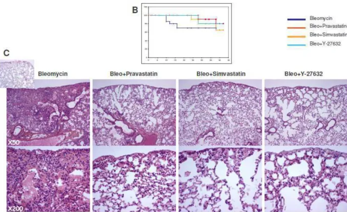
In our studies, we showed the role of Statin (Pravastatin e Simvastatin) and a specific inhibitor ROCK inhibitors (Y-27632) in a mice model of pulmonary induced-fibrosis obtained by a pharmacological approach (Bleomycin – BLM). Indeed, we developed a model of lung fibrosis by complete irradiation of chest and tested Pravastatin action. In this model and in a model of radiation induced gut fibrosis, we analysed, from a immunohistological point of view, the underlying mechanisms of the antifibrotic action of Pravastatin via MMP2-TIMP2 axis. Finally, in vitro, we investigate by zymography the expression of Gelatinases (MMP2 and MMP9) in primary lung fibroblasts cultures exposure at the different radiation and Pravastatin doses.
In our animal model of pulmonary fibrosis, Pravastatin reverts the fibrotic process and,
in vivo and in vitro, metalloproteases would appear to be in turn involved in pro-fibrolytic
mechanisms induced by statin.
The multiplicity of actors involved in the pathogenesis of fibrotic lesions explains why the definition of an effective therapeutic strategy is so complex.
Researches in mechanistic processes of normal tissue damage paved the way for new therapeutic approaches. These new targets include reduction of vascular activation, inflammation and thrombosis and new molecular targets definition. Effective strategies are multiple on preclinical models, but numerous efforts have to be made to achieve the complicated goal of protection of normal tissues from the side effects of radiation therapy.
RIASSUNTO
La radioterapia è la seconda modalità di trattamento più importante dopo chirurgia nel trattamento delle neoplasie. I recenti progressi tecnici, come la terapia ad intensità modulata (IMRT) o l’image-guided radioterapia (IGRT), in combinazione con nuovi farmaci ad azione mirata come gli anticorpi monoclonali, costituiscono ulteriore garanzia di incremento dell’indice terapeutico. Tuttavia il trattamento radiante può causare un’alterazione del normale processo di riparazione e indurre lo sviluppo di un quadro di fibrosi in un sottogruppo di pazienti sensibili e nei lungo-sopravviventi al cancro. La caratteristica cardinale della fibrosi radioindotta è l’eccessivo ed anomalo accumulo di collagene composto principalmente di componenti fibrillari e immature della matrice extracellulare (ECM).
Gli organi che possono essere interessati da questo fenomeno sono fegato, pelle, intestino, reni e polmoni. Da un punto di vista clinico, la fibrosi può essere vista come una condizione irreversibile, senza soluzione. Noi ed altri recentemente abbiamo mostrato che accanto alla attivazione della via canonica TGF-β/Smad, altre vie vengono attivate nei tessuti fibrotici come la cascata di segnalazione intracellulare della via Rho/ROCK. Interessante notare che la via Rho/ROCK sembra specificatamente attivata nella radiazione indotta fibrosi intestinale, fornendo così una spiegazione razionale per una specifica, mirata strategia anti-fibrotica. L'inibizione farmacologica di Rho con le statine infatti è in grado di prevenire e addirittura invertire i fenomeni di fibrosi intestinale post-attinica.
Grazie a queste premesse, nei nostri studi, abbiamo mostrato il ruolo delle statine (Pravastatina e Simvastatina) e di uno specifico inibitore di ROCK (Y-27632) in un modello murino di fibrosi polmonare indotta ottenuto con un approccio farmacologico (bleomicina - BLM). In seguito, abbiamo sviluppato un modello di fibrosi polmonare indotta dall’irradiazione completa del torace e valutata la risposta alla somministrazione della Pravastatina. In questo modello ed in un modello di fibrosi intestinale indotto da radiazioni, abbiamo analizzato, da un punto di vista immunoistologico, i meccanismi sottostanti l'azione
antifibrotica della pravastatina e il ruolo delle metalloproteasi (MMP2 e TIMP2). Infine, in
vitro, abbiamo indagato, mediante zimografia, l'espressione delle gelatinasi (MMP2 e MMP9)
in culture primarie di fibroblasti polmonari murini esposti a differenti dosi di radiazione e pravastatina.
Nel nostro modello animale di fibrosi polmonare, la Pravastatina è in grado di rendere reversibile il processo fibrotico e le metalloproteasi parrebbero essere a loro volta coinvolte,
in vivo and in vitro, nei meccanismi pro-fibrolitici indotti dal farmaco.
La molteplicità di attori coinvolti nella patogenesi delle lesioni fibrotiche spiega perché la definizione di una strategia terapeutica efficace è così complessa. Ricerche nei processi meccanicistici di danno ai tessuti normali hanno aperto la strada a nuovi approcci terapeutici. Questi nuovi obiettivi comprendono la riduzione dell’ attivazione vascolare, dell'infiammazione e della trombosi, oltre alla definizione di nuovi target molecolari. Esistono molteplici ed efficaci strategie su modelli preclinici, ma numerosi sforzi devono essere fatti per raggiungere il complicato obiettivo di proteggere i tessuti normali dagli effetti collaterali della radioterapia.
RESUME
La Radiothérapie occupe la deuxième place dans la liste de traitement du cancer le plus important après chirurgie. Le progrès technique récent, comme la radiothérapie avec modulation d'intensité (IMRT) ou la radiothérapie guidée par l'image (IGRT), en combinaison avec de nouveaux médicaments à action spécifique tels que les anticorps monoclonaux, sont une garantie d'augmentation de l'index thérapeutique. Cependant, la radiothérapie peut provoquer un’ altération du processus normal de réparation et d'induire le développement d'un cadre de fibrose dans un sous-ensemble de patients sensibles et dans les survivants à long terme de cancer. La principale caractéristique de la fibrose radio-induit est l'accumulation excessive et anormale de collagène composé principalement des éléments fibrillaire et immatures de la matrice extracellulaire (ECM).
Les organes qui peuvent être touchés par ce phénomène sont le foie, la peau, les intestins, les reins et les poumons. D'un point de vue clinique, la fibrose peut être considérée comme une condition irréversible, sans solution. Nous et d'autres ont récemment montré que, outre l'activation de la TGF-β/Smad canonique, d'autres voies sont activées dans les tissus fibreux tels que la cascade de signalisation intracellulaire Rho/ROCK. Fait intéressant, la façon dont Rho/ROCK semble spécifiquement activé dans la fibrose intestinale radio-induite, fournis une justification pour un stratégie anti-fibrotique ciblé. L’ inhibition pharmacologique de Rho avec les statines, en fait, est en mesure de prévenir et même inverser les phénomènes de fibrose post-actinique intestinale.
Avec ces prémisses, dans nos études, nous avons montré le rôle des statines (Simvastatine et Pravastatine) et d'un inhibiteur spécifique de ROCK (Y-27632) dans un modèle murin de fibrose pulmonaire obtenue avec une approche pharmacologique (Bléomycine - BLM) . Par la suite, nous avons développé un modèle de fibrose pulmonaire induite par l'irradiation complet du thorax et évalué la réponse à l'administration de la Pravastatine. Dans ce modèle, et dans un modèle de fibrose intestinale radio-induite, nous
avons analysé, grâce à l’immunohistochimie, les mécanismes sous-jacents l'action antifibrotique de la Pravastatine et le rôle des métalloprotéases (MMP2 et TIMP2). Enfin, in
vitro, nous avons étudié par zymographie, l'expression des gélatinases (MMP2 et MMP9)
dans des cultures primaires de fibroblastes pulmonaires murins exposées à différentes doses de rayonnement et de Pravastatine.
Dans notre modèle animal de fibrose pulmonaire, la Pravastatine est capable d'inverser le processus fibrotique et les métalloprotéases semblent être impliqués à leur tour, in vivo et in
vitro, dans les mécanismes pro-fibrolyse induits par le médicament.
La multiplicité des acteurs impliqués dans la physiopathologie de lésions fibrotiques explique pourquoi la mise en place d'une stratégie thérapeutique efficace est si complexe. La recherche dans les processus mécaniques de dommages aux tissus normaux ont ouvert la voie à de nouvelles approches thérapeutiques. Ces nouvelles cibles comprennent la réduction de l’inflammation, de l'activation vasculaire et de la thrombose, ainsi que la découverte de nouvelles cibles moléculaires. Il existe une variété de modèles précliniques et des stratégies efficaces, mais de nombreux efforts doivent être déployés pour atteindre l'objectif difficile de protéger les tissus normaux des effets secondaires de la radiothérapie.
Radiation oncology is a cornerstone of modern multidisciplinary cancer treatment. It has a place in the management of most common types of cancer, either as a single modality and organ-preserving alternative to surgery, for example, in organ-confined prostate cancer, or as an element in a sequence of treatment steps, such as in adjuvant radiotherapy after breast-conserving surgery for breast cancer1.
Strategies to improve the outcome of radiotherapy have aimed to improve tumor control rates, thereby increasing the chances of cure in radical or adjuvant therapy and the rates of symptom response in palliative situations. Thanks to technological advances like Image Guided Radiotherapy (IGRT) and Intensity Modulation Radiation Therapy (IMRT), reduction of toxicity and late effects on healthy tissue was also partially gained. At the same time, evolution of the standard of care toward combination therapies (radiotherapy + chemotherapy + targeted drugs) does enhance the rate and severity of both acute and delayed complications at the normal tissue level2.
The physiopathology of normal tissue injury has been extensively studied by radiation oncologist and radiation biologist. Today, normal tissue toxicity is known to be the result of treatment modalities, tumor type and patient’s characteristics that include biological heterogeneity, genetic factors, co-morbidities3. Tissue response to radiotherapy depends upon the total dose delivered and the volume irradiated. In addition, radiation therapy is delivered fractionated and thus causes multiple and iterated cellular injuries. Radiation produces free radicals, damages to the vasculature, induces a cascade of local and systemic cytokine and chemokine expression, elicits inflammatory response, and causes death of cells. The tumor itself also leads to architectural destruction of the organ affected, releases cytokines, affects the immune system, and alters vascular permeability. Patients may have underlying medical conditions predisposing to injury (e.g., diabetes, poor underlying pulmonary function, or certain collagen vascular diseases) or have genetic susceptibility. All these factors may contribute to individual radiation sensitivity and their relative importance is difficult to assess.
Nonetheless, it is becoming clear that abnormal microenvironmental conditions exist that are sustained long after the beam is turned off, the drugs are discontinued, and the tumor is eradicated, which appear to be responsible for the perpetuation of the tissue atrophy or hypertrophy, loss of epithelial/vascular/parenchymal cells, and excessive fibrosis characteristic of late normal tissue injury after cancer therapy.
Research over the past 15 years has led to a better understanding of the underlying molecular events responsible for the development of normal tissue injury after cancer therapy4,5,6,7. Characterization of the signalling pathways involved in radiation-induced fibrogenesis has mostly focused on one potent fibrogenic growth factor: the transforming growth factor β1 (TGF-β1)8 and today, inhibitors of TGF-b1 such as Pirfenidone are used in the clinic to halt progression of Idiopathic Pulmonary Fibrosis (IPF)9 More particularly, in the context of radiation-induced fibrosis several strategies have also been used in experimenatl models and in the clinic.
Amongst these various strategies, some have been used in patients. Delanian S. et al. have shown that the combination of pentoxifylline, vitamin E and clodronate (PENTOCLO) was
useful in healing sternocostal and some mandibular osteoradionecrosis10 Moreover, treatment combining PENTOCLO significantly improved on a clinical point of view, neurological sensorimotor symptoms of two patients with radiation-induced lumbosacral polyradiculopathy11. Cohen et al.12 suggested that ACE inhibitors might be used after completion of radiation therapy, but before expression of injury, to mitigate the later development of renal failure after radiation-based hematopoietic stem cell transplantation (HSCT).
Another strategy conducted in our lab and others, was to characterize the molecular signals involved in fibrogenesis to provide new therapeutic targets13,14,15,16,17,18,19,20,21,22. Using molecular profiling approaches, we have shown that the activation of Rho/ROCK/CTGF pathway was controlling the fibrogenic differentiation in several organs: the gut (human radiation-induced enteropathy) 13,14,16, the heart and lungs (experimental models in C57Bl6 mice)19. Then we showed that it was possible to target it to prevent and reverse radiation fibrosis in the organs mentioned above using Statins17,18,19,20,21,22,23.
Inhibitors of Rho/ROCK/CTGF pathway such as statins that inhibits Rho isoprenylation; Y-27632 an allosteric inhibitor of ROCK and monoclonal antibody against CTGF have shown anti-fibrotic propertiesErrore. L'origine riferimento non è stata trovata.,Errore. L'origine riferimento non è stata
trovata.,Errore. L'origine riferimento non è stata trovata.,Errore. L'origine riferimento non è stata trovata.,Errore. L'origine riferimento non è stata trovata.,22,23,24
.
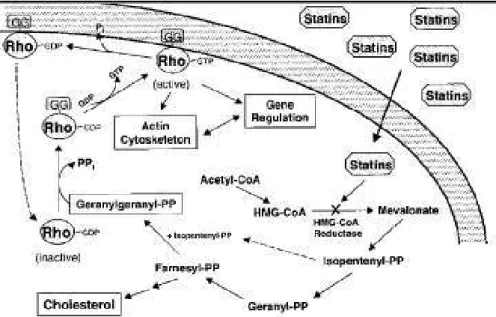
My thesis work was specifically focused on the anti-fibrotic mechanisms of action of one hydrophilic statin, Pravastatin. The pleiotropic actions of statins are mediated by inhibition of the production of isoprenoid residues and subsequent modulation of posttranslational protein prenylation, including that of Rho20. Pre-clinical studies showed that Pravastatin inhibited the Rho/ROCK/CTGF cascade in human samples ex vivo and reversed intestinal radiation-induced fibrosis in vivo17,21. In addition, Pravastatin protected normal intestine and cutaneous22,23 from radiation damage without interfering with the anticancer action of
irradiation in experimental models, both in vitro and in vivo. Other studies using Simvastatin confirmed these results18,24. Additional mechanism may also be involved, such as preservation of endothelial barrier function, anti-inflammatory action, modulation of platelet activation and anti-thrombotic action, antioxidant properties and may certainly contribute to the anti-fibrotic action of Pravastatin. Based on this biological rational, a phase II clinical trial supported by the French Ministry of Health (PHRC 2010) is currently conducted at IGR to assess the anti-fibrotic efficacy of Pravastatin in patients with cutaneous fibrosis after treatment for Head&Neck tumors.
My thesis work was to characterize further the anti-fibrotic action of Pravastatin from the molecular point of view. Our main hypothesis was that persistent alteration of the cell phenotype induced by irradiation depended, at least in part, upon the Rho/ROCK/CCN2 pathway in various organs. In the first part, we showed that the pathological activation of Rho/ROCK pathway was not specific of radiation-induced intestinal fibrosis but could be considered as a general mechanism as it was also observed in lungs (and heart). In this manuscript we will develop our findings in details. Briefly, we showed activation of the two important fibrogenic pathways: TGF-b/Smad and Rho/ROCK, in response to radiation-exposure in vivo in lungs of mice, 15 and 30 weeks post-irradiation. This fibrogenic molecular imprint was associated with long-term remodelling of pulmonary histological structures was successfully modulated using Rho/ROCK inhibitors (statins and Y-27632) that induces a normalization of fibrogenic markers.
The second part of my work focused on ECM remodelling and fibrolysis induced by Pravastatin in vivo and in vitro. To investigate this question, we used the two long-term experimental models of radiation-induced (RI) fibrosis available in our laboratory that model fibrosis in two majors dose limiting organs: the intestine and the lung. Then, we studied Gelatinase and TIMP regulations and showed that Pravastatin administered as a mitigator induces a persistent activation of the MMP2-TIMP2 axis in the mucosal and muscular
compartment of the gut. When Pravastatin was administered with curative intent the mechanism seemed different since no significant modulation of MMP2-TIMP2 was observed in the gut whereas local fibrolytic process was observed in lungs. Our results suggest that Pravastatin anti-fibrotic action is mediated by Rho/ROCK pathway inhibition and gelatinase-mediated fibrolytic induction.
In conclusion, our findings extend the biological rational for using Pravastatin to treat radiation-induced fibrosis in the patients. Pravastatin offers a safe and efficient therapeutic opportunity potentially usable either before or after radiation exposure. This approach is especially attractive in (1) the radiation oncology setting, as it does not interfere with prior anti-cancer treatment and in (2) radioprotection, as applicable to the treatment of established radiation injury, for example in the case of radiation accidents or acts of terrorism.
PRINCIPLES OF RADIATION EFFECTS ON NORMAL
TISSUES
The modern era of cancer therapy involves safe intensification of radiation, chemotherapy, and biologic adjuvants. Yet, the impact of such newer radiation technologies and normal tissue constraints have been offset in some cancers by escalation of the radiation dose and concurrent chemotherapy aimed at improving the tumor response. This has resulted in a markedly increased survivorship, which now exceeds 64% overall, and is much higher for selected malignancies, such as 87% for breast cancer and 80% for all childhood cancers25. Malignancies resistant to therapy required very aggressive treatment approaches often at the edge of normal tissue tolerance, or that even exceeds tolerance to some “acceptable” degree. Definition of normal tissue tolerance has evolved and led to a deeper understanding of the multiparametric inputs that influence toxicity26. There is abundant evidence that clinicians underestimate the frequency and severity of patients’ symptoms and, therefore, the published data underestimate the true toxicity burden27.
Patients treated with radiation may experience severe and potentially life-threatening late intestinal complications, such as fistulation, sepsis, intestinal failure, perforation, obstruction and bleeding, which required surgery in 5% of cases at 5 years13. Recent reports suggest that, in more than half of patients, irradiation of pelvic tumours leads to permanent gastrointestinal damage (i.e. alternating episodes of diarrhoea and constipation, abdominal pain, malabsorption, faecal urgency)28, for which no efficient treatment is available today.
Clearly, the potential to ameliorate or prevent such normal tissue damage, or to manage and rehabilitate affected patients, requires an understanding of tissue tolerance to therapy. Because “late effects” can manifest months or years after cessation of treatment, therapeutic decisions intended to obviate such effects can be based only on the probability, not the certainty, that such effects will develop. In making such decisions, the balance between efficacy and potential for toxicity should be considered and may be influenced by host, disease, and
treatment-related risk factors. Recently, many agents have been identified that target molecular pathways that can mitigate radiation toxicity. To date, no drugs have been approved as radiation injury mitigators (RIM), defined as agents administered after irradiation but before toxicity is manifest. Movsav B et al29have present an algorithm to guide clinical trials for such agents in patients receiving radiotherapy or radiochemotherapy. The goal is to be able to apply such promising agents to improve the quality of life for all these patients. Patients have a central role in reported outcomes They reviewed the mechanisms of radiation injury and the clinical problem related to radiation effects, the preclinical and clinical development of candidate agents, and how to design and conduct clinical trials.
Normal tissue responses to radiation can be divided into two categories: those that occur during the first days and weeks after treatment, often called early effects, and those that occur months, years, or even decades after irradiation, called late effects. “Consequential late effects” result from the host’s reaction to severe acute toxicity. Some organs are prone to late toxicity (often termed late responding tissues) while others are prone to acute toxicity (early
responding tissues), although in any organ both acute and/or late effects occur. The type of
effect expressed by an organ is generally a function of the tissue’s renewal properties, but the clinical importance of the response typically depends on the biology of the organ. The most important parameters that control toxicity occurrence in patients are the total radiation dose and fraction size, the duration of time during which the course of radiation is delivered, the interval between radiation fractions, the rate at which the radiation is given (dose rate), the specific organ being irradiated, and the volume. As previously stated, the organizational structure and the repair or compensatory capacities of the organ influence its tolerance to partial or whole-organ irradiation.
The classic concept of a single target cell explaining the dynamic sequence of events leading to normal tissue damage has been supplanted by a more complex vision in which the
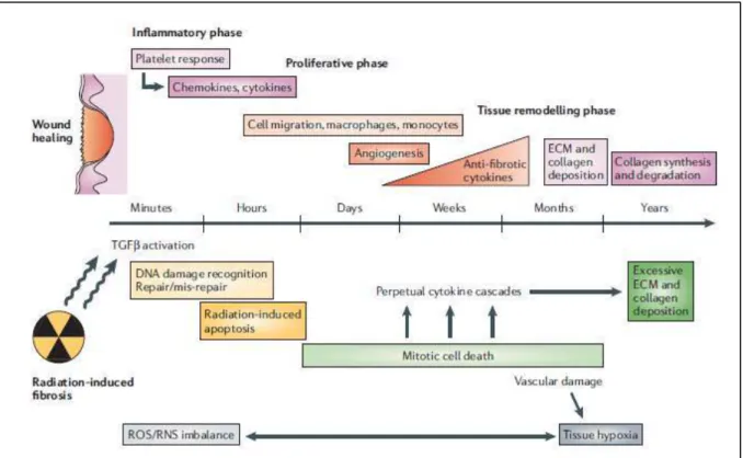
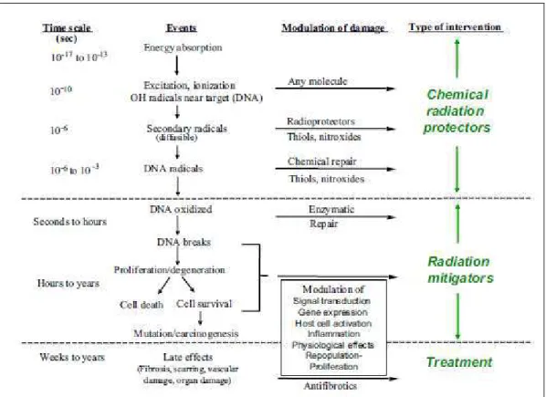
interaction of multiple cellular systems through various molecular signalling and paracrine factors occurs. Moreover, the perceived acute and late phases of adverse effects now are seen as manifestations of an ongoing sequence of events perpetuated through autocrine, paracrine, and endocrine messages that are initiated immediately after injury and persist until the clinical late effect events themselves. After irradiation, a variety of growth stimulatory and inhibitory factors are released, cell receptors are altered, and the resulting dysregulation of the tissue environment is translated ultimately into postreceptor cytoplasmic, nuclear, and interstitial events. Thus, a combination of cell death, the production of reactive oxygen species, alterations in gene expression, and the expression of both proinflammatory and profibrotic cytokines are viewed as integral in the pathogenesis of late effects (Fig.1)30,31.
Fig. 1.: From “Preventing or reducing late side effects of radiation therapy: radiobiology meets molecular pathology. Bentzen SM. Nat Rev Cancer. 2006 Sep;6(9):702-13. Review”.
Despite optimum conformation of the treatment fields to the tumour and precise treatment planning and application, the target volume in curative radiotherapy necessarily includes a substantial amount of normal tissue, for several reasons.
First, malignant tumours infiltrate microscopically into normal structures, which hence must be included into the high-dose volume as a tumour margin. Second, normal tissues within the tumour, such as soft tissue and blood vessels, are exposed to the full tumour dose. Third, normal structures in the entrance and exit channels of the radiation beam may be exposed to clinically relevant doses. Therefore, effective curative radiotherapy is unavoidably associated with an accepted risk for early and late radiation side-effects (‘adverse events’) in order to achieve adequate tumour cure rates.
Radiotherapy is a localised treatment. The definition of tumour and target volumes for radiotherapy is vital to its successful execution. This requires the best possible characterisation of the location and extent of tumour. Diagnostic imaging, including help and advice from diagnostic specialists, is therefore essential for radiotherapy planning.
There are three main volumes in radiotherapy planning (Fig.2, 3)
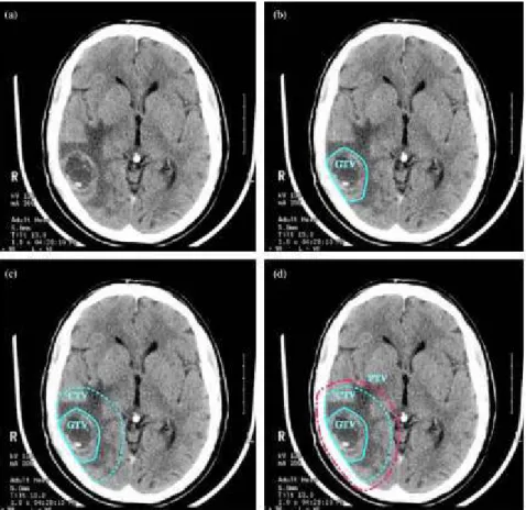
First volume is the position and extent of gross tumour, i.e. what can be seen, palpated or imaged; this is known as the gross tumour volume (GTV). Developments in imaging have contributed to the definition of the GTV. The second volume contains the GTV, plus a margin for sub-clinical disease spread which therefore cannot be fully imaged; this is known as the clinical target volume (CTV). It is the most difficult because it cannot be accurately defined for an individual patient, but future developments in imaging, especially towards the molecular level, should allow more specific delineation of the CTV. The CTV is important because this volume must be adequately treated to achieve cure. The third volume, the planning target volume (PTV), allows for uncertainties in planning or treatment delivery. It is a geometric concept designed to ensure that the radiotherapy dose is actually delivered to the CTV. Radiotherapy planning must always consider critical normal tissue structures, known as organs at risk (ORs). In some specific circumstances, it is necessary to add a margin analogous to the PTV margin around an OR to ensure that the organ cannot receive a higher-than-safe dose; this gives a planning organ at risk volume. This applies to an organ such as the spinal cord, where damage to a small amount of normal tissue would produce a severe clinical manifestation. The concepts of GTV, CTV and PTV have been enormously helpful in developing modern radiotherapy.
Fig. 3: from: Cancer Imaging 2004 Oct 21;4(2):153-61.
Planning volumes for a patient with WHO Grade 4 glioma (glioblastoma). (a) Planning CT showing contrast-enhancing tumour. (b) The GTV is the visible tumour. (c) A margin for microscopic spread has been added to make the CTV; the margin is the same in all directions except that it is restricted by the skull. (d) The PTV has been added outside the CTV to account for uncertainties in planning and execution of treatment; this extends beyond the inner table of the skull.
Late radiation effects, in particular radiation-induced fibrosis (RIF) and atrophy, hence represent a multifaceted, orchestrated response with various components32.
Each cellular components of an organ does respond to radiation injury with a specific dose-dependence, which, in that orchestrated response, then defines the overall dose–response for the different clinical endpoints of the entire tissue. RIF is a relatively rare, late, local consequence of high-dose RT.
It is estimated that, in pelvic tumor irradiation, most patients present digestive effects in the short term (80%) and that 5 to 10% develop late complications due to irradiation of non-tumor tissues.
Co-morbidity factors such as cardiovascular disease; preexisting collagen vascular diseases; and hypersensitivity or very rare congenital diseases are confounding factors that enhance the rate of radiation-induced fibrosis33.
At the cellular level, fibrosis is characterized by fibroblast proliferation and differentiation. Excessive extracellular matrix deposition is also a major hallmark of the disease, amplified by the action of cytokines and growth factors leading to abnormal reactive oxygen species (ROS) and reactive nitrogen species concentrations.
All tissues affected by advanced RIF are at risk to develop radionecrosis that might be seen as to ultimate evolution of the disease. One of more severe clinical manifestations of this cellular and molecular impaired response to radiation is Osteoradionecrosis (ORN)34.
RADIATION-INDUCED FIBROSIS
Radiation-induced fibrosis is a dynamic process, which varies both in its intensity and in its qualitative effect on the structure and function of the affected organ or tissue. Schematically, spontaneous RIF is divided into 3 histopathological phases, each of which is predominantly cellular, extracellular, or a mixture of both and is characterized by gradual worsening over several years: (1) a prefibrotic phase often asymptomatic, marked by signs of chronic inflammation, where endothelial cells play an important role; (2) a phase of organized fibrosis, characterized by a patchwork of areas of active fibrosis containing a high density of myofibroblasts in an unorganized matrix, surprisingly adjacent to poorly cellularized fibrotic areas consisting of senescent fibrocytes in a dense sclerotic matrix; and (3) a late fibroatrophic phase, with retractile fibrosis and gradual loss of parenchymal cells.
General mechanisms of wound healing and fibrosis
Acute and late normal tissue injury occurs from a complex interaction between radiation-induced death of parenchymal cells, damage to the supporting vasculature, and associated inflammatory and fibrotic reactions. Long-term depletion of tissue-specific stem cells or progenitor cells can lead to fibrosis, organ dysfunction, and necrosis6.
A wound-healing response is often described as having three distinct phases: injury, inflammation and repair35.
Phase I: injury
Cells acknowledge damage from radiation exposure through multiple sensor molecules and structures. Not surprisingly, chemokinesand proinflammatory cytokinesare highly prominent among the panoply of molecules expressed in tissues after irradiation. Perhaps less obvious is the fact that anti-inflammatory cytokines can also be increased, some of which may
participate in angiogenesis. This cytokine balance is a common feature of inflammatory responses. Cytokines, in turn, amplify further coordinated changes in additional cytokines, cell adhesion molecules, prostaglandins and leukotriene species, redox regulating enzymes and pro- and antioxidant species (manganese superoxide dismutase, inducible nitric oxide synthase, metallothionein, heme oxygenase, gamma-glutamylcysteine synthetase, and myeloperoxidase), matrix remodeling enzymes and inhibitors, plasminogen activators and inhibitors, and heat shock proteins. These components also often have mutually antagonistic aspects that allow tight control on the inflammatory and tissue-healing responses.
In many respects, the tissue responses to irradiation mimic the cytokine storms generated by many other tissue-damaging insults.
Phase II: inflammation
Once access to the site of tissue damage has been achieved, chemokine gradients recruit inflammatory cells. Neutrophils, eosinophils, lymphocytes, and macrophages are observed at sites of acute injury with cell debris and areas of necrosis cleared by phagocytes. The timing of inflammatory events may determine the role played by the inflammatory process. Early inflammation that is diminished at the later stages of disease may promote wound healing and may contribute to fibrosis. For example the early recruitment of eosinophils, neutrophils, lymphocytes, and macrophages providing inflammatory cytokines and chemokines can contribute to local TGFβ and IL-1336.
However, following the initial insult and wave of inflammatory cells, a late-stage recruitment of inflammatory cells may assist in phagocytosis, clear cell debris, and control excessive cellular proliferation, which together may contribute to normal healing. Thus late-stage inflammation may in fact serve an anti-fibrotic role and could be required for successful resolution of wound-healing responses.
The nature of the insult or causative agent often dictates the character of the ensuing inflammatory response and the nature of the inflammatory response dramatically influences resident tissue cells and the ensuing inflammatory cells. Inflammatory cells themselves also propagate further inflammation through the secretion of chemokines, cytokines, and growth factors. Many cytokines are involved throughout a wound-healing and fibrotic response, with specific groups of genes activated in various conditions37. Each of these cytokines can exhibit significant pro-fibrotic activity, acting through the recruitment, activation and proliferation of fibroblasts, macrophages, and myofibroblasts.
Phase III: tissue repair and contraction
The closing phase of wound healing consists of an orchestrated cellular re-organization guided by a fibrin-rich scaffold formation, wound contraction, closure and re-epithelialization. Myofibroblast-derived collagens and α-SMA form the provisional extracellular matrix, with macrophage, platelet, and fibroblast-derived fibronectin38 forming a fibrin scaffold. Collectively, these structures are commonly referred to as granulation tissues.
Growth factor and TGFβ-activated fibroblasts migrate along the extracellular matrix network and repair the wound. Fibroblast to myofibroblast differentiation, as discussed above, also creates stress fibers and the neo-expression of α-SMA, both of which confer the high contractile activity within myofibroblasts35. The attachment of myofibroblasts to the extracellular matrix at specialized sites pull the wound together, reducing the size of the lesion during the contraction phase. The degree of extracellular matrix laid down and, the quantity of activated myofibroblasts39 determines the amount of collagen deposition. To this end, the balance of MMPs to TIMPs40 and collagens to collagenases vary throughout the response, shifting from pro-synthesis and increased collagen deposition, towards a controlled balance, with no net increase in collagen. For successful wound healing, this balance often occurs when fibroblasts undergo apoptosis, inflammation begins to subside, and granulation tissue
recedes, leaving a collagen-rich lesion. The removal of inflammatory cells and especially α-SMA+ myofibroblasts is essential to terminate collagen deposition41.
Collectively, the degree of inflammation, angiogenesis, and amount of extracellular matrix deposition all contribute to the net collagen deposition and ultimately whether a fibrotic lesion develops. Therapeutic intervention, interfering with fibroblast activation, proliferation or apoptosis requires a thorough understanding and appreciation of all of the phases of wound repair. Although these three phases are often presented sequentially, during chronic or repeated injury these processes function in parallel, placing significant demands on regulatory mechanisms.
THE LUNGS AND THE SMALL INTESTINE: TWO MAJOR
DOSE-LIMITING ORGANS IN RADIOTHERAPY
An important challenge to modern radiation therapy is to increase the tolerance of normal tissues, in order to improve the quality of life of the patients, and to enhance local tumor control using dose escalation and/or new biological radiosensitizers42.
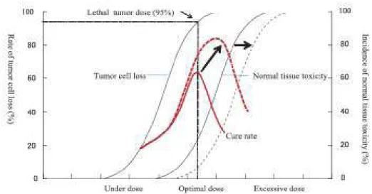
The recent progress made by intensity-modulated radiation therapy (IMRT) and image-guided radiation therapy (IGRT) has reduced radiation-induced complications43 especially in dose-limiting organs like the intestine44and lungs that still remains major dose limiting organs45. The dose effect relationship in both tumor and normal tissue in characterized by a sigmoid curve (Fig.4).
Fig. 4: Dose effect on tumor and normal tissue. From “Ikushima H. Radiation therapy: state of the art and the
future. J Med Invest. 2010 Feb;57(1-2):1-11. Review”.
If we can reduce the radiation dose to the surrounding normal tissues adjacent to the tumor, by improving dose conformity, the sigmoid curve for normal tissue damage can be shifted to a higher dose area. This makes it possible to escalate the tumor dose, resulting in an improvement in the cure rate. Technical innovation in RT always aims to improve dose distribution conformity with the objective of decreasing normal tissue toxicity.
Despite technological progress, give the preferable high tumor dose is not always achievable (due the presence of organs at risk), with consequent reduction in probability of regional tumor control.
Reducing the dose delivered to the bowel is also required in prostate46, bladder47, and gynaecological48 tumor treatment.
Lungs sparing is necessary not only during lung cancer treatment49but also in breast cancer50, Hodgkin Lymphoma45, and oesophagus neoplasm irradiation51.
An alternative mechanism to reduce normal tissue toxicity is the use of radiation modifiers/protectors, agents that when present prior to or shortly after radiation exposure alter the response of normal tissues to irradiation. This approach has also been viewed as an attractive countermeasure for possible nuclear/radiological terrorism. To be useful in the radiotherapy clinic, radioprotectors should ideally have several characteristics that relate to the ability of the agent to improve the therapeutic ratio. First, the agent should be selective in protecting normal tissues from radiotherapy without protecting tumor tissue, otherwise no benefit in the therapeutic index will be realized. Second, the agent should be delivered with relative ease and with minimal toxicity. Finally, the agent should protect normal tissues that are considered sensitive such that acute or late toxicities in these tissues are either dose-limiting or responsible for a significant reduction in quality of life (i.e., mucositis, pneumonitis, myelopathy, xerostomia, proctitis, and leukencephalopathy)52.
In effect, low and mild grade chronic gastrointestinal and lung side effects continue to influence the patient’s quality of life. Acute enteritis affects most patients treated with pelvic radiotherapy.
The symptoms occur during or immediately after radiotherapy: diarrhea, abdominal pain and incontinence, with, more rarely, constipation, bleeding and discharge of mucus53.
The epithelial damage promotes bacterial translocation and septic risks, bleeding, and reduce the absorption capacity of the digestive mucosa.
Therapeutic management associates symptomatological and support treatment. Acute enteritis resolves most of the time by itself during the weeks following the end of treatment.
Acute symptoms may be followed by a phase of evolution and a progressive worsening of the patient's clinical status. The most common symptoms are chronic recurrent episodes of diarrhoea and constipation, with violent abdominal pain. The wall thickening due to tissue fibrosis and the restriction of the intestinal lumen disrupt transit, promote stenosis and can lead to a total bowel obstruction. Ulceration and tissue necrosis can cause severe gastrointestinal bleeding, perforation of the intestinal wall and create entero-cutaneous, entero-enteric or entero-urinary fistulae.
Acute and subacute radiation pneumonitis, with late occurrence of fibrosis, are well-known risk factors for quality of life and survival of patients receiving radiotherapy to the thoracic region. Although 30 to 40% of the patients with lung cancer can benefit from radiotherapy, approximately 20% of these patients develop radiation-induced pulmonary injury.
The occurrence and severity of damage are semiquantitatively related to the volume of lung irradiated, and the dose rate of irradiation. The clinical syndrome occurs in up to about 10% of patients and consists of an acute transient phase, radiation pneumonitis, usually occurring 6 to 12 weeks after radiation therapy. Symptoms of radiation pneumonitis, including low-grade fever, congestion, dry cough, pleuritic chest pain, and a sensation of chest fullness, usually develop one to three months after completion of radiation therapy. Diagnosis is difficult, often complicated by comorbid conditions and radiation injury to adjacent structures (e.g., esophagus, pericardium). Although patients with acute pneumonitis may exhibit complete resolution of their signs and symptoms, unfortunately, the majority of them will go on to develop progressive pulmonary fibrosis; interestingly, this chronic condition also has been shown to occur in the absence of a preceding acute phase. In general, pulmonary fibrosis evolves between 6 to 24 months post-treatment, but then stabilizes after 2 years54. The
condition can result in a chronic pulmonary insufficiency, although this will be dependent upon the volume of lung treated, since fibrosis, like radiation pneumonitis, is characteristically restricted in its appearance to within the portal field. Where a large volume has been irradiated, the chronic insufficiency may progress to chronic cor pulmonale from the resultant pulmonary hypertension and othropnea, with associated cyanosis, hepatomegaly, or liver tenderness54.
Concomitant chemotherapy, repeat courses of radiation, and steroid withdrawal are exacerbating factors. Because the clinical evolution of delayed toxicity is progressive and inevitable, these complications are of much concern in clinical practice and further improvement in the management of such patients is required.
THE LUNGS
Basic structure and function
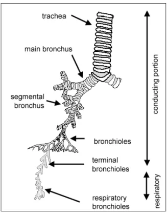
Lungs, in air-breathing vertebrates, are two large organs of respiration located in the chest cavity and responsible for adding oxygen to and removing carbon dioxide from the blood. In humans each lung is encased in a thin membranous sac called the pleura, and each is connected with the trachea (windpipe) by its main bronchus (large air passageway) and with the heart by the pulmonary arteries.
Diagram in Fig.5 shows the respiratory system.
Fig.5
It can be divide into two major components:
1. Conducting portion
The main function of the conducting system is to 'condition' the inspired air to humidify (by serous and mucous secretions) warm (by underlying blood vessels) and filter (by particles being trapped in mucous secretions, and transported towards the throat, where the mucous is
swallowed). The upper regions of the respiratory system (in the nasal passages) are covered with respiratory mucosa, and in some regions, olfactory mucosa.
The conduction portion is made up of: nasal cavities, nasopharynx, larynx, trachea, bronchi and bronchioles.
2. Respiratory portion
The interphase for passive exchange of gases between the atmosphere and blood.
The respiratory portion is made up of: respiratory bronchioles, alveolar ducts, alveolar sacs and alveoli. The epithelium of the alveoli (Fig.5), contains two main types of cells:
1. Type I pneumocytes: large flattened cells (95% of the total alveolar area) which present a
very thin diffusion barrier for gases. They are connected to each other by tight junctions.
2. Type II pneumocytes (making up 5% of the total alveolar area, but 60% of total number of
cells). These cells secrete 'surfactant' which decreases the surface tension between the thin alveolar walls, and stops alveoli collapsing during expiration.
Physiopathology of Radiation induced lung fibrosis
Pneumonitis and fibrosis are distinct features of the lung’s response to radiation damage. The former may occur after 6 to 16 weeks and is typified by inflammation and interstitial pneumonia. The latter is a more chronic response lasting months to years with progressive obliterative fibrosis55.
Multiple mechanisms have been identified in RT-induced fibrosis, including alveolar damage, increased reactive oxygen species (ROS) and the toxic effects of ROS on parenchymal cells, disruption of proliferation-associated transcription factors, and the influx of inflammatory cells, such as macrophages and lymphocytes35.
Type II pneumocytes have traditionally been considered “target” cells for radiation whose loss leads to inflammation, desquamation of epithelial cells from the alveolar surface, edema, and discharge of proteinaceous material into the alveoli. More recently, the cellular response has come to be viewed more in its entirety as involving multiple cell types with the outcome being dependent on the genetically determined molecular profile that drives the wound-healing process. Similar to other tissues, the lung’s response to radiation involves an inflammatory response.
After initial decreases in total cell number, neutrophils then lymphocytes infiltrate and are found elevated in bronchoalveolar lavage (BAL) for months.
Despite the evidence for dynamic changes within the BAL population, they contribute less to radiation-induced cytokine alterations than interstitial cells.
Rubin and coworkers56first used the cytokine “cascade” in the lung after irradiation to argue that there was no real “latent period” before the genesis of fibrosis and suggested that this was a self-sustaining continuous process.
Dysregulated pro-inflammatory and pro-fibrotic cytokines, TGF-β1, IL-6, MMPs57 and chemokines, in addition to reduced anti-inflammatory cytokines following radiation can further exacerbate the inflammatory and wound-healing response. TGF-β1 drives procollagen
1 production by fibroblasts, myofibroblasts, and other reparative cells through the Smad transcription factor pathway in addition to controlling many other aspects of extracellular matrix homeostasis. TGF-β1 has, however, other numerous biological functions. These include inhibiting proliferation of many cell types including lymphocytes and type II pneumocytes5.
Other cytokines have, however, been implicated in radiation-induced pulmonary injury as IL-1α, tumor necrosis factor-α (TNF-α) and interpheron-γ (INF-γ).
RT of the thoracic region can cause significant damage to radiation-sensitive alveolar regions of the lung invoking a dysregulated inflammatory cascade, rich in inflammatory and pro-fibrotic mediators. Dysregulated chemokines, transcription factors, and anti-inflammatory pathways can further compound this uncontrolled response, leading to pulmonary fibrosis.
THE SMALL INTESTINE
Basic structure and function
The main functions of the small intestine are digestion, absorption of food and production of gastrointestinal hormones. The small intestine is 4-6 metres long in humans. Structure is represented in Fig.7.
Fig. 7
To aid in digestion and absorption:
1) the small intestine secretes enzymes and has mucous producing glands. The pancreas and liver also deliver their exocrine secretions into the duodenum.
2) The mucosa is highly folded.
a) large circular folds called plicae circulares (shown in the diagram to the right), most numerous in the upper part of the small intestine,
b) smaller folds called villi, which are finger like mucosal projections, about 1mm long.
c) the lining columnar epithelial cells have fine projections on their apical surfaces called microvilli.
Together, these folds provide a huge surface area for absorption. Between the villi there are crypts, called crypts of Lieberkuhn, which extend down to the muscularis mucosae. These crypts are short glands.
The lamina propria which underlies the epithelium has a rich vascular and lymphatic network, which absorbs the digestive products, and there is a muscularis mucosae layer immediately at the base of the crypts. The lymphatic capillaries are called lacteals, and absorb lipids. The vascular capillaries are fenestrated to aid absorption.
The muscularis externa layer contains two layers of smooth muscle, an inner circular and outer longitudinal, for continuous peristaltic activity of the small intestine. There are around 200 or so lymphoid aggregations called Peyer's patches in the mucosa.
The external serous membrane is composed of connective tissue and epithelium.
Physiopathology of Radiation induced intestinal fibrosis
Pathological changes observed in severe late intestinal lesions have been widely explored using a descriptive approach 30 years ago. The main histopathological hallmarks of radiation-induced late intestinal damages in radiotherapy patients are fibrosis associated with inflammation and vascular sclerosis and, although to a lesser extent, epithelial lesions. Bowel fibrosis is characterized by transmural accumulation of extracellular matrix within intestinal layers that induces loss of compliance, impairs intestinal function and leads to obstructive syndromes.
Initial responses to radiation in the gut are characterized by radiation-induced cytokine expression, as in other tissues5.
Within hours of radiation exposure the rat ileal muscularis layer expresses IL-1β, TNF-α, and IL-658.
TGF-β1 is activated at 24 hours after irradiation and remains high throughout later responses, whereas IL-10 decreased. At this time, TGF-β1 is found in the inflammatory cells and surrounding extracellular matrix, and fibroblasts. Additional studies showed TGF-β1 association with mucosal ulceration, membrane thickening, and epithelial atypia 2 weeks after irradiation. A significant increase in the number of inflammatory cells in the mouse intestine can be observed at 24 hours after irradiation and IL-8 is upregulated at about 3 days after irradiation58.
Long-term changes in cytokine expression in the bowel of mice after irradiation implicate TNF-α, IL-1, IL-6, and TGF-β1 in late radiation-induced bowel fibrovascular toxicity.99 Connective tissue damage and increased collagen deposition is accompanied by high expression of smooth muscle actin and increased levels of the fibrogenic growth factor connective tissue growth factor (CTGF)16. CTGF may be involved in radiation-induced fibrogenic differentiation in intestinal smooth muscle cells. The Rho/ROCK pathway has been shown to regulate CTGF expression and may serve as a target for intervention.13,14,17
Microvascular injury in both acute and chronic radiation injury to the gut has been investigated in some depth and ascribed to dysfunction of the thrombomodulin (TM)-protein C (PC) system18. The TM-PC system is a critical physiological anticoagulation system in which TM forms a complex with thrombin to promote anticoagulation. TM and activated PC have important anti-inflammatory properties. Cytokines such as IL-1, TNF-α, and TGF-β1, all induced after radiation, reduce the transcription of TM5.
TM deficiency is found early postirradiation and continues to persist into chronic radiation injury, paralleling cytokine expression. This mechanism may allow persistence of the late cytokine cascade and damage.
CELLS AND MOLECULAR MEDIATORS INVOLVED IN
RADIATION INDUCED FIBROSIS
Fibroblasts and Myofibroblasts
One of the most important players in fibrosis is the myofibroblast. First described by Gabbiani in 197159, myofibroblasts display an intermediate phenotype between fibroblasts and smooth muscle cells. Typically, they have an hyper-contractile cytosqueleton suitable to achieve wound contraction, express α-SMA and secrete large amounts of extracellular matrix proteins in particular fibrillar collagens60. In physiological remodeling such as during dermal wound healing, the contractile activity of myofibroblasts is terminated when the tissue is repaired; a-SMA expression then decreases and myofibroblasts disappear by apoptosis.61 In pathological wound healing, myofibroblast activity persists and leads to tissue deformation, which is particularly evident, for example, in hypertrophic scars that occurs in scleroderma or after burn injuries61
Myofibroblast-generated contractures are also characteristic of fibrosis affecting vital organs such as the liver and kidney62,63 heart64,65 and lung66,67. In addition, myofibroblast participation to the process called stromal reaction does promote cancer progression (in that case they are called CAF Carcinoma associated fibroblasts) by creating a stimulating microenvironment for epithelial tumor cells68,69. Recent findings showed that overload with interstitial fluids cause fibroblast-mediated stromal remodeling that facilitates tumor invasion70.
In fibrotic lungs, myofibroblasts are key mediators of extracellular matrix deposition, structural remodeling, and destruction of alveocapillary units66 understanding their origin would therefore be critical to understand the pathogenesis of lung fibrosis.
In intestinal fibrosis, the role of intestinal mesenchymal cells constitution and maintenance is increasingly recognized nowadays13. In healthy bowel, subepithelial myofibroblasts, located
in the mucosa, and smooth muscle cells, located in muscular layers, are involved in the maintenance of tissue structure and function (intestinal contractility, extracellular matrix homeostasis). In fibrotic conditions as in lungs, their role is enhanced as they are responsible for the excessive collagen secretion, impaired motility and secretion of the fibrogenic growth factors.
Several origins for myofibroblasts have been proposed that may differ depending on the affected organ and the initiating event. However, three general mechanisms can be mentioned:
I) Proliferation and activation of tissue resident fibroblasts or perivascular and vascular adventitial fibroblasts can be activated into myofibroblasts in response to specific pro-fibrotic stimuli coming from infiltrating inflammatory cells leading to a progressive evolution from quiescent fibroblasts to cells expressing a myofibroblast phenotype71.
II) Recruitment of fibroblast precursor cells from bone marrow as a result of the local release of activated chemokines. These bone marrow precursor cells are fibrocytes, a unique cell population expressing bone marrow cellular surface markers (CD34 protein) and capable of production of extracellular matrix proteins (type I procollagen). These cells are able to migrate from the bloodstream in response to specific chemokine gradients and chemoattractant, to niche in damaged tissue to ensure physiological wound healing process or fibrogenesis72,73.
III) Transition of epithelial cells and endothelial cells to fibroblasts, a process known as EMT which is induced by transforming growth factor β (TGF-β) and perhaps other polypeptides such as endothelin-1 (ET-1) or insulin growth factor74,75,76.
Changes in cell phenotype between epithelial and mesenchymal states, defined as epithelial– mesenchymal (EMT) and mesenchymal–epithelial (MET) transitions, not only occurs during embryonic development or as a physiological response to injury, but is also an important
element in cancer progression and other pathologies that involve organ degeneration, such as fibrosis. At the cellular level, pathological EMTs are very similar to physiological EMTs in that they are governed by similar signaling pathways, regulators, and effector molecules75. Recently, Endothelial-Mesenchymal Transition (EndoMT), another type of cellular transition, has emerged as a possible mechanism in pathological fibrosis. EndoMT is a complex biological process in which endothelial cells lose their specific endothelial cell markers, such as vascular endothelial cadherin (VE cadherin), and acquire a mesenchymal or myofibroblastic phenotype initiating expression of mesenchymal cell products such as α-SMA, vimentin, and type I collagen. Besides acquisition of an activated pro-fibrogenic phenotype, these cells also become motile and are capable of migrating into surrounding tissues. Similar to EMT, EndoMT can be induced by TGF-β77.
For instance in lungs, ionizing irradiation induces production of radical oxygen species (ROS) including superoxide, hydroxyl radical, nitric oxide and. ROS interaction with pyrimidine and purine bases nuclear DNA produces single and double strand breaks, initiation of DNA repair, communication of DNA damage through the cell cytoplasm to the mitochondrial membrane via (stress activated protein (SAP) kinases) and then translation of pro-apoptotic, BCL2 family members from nucleus to mitochondria. Then, mitochondrial membrane permeability is enhanced. Cytochrome c dissociation from cardiolipin, and cytoplasmic leakage of cytochrome c leads to activation of the caspase-3 pathway and apoptosis. Both dying and recovering cells release ROS and inflammatory cytokines including IL-1b, TNFα and TGFβ, do produce acute local tissue and systemic effects78. Within the irradiated tissue differences in radiosensitivity of various cell phenotypes (endothelial cells, alveolar pneumocytes, alveolar macrophages and bronchopulmonary “stem” cells) contribute to the magnitude of tissue damage.
Role of immune system, cytokines and chemokines
Contribution of T lymphocytes is most important in fibrosis. Prolonged inflammation induces a specific lymphocyte T helper (TH) polarization. Local production of specific cytokines associated with this polarization have been well investigated in fibrosis. On the one hand TH1 orientation, notably characterized by the secretion of interferon-γ, is associated with resolution of the wound healing process. On the other hand TH2 orientation, characterized by the secretion of IL-4, IL-13 and TGF-b1, triggers tissue response toward fibrosis probably mediated by the pro-fibrotic growth factor: TGF-b179,80,81. Exposure of intestinal stroma to bacteria is known to induce a TH1 polarization82, but in the lung persistent exposure to bacterial antigens reorients TH1 polarization toward a TH2 profile suggesting that chronic epithelial depletion is fibrosis-prone83.
Macrophage infiltration into inflamed tissues has been implicated in chronic inflammation-induced lung fibrosis84. In addition to their roles in immune regulation, macrophages play a pivotal role in matrix regression during the recovery phase of fibrosis and in the regulation of stellate cell proliferation85. The phenotype of these macrophages is generally reported to match that of alternatively activated cells (M2) rather than classically activated cells (M1). M2 macrophages express immunosuppressive molecules such as IL-10 and arginase I, which suppress the induction of Th1 cells that produce the anti-fibrotic cytokine IFNγ. On the other hand, M1 macrophages express IL-1, IL-12, IL-23, and induce Th1 cell infiltration and activation. However, it remains to be established whether a particular macrophage subset with M2-type properties preferentially infiltrates into fibrotic tissues, or whether it is the pro-fibrotic microenvironment that drives macrophage polarization toward an M2 phenotype.
Pro-inflammatory mediators involved in fibrogenesis
There is a variety of pro-inflammatory chemokines have non-redundant roles of recruiting macrophages and other effector cells to the sites of inflammatory injury86. Chemokines, especially macrophage inflammatory protein-1α (MIP-1α, also known as CCL3) and related CC-chemokines, act as signal transducers in inflammatory injury, and perform important regulatory functions. Different activated macrophages have different behaviour related to MIP-1α secretion: M1 stimulated by LPS and IFN-γ promotes MIP-1a-generation, while IL-4 and IL-10 inhibit MIP-1a production induced by LPS or IL-1β87.
IL-4, the archetypal type-2 cytokine, has been firmly established as a pro-fibrotic cytokine and is elevated in radiation-induced pneumonitis and pulmonary fibrosis35. IL-4 receptors are present on lung fibroblasts88 with IL-4 signaling increasing extra cellular matrix proteins and collagen deposition. Surprisingly, some studies have suggested that IL-4 is superior to TGF-β1 at inducing collagen synthesis from fibroblasts88. Indirect mechanisms of IL-4 include its ability to promote the alternative activation of macrophages.
Finally, one of the most renowned properties of IL-4 is its ability to promote the differentiation of T cells into Th2 cells, providing a source of many type-2 cytokines in this inflammatory axis (IL-5, IL-9, IL-13, and IL-21). The Th2 cytokines interact in dramatic ways propagating wound healing and potentially pro-fibrotic responses. For example, IL-5 mobilizes, matures, and recruits eosinophils89 and promoting TGF-β production.
IL-5 can also augment IL-13 production and increase IL-13-dependent fibrosis90. IL-9 can selectively recruit and activate mast cells that increasing TGFβ activity and contributing to pulmonary fibrosis91. Mast cells can also promote fibroblast proliferation, collagen, and MMP production, and may be involved in subepithelial fibrosis following allergen challenge. IL-21 can also amplify Th2 pulmonary responses and 13-associated fibrosis by upregulating IL-4/IL-13 receptor expression.