Advance Access publication March 6, 2006 doi:10.1093/molehr/gal023
© The Author 2006. Published by Oxford University Press on behalf of the European Society of Human Reproduction and Embryology. All rights reserved. For
Involvement of MAPK pathway in TNF-a-induced MMP-9
expression in human trophoblastic cells
Marie Cohen, Arielle Meisser, Luise Haenggeli and Paul Bischof
1
Department of Obstetrics and Gynaecology, Maternity, Hormone Laboratory, University of Geneva, Boulevard de la Cluse, Geneva,
Switzerland
1
To whom correspondence should be addressed at: Laboratoire d’Hormonologie, 32 bd de la Cluse, 1211 Genève 14, Switzerland. E-mail:
[email protected]
The aim of this article was to investigate the signalling pathways involved in metalloproteinase-9 (MMP-9) expression induced by
tumour necrosis factor-
a (TNF-a) in first-trimester trophoblastic cells. TNF-a-induced MMP-9 expression, secretion and activity
were completely blocked by stress-activated protein kinase/jun kinase (SAPK/JNK) and Erk inhibitors (SP600 125 and U0126
respectively) but not by p38 mitogen-activated protein kinase (MAPK) inhibitors (SB203 580 and SB202 190). Stimulation of
HIPEC 65 cells with TNF-
a caused phosphorylation of JNK and extracellular signal-regulated kinase 1/2 (Erk1/2), with a peak
after 20 min of treatment. Transcription factors nuclear factor-
kB (NF-kB) and activator protein 1 (AP-1)-binding site were
iden-tified as the cis-elements involved in TNF-
a activation as determined by electromobility shift assays. TNF-a-induced
transactiva-tion of NF-
kB was inhibited by U0126, whereas TNF-a-induced transactivation of AP-1 was inhibited by SP600 125. Taken
together, these results indicate that in trophoblastic cells, TNF-
a probably activates two different pathways leading to MMP-9
expression: (a) Erk1/2 pathway which in turn initiates NF-
kB activation and (b) SAPK/JNK pathway that activates AP-1.
Key words: AP-1/cytotrophoblastic cells/Erk1/2/JNK/MMP-9/NF-κB/TNF-α
Introduction
Cytotrophoblastic cells (CTB) of the human placenta proliferate,
migrate and invade the pregnant uterus to allow implantation and
pla-centation (Red-Horse et al., 2004). The invasive property of
trophob-lastic cells is due to their ability to secrete proteases. Serine proteases,
cathepsin and metalloproteinases (MMPs) are involved in this
inva-sive process (Fisher et al., 1985; Bischof et al., 1995; Divya Chhikara
et al., 2002). Among the MMPs, MMP-9 and MMP-2 have been
shown to mediate invasion of CTB or CTB cell line into matrigel
(Fisher et al., 1985, 1989, Staun-Ram et al., 2004). Trophoblastic
expression of MMP-2 and MMP-9 has been studied during the first
trimester of pregnancy (Xu et al., 2000). It was shown that the level of
expression of MMP-9 was very small as compared with MMP-2,
suggesting that MMP-2, rather than MMP-9, plays an important role
in human embryo implantation. After the eighth week, MMP-9
secre-tion increases gradually, while MMP-2 producsecre-tion declines during the
same period Expression of MMP-9 coincided with the maximal
inva-sive potential of CTB, suggesting that MMP-9 is implicated in the
invasiveness of these cells (Fisher et al., 1985; Librach et al., 1991).
The invasive behaviour of CTB is limited in time and in space and
could be mediated in an autocrine way by trophoblastic factors and in
a paracrine way by uterine factors. It has already been suggested that
tumour necrosis factor (TNF-
α) could serve as an autocrine growth
factor in choriocarcinoma cells and might thus facilitate proliferation
of CTB (Yang et al., 1993). Since MMP-9 is instrumental to
trophob-last invasion (Librach et al., 1991) and since TNF-
α is a known
regulator of MMP-9 (Meisser et al., 1999), it was interesting to
inves-tigate the signalling pathways involved in the regulation of MMP-9 by
TNF-
α in CTB.
TNF-
α can activate three known mitogen-activated protein kinase
(MAPK) cascades in mammalian cells: stress-activated protein
kinase/jun kinase (SAPK/JNK), p38 MAPK and Erk/MAPK (Baud
and Karin, 2001; Wajant et al., 2003). These cascades of kinases have
different functions and can cross-react at several levels. The SAPK/
JNK pathway is involved in the regulation of TNF-
α-induced gene
expression by phosphorylation of transcription factors mainly c-jun
and ATF-2, leading to increased activity of activator protein 1 (AP-1).
The p38 MAPK enhances also the function of AP-1 but mainly
through other transcription factors such as Elk-1 or CREB, whereas
the Erk/MAPK pathway leads essentially (but not exclusively) to
enhancing the function of nuclear factor-
κB (NF-κB).
MAPK signalling pathways leading to increased expression of MMP-9
have already been studied in different TNF-stimulated cells (Holvoet
et al., 2003; Moon et al., 2004; Wiehler et al., 2004). It was shown that
TNF-
α induced the secretion of MMP-9 from eosonophiles via p38
MAPK but not the extracellular signal-regulated kinase 1/2 (Erk1/2)
pathway (Wiehler et al., 2004). In contrast, TNF-
α-induced MMP-9
expression in keratinocytes involved the Erk1/2 cascade and AP-1
activa-tion but no p38MAPK nor SAPK/JNK activaactiva-tion (Holvoet et al., 2003).
Since the AP-1 and NF-
κB are known regulators of MMP-9
pro-moter (Van den Steen et al., 2002), this article investigates the
signal-ling pathways induced by TNF-
α that regulate MMP-9 expression in
human trophoblastic cells.
Materials and methods
Reagents
Dulbecco’s modified Eagle’s medium high glucose (DMEM), Ham’s nutrient mixture F-12 (Ham F12) and antibiotic mixture (penicillin and streptomycin)
were products of Invitrogen (Basel, Switzerland). Fetal bovine serum was from Biochrom AG (Oxoid AG, Basel, Switzerland). TNF was from R&D Systems (Buehlmann AG Basel, Switzerland). Tocriset MAPK inhibitor was from Toc-ris (Avonmouth, UK). Lysis buffer 5× was from Promega (Catalys AG, Wallisellen, Switzerland). BIO-RAD protein assay, Trans-Blot transfer medium were from Bio-Rad (Munich, Germany). Hybond-N+ membrane, Rainbow-stained protein molecular weight markers and enhanced chemilumi-nescence (ECL) western blotting detection system were from Amersham Bio-sciences (Buckinghamshire, UK). Electromobility shift assay (EMSA) and biotin 3´-end DNA-labelling kits were from Pierce (Rockford, IL, USA). Rabbit polyclonal phospho-SAPK/JNK, SAPK-JNK, phospho-p38MAPK, p38MAPK, phospho-Erk1/2, Erk1/2 antibodies and p38 MAPK control cell extracts were from Cell Signaling (Beverly, MA, USA). Sheep polyclonal MMP-9 antibody was from The Binding Site (Birmingham, UK). Goat poly-clonal GAPDH-specific antibody was from Santa Cruz (Santa Cruz, CA, USA). The NF-κB and AP-1-specific oligonucleotides (5´-CCAGTGGAAT-TCCCCAGCCT-3´ and 5´-GAAGCTGAGTCAAAGAAGGC-3´ respectively) were purchased from Mycrosynth GmbH (Balgach, Switzerland).
Cell culture
The human invasive, proliferative extravillous cytotrophoblast cell line (HIPEC) 65 (a generous gift from Pr. D Evain-Brion, Paris; Pavan et al. 2003) and first-trimester CTB (isolation as described elsewhere, Bischof et al., 1995) were grown in DMEM high glucose/Ham F-12 (1/1) containing 10% fetal bovine serum (FBS) and antibiotics (100 U/ml penicillin, 100 μg/ml strepto-mycin) at 37°C in a humidified, 5% CO2 atmosphere. Consent was obtained
for the use of discarded tissue for research purposes using procedures approved by the Geneva Hospital Ethics Committee.
Effect of MAPK inhibitors on MMP-9 expression and secretion
HIPEC 65 and CTB were seeded in 12-well plates. One day later, they were cultured for 24 h in a serum-free medium before being treated with/without inhibitors (100 nM SB203 580, 100 nM SP600 125, 100 nM SB202 190 or 500 nM
UO126). TNF-α (100 ng/ml) was added 1 h after the inhibitors and the cells incubated for 48 h. Culture supernatants were collected for analysis and cells washed with ice cold PBS, lysed with lysis buffer ×1, scraped and centrifuged at 7600 g for 10 min at 4°C.
Phosphorylation kinetic of p38MAPK, JNK and Erk1/2
HIPEC 65 cells were seeded in 6-well plates. After 24 h, the cells were cul-tured for 24 h in a serum-free medium before being treated for 1 h with or without inhibitors (same as above). TNF-α (100 ng/ml) was then added for 0, 10, 20, 30 and 60 min. Culture supernatants were collected for analysis and cells washed with ice cold PBS. Then, they were lysed with lysis buffer ×1, scraped, centrifuged at 7600 g for 10 min at 4°C and supernatants stored at –20°C until western blot analysis.
Zymography
Proteolytic activity of culture supernatants were assayed using gelatin-substrate gel electrophoresis as described previously (Martelli et al., 1993). Zymograms were scanned with an Epson Perfection 1 200 Photo scanner, and the surface of the digestion bands was measured by the Kodak 1D Image ana-lysis software (Kodak, Rochester, NY, USA).
ELISA for MMP-2 and MMP-9
MMP-2 and MMP-9 concentrations were measured in the cell supernatants using our own enzyme immunoassays (EIA) as described and validated else-where (Meisser et al., 1999).
Western blot analysis
Protein concentration of cell extracts was determined by the Bio-Rad protein assay according to the instructions of the manufacturer. Proteins (50 μg) were denatured and subjected to SDS-PAGE using a 10% running gel. Rainbow-stained molecular weight markers were used as standards. Proteins were elec-trotransferred to nitrocellulose membranes. Nonspecific binding was blocked for 30 min at 37°C with 5% powdered milk in 0.2% NP40 buffer. MMP-9-specific polyclonal antibody (1/1000), GAPDH-MMP-9-specific antibody (1/750),
phospho-JNK, phospho-p38 or phospho-Erk1/2-specific antibodies (1/800) and p38 or Erk1/2-specific antibodies (1/1000) were incubated overnight with the nitrocellulose membrane. After washing, the membranes were incubated with the appropriate horse-radish peroxidase (HRP)-linked secondary antibody (2 h, room temperature). After washing, the bands were revealed by chemilu-minescence (ECL detection kit). Films were scanned with an Epson Perfection 1 200 Photo scanner, and the surface of bands was measured by the Kodak 1D Image analysis software (Kodak).
Preparation of nuclear extracts and EMSA
HIPEC 65 cells were plated in 25 cm2 flasks and made quiescent at conflu-ence by incubation in serum-free medium for 24 h. Cells were then incu-bated with or without TNF-α (100 ng/ml) at 37°C for 48 h. When inhibitors (SP600 125 or U0126) were used, they were added 1 h prior to the TNF-α treatment. After incubation, cells were washed twice with ice-cold PBS, then lysed with 0.5 ml of Hepes buffer pH 7.9 (10 mM Hepes, 10 mM KCl, 0.1 mM EDTA, 1 mM DTT, 0.5 mM PMSF, 0.4% NP40 and Roche pro-tease inhibitor cocktail) for 10 min at room temperature. Cells were scraped and transferred to a tube placed on ice before being centrifuged at 4°C at 15 000 g for 3 min. The resultant pellet was resuspended in extrac-tion buffer (20 mM Hepes, pH 7.9, 0.4 M NaCl, 1 mM EDTA, 10% glyc-erol, 1 mM DTT, 0.5 mM PMSF and Roche protease inhibitor cocktail) and vigorously shaken at 4°C for 2 h on a shaking platform. The nuclear extracts were centrifuged at 4°C, 15 000 g for 5 min, and their protein con-centration was determined before being aliquoted and stored at –80°C.
Biotin 3´-end DNA labelling was performed according to the instruc-tions of the manufacturer. Binding reacinstruc-tions were conducted by incubation of 6 or 4 μg of nuclear protein from HIPEC 65 with 20 fmol of biotin end-labelled AP-1 or NF-κB probes respectively at room temperature for 20 min according to the instructions of the manufacturer. Subsequently, reaction mix-tures were separated on a native 6% polyacrylamide gel and electrotrans-ferred to nylon membranes at 4°C. The membranes were then cross-linked on a transilluminator for 15 min before detection of biotin-labelled DNA by chemiluminescence.
Invasion assay
Cell invasion assay was performed in an invasion chamber based on the Boyden chamber principle. Each insert is fitted with an 8 μM pore size poly-carbonate membrane (Costar, Corning, NY, USA) precoated with rat tail colla-gen I (5 μg/cm2). Inserts were washed in DMEM and incubated for 30 min at room temperature. For each well, 15 × 104 cells in 100 μl of serum-free media,
in the presence or absence of TNF-α (100 ng/ml), were added to the upper compartment of the transwell chambers. Five hundred microlitres of media was added to the lower chamber. Cells were incubated for 48 h at 37°C in a CO2 (5%) incubator. After incubation, the supernatant was discarded, and
via-ble cells were stained with Chemicon cell stain (collagen-based cell invasion assay, Chemicon, Temecula, CA, USA). After washing, the stain was extracted with a solution of 1% acetic acid : 50% ethanol for 15 min at room tempera-ture. Hundred microlitres of the dye mixture was transferred to a 96-well microtitre plate for colorimetric measurement at 560 nm. Data are expressed as the percentage of TNF-α-treated cells that invaded the collagen-coated mem-brane relative to the untreated (controls) cells.
Analysis of data
Data are expressed as the mean ± SEM and analysed with a Student’s t-test at a P < 0.05 level of significance.
Results
Effect of TNF on 8–9 weeks CTB invasiveness
Invasion assays performed on 8–9 weeks CTB in the presence or
absence of TNF-
α (Figure 1) showed that addition of this cytokine
increases relative invasion of these cells (about 42%, P = 3 · 10
−6).
Since MMP-9 has been shown to mediate invasion of CTB in matrigel
and since TNF-
α is a known regulator of MMP-9, we next
investi-gated the signalling pathways involved in the regulation of MMP-9 by
TNF-
α in CTB and HIPEC 65 cell line.
Inhibition of TNF-a -induced increase in MMP-9 activity and
expression by MAPK inhibitors in trophoblastic cells
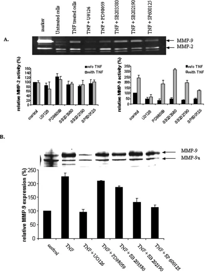
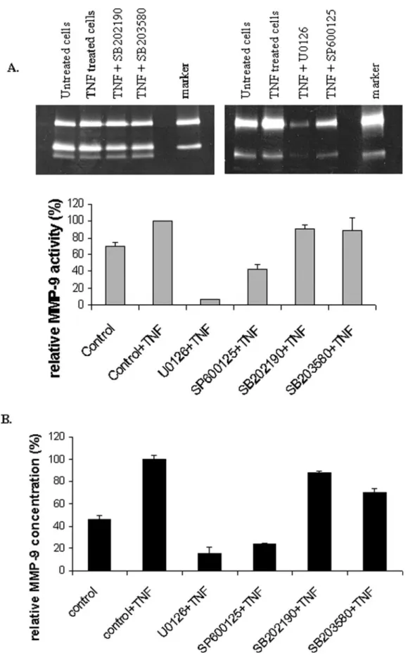
Treatment of trophoblastic cells (HIPEC65, Figure 2 and CTB, Figure 3)
with TNF-
α induced a two-fold increase of MMP-9 activity (Figures 2A
and 3A), secretion (Figure 3B) and expression (Figure 2B), whereas
the activity of MMP-2 was not modified (Figure 2A). To examine
whether Erk1/2, JNK and p38 MAPK activations are involved in the
signal transduction pathway leading to MMP-9 expression caused by
TNF-
α, different MAPK inhibitors were added 1 h before TNF-α
treatment in HIPEC 65. While pretreatment of HIPEC 65 with p38
MAPK inhibitors (SB203 580 and SB202 190) had no marked effect
on TNF-
α-induced MMP-9 expression and activity (Figure 2),
inhibi-tors of JNK and MEK, SP600 125 and U0126 respectively, decreased
significantly the TNF-
α-induced MMP-9 activity (P = 0.003 and
P = 4
× 10
−5respectively) and expression (P = 0.03 and P = 0.02
respectively), suggesting that JNK and MEK but not p38 MAPK
are implicated in these responses to TNF-
α (Figure 2). Since similar
results were obtained from primary CTB (Figure 3), HIPEC 65
represents a good model for TNF-
α signalling studies in extravillous
CTB.
Since the activation of Erk1/2 and JNK requires phosphorylation,
western blots were performed to examine Erk1/2 and JNK
phosphor-ylation using specific antibody for phospho-Erk1/2 and phospho-JNK.
p38 MAPK phosphorylation induced by TNF-
α was also investigated.
HIPEC 65 cells were stimulated with TNF-
α (100 ng/ml) for various
periods of time (10, 20, 30 and 60 min). While p38 MAPK protein
was present in HIPEC 65 and phospho-p38 MAPK was detected in
positive control extracts, phospho-p38 MAPK was not detected in
extracts from TNF-
α-stimulated cells (Figure 4A). This result confirms
that p38 MAPK is not activated by TNF-
α and consequently is not
involved in the signal transduction pathway leading to MMP-9
expres-sion induced by TNF-
α. When cells were stimulated with TNF-α, JNK
phosphorylation began at 10 min, peaked at 20 min and then gradually
decreased after 30 min of treatment (Figure 4B). Pretreatment with
JNK inhibitor, SP600 125 (10 nm), 1 h before TNF-
α treatment,
delayed and decreased phosphorylation of JNK. The protein level of
JNK protein was not affected by TNF-
α addition. Treatment of
HIPEC 65 cells with TNF-
α also caused Erk1/2 activation (Figure 4C).
This activation began at 10 min, with a maximum effect at 20 min and
then gradually declined after 30 min of treatment. The protein level of
Erk1/2 was not affected by TNF-
α addition. When cells were
pre-treated with U0126 (50 nm) 1 h before TNF-
α treatment,
TNF-α-induced activation of Erk1/2 was markedly inhibited.
Involvement of AP-1 and NF-kB in TNF-a-induced MMP-9
expression
To elucidate whether AP-1 and NF-
κB are involved in the
TNF-α-induced signal transduction pathway leading to MMP-9 expression,
binding of nuclear protein to AP-1 or NF-
κB-specific DNA consensus
sequences was estimated by EMSA. As shown in Figure 5, TNF-
α
induced the activation of AP-1 (compare lanes 3 and 4). This
activa-tion was completely inhibited in the presence of SP600 125 (lane 5),
in contrast to in presence of U0126, and specific to AP-1 (lane 1). In
the absence of nuclear proteins (lane 2), no chemiluminescence was
observed, showing that only biotin-labelled DNA-binding proteins
were detected. These results suggest that JNK is involved in TNF-
α-induced activation of AP-1.
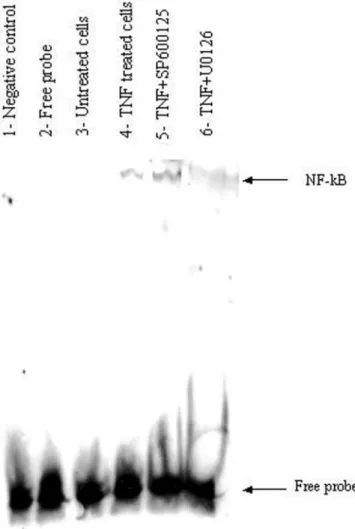
Treatment with U0126 (lane 6 compared to lane 4, Figure 6), but
not SP600 125 (lane 5 compared to lane 4, Figure 6), inhibited the
TNF-
α-induced increase in NF-κB-binding activities, suggesting that
NF-
κB is involved in the Erk1/2 pathway leading to TNF-α-induced
MMP-9 expression.
Discussion
TNF-
α was first identified as a product of activated macrophages. It is
now known that TNF-
α is produced by many types of cells and could
serve as an endogenous modulator of normal tissue homeostasis and
physiology (Tovey, 1989; Hunt et al., 1992). Many studies showed
that protein and TNF transcripts are present in human placental
tro-phoblast cells (Chen et al., 1991; Haynes et al., 1993; King et al.,
1995). Two types of receptor were described for TNF, and both are
identified in choriocarcinoma cell lines and in lysates of human term
placentas. These observations suggest that TNF could play a
regula-tory role in trophoblast invasiveness (Yang et al., 1993). This property
could be due in part to its stimulating effect on MMP-9 activity in
CTB (Meisser et al., 1999). However, in contrast to these
consider-ations, it was observed that in first-trimester placental explant cultures
(Bauer et al., 2004) or in the trophoblast cell line HTR-8/SVneo
(Renaud et al., 2005), exogenous TNF decreased trophoblast
inva-siveness. In the present study, we observed that exogenous TNF-
α
increased invasion of 8–9 weeks CTB. This discrepancy suggests that
trophoblastic cell lines do not respond in the same way as primary
CTB. Trophoblastic cell lines are very useful models, but results
obtained with these cells should always be verified with primary cells.
MMP-9 has been observed to be instrumental to trophoblast
inva-sion (Librach et al., 1991); in this article, we show that treatment of
CTB or a trophoblast cell line (HIPEC 65) by TNF-
α induces a
two-fold increase in MMP-9 expression, secretion and activity via the
Erk1/2 and SAPK/JNK pathways. The results obtained from HIPEC
65 cells and CTB are very similar, and we may conclude that results
Figure 1. Increased in vitro invasion of 8–9 weeks cytotrophoblastic cells (CTB) by exogenous tumour necrosis factor-α (TNF-α). Primary cell invasion assay was performed in an invasion chamber based on the Boyden chamber principle. Cells were added in 100 μl of serum-free media, in the presence or absence of TNF-α (100 ng/ml), to the upper compartment of the transwell chambers. Media was added to the lower chamber. Cells were incubated for 48 h at 37°C in a CO2 (5%) incubator. After incubation, the supernatant was
dis-carded, and viable cells were stained with Chemicon cell stain. After washing, the stain was extracted with a solution of 1% acetic acid : 50% ethanol for 15 min at room temperature. Hundred microlitres of the dye mixture was trans-ferred to a 96-well microtitre plate for colorimetric measurement at 560 nm. Experiment was performed three times with different cell preparations and samples run in triplicate. Statistics were performed with Student’s t-test. Data are expressed as the mean ± SEM and analysed with a Student’s t-test.
Figure 2. Down-regulation of tumour necrosis factor-α (TNF-α)-induced metalloproteinase-9 (MMP-9) activity and expression by mitogen-activated protein kinase (MAPK) inhibitors in HIPEC 65. Cells were treated with or without TNF-α (100 ng/ml) for 48 h. When inhibitors were used, they were added 1 h before TNF-α treatment. After incubation, culture supernatants were subjected to gelatin-substrate gel electrophoresis (A) and cell lysates to 10% SDS–PAGE and trans-ferred to nitrocellulose membrane. Western blot analysis was performed using an MMP-9-specific polyclonal antibody (B). Bands of western blot visualized by an ECL method and surface of the digestion bands of zymogram were scanned and quantified by the Kodak 1D image analysis software. The results are expressed as the ratio of MMP-9 or MMP-2 band intensity of treated cells versus that of untreated cells used as control. Each experiment was performed two times with different cell preparations and samples run in triplicate. Statistics were performed with Student’s t-test. *P < 0.05, as compared with TNF-α-treated control cells.
Figure 3. Tumour necrosis factor-α (TNF-α)-induced down-regulation of secreted metalloproteinase-9 (MMP-9) concentration and activity by mitogen-activated protein kinase (MAPK) inhibitors in cytotrophoblastic cells (CTB). Cells were treated with/without TNF-α (100 ng/ml) for 48 h. When inhibitors were used, they were added 1 h before TNF-α treatment. After incubation, culture supernatants were subjected to gelatin-substrate gel electrophoresis (A) or quantified by ELISA (B). Experiment was performed two times with different cell preparations and samples run in triplicate. Statistics were performed with Student’s t-test. *P < 0.05, as compared with TNF-α-treated control cells.
obtained from HIPEC 65 cells could correspond to the in vivo
situ-ation of extravillous CTB.
The signalling pathway of TNF-induced MMP-9 expression has
already been studied in a number of cell types (Holvoet et al., 2003;
Moon et al., 2004; Nee et al., 2004; Wiehler et al., 2004) but never, to
the best of our knowledge, in trophoblastic cells. The study of Wiehler
et al. (2004) showed that TNF-
α stimulates MMP-9 release from
human eosinophils and found that the signalling pathway leading to
TNF-
α-induced MMP-9 release has unique features relative to other
granule components from eosinophils and relative to the specific
mechanism of TNF-
α-induced MMP-9 release from neutrophils
(Chakrabarti et al., 2006). Wiehler et al. suggest that each MAP
kinase may be responsible for mediating a unique set of functional
responses. Therefore, it will be important to characterize the
mecha-nisms that regulate TNF-
α-induced MMP-9 production and release in
each cell type. In HIPEC 65 cells, activation of the MAPK pathways
by TNF-
α induced both phosphorylation of Erk1/2, followed by
acti-vation of NF-
κB, and phosphorylation of SAPK/JNK, followed by
activation of AP-1 (Figure 7). Inhibition of the Erk pathway by
treat-ment with U0126, or inhibition of SAPK/JNK pathway by treattreat-ment
with SP600 125, resulted in a concomitant decrease of MMP-9
activ-ity, expression and secretion. Interestingly, MMP-2 promoter, which
has also an AP-1 site but no NF-
κB site (Van den Steen et al., 2002),
was not activated under these experimental conditions. We have
pre-viously shown that AP-1 was clearly implicated in the expression of
MMP-9 in primary human CTB (Bischof et al., 2003), and we concluded
that AP-1 was necessary but probably not sufficient for a complete
Figure 4. Erk1/2 and JNK, but not p38 mitogen-activated protein kinase (MAPK), are involved in tumour necrosis factor-α (TNF-α)-induced metallo-proteinase-9 (MMP-9) expression in HIPEC 65 cells. (A) Cells were treated with TNF-α (100 ng/ml) for different time intervals, and p38 phosphorylation was evaluated by immunoblotting with an antibody specific for phosphorylated p38 (P-p38 MAPK). Equal loading in each lane is shown by the similar intensities of p38 MAPK. Negative and positive cell extract controls of phosphorylated p38 MAPK from Cell Signaling were used to confirm the specificity of anti-phosphorylated p38 MAPK antibody. (B) Cells were pretreated with SP600 125 or not for 1 h before TNF-α treatment for various periods of times. Effect of MAPK inhibitor on TNF-α-induced JNK phosphorylation was shown by immunoblot-ting with an antibody specific for phosphorylated JNK (P-JNK). (C) Cells were pretreated or not with U0126 for 1 h before TNF-α treatment for various periods of times. Effect of U0126 on TNF-α-induced Erk1/2 phosphorylation was shown by immunoblotting with an antibody specific for phosphorylated Erk1/2 (P-Erk1/2). Equal loading in each lane is shown by the similar intensities of Erk1/2.
Figure 5. Nuclear factor-κB (NF-κB) DNA-binding capacity in TNF-α-stimu-lated HIPEC 65 cells. In the absence of nuclear protein (lane 2), no chemilumi-nescence was detected, demonstrating that only biotin-labelled DNA-binding proteins were detected. Unlabeled activator protein 1 (AP-1) consensus oligo-nucleotides (lane 1) were added in excess (4 pmoles) for specific competitive assay with biotin end-labelled AP-1 oligonucleotides. NS, nonspecific binding.
transactivation of MMP-9. We would now propose that NF-
κB is also
an essential transcription factor in the process of MMP-9 induction by
TNF-
α in trophoblastic cells. The situation in human trophoblastic
cells is not unique, since it is known that TNF-
α induced MMP-9 via
activation of NF-
κB in glioma cells (Esteve et al., 2002) and via the
activation of NF-
κB and AP-1 in osteosarcoma OST cells (Sato and
Seiki, 1993).
We also reported here that p38 MAPK is not involved in TNF-
α-induced MMP-9 expression in trophoblastic cells, in contrast to other
cells (Cho et al., 2000; Wiehler et al., 2004). Inhibition of the Erk1/2
and SAPK/JNK pathways totally blocked the MMP-9 expression,
secretion and activity induced by TNF-
α in CTB and HIPEC 65 cells.
Thus, for MMP-9 induction by TNF-
α in trophoblastic cells, both AP-1
and NF-
κB activation seem to be essential, confirming that, acting
together, AP-1 and NF-
κB synergistically up-regulated gelatinase B,
whereas the expression was rather unaffected by the individual
tran-scription factors (Bond et al., 2001).
Acknowledgements
The authors express their gratitude to the Swiss National Science Foundation for their financial support and to Pr. Evain-Brion for the donation of HIPEC 65 cell line.
References
Baud V and Karin M (2001) Signal transduction by tumor necrosis factor and its relatives. Trends Cell Biol 11,372–377.
Bauer S, Pollheimer J, Hartmann J, Husslein P, Aplin JD and Knöfler M (2004) Tumor necrosis factor-alpha inhibits trophoblast migration through elevation of plasminogen activator inhibitor-1 in first-trimester villous explant cultures. J Clin Endocrinol Metab 89,2812–2822.
Bischof P, Martelli M, Campana A, Itoh Y, Ogata Y and Nagase H (1995) Importance of matrix metalloproteinases in human trophoblast invasion. Early Pregnancy 1,263–269.
Bischof P, Truong K and Campana A (2003) Regulation of trophoblastic gela-tinases by proto-oncogenes. Placenta 24,155–163.
Bond M, Chase AJ, Baker AH and Newby AC (2001) Inhibition of transcrip-tion factor NF-kappaB reduces matrix metalloproteinase-1-3, and -9 produc-tion by vascular smooth muscle cells. Cardiovasc Res 50,556–565. Chakrabarti S, Zee JM and Patel KD (2006) Regulation of matrix
metallopro-teinase-9 (MMP-9) in TNF-stimulated neutrophils: novel pathways for terti-ary granule release. J Leukoc Biol 79 (1),214–222.
Chen HL, Yang YP, Hu XL, Yelavarthi KK, Fishback JL and Hunt JS (1991) Tumor necrosis factor alpha mRNA and protein are present in human placental and uterine cells at early and late stages of gestation. Am J Pathol 139,327–335.
Cho A, Graves J and Reidy MA (2000) Mitogen-activated protein kinases mediate matrix metalloproteinase-9 expression in vascular smooth muscle cells. Arterioscler Thromb Vasc Biol 20,2527–2532.
Divya Chhikara P, Mahajan VS, Datta Gupta S and Chauhan SS (2002) Differ-ential activity of cathepsin L in human placenta at two different stages of gestation. Placenta 23,59–64.
Esteve PO, Chicoine E, Robledo O, Aoudjit F, Descoteaux A, Potworowski EF and St-Pierre Y (2002) Protein kinase C-zeta regulates transcription of the matrix metalloproteinase-9 gene induced by IL-1 and TNF-alpha in glioma cells via NF-kappa B. J Biol Chem 277,35150–35155.
Fisher SJ, Leitch MS, Kantor MS, Basbaum CB and Kramer RH (1985) Degra-dation of extracellular matrix by the trophoblastic cells of first-trimester human placentas. J Cell Biochem 27,31–41.
Fisher SJ, Cui TY, Zhang L, Hartman L, Grahl K, Zhang GY, Tarpey J and Damsky CH (1989) Adhesive and degradative properties of human placental cytotrophoblast cells in vitro. J Cell Biol 109,891–902.
Haynes MK, Jackson LG, Tuan RS, Shepley KJ and Smith JB (1993) Cytokine production in first trimester chorionic villi: detection of mRNAs and protein products in situ. Cell Immunol 151,300–308.
Holvoet S, Vincent C, Schmitt D and Serres M (2003) The inhibition of MAPK pathway is correlated with down-regulation of MMP-9 secretion induced by TNF-alpha in human keratinocytes. Exp Cell Res 290,108–119. Hunt JS, Chen HL, Hu XL, Chen TY and Morrison DC (1992) Tumor necrosis
factor-alpha gene expression in the tissues of normal mice. Cytokine 4,340–346. Figure 6. Nuclear factor-κB (NF-κB) DNA-binding capacity in tumour
necro-sis factor-α (TNF-α)-stimulated HIPEC 65 cells. In the absence of nuclear pro-teins (lane 2), no chemiluminescence was detected, demonstrating that only biotin-labelled DNA-binding proteins were detected. Unlabeled NF-κB con-sensus oligonucleotides (lane 1) were added in excess (4 pmoles) for specific competitive assay with biotin-end-labelled NF-κB oligonucleotides. NS, non-specific binding.
Figure 7. Schematic summary of signal transduction by tumour necrosis factor-α (TNF-α) on metalloproteinase-9 (MMP-9) expression in human tro-phoblastic cells (HIPEC 65). TNF-α activates different mitogen-activated pro-tein kinase (MAPK) pathways, Erk1/2 and JNK, which in turn initiate nuclear factor-κB (NF-κB) and activator protein 1 (AP-1) activation respectively and finally induce MMP-9 expression. In contrast TNF-α does not activate p38 MAPK in HIPEC 65 cells.
King A, Jokhi PP, Smith SK, Sharkey AM and Loke YW (1995) Screening for cytokine mRNA in human villous and extravillous trophoblasts using the reverse-transcriptase polymerase chain reaction (RT-PCR). Cytokine 7,364–371.
Librach CL, Werb Z, Fitzgerald ML, Chiu K, Corwin NM, Esteves RA, Grobelny D, Galardy R, Damsky CH and Fisher SJ (1991) 92-kD type IV collagenase mediates invasion of human cytotrophoblasts. J Cell Biol 113,437–449.
Martelli M, Campana A and Bischof P (1993) Secretion of matrix metallopro-teinases by human endometrial cells in vitro. J Reprod Fertil 98,67–76. Meisser A, Chardonnens D, Campana A and Bischof P (1999) Effects of
tumour necrosis factor-alpha, interleukin-1 alpha, macrophage colony stim-ulating factor and transforming growth factor beta on trophoblastic matrix metalloproteinases. Mol Hum Reprod 5,252–260.
Moon SK, Cha BY and Kim CH (2004) ERK1/2 mediates TNF-alpha-induced matrix metalloproteinase-9 expression in human vascular smooth muscle cells via the regulation of NF-kappaB and AP-1: Involvement of the ras dependent pathway. J Cell Physiol 198,417–427.
Nee LE, McMorrow T, Campbell E, Slattery C and Ryan MP (2004) TNF-alpha and IL-1beta-mediated regulation of MMP-9 and TIMP-1 in renal proximal tubular cells. Kidney Int 66,1376–1386.
Pavan L, Tarrade A, Hermouet A, Delouis C, Titeux M, Vidaud M, Thérond P, Evain-Briond D and Fournier T (2003) Human invasive trophoblasts trans-formed with simian virus 40 provide a new tool to study the role of PPAR-gamma in cell invasion process. Carcinogenesis 24,1325–1336.
Red-Horse K, Zhou Y, Genbacev O, Prakobphol A, Foulk R, McMaster M and Fisher SJ (2004) Trophoblast differentiation during embryo implan-tation and formation of the maternal–fetal interface. J Clin Invest 114,744–754.
Renaud SJ, Postovit LM, Macdonald-Goodfellow SK, McDonald GT, Caldwell JD and Graham CH (2005) Activated macrophages inhibit human cytotrophoblast invasiveness in vitro. Biol Reprod 73,237–243.
Sato H and Seiki M (1993) Regulatory mechanism of 92 kDa type IV colla-genase gene expression which is associated with invasiveness of tumor cells. Oncogene 8,395–405.
Staun-Ram E, Goldman S, Gabarin D and Shalev E (2004) Expression and importance of matrix metalloproteinase 2 and 9 (MMP-2 and -9) in human trophoblast invasion. Reprod Biol Endocrinol 2,59.
Tovey MG (1989) Expression of the genes of interferons and other cytokines in normal and diseased tissues of man. Experientia 45526–535.
Van den Steen PE, Dubois B, Nelissen I, Rudd PM, Dwek RA and Opdenakker G (2002) Biochemistry and molecular biology of gelatinase B or matrix metal-loproteinase-9 (MMP-9). Crit Rev Biochem Mol Biol 37,375–536. Wajant H, Pfizenmaier K and Scheurich P (2003) Tumor necrosis factor
sig-nalling. Cell Death Differ 10,45–65.
Wiehler S, Cuvelier SL, Chakrabarti S and Patel KD (2004) p38 MAP kinase regulates rapid matrix metalloproteinase-9 release from eosinophils. Bio-chem Biophys Res Commun 315,463–470.
Xu P, Wang YL, Zhu SJ, Luo SY, Piao YS and Zhuang LZ (2000) Expression of matrix metalloproteinase-2, -9, and -14, tissue inhibitors of metalloprotei-nase-1, and matrix proteins in human placenta during the first trimester. Biol Reprod 62,988–994.
Yang Y, Yelavarthi KK, Chen HL, Pace JL, Terranova PF and Hunt JS (1993) Molecular, biochemical, and functional characteristics of tumor necrosis factor-alpha produced by human placental cytotrophoblastic cells. J Immu-nol 150,5614–5624.