HAL Id: hal-02965130
https://hal.inrae.fr/hal-02965130
Submitted on 13 Oct 2020
HAL is a multi-disciplinary open access
archive for the deposit and dissemination of
sci-entific research documents, whether they are
pub-lished or not. The documents may come from
teaching and research institutions in France or
abroad, or from public or private research centers.
L’archive ouverte pluridisciplinaire HAL, est
destinée au dépôt et à la diffusion de documents
scientifiques de niveau recherche, publiés ou non,
émanant des établissements d’enseignement et de
recherche français ou étrangers, des laboratoires
publics ou privés.
death prevent and cure rotavirus infection
Zhan Zhang, Jun Zou, Zhenda Shi, Benyue Zhang, Lucie Etienne-Mesmin,
Yanling Wang, Xuyan Shi, Feng Shao, Benoit Chassaing, Andrew Gewirtz
To cite this version:
Zhan Zhang, Jun Zou, Zhenda Shi, Benyue Zhang, Lucie Etienne-Mesmin, et al.. IL-22-induced cell
extrusion and IL-18-induced cell death prevent and cure rotavirus infection. Science Immunology,
American Association for the Advancement of Science, 2020, 5 (52), pp.eabd2876.
�10.1126/sciim-munol.abd2876�. �hal-02965130�
V I R A L I M M U N O L O G Y
IL-22–induced cell extrusion and IL-18–induced cell
death prevent and cure rotavirus infection
Zhan Zhang1, Jun Zou1, Zhenda Shi1, Benyue Zhang1, Lucie Etienne-Mesmin1,2, Yanling Wang1,
Xuyan Shi3, Feng Shao3, Benoit Chassaing1,4,5,6, Andrew T. Gewirtz1*
Bacterial flagellin can elicit production of TLR5-mediated IL-22 and NLRC4-mediated IL-18 cytokines that act in concert to cure and prevent rotavirus (RV) infection. This study investigated the mechanism by which these cyto-kines act to impede RV. Although IL-18 and IL-22 induce each other’s expression, we found that IL-18 and IL-22 both impeded RV independently of one another and did so by distinct mechanisms that involved activation of their cognate receptors in intestinal epithelial cells (IEC). IL-22 drove IEC proliferation and migration toward villus tips, which resulted in increased extrusion of highly differentiated IEC that serve as the site of RV replication. In contrast, IL-18 induced cell death of RV-infected IEC thus directly interrupting the RV replication cycle, resulting in spewing of incompetent virus into the intestinal lumen and causing a rapid drop in the number of RV-infected IEC. Together, these actions resulted in rapid and complete expulsion of RV, even in hosts with severely compro-mised immune systems. These results suggest that a cocktail of IL-18 and IL-22 might be a means of treating viral infections that preferentially target short-lived epithelial cells.
INTRODUCTION
Rotavirus (RV) remains a scourge to humanity, causing severe dis-tress (“morbidity”) to many children and contributes to thousands of childhood deaths annually, particularly in developing countries wherein RV vaccines have only moderate efficacy (1). RV is a double- stranded RNA virus that primarily infects intestinal epithelial cells (IEC) that line the villus tips of the ileum, resulting in severe life-threatening diarrhea in young children and moderate gastro-intestinal distress in adults (2–4). Such tropism and pathogenesis is faithfully recapitulated in RV-infected mice, making the mouse model of RV useful for studying basic aspects of RV immunity and disease pathophysiology. Furthermore, the RV mouse model may prove a useful platform for discovery of novel means to treat and prevent RV infection, especially in scenarios when adaptive immu-nity, which normally plays an essential role in clearing RV, is not functioning adequately. Toward this end, we previously reported that administration of bacterial flagellin rapidly cured, and/or pro-tected against, RV infection. Such protection was independent of interferon and adaptive immunity and dependent on the genera-tion of both Toll-like receptor 5 (TLR5)–mediated interleukin-22 (IL-22) and nucleotide-binding oligomerization domain–like re-ceptor C4 (NLRC4)–mediated IL-18, which together resulted in prevention and/or cure of RV infection, and its associated diarrhea (5). However, the mechanisms by which these cytokines impede RV infection remained unknown. Herein, we report that IL-22 acts upon IEC to drive proliferation, migration, and ultimately extru-sion of infected IEC into the intestinal lumen, whereas IL-18 drives rapid death of RV-infected IEC. The combined actions of IL-22 and IL-18 eliminate RV from the intestine independent of adaptive immunity.
RESULTS
IL-22 and IL-18 activate their receptors on epithelial cells to protect against RV
We previously reported that systemic administration of bacterial flagellin elicits TLR5-mediated production of IL-22 and NLRC4- mediated generation of IL-18 that can act in concert to prevent or treat RV and some other enteric viral infections (5). Specifically, as shown in fig. S1 and our previous work, chronic RV infections that developed in RV-inoculated immune-deficient C57BL/6 Rag-1−/− mice were cured by combined systemic treatment with IL-18 and IL-22, whereas injection of either cytokine alone reduced RV loads but did not clear the virus, regardless of cytokine dose and duration of administration. In these particular experiments, RV infection was assayed by measuring fecal RV antigens by enzyme-linked im-munosorbent assay (ELISA), but measurement of RV genomes in the intestine yields similar results (5). In wild-type (WT) mice, a sufficiently high doses of recombinant IL-22 can, by itself, fully pre-vent RV infection, whereas lower doses of exogenously adminis-tered IL-22 and IL-18 markedly reduced the extent of RV infection, while the combination of these cytokines eliminated evidence of infection (Fig. 1A). The central goal of this study was to elucidate mechanisms by which these cytokines act in concert to control and prevent RV infection.
In the context of parasitic infection, both IL-18 and IL-22 pro-mote expression of each other, and loss of either impairs immunity to Toxoplasma gondii (6). We thus hypothesized that administra-tion of IL-18 might impede RV as a result of its ability to induce IL-22 expression. This hypothesis predicted that the ability of IL-18 to protect against RV infection would be largely absent in IL-22−/− mice. However, administration of IL-18 upon RV inoculation clearly reduced the extent of RV infection in IL-22−/− mice, which argued strongly against this hypothesis (Fig. 1B). We considered the con-verse hypothesis, namely, that IL-22 might impede RV infection by elicitation of IL-18, but we observed that recombinant IL-22 markedly prevented RV infection in IL-18−/− mice (Fig. 1C). Although IL-18 and IL-22 may play important roles in inducing each other’s expression, our results indicate that they each activate distinct signaling
1Center for Inflammation, Immunity and Infection, Institute for Biomedical Sciences
Georgia State University, Atlanta, GA 30303 USA. 2Université Clermont Auvergne,
INRAe, UMR 454 MEDIS, F-63000 Clermont-Ferrand, France. 3National Institute of
Biological Sciences, Beijing 102206, China. 4INSERM, U1016, team “Mucosal
micro-biota in chronic inflammatory diseases,” Paris, France. 5Université de Paris, Paris,
France. 6Neuroscience Institute, Georgia State University, Atlanta, GA 30303 USA.
*Corresponding author. Email: agewirtz@gsu.edu
Copyright © 2020 The Authors, some rights reserved; exclusive licensee American Association for the Advancement of Science. No claim to original U.S. Government Works by guest on October 12, 2020 http://immunology.sciencemag.org/ Downloaded from
pathways that cooperate to impede RV infection.
Next, we examined the extent by which IL-18 and IL-22 acted upon the hemato-poietic or nonhematohemato-poietic compart-ment to impede RV infection. We used WT, IL-18-R−/−, and IL-22-R−/− mice to gen-erate irradiated bone marrow chimeric mice that expressed the receptors for IL-22 or IL-18 in only bone marrow–derived or radioresistant cells. Such mice were in-oculated with RV, treated with recom-binant IL-22 or IL-18, and RV infection was monitored via measuring fecal RV antigens by ELISA. Figure 1 used a rela-tively low dose of cytokine that highlighted the cooperativity of IL-18 and IL-22, but suc-cessive experiments used fivefold higher doses to enable a robust effect that could be dissected via bone marrow chimeric mice. Mice that expressed the IL-22 re-ceptor only in bone marrow–derived cells were not protected from RV infec-tion by treatment with IL-22 (Fig. 2A), whereas mice with IL-22 receptor only in radioresistant cells were almost com-pletely protected by this cytokine (Fig. 2B). These results suggest that IL-22 pro-tects mice from RV infection by acting on IEC, which are known to be populated from radioresistant stem cells and re-sponsive to IL-22 (7). In accord with this notion, we observed that multiple IEC cell lines are responsive to IL-22 in vitro via STAT3 phosphorylation, although
IL-22, like flagellin and IL-18, did not affect RV infection in vitro (fig. S2). Studies with IL-18-R chimeric mice similarly revealed that expression of this receptor in only bone marrow–derived cells con-ferred only a modest nonsignificant reduction (12 ± 3.8%) in the
extent of RV infection upon IL-18 administration (Fig. 2C). In con-trast, in mice that expressed IL-18-R in only radioresistant cells, IL-18 reduced extent of RV infection by 76 ± 8.7% (Fig. 2D). To-gether, these results suggest that agonizing IL-18 and IL-22 receptors Fig. 1. IL-22 and IL-18 elicit distinct antiviral activities against mRV invasion. Mice were administered PBS, IL-22 (2 g), and/or IL-18 (1 g) via intraperitoneal injection, 2 hours before, or 2, 4, 6, or 8 days after (indicated by arrows) oral inoculation with mRV. Fecal RV levels were measured over time by ELISA. (A) C57BL/6 mice n = 4. (B) IL-22−/− mice, n = 5 and 7 for PBS and IL-18, respectively. (C) IL-18−/− mice, n = 5. * indicates significantly different from PBS by two-way analysis of variance (ANOVA),
P < 0.0001. dpi, days post-inoculation.
Fig. 2. Both IL-22– and IL-18–mediated antiviral pathway use their cognate nonhematopoietic cell receptors. In-dicated bone marrow–irradiated chimeric mice were administered PBS (control), IL-22 (10 g), or IL-18 (2 g) via intraper-itoneal injection, 2 hours before or 2, 4, 6, or 8 days after oral inoculation with mRV. Fecal RV levels were measured over time by ELISA. Differences between control and cytokine groups for each chimera/panel were analyzed by two-way ANOVA. (A) n = 7, P = 0.7715. (B) n = 4 and 7 for PBS and IL-22, respectively. (C) n = 7 and 6 for PBS and IL-18, respectively. (D) n = 4 and 6 for PBS and IL-18, respectively. * indicates significantly different from PBS by two-way ANOVA, P < 0.0001.
by guest on October 12, 2020
http://immunology.sciencemag.org/
on IEC result in generation of signals that impede RV in vivo but not in vitro.
IL-22 and IL-18 promote IEC proliferation/migration
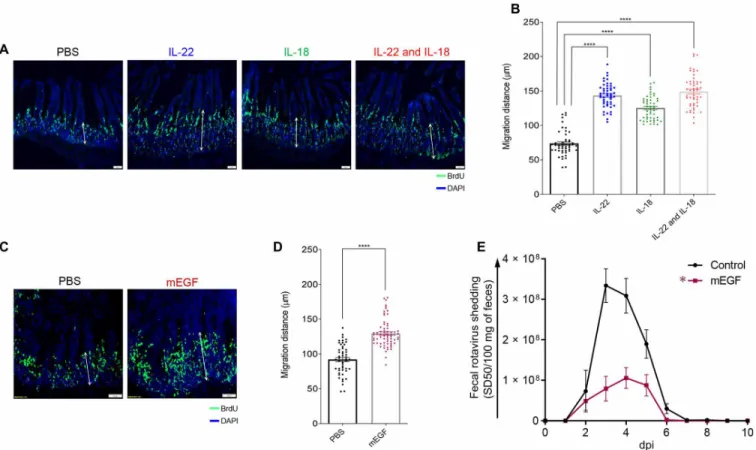
In cell culture and organoid models, IL-22 promotes IEC proliferation, migration, and stem cell regeneration (8–10), which together are thought to contribute to ability of IL-22 to promote healing in response to an array of insults, including exposure to radiation and dextran sodium sulfate in vivo (11–14). In contrast to such severe injuries, RV infection is generally characterized by a lack of overt intestinal inflammation (15, 16). We hypothesized that IL-22 may promote IEC prolifera-tion and/or migraprolifera-tion that might reduce the extent of RV infecprolifera-tion by increasing the rate of IEC turnover, especially near villus tips, which is the predominant site of RV infection (2–4). We further rea-soned that IL-18 might trigger the same kind of response and further increase IEC proliferation and turnover. Mice were administered 5-bromo-2´-deoxyuridine (BrdU) and treated with IL-22 and/or IL-18. Sixteen hours later, mice were euthanized, and intestines were sub-jected to fluorescence microscopy to measure rates at which IEC migrated toward villus tips (17). In accord with our hypothesis, ad-ministration of IL-22 approximately doubled the rate at which IEC migrated toward villus tips (Fig. 3, A and B). IL-18 administration also increased the rate of IEC migration to a lesser extent. The
com-bination of these cytokines did not result in a faster rate of IEC mi-gration relative to IL-22 alone. Epidermal growth factor (EGF) is known to promote IEC proliferation and migration (18, 19), so we tested whether this cytokine might protect against RV infection. In accord with EGF promoting proliferation in a variety of tissues, EGF treatment induced IEC migration up the crypt villus axis (Fig. 3, C and D), albeit not quite as robustly as IL-22 (1.43- versus 1.95-fold increase respectively). Moreover, EGF had the ability to reduce the extent of RV infection (Fig. 3E), but not as completely as IL-22. To-gether, these results support the hypothesis that IL-22 and IL-18 promote IEC replication and migration, which contributes to pro-tection against RV infection.
IL-22 promotes extrusion of IEC into small intestinal lumen
We next considered how promoting IEC proliferation might im-pede RV infection. Increased extrusion of IEC into the lumen is a likely consequence of increased IEC proliferation/migration, which is thought to occur such that cells remain alive until extrusion is com-pleted to preserve the gut barrier (20). We hypothesized that increased proliferation/migration induced by IL-22 and/or IL-18 treatments might result in increased extrusion of villus tip cells, which are the site of RV infection. We investigated this hypothesis using a previously described method (21) in which cross sections of hematoxylin and
Fig. 3. Accelerated proliferation rate and migration levels of IEC are correlated with debilitation of mRV infectivity. Mice were intraperitoneally injected with PBS, IL-22, (10 g) IL-18 (2 g), both cytokines, or mEGF. One hour later, mice were administered BrdU. Mice were euthanized 16 hours after BrdU administration, and BrDU was visualized (A and C) and migration was measured (B and D) by microscopy and image analysis, respectively. Images shown in (A) and (C) are representative. Scale bar equals 50 m. For (B) and (D), sections were scored at least from 50 villus per group of mice (n = 5). Distance of the foremost migrating cells along the crypt-villus axis was measured with ImageJ software. Results are presented as means ± SEM. Statistical significance was evaluated by Student’s t test (****P < 0.0001). (E) Mice were intraperitone-ally injected with PBS or EGF (10 g) mEGF 2 hours before or 2, 4, 6, or 8 days after oral inoculation with mRV. Fecal RV levels were measured over time by ELISA. Data are means ± SEM, n = 5 * indicates significantly different from PBS by two-way ANOVA, P < 0.0001.
by guest on October 12, 2020
http://immunology.sciencemag.org/
eosin–stained pieces of ileum are examined for visual evidence of cell shedding. We were unable to consistently distinguish IEC from other luminal contents, so we visualized cells using the DNA stain. This approach suggested a greater presence of IEC in the lumen of mice treated with cytokines, particularly IL-22 (Fig. 4A), but it was diffi-cult to quantitate such a difference via cell counting, so we sought to evaluate levels of host cells via quantitative polymerase chain reac-tion (qPCR) of 18S DNA in the ileum. The highly degradative envi-ronment of the intestine would likely degrade IEC shed into the lumen, but because such cells are extruded in a relatively intact state, their DNA might survive long enough to enable quantitation by qPCR. Small intestinal contents were extracted, and 18S DNA quantitated and ex-pressed as number of cells per 100 mg of luminal content using known numbers of mouse epithelial cells to generate a standard curve. This approach indicated that IL-22 treatment markedly increased the level of IEC present in the lumen (Fig. 4B), suggesting increased IEC shedding. IL-18 induced only a modest level of IEC shedding that appeared to be additive to the shedding induced by IL-22. A gener-ally similar pattern was observed in the cecum (Fig. 4C). In contrast, these cytokines did not affect levels of 18S DNA present in the lu-men of the colon (Fig. 4D), perhaps reflecting that the impact of these cytokines on IEC shedding is specific to the ileum/cecum and/or
that the DNA of shed IEC is quickly degraded in the bacterial- dense colon. An even greater amount of shedding of IEC into the ileum was induced by treating mice with flagellin, although two treatments of IL-18/22 could match this level, which suggested that production of these cytokines might be sufficient to recapitulate the IEC shedding induced by flagellin (Fig. 4E). The greater potency of flagellin may reflect ability of IL-18 and IL-22 to promote each other’s expression. Use of IL-22−/− and IL-18−/− mice revealed that these cytokines, both of
which are necessary for flagellin’s anti-RV action (5), were both nec-essary for flagellin-induced cell shedding (Fig. 4F). Collectively, these results support the notion that increased extrusion of IEC, particularly in response to IL-22, might be central to this cytokine’s ability to im-pede RV infection, but these data did not offer insight into how IL-22 and IL-18 cooperate to offer stronger protection against this virus.
IL-18 induces death of RV-infected IEC
Next, we examined how IL-22 and IL-18 might affect IEC in the pres-ence of an active RV infection. We used WT mice 3 days after inocula-tion with RV, a time approaching peak levels of RV shedding (Fig. 1A). RV-infected and uninfected mice were administered IL-22 and/or IL-18 and euthanized 6 hours later, and small intestinal content was isolated. Like IL-18/22 administration, RV infection up-regulated
Fig. 4. IL-22 promotes cell extrusion into intestinal lumen. Mice [WT or indicated knockout (KO) strain] received a single (except where indicated otherwise) intraper-itoneal injection of PBS, IL-22, (10 g), IL-18 (2 g), both cytokines or bacterial flagellin, FliC (15 g). Eight hours later, mice were euthanized, intestine was isolated, and luminal content was collected. (A) Microscopic appearance of DAPI-stained section to visualize shed cells in lumen. Scale bar equals 50 m. (B to F) Measurements of shed cells in different regions of the gastrointestinal tract via 18s by q-PCR (B, E, and F) small intestine, (C) cecum, (D) colon [double doses of IL-22 and IL-18 in (E) were 12 hours apart]. Data in (B) to (F) are means ± SEM (B), with significance assessed by Student’s t test, n = 5 to 15 mice as indicated by number of data points. *P < 0.05, **P < 0.01, ***P < 0.001, and ****P < 0.0001. n.s., not significant; SI, small intestine.
by guest on October 12, 2020
http://immunology.sciencemag.org/
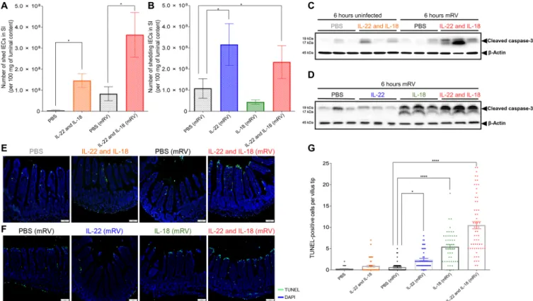
IEC extrusion, with a marked further increase in IEC extrusion be-ing observed by administration of IL-18/22 to RV-infected mice (Fig. 5A). This suggests that increased IEC extrusion may normally contribute to innate defense against RV (2) and that exogenously ad-ministered IL-18/22 (or flagellin) may enhance this protective mech-anism. Yet, like the case in uninfected mice, the promotion of IEC extrusion appeared to be driven by IL-22 and not IL-18 (Fig. 5B).
Next, we sought to investigate events in IEC that remained part of the small intestine at the time of increased IEC extrusion. Specif-ically, we examined whether IL-18 and/or IL-22 might affect cell death. We observed that IL-18/22 or RV induced modest and variable induc-tion of cleaved caspase-3. In contrast, administrainduc-tion of these cyto-kines to RV-infected mice induced marked elevations in cleaved caspase-3 (Fig. 5C). Caspase-3 cleavage was also observed in response to IL-18 but not IL-22 (Fig. 5D). Quantitation of cell death by termi-nal deoxynucleotidyl transferase–mediated deoxyuridine triphos-phate nick end labeling (TUNEL) yielded a similar pattern of re-sults. Specifically, both IL-18/22 and RV by themselves resulted in a modest increase in TUNEL-positive cells, which appeared sporadi-cally throughout the villi (Fig. 5E and fig. S3, A and B). In contrast, treating RV-infected mice with IL-18 or the combination of IL-18 and IL-22, but not IL-22 itself, resulted in notable TUNEL positivity at the villus tips (Fig. 5, E to G), known sites of RV infection. Cytokine- induced TUNEL positivity, which did not occur in the absence of RV, appeared to localize in the villus tip, where RV was localized before
cytokine treatment, thus suggesting that IL-18 was promoting cell death in RV-infected cells (fig. S3C).
Cell death can occur via numerous pathways, so we hypothesized that IL-18–induced cell death might occur via pyroptosis, which ap-pears to be a frequent form of cell death for infected cells (22). In accord with this possibility, IL-18 administration to RV-infected mice results in cleaved gasdermin D (Fig. 6A), whose activity is essential for pyroptosis. To test the role of gasdermin D activation in IL-18–induced cell death, we performed experiments in mice lacking gasdermin D and gasdermin E, the latter of which is thought to compensate for lack of gasdermin D in some scenarios. Our initial experiments found that gasdermin-deficient mice were highly resistant to RV infection (fig. S4). However, such resistance was associated with high levels of segmented filamentous bacteria (SFB), which we have recently shown drives spontaneous resistance to RV in Rag1−/− mice (23). Cross-fostering on gasdermin-deficient mice removed SFB and restored susceptibil-ity to RV infection, thus extending our recent findings to mice with functional adaptive immunity. This model could also address if the IL-18–induced cell death that associates with clearance of RV is me-diated by pyroptosis. IL-18 administration did not induce cleaved gasdermin D in mice lacking this gene (Fig. 6A), thus verifying the specificity of the antibody we used. IL-18–induced cell death of RV- infected mice proceeded at least as robustly as had been observed in WT mice (Fig. 6B). Specifically, although gasdermin-deficient mice had mild elevations in basal caspase-3, they still up-regulated this
Fig. 5. IL-18 induced TUNEL-positive cell death in villus tips of RV-infected mice. Mice were orally inoculated with mRV, or not(sham?) and were intraperitoneally injected at 3 dpi with PBS, IL-22, (10 g) IL-18 (2 g), or both cytokines. Mice were euthanized 6 hours later and following assays were carried out. (A and B) Assay of cell extrusion (i.e., measure of cells in lumen) as performed in response to cytokines in Fig. 4. (C and D) Assay cleaved caspase-3 in IEC was assayed by SDS-PAGE immunoblotting. (E and F) Visualization of cell death by TUNEL staining, counterstained with DAPI. (G) Quantitation of TUNEL-positive cells at villus tip region based on visual counts. Data in (A), (B), and (G) are means ± SEM. Panels (A) and (B) used five mice per condition to generate one value per mouse. Panel (G) used five mice per condition and assayed 6 to 10 villi per mouse, which are indicated by data points. Significance was determined by Student’s t test. *P < 0.05 and ****P < 0.0001.
by guest on October 12, 2020
http://immunology.sciencemag.org/
caspase in response to IL-18, albeit at markedly lower levels com-pared with WT mice. IL-18 induced marked TUNEL positivity in these mice (Fig. 6, C and D) and fully protected gasdermin-deficient mice against RV infection (Fig. 6E). These results argue that IL-18– induced cell death and associated clearance of RV are not mediated by pyroptosis.
IL-18 interrupts viral replication
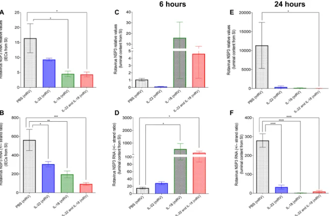
We examined the extent by which IL-22–induced IEC extrusion and IL-18–induced IEC death were associated with RV reduction in the ileum at 6 and 24 hours after administration of these cytokines. We measured the levels of RV genomes and the ratio of positive to negative (+/−) RV strands in both the lumen and IEC, which re-flects levels of active replication because most positive strands en-code RV proteins and do not get incorporated into virions (24). In accord with our previous work, we observed that, in the epithelium, both IL-22 and IL-18 led to a clear reduction in both the level of RV genomes and RV replication by 6 hours (Fig. 7, A and B). In con-trast, the small intestinal lumen had a marked but variable increase in the level of RV genomes and a stark increase in RV +/− strand ratios 6 hours after administration of IL-18 with the combination of IL-18 and IL-22 but not IL-22 alone (Fig. 7, C and D). By 24 hours, levels of RV in the lumen had dropped markedly, whereas the miniscule levels of remaining virus appeared to not be actively replicating (Fig. 7, E and F). Collectively, these results support a model wherein IL-18–induced cell death interrupts active RV replication, spewing incompletely replicated virus into the lumen while IL-22 induces IEC migration and subsequent extrusion of the mature IEC that RV targets, thus together working in concert to resolve RV infection.
DISCUSSION
The central focus of this study was to determine the mechanisms by which IL-18 and IL-22, which are elicited by bacterial flagellin, con-tribute to preventing or curing RV infection. We initially considered that the ability of IL-18 and IL-22 to promote each other’s expres-sion allowed them to use a shared mechanism to promote RV clear-ance. We found that irrespective of such mutual promotion, IL-18 and IL-22 both impeded RV independent of each other and did so by distinct mechanisms, which is illustrated in Fig. 8. Specifically, IL-22 drove IEC proliferation and migration toward villus tips, thus accelerating the ongoing process of extrusion of highly differentiated IEC at the major site of RV replication. In contrast, administration of IL-18 to RV-infected mice induced rapid cell death, as defined by TUNEL, at villus tips where RV is localized. Such induction of TUNEL positivity, which is not typically seen at significant levels in the in-testine, was associated with rapid abortion of the RV replication cycle followed by a marked reduction of RV antigens in the intesti-nal tract. These actions of IL-22 and IL-18 together resulted in rapid and complete expulsion of RV, thus providing a mechanism that explains how this combination of cytokines prevents and cures RV infection.
RV does not induce detectable increases in IL-22 expression nor does genetic deletion of IL-22 appear to markedly augment RV in-fection (5), thus arguing that IL-22 does not normally play a major role in clearance of this pathogen. The known cooperation of IL-22 and interferon- in activating antiviral gene expression (3) suggests the possibility that RV may have evolved strategies to deliberately avoid or block IL-22 induction. Nonetheless, the downstream ac-tion of IL-22, particularly its promoac-tion of IEC turnover, may be Fig. 6. Pyroptosis mediator gasdermin is not required for IL-18–induced cell death or its protection against mRV infection. (A to D) Gasdermin-deficient, or WT, mice were administered PBS or IL-18 (2 g) 3 days after mRV inoculation. Mice were euthanized 6 hours later and jejunums were analyzed. (A and B) IEC were analyzed by SDS-PAGE immunoblotting for detection of gasdermin D, cleaved gasdermin D, and cleaved caspase-3, respectively. (C) Cell death by TUNEL, counterstained with DAPI. (D) Quantitation of TUNEL-positive cells at villus tip region based on visual counts. Experiments included five mice per condition. Data in (D) was based on assay 6 to 8 villi per mouse, which are indicated by data points ****P < 0.0001 by Student’s t test. (E) Gasdermin-deficient mice were administered PBS or IL-18 (2 g) via intraperito-neal injection, 2 hours before, or 2, 4, 6 or 8 days after (indicated by arrows), oral inoculation with mRV. Fecal RV levels were measured over time by ELISA. Data are means ± SEM. n = 5. * indicates significantly different from control by two-way ANOVA, P < 0.0001.
by guest on October 12, 2020
http://immunology.sciencemag.org/
shared by endogenous anti-RV host de-fense mechanisms. The role of adaptive immune-independent host defense against RV is most easily appreciated in immune compromised mice wherein RV loads decline markedly from their peak levels, but it may also play a role in protecting against RV even in immune competent mice. Innate host defense against RV is likely multifactorial and may involve type III interferon (3), particularly in neo-nate mice. Our observations in adult mice indicate that RV infection increases IEC extrusion, and this mechanism combined with previous observations that RV in-fection activates intestinal stem cell pro-liferation suggests that increased IEC turnover may limit RV infection (2). We do not think that such a mechanism is nec-essarily unique to IL-22 as EGF has ability to drive similar events. Moreover, we re-cently showed that SFB also drives entero-cyte proliferation independent of IL-22 and is not required for adaptive immu-nity (23). Hence, we presume that IL-22 can Fig. 7. Administration of IL-18 rapidly releases the replicating virus into the luminal side. mRV-infected mice were intraperitoneally injected with PBS, IL-22 (10 g), IL-18 (2 g), or both cytokines on day 3 post-mRV inoculation. Six or 24 hours later, mice were euthanized, and contents of jejunums were isolated. RNA was extracted and used to measure of mRV genomes and replication status as reflected by NSP3 RNA levels and the ratio of NSP3 (+) RNA strand to complimentary NSP3 (−) RNA strand. (A and B) The overall mRV genome and efficacy of virus replication in small intestinal epithelial cells. (C to F) The overall mRV genome and efficacy of virus replication in luminal content from small intestine (one-way ANOVA, n = 5 to 10, *P < 0.05, **P < 0.01, ***P < 0.001, and ****P < 0.0001).
Fig. 8. Proposed mechanism by which IL-22 and IL-18 prevent and cure RV infection. IL-22 increases epithelial proliferation thus increasing extrusion of epithelial cells, including RV-infected cells. Into lumen the intestinal lumen, i.e., anoikis. IL-18 induces rapid cell death, associated with loss of cell rupturing of RV-infected cells.
by guest on October 12, 2020
http://immunology.sciencemag.org/
activate a primitive mechanism of host defense against a variety of challenges, especially those affecting IEC.
IEC are rapidly proliferating cells with average lifetimes of about 3 days (24), which means that the intestine must eliminate vast num-bers of cells continuously. The overwhelming majority of IEC are eliminated via cell extrusion at villus tips through a process termed anoikis. A central tenet of anoikis is that cells remain alive at the time of extrusion followed by the lack of attachment to other cells result-ing in induction of a programmed death process (25). A key aspect of this process is that cells can be eliminated without comprising gut barrier function, thus avoiding infection and inflammation that might otherwise occur. Accordingly, administration of IL-22 is associated with few adverse effects and has been shown to resolve inflammation in several different scenarios. (26). Moreover, IL-22 plays a broad role of maintaining gut health in the intestinal tract, including mediat-ing microbiota-dependent impacts of dietary fiber (27). It is possible that increasing anoikis via IL-22 results in extrusion of RV-containing cells in a manner that prevents viral escape and, consequent infection of other IEC. However, inability of IL-22 to induce detectable increases in luminal RV argues against this possibility. Rather, we envisage that the cell death process after IEC extrusion might result in destruc-tion of RV in these cells. We also hypothesize that the accelerated IEC turnover induced by IL-22 may result in villus IEC being less differ-entiated and less susceptible to RV infection. In accord with this possibility, we observed that that flagellin administration resulted in an IL-22–dependent increase in CD44+26− IEC (fig. S5), which are known to be RV resistant (28). It is difficult to discern the relative im-portance of IL-22 in the induction of IEC extrusion versus its impact on differentiation state of villus IEC. IL-22–induced reduction in RV levels in chronically infected Rag-1−/− mice occurs over a course
of several days that supports a role for the latter mechanism. Use of IL-22 receptor bone marrow chimera mice demonstrated that IL-22 acts directly on IEC to affect RV infection. (7). IL-22–induced signal-ing is generally thought to be mediated by STAT3 (5, 10), and IL-22 in-duced phosphorylation of STAT3 in IEC in vivo. However, we observed that IEC-specific STAT3-knockout mice could still be protected against RV by IL-22, suggesting that this mechanism of action may not pro-ceed by a characterized signaling mechanism (fig. S6). Thus, how the IL-22 receptor signals to affect IEC phenotype remains incom-pletely understood.
In contrast to IL-22, recent work indicates that induction of IL-18 plays a role in endogenous immunity against RV, wherein caspase-1– mediated IL-18 production results from activation of the NLR9pb inflammasome. Such IL-18 induction paralleled gasdermin-dependent cell death, the absence of which resulted in delayed clearance of RV (29, 30). On the basis of this work, we hypothesized that exogenously administered IL-18 might enhance RV-induced death of RV-infected cells and/or increase IEC turnover analogous to IL-22. Administra-tion of IL-18 in the absence of RV elicited a modest increase in the number of TUNEL-positive cells as well as a modest increase in IEC proliferation/migration that was not accompanied by increased IEC extrusion, suggesting the increased proliferation compensated for cell death. However, TUNEL-positive cells were scattered along the villus. In RV-infected mice, IL-18 led to TUNEL-positive cells at the villus tips, which is also the primary site of RV infection. It is tempting to envisage localized impacts of IL-18 reflect the pattern of expression of the IL-18 receptor, including localization to villus tips and/or induced by RV, but limited knowledge of the determinants of its expression and lack of available reagents to study it render these ideas as speculative.
The manner of IL-18–induced cell death, namely, its notable TUNEL induction, which was associated with spewing of RV repli-cation intermediates, suggested pyroptotic cell death. However, we found that lack of gasdermin D and E, which are thought to be es-sential for pyroptosis, did not impede IL-18–induced cell death in RV-infected cells thus arguing such cell death does not fit perfectly into any known cell death pathways. Induction of IL-18 receptor- mediated signaling by itself is not sufficient to induce cell death in villus tip epithelial cells but triggers death in cells primed as a result of RV infection. The nature of such priming is not understood but may involve IEC signaling pathways, including NLR9pb, TLR3, and RNA- activated protein kinase, which are capable of recognizing RV compo-nents and/or responding to intracellular stress in general (30–32). In this context, the ability of IL-22 to enhance IL-18–induced TUNEL positivity in RV-infected cells may reflect an intersection of IL-22-R and IL-18-R signaling or be a manifestation of these cytokines to pro-mote each other’s expression.
The central limitation in our study was that our approaches were largely correlative. Specifically, we lacked modalities to specifically block IEC migration or cell death in response to IL-22 and IL-18, re-spectively. Another limitation is that we were not able to demonstrate that the TUNEL-positive cells actually contained RV. Our attempts to do so via double-staining were not successful, possibly reflecting that the disappearance of RV after cytokine treatment likely occurs early in the cell death process while the DNA fragmentation that un-derlies TUNEL positivity is considered a late event in the cell death process. Thus, more specific identification of processes that mediate cell death of RV-infected IEC in response to IL-18 is an important target of future studies.
The improved understanding of the mechanism by which IL-18/22 controls RV infection reported herein should inform use of these cytokines to treat viral infection in humans. Chronic RV infec-tions can occur in immune compromised humans, suggesting that IL-18/22 may be explored as a possible treatment for this and other chronic viral infections. Our results suggest that this cytokine treat-ment may be effective for viruses that preferentially infect villus ep-ithelial cells and possibly other epithelia that have high cell turnover rates. In contrast, this combination of cytokines seems unlikely to affect viruses that inhabit more long-lived cells, including hemato-poietic cells that are generally not responsive to IL-22. We observed that flagellin and IL-18/22 has some efficacy against reovirus, par-ticularly early in infection when it infects gut epithelial cells, as well as some efficacy against influenza, which initially infects lung epithe-lial cells, but did not show any impact on hepatitis C virus as assayed in mice engrafted with human hepatocytes, which are thought to be long-lived cells. IL-18/22 can protect mice against norovirus in-fection, which infects B cells and tuft cells (33, 34), but human nor-ovirus is thought to primarily infect epithelial cells, particularly in immunocompromised persons who develop chronic norovirus in-fections (35). SARS-CoV-2, the causative agent of coronavirus dis-ease 2019 (COVID-19) has also been observed to replicate in IEC (36), and like RV, appears to replicate in mature IEC, which express the SARS-CoV-2 receptor angiotensin-converting enzyme 2. Intes-tinal replication of SARS-CoV-2 is thought to contribute to extra-respiratory pathologies associated with COVID-19 (37). As such, the use of IL-18/22–based therapy may be a potential strategy to treat chronic RV and/or norovirus infections in person with im-mune dysfunction and, moreover, might serve to mitigate severe cases of COVID-19.
by guest on October 12, 2020
http://immunology.sciencemag.org/
MATERIALS AND METHODS
Study design
This study sought to ascertain the mechanism by which IL-22 and IL-18 prevent and cure RV infection. Mice were orally administered RV. Extent of infection was assayed my measuring viral genomes and proteins in the intestine. IL-18 and or IL-22 were administered to mice with various genetic deficiencies. Cell extrusion and cell death were measured. All procedures involving mice were approved by GSU’s animal care and use committee (Institutional Animal Care and Use Committee no.17047).
Mice
All mice used herein were adults (i.e., 4 to 8 weeks old) on a C57BL/6 background bred at Georgia State University (GSU) (Atlanta, GA). RV-infected mice were housed in an animal biosafety level 2 facility. WT,
Rag-1−/−, IL-18−/−, IL-18-R−/−, Stat3flox, and Villin-cre were purchased from the Jackson laboratory (Bar Harbor, ME, USA). NLRC4−/−, IL-22−/−,
and IL-22-R−/− mice were provided by Genentech (South San Francisco, CA, USA). TLR5−/− and TLR5−/−/NLRC4−/− and WT littermates were
maintained as previously described (5). Gasdermin D/E−/− mice, whose generation and initial characterization were previously described (22), were shipped to GSU and studied in original and cross-fostered state as indicated in results.
Materials
Murine Fc-IL-22 was provided by Genentech Inc. Murine IL-18 was purchased from Sino Biological Inc. (Beijing, China). Procedures for isolation of flagellin and verification of purity were described pre-viously (5). Recombinant murine EGF (mEGF) was purchased from PeproTech.
RV infection Acute Model
Age- and sex-matched adult mice (8 to 12 weeks of age) were orally administered 100 l of 1.33% sodium bicarbonate (Sigma-Aldrich) and then inoculated with 105 SD50 of murine RV EC strain. Approach used to determine SD50 has been described previously (5).
Chronic model
Five-week-old Rag-1−/− mice were infected with murineRV (same infection procedure as described in the “Acute models” section). Feces were collected 3 weeks after RV inoculation to confirm the establishment of chronic infection.
In vitro model
Cell culture-adapted rhesus RV (RRV) was trypsin-activated [trypsin (10 g/ml)] in serum-free RPMI-1640 (cellgro) at 37°C for 30 min. The basolateral side of the polarized Caco-2 cells was stimulated with cytokines, 1.5 hours before expose to RRV infection as previously de-scribed (5). The upper chamber of transwells was infected with trypsin- pretreated RRV and allowed to adsorb at 37°C for 40 min before being washed with serum-free medium. The presence of cytokines was maintained at a constant level throughout the experiment.
Fecal RV antigen detection
Fecal pellets were collected daily from individual mouse on days 0 to 10 after RV inoculation. Samples were suspended in phosphate- buffered saline (PBS) [10% (w/v)], after centrifugation, supernatants of fecal homogenates were analyzed by ELISA, and after multiple serial dilutions, more detailed descriptions of experimental proce-dures are previously described (5).
Generation of bone marrow chimeric mice
Mice were subjected to x-ray irradiation using an 8.5 gray (Gy) equiva-lent followed by injection of 2 × 10⁷ bone marrow cells adminis-tered intravenously as previously described (5). All mice were afforded an 8-week recovery period before experimental use. For the first 2 weeks after transfer, mice were maintained in sterile cages and supplied with drinking water containing neomycin (2 mg/ml) (Mediatech/Corning).
Visual assessment of IEC shed into small intestinal lumen
Intestinal sections were fixed in methanol-Carnoy’s fixative solu-tion (60% methanol, 30% chloroform, and 10% glacial acetic acid) for 48 hours at 4°C. Fixed tissues were washed two times in dry methanol for 30 min each, followed by two times in absolute ethanol for 20 min each, and then incubated in two baths of xylene before proceeding to paraffin embedding. Thin sections (4 m) were sliced from paraffin- embedded tissues and placed on glass slides after floating on a water bath. The sections were dewaxed by initial incubation at 60°C for 20 min, followed by two baths in prewarmed xylene substitute solution for 10 min each. Deparaffinized sections were then hydrated in solutions with decreasing concentration of ethanol (100, 95, 70, 50, and 30%) every 5 min in each bath. Last, slides allowed to dry almost completely and were then mounted with ProLong antifade mounting media containing 4′,6-diamidino-2-phenylindole (DAPI) before analysis by fluorescence microscopy.
Immunohistochemistry for TUNEL staining
Intestinal sections were fixed in 10% buffered formalin at room tem-perature for 48 hours and then embedded in paraffin. Tissues were sectioned at 4 m thickness, and IEC death was detected by TUNEL assay using the In Situ Cell Death Detection Kit, Fluorescein (Roche) according to the manufacturer’s instructions.
Immunoblot analysis for assay of cleaved caspase-3, phospho-STAT3, and cleaved gasdermin D
IECs lysate (20 g per lane) was separated by SDS–polyacrylamide gel electrophoresis through 4 to 20% Mini-PROTEAN TGX gel (Bio-Rad, USA), transferred to nitrocellulose membranes, and analyzed by im-munoblot, as previously described (5). Briefly, isolated IEC was in-cubated with radioimmunoprecipitation assay lysis buffer (Santa Cruz Biotechnology, USA) for 30 min at room temperature. Subsequently, cell lysates were homogenized by pipette and subjected to full-speed centrifugation. Protein bands were detected for cleaved caspase-3, phosphor-STAT3, and anti–-actin (Cell Signaling Technology) and incubated with horseradish peroxidase–conjugated anti-rabbit second-ary antibody. Immunoblotted proteins were visualized with Western blotting detection reagents (GE Healthcare) and then imaged using the ChemiDoc XRS+ system (Bio-Rad).
Isolation of IECs
The entire small intestine was harvested from mice according to indicated experimental design and sliced longitudinally before be-ing washed gently in PBS to remove the luminal content. Tissues were processed and maintained at 4°C throughout. Cleaned tissue samples were further minced into 1- to 2-mm3 pieces and shaken in 20 ml of Hanks’ balanced salt solution (HBSS) containing 2 mM EDTA and 10 mM Hepes for 30 min. An additional step of vigorous vortexing in fresh HBSS (10 mM Hepes) after EDTA incubation fa-cilitated cell disaggregation. IECs were then filtered through 70-m
by guest on October 12, 2020
http://immunology.sciencemag.org/
nylon mesh strainer (BD Biosciences), centrifuged, and resuspend-ed in PBS.
Antibody staining and flow cytometry analysis
Bulk leukocytes and IECs isolated above were incubated with suc-cinimidyl esters (NHS ester)–Alexa Fluor 430, which permitted deter-mination of cell viability. Cells were then blocked by incubation with anti-CD16/anti-CD-32 (10 g/ml) (clone 2.4G2, American Type Cul-ture Collection). Twenty minutes later, cells were stained with fluores-cently conjugated antibodies: CD26-PE (clone H194-112, eBioscience), CD44-PECy7 (clone IM7, eBioscience), CD45–fluorescein isothiocyanate (clone, 30-F11, eBioscience), and CD326-allophycocyanin (clone G8.8, eBioscience). Last, stained cells were fixed with 4% formaldehyde for 10 min before whole-cell population was analyzed on a BD LSR II flow cytometer. Collected data were analyzed using FlowJo.
Quantification of IEC shedding from luminal content
Host DNA was quantitated from 100 mg of luminal content (100 mg) from small intestine by using the QIAamp DNA Stool Mini kit (Qiagen) and subjected to qPCR using QuantiFast SYBR Green PCR kit (Bio-Rad) in a CDX96 apparatus (Bio-Rad) with specific mouse 18S oligonu-cleotides primers. The sense and antisense oligonuoligonu-cleotides primers used were: 18s-1F: 5′-GTAACCCGTTGAACCCCATT-3′ and 18s-1R: 5′-CCATCCAATCGGTAGTAGCG-3′. PCR results were expressed as actual numbers of IEC shedding per 100 mg of luminal content, calculated using a standard curve, which was generated using two-fold serial dilutions of mouse colon carcinoma cell line MC26. DNA was extracted from each vial with known number of MC26 cells af-ter serial dilutions, and then real-time qPCR was performed. The cycle quantification (Cq) values (x axis) are inversely proportional to the amount of target genes (18S) (y axis), and a standard curve is applied to estimate the numbers of cell shedding from luminal con-tent based on the quantity of target copies (18S) from each sample.
Quantification of RV genomes and replication in IEC and luminal content
To extract RNA, cell pellets were homogenized with TRIzol (Invitrogen), and chloroform was added to the homogenate to separate RNA (an upper aqueous layer) from DNA and proteins (a red lower organic layer). Isopropanol facilitated the precipitation of RNA out of solution, and after centrifugation, the impurities were removed by washing with 75% ethanol. RNA pellets were resuspended in ribonuclease- free water and underwent quantitative reverse transcription PCR. Total RNA from luminal content was purified from the RNeasy PowerMicrobiome Kit according to the manufacturer’s instructions. Primers that target non-structural protein 3 region: EC.C (+) (5′-GTTC-GTTGTGCCTCATTCG-3′ and EC.C (−) (5′-TCGGAACGTACTTCT-GGAC-3′) were applied to quantify viral genomes from IEC and luminal content. RV replication was quantitated as previously described (38).
BrdU pulse-chase labeling analysis of intestinal enterocyte migration
A pulse-chase experimental strategy was used to label intestinal en-terocytes with BrdU to estimate the IEC migration rate along the crypt-villus axis over a defined period of time. Briefly, 8-week-old mice were intraperitoneally injected with either PBS or cytokine(s) (IL-22 and/or IL-18) 1 hour before BrdU treatment (50 g/mg of mice body weight, ip). After 16 hours, mice were euthanized, and a segment of the jejunum were resected, immediately embedded in
optimal cutting temperature compound (OCT) (Sigma-Adrich) and then underwent tissue sectioning. Tissue sections (4 m) were firstly fixed in 4% formaldehyde for 30 min at room temperature and then washed three times in PBS. DNA denaturation was performed by in-cubating the sections in prewarmed 1.5 N HCl for 30 min at 37°C, and then acid was neutralized by rinsing sections three times in PBS. Before BrdU immunostaining, sections were blocked with rabbit serum (Bio-Genex, Fremont, CA) for 1 hour at room temperature, then incubated with anti-BrdU (Abcam) 2 hours at 37°C, and counterstained with DAPI. The BrdU-labeled cells were visualized by fluorescence microscopy.
mRV antigen detection by fluorescence immunohistochemistry
The proximal jejunum was imbedded into OCT compound, and then sliced into 6-m-thin sections. Tissue slides were incubated in 4% paraformaldehyde for 15 min, followed by 5 min washing of PBS twice. Autofluorescence caused by free aldehydes was quenched by incubating slides in 50 mM NH4Cl in PBS or 0.1 M glycine in PBS for 14 min at room temperature, followed by 5 min PBS washing three times. Bovine serum albumin–PBS (3%) was used to block the tissue samples for 1 hour at room temperature. The slides were then washed with PBS for 5 min, followed by incubation with primary anti-body (1:100; hyperimmune guinea pig anti-RRV serum) in blocking buffer overnight at 4°C. After slides were washed three times with PBS, secondary antibody (donkey anti–guinea pig immunoglobulin G, Jackson ImmunoResearch, 706-586-148) was applied to the sample slides for 1 to 2 hours at room temperature. The fluorescence emis-sion of mRV antigen was detected by fluorescence microscopy.
Quantification and statistical analysis
Significance was determined using the one-way analysis of variance (ANOVA) or student’s t test (GraphPad Prism software, version 6.04). Differences were noted as significant *P < 0.05, **P < 0.01, ***P < 0.001, and ****P < 0.0001.
SUPPLEMENTARY MATERIALS
immunology.sciencemag.org/cgi/content/full/5/52/eabd2876/DC1
Fig. S1. Impact of IL-22 and IL-18 on RV infection in immune-deficient, Rag-1−/− mice. Fig. S2. Lack of effect of IL-22 and IL-18 on RV infection in cultured IECs.
Fig. S3. Impact of IL-18 on IEC death.
Fig. S4. Cross-fostering on gasdermin-deficient mice conferred susceptibility to RV infection. Fig. S5. Flagellin-mediated changes of cell subpopulations along intestinal villus-crypt axis. Fig. S6. Impact of IL-22 and flagellin on RV infection in mice lacking STAT3 in IEC.
View/request a protocol for this paper from Bio-protocol.
REFERENCES AND NOTES
1. J. E. Tate, A. H. Burton, C. Boschi-Pinto, U. D. Parashar; World Health Organization-Coordinated Global Rotavirus Surveillance Network, Global, regional, and national estimates of rotavirus mortality in children <5 years of age, 2000-2013. Clin. Infect. Dis. 62, S96–S105 (2016).
2. W. Y. Zou, S. E. Blutt, X. L. Zeng, M. S. Chen, Y. H. Lo, D. Castillo-Azofeifa, O. D. Klein, N. F. Shroyer, M. Donowitz, M. K. Estes, Epithelial WNT ligands are essential drivers of intestinal stem cell activation. Cell Rep. 22, 1003–1015 (2018).
3. P. P. Hernandez, T. Mahlakoiv, I. Yang, V. Schwierzeck, N. Nguyen, F. Guendel, K. Gronke, B. Ryffel, C. Hoelscher, L. Dumoutier, J. C. Renauld, S. Suerbaum, P. Staeheli, A. Diefenbach, Interferon- and interleukin 22 act synergistically for the induction of interferon-stimulated genes and control of rotavirus infection. Nat. Immunol. 16, 698–707 (2015).
4. A. Z. Kapikian, R. E. Shope, Rotaviruses, reoviruses, coltiviruses, and orbiviruses, in Medical Microbiology, S. Baron, Ed. (Galveston, 1996).
5. B. Zhang, B. Chassaing, Z. Shi, R. Uchiyama, Z. Zhang, T. L. Denning, S. E. Crawford, A. J. Pruijssers, J. A. Iskarpatyoti, M. K. Estes, T. S. Dermody, W. Ouyang, I. R. Williams,
by guest on October 12, 2020
http://immunology.sciencemag.org/
M. Vijay-Kumar, A. T. Gewirtz, Viral infection. Prevention and cure of rotavirus infection via TLR5/NLRC4-mediated production of IL-22 and IL-18. Science 346, 861–865 (2014). 6. M. Munoz, C. Eidenschenk, N. Ota, K. Wong, U. Lohmann, A. A. Kuhl, X. Wang,
P. Manzanillo, Y. Li, S. Rutz, Y. Zheng, L. Diehl, N. Kayagaki, M. van Lookeren-Campagne, O. Liesenfeld, M. Heimesaat, W. Ouyang, Interleukin-22 induces interleukin-18 expression from epithelial cells during intestinal infection. Immunity 42, 321–331 (2015).
7. K. Wolk, S. Kunz, E. Witte, M. Friedrich, K. Asadullah, R. Sabat, IL-22 increases the innate immunity of tissues. Immunity 21, 241–254 (2004).
8. C. A. Lindemans, M. Calafiore, A. M. Mertelsmann, M. H. O’Connor, J. A. Dudakov, R. R. Jenq, E. Velardi, L. F. Young, O. M. Smith, G. Lawrence, J. A. Ivanov, Y. Y. Fu, S. Takashima, G. Hua, M. L. Martin, K. P. O'Rourke, Y. H. Lo, M. Mokry, M. Romera-Hernandez, T. Cupedo, L. Dow, E. E. Nieuwenhuis, N. F. Shroyer, C. Liu, R. Kolesnick, M. R. M. van den Brink, A. M. Hanash, Interleukin-22 promotes intestinal-stem-cell-mediated epithelial regeneration. Nature 528, 560–564 (2015).
9. G. Pickert, C. Neufert, M. Leppkes, Y. Zheng, N. Wittkopf, M. Warntjen, H. A. Lehr, S. Hirth, B. Weigmann, S. Wirtz, W. Ouyang, M. F. Neurath, C. Becker, STAT3 links IL-22 signaling in intestinal epithelial cells to mucosal wound healing. J. Exp. Med. 206, 1465–1472 (2009). 10. M. L. Nagalakshmi, A. Rascle, S. Zurawski, S. Menon, R. de Waal Malefyt, Interleukin-22
activates STAT3 and induces IL-10 by colon epithelial cells. Int. Immunopharmacol. 4, 679–691 (2004).
11. J. L. Bishop, M. E. Roberts, J. L. Beer, M. Huang, M. K. Chehal, X. Fan, L. A. Fouser, H. L. Ma, J. T. Bacani, K. W. Harder, Lyn activity protects mice from DSS colitis and regulates the production of IL-22 from innate lymphoid cells. Mucosal Immunol. 7, 405–416 (2014). 12. J. A. Dudakov, A. M. Hanash, M. R. van den Brink, Interleukin-22: Immunobiology
and pathology. Annu. Rev. Immunol. 33, 747–785 (2015).
13. A. M. Hanash, J. A. Dudakov, G. Hua, M. H. O’Connor, L. F. Young, N. V. Singer, M. L. West, R. R. Jenq, A. M. Holland, L. W. Kappel, A. Ghosh, J. J. Tsai, U. K. Rao, N. L. Yim, O. M. Smith, E. Velardi, E. B. Hawryluk, G. F. Murphy, C. Liu, L. A. Fouser, R. Kolesnick, B. R. Blazar, M. R. van den Brink, Interleukin-22 protects intestinal stem cells from immune-mediated tissue damage and regulates sensitivity to graft versus host disease. Immunity 37, 339–350 (2012).
14. Y. Zheng, P. A. Valdez, D. M. Danilenko, Y. Hu, S. M. Sa, Q. Gong, A. R. Abbas, Z. Modrusan, N. Ghilardi, F. J. de Sauvage, W. Ouyang, Interleukin-22 mediates early host defense against attaching and effacing bacterial pathogens. Nat. Med. 14, 282–289 (2008). 15. O. Lundgren, L. Svensson, Pathogenesis of rotavirus diarrhea. Microbes Infect. 3,
1145–1156 (2001).
16. A. P. Morris, M. K. Estes, Microbes and microbial toxins: paradigms for microbial-mucosal interactions. VIII. Pathological consequences of rotavirus infection and its enterotoxin. Am. J. Physiol. Gastrointest. Liver Phtysiol. 281, G303–G310 (2001).
17. L. W. Peterson, D. Artis, Intestinal epithelial cells: Regulators of barrier function and immune homeostasis. Nat. Rev. Immunol. 14, 141–153 (2014).
18. X. Tang, H. Liu, S. Yang, Z. Li, J. Zhong, R. Fang, Epidermal growth factor and intestinal barrier function. Mediators Inflamm. 2016, 1927348 (2016).
19. M. R. Frey, A. Golovin, D. B. Polk, Epidermal growth factor-stimulated intestinal epithelial cell migration requires Src family kinase-dependent p38 MAPK signaling. J. Biol. Chem. 279, 44513–44521 (2004).
20. G. T. Eisenhoffer, P. D. Loftus, M. Yoshigi, H. Otsuna, C. B. Chien, P. A. Morcos,
J. Rosenblatt, Crowding induces live cell extrusion to maintain homeostatic cell numbers in epithelia. Nature 484, 546–549 (2012).
21. J. M. Williams, C. A. Duckworth, A. J. Watson, M. R. Frey, J. C. Miguel, M. D. Burkitt, R. Sutton, K. R. Hughes, L. J. Hall, J. H. Caamano, B. J. Campbell, D. M. Pritchard, A mouse model of pathological small intestinal epithelial cell apoptosis and shedding induced by systemic administration of lipopolysaccharide. Dis. Model. Mech. 6, 1388–1399 (2013).
22. J. Sarhan, B. C. Liu, H. I. Muendlein, P. Li, R. Nilson, A. Y. Tang, A. Rongvaux, S. C. Bunnell, F. Shao, D. R. Green, A. Poltorak, Caspase-8 induces cleavage of gasdermin D to elicit pyroptosis during Yersinia infection. Proc. Natl. Acad. Sci. U.S.A. 115, E10888–E10897 (2018).
23. Z. Shi, J. Zou, Z. Zhang, X. Zhao, J. Noriega, B. Zhang, C. Zhao, H. Ingle, K. Bittinger, L. M. Mattei, A. J. Pruijssers, R. K. Plemper, T. J. Nice, M. T. Baldridge, T. S. Dermody, B. Chassaing, A. T. Gewirtz, Segmented filamentous bacteria prevent and cure rotavirus infection. Cell 179, 644–658.e13 (2019).
24. J. H. Park, T. Kotani, T. Konno, J. Setiawan, Y. Kitamura, S. Imada, Y. Usui, N. Hatano, M. Shinohara, Y. Saito, Y. Murata, T. Matozaki, Promotion of intestinal epithelial cell turnover by commensal bacteria: Role of short-chain fatty acids. PLOS ONE 11, e0156334 (2016).
25. A. P. Gilmore, Anoikis. Cell Death Differ. 12, 1473–1477 (2005).
26. S. Y. Alabbas, J. Begun, T. H. Florin, I. Oancea, The role of IL-22 in the resolution of sterile and nonsterile inflammation. Clin. Trans. Immunol. 7, e1017 (2018).
27. J. Zou, B. Chassaing, V. Singh, M. Pellizzon, M. Ricci, M. D. Fythe, M. V. Kumar, A. T. Gewirtz, Fiber-mediated nourishment of gut microbiota protects against diet-induced obesity by restoring IL-22-mediated colonic health. Cell Host Microbe 23, 41–53.e4 (2018). 28. A. Sen, M. E. Rothenberg, G. Mukherjee, N. Feng, T. Kalisky, N. Nair, I. M. Johnstone,
M. F. Clarke, H. B. Greenberg, Innate immune response to homologous rotavirus infection in the small intestinal villous epithelium at single-cell resolution. Proc. Natl. Acad. Sci. U.S.A. 109, 20667–20672 (2012).
29. W. T. He, H. Wan, L. Hu, P. Chen, X. Wang, Z. Huang, Z. H. Yang, C. Q. Zhong, J. Han, Gasdermin D is an executor of pyroptosis and required for interleukin-1 secretion. Cell Res. 25, 1285–1298 (2015).
30. S. Zhu, S. Ding, P. Wang, Z. Wei, W. Pan, N. W. Palm, Y. Yang, H. Yu, H. B. Li, G. Wang, X. Lei, M. R. de Zoete, J. Zhao, Y. Zheng, H. Chen, Y. Zhao, K. A. Jurado, N. Feng, L. Shan, Y. Kluger, J. Lu, C. Abraham, E. Fikrig, H. B. Greenberg, R. A. Flavell, Nlrp9b inflammasome restricts rotavirus infection in intestinal epithelial cells. Nature 546, 667–670 (2017). 31. M. A. Garcia, J. Gil, I. Ventoso, S. Guerra, E. Domingo, C. Rivas, M. Esteban, Impact of protein
kinase PKR in cell biology: From antiviral to antiproliferative action. Microbiol. Mole. Biol. Rev. 70, 1032–1060 (2006).
32. E. Vercammen, J. Staal, R. Beyaert, Sensing of viral infection and activation of innate immunity by toll-like receptor 3. Clin. Microbiol. Rev. 21, 13–25 (2008).
33. C. B. Wilen, S. Lee, L. L. Hsieh, R. C. Orchard, C. Desai, B. L. Hykes Jr., M. R. McAllaster, D. R. Balce, T. Feehley, J. R. Brestoff, C. A. Hickey, C. C. Yokoyama, Y. T. Wang, D. A. MacDuff, D. Kreamalmayer, M. R. Howitt, J. A. Neil, K. Cadwell, P. M. Allen, S. A. Handley, M. van Lookeren Campagne, M. T. Baldridge, H. W. Virgin, Tropism for tuft cells determines immune promotion of norovirus pathogenesis. Science 360, 204–208 (2018).
34. M. K. Jones, M. Watanabe, S. Zhu, C. L. Graves, L. R. Keyes, K. R. Grau, M. B. Gonzalez-Hernandez, N. M. Iovine, C. E. Wobus, J. Vinje, S. A. Tibbetts, S. M. Wallet, S. M. Karst, Enteric bacteria promote human and mouse norovirus infection of B cells. Science 346, 755–759 (2014).
35. U. C. Karandikar, S. E. Crawford, N. J. Ajami, K. Murakami, B. Kou, K. Ettayebi, G. A. Papanicolaou, U. Jongwutiwes, M. A. Perales, J. Shia, D. Mercer, M. J. Finegold, J. Vinje, R. L. Atmar, M. K. Estes, Detection of human norovirus in intestinal biopsies from immunocompromised transplant patients. J. Gen. Virol. 97, 2291–2300 (2016). 36. R. Zang, M. F. Gomez Castro, B. T. McCune, Q. Zeng, P. W. Rothlauf, N. M. Sonnek, Z. Liu,
K. F. Brulois, X. Wang, H. B. Greenberg, M. S. Diamond, M. A. Ciorba, S. P. J. Whelan, S. Ding, TMPRSS2 and TMPRSS4 promote SARS-CoV-2 infection of human small intestinal enterocytes. Sci. Immunol. 5, eabc3582 (2020).
37. L. Pan, M. Mu, P. Yang, Y. Sun, R. Wang, J. Yan, P. Li, B. Hu, J. Wang, C. Hu, Y. Jin, X. Niu, R. Ping, Y. Du, T. Li, G. Xu, Q. Hu, L. Tu, Clinical characteristics of COVID-19 patients with digestive symptoms in Hubei, China. Am J Gastroenterol 115, 766–773 (2020). 38. M. Fenaux, M. A. Cuadras, N. Feng, M. Jaimes, H. B. Greenberg, Extraintestinal spread
and replication of a homologous EC rotavirus strain and a heterologous rhesus rotavirus in BALB/c mice. J. Virol. 80, 5219–5232 (2006).
Funding: This work was supported by NIH grants DK083890 and DK099071 (to A.T.G.). J.Z. is supported by career development award from American Diabetes Association. B.C. is supported by a Starting Grant from the European Research Council, an Innovator Award from the Kenneth Rainin Foundation, and a Chaire d’Excellence from Paris University. Author contributions: Z.Z. led performance of all experiments. J.Z. and Z.S. helped with specimen analysis. B.Z., L.E.-M., Y.W., and B.C. advised in experimental design and data interpretation. X.S. and F.S. provided advice and key reagents. A.G. helped design study and drafted manuscript. Competing interests: A.T.G. and B.Z. are inventors on patent application (WO2015054386A1 WIPO) held by GSU that covers “Prevention and treatment of rotavirus infection using IL-18 and IL-22”. Data and materials availability: All data needed to evaluate the conclusions in the paper are present in the paper or the Supplementary Materials. All mice are either commercially available or available under a material transfer agreement.
Submitted 12 June 2020 Accepted 10 September 2020 Published 2 October 2020 10.1126/sciimmunol.abd2876
Citation: Z. Zhang, J. Zou, Z. Shi, B. Zhang, L. Etienne-Mesmin, Y. Wang, X. Shi, F. Shao, B. Chassaing, A. T. Gewirtz, IL-22–induced cell extrusion and IL-18–induced cell death prevent and cure rotavirus infection. Sci. Immunol. 5, eabd2876 (2020).
by guest on October 12, 2020
http://immunology.sciencemag.org/
Chassaing and Andrew T. Gewirtz
DOI: 10.1126/sciimmunol.abd2876 , eabd2876. 5
Sci. Immunol.
support clearance of other enteric viral infections.
these mechanisms accelerate clearance of RV from the intestine. These findings suggest that these cytokines may extrusion of differentiated IEC, which are sites of RV replication. IL-18 induces cell death of infected IEC, and together,
. show that IL-22 drives intestinal epithelial cell (IEC) proliferation and migration toward villus tips resulting in et al
Zhang
IL-22 production and NLRC4-mediated generation of IL-18 can work together to prevent or treat RV infection. Here, resource-poor countries where the existing vaccine is of moderate efficacy. A previous study showed that TLR5-driven
Rotavirus (RV) is an enteric infection that can cause severe morbidity and contributes to childhood mortality in Revenge against rotavirus
ARTICLE TOOLS http://immunology.sciencemag.org/content/5/52/eabd2876
MATERIALS SUPPLEMENTARY http://immunology.sciencemag.org/content/suppl/2020/09/28/5.52.eabd2876.DC2 http://immunology.sciencemag.org/content/suppl/2020/09/28/5.52.eabd2876.DC1 REFERENCES http://immunology.sciencemag.org/content/5/52/eabd2876#BIBL This article cites 37 articles, 11 of which you can access for free
PERMISSIONS http://www.sciencemag.org/help/reprints-and-permissions
Terms of Service Use of this article is subject to the
is a registered trademark of AAAS. Science Immunology
New York Avenue NW, Washington, DC 20005. The title
(ISSN 2470-9468) is published by the American Association for the Advancement of Science, 1200 Science Immunology
Science. No claim to original U.S. Government Works
Copyright © 2020 The Authors, some rights reserved; exclusive licensee American Association for the Advancement of
by guest on October 12, 2020
http://immunology.sciencemag.org/

![Fig. 4. IL-22 promotes cell extrusion into intestinal lumen. Mice [WT or indicated knockout (KO) strain] received a single (except where indicated otherwise) intraper- intraper-itoneal injection of PBS, IL-22, (10 g), IL-18 (2 g), both cytokines or b](https://thumb-eu.123doks.com/thumbv2/123doknet/14651040.551537/5.891.74.818.487.965/promotes-extrusion-intestinal-indicated-knockout-indicated-injection-cytokines.webp)