HAL Id: hal-01871212
https://hal.uca.fr/hal-01871212
Submitted on 10 Sep 2018
HAL is a multi-disciplinary open access
archive for the deposit and dissemination of
sci-entific research documents, whether they are
pub-lished or not. The documents may come from
teaching and research institutions in France or
abroad, or from public or private research centers.
L’archive ouverte pluridisciplinaire HAL, est
destinée au dépôt et à la diffusion de documents
scientifiques de niveau recherche, publiés ou non,
émanant des établissements d’enseignement et de
recherche français ou étrangers, des laboratoires
publics ou privés.
Using acceleration to quantify symptoms during deep
brain stimulation surgery
Ashesh Shah, Jerome Coste, Erik Schkommodau, Jean-Jacques Lemaire,
Simone Hemm-Ode
To cite this version:
Ashesh Shah, Jerome Coste, Erik Schkommodau, Jean-Jacques Lemaire, Simone Hemm-Ode. Using
acceleration to quantify symptoms during deep brain stimulation surgery. 47th annual conference of
the German Society for Biomedical Engineering, Sep 2013, Graz, Austria. 58 (Suppl. 1-A), pp.4007,
2013, Biomedizinische Technik. Biomedical engineering. �10.1515/bmt-2013-4007�. �hal-01871212�
Using acceleration to quantify symptoms during
deep brain stimulation surgery.
Shah A.
1, Coste J.
2, Schkommodau E.
1, Lemaire J.J.
2, Hemm-Ode S.
11
Institute for Medical and Analytical Technologies, University of Applied Sciences and Arts Northwestern
Swit-zerland, Switzerland
2
Centre Hospitalier Universitaire de Clermont-Ferrand, Image-Guided Clinical Neurosciences and Connectomics
(EA 7292, IGCNC), Université d'Auvergne, France
[email protected]
Abstract: The use of Deep Brain Stimulation (DBS) sur-gery is increasing as a treatment for movement related disorders. One of the important areas of improvement is the target selection procedure. To do so, we measured the acceleration of tremor by sensors in 6 patients during their DBS surgeries to evaluate the changes quantita-tively. The post-operative data analysis revealed that acceleration measurements are very sensitive to the changes in tremor and that they can be used to identify clinically effective stimulation amplitudes. With the aim to increase objectivity in symptom evaluation, we intend to introduce real-time analysis so as to provide more infor-mation to the neurosurgeon to aid him in his target selec-tion during the surgery.
Keywords: movement related disorders, acceleration measurements, tremor quantification, deep brain stimula-tion
Introduction
The usage of Deep brain stimulation (DBS) of basal ganglia to treat neurological movement related disorders like Parkinson's disease (PD) and Essential Tremor (ET) has increased considerably in the recent years. However, due to incomplete understanding of the mechanism of action of DBS, optimal target definition is difficult. To overcome this, intraoperative stimulation tests are per-formed along the predetermined trajectories to semiquan-titatively evaluate the clinical effects on tremor while gradually increasing the stimulation parameters (volta-ge/current), determining the thresholds for clinical effects (subjective threshold) and side effects at each anatomical measurement position. Various methods have been propo-sed to quantitatively evaluate tremor using accelerometer ([1], [2]) as well as other sensing techniques ([3], [4]), but not specifically during DBS surgery. Methods to quantita-tively evaluate tremor intra-operaquantita-tively ([5], [6]) using accelerometers
have been proposed, but they were not made a part of the routine surgical protocol. Our aim is to measure the ac-celeration of the patient's wrist before, during and after the DBS surgery, to extract multiple parameters to quanti-fy the changes in the tremor and use these parameters to aid the neurosurgeons in optimizing the final target for implanting the electrodes.
Methods
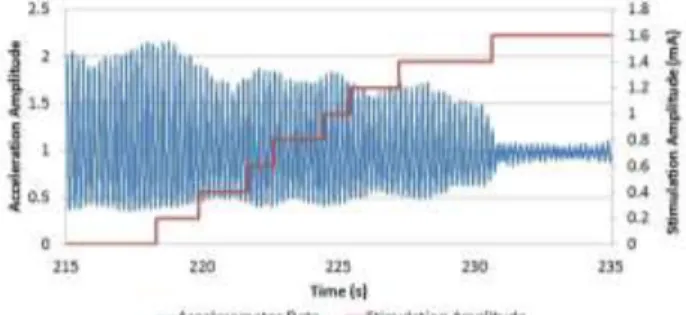
We have recorded acceleration data from 6 bilateral DBS implantations for PD (n=2) and ET (n=4), under an on-going clinical study at the University Hospital Clermont-Ferrand, France. A 3-axis accelerometer is placed inside an in-house developed plastic case and tied to the patient's wrist (Fig. 1) to measure its acceleration during test sti-mulations. Acceleration data is recorded at all the test stimulation positions on all the planned trajectories. The acceleration recording is performed by connecting the accelerometer via a USB cable to a laptop using in-house developed application. The acceleration recording is started earlier than the test stimulation and continues while the stimulation amplitude is varied (Fig 2). No specific instructions are given to the neurosurgeon or the patient for the posture of the arm or movements. The data is recorded while the neurosurgeon performs his routine evaluation. The data recorded without any test stimulation is used as a baseline for comparison with data recorded during the test stimulation. The amplitudes of stimulation at which an effect is visually observed on the symptoms (subjective threshold) and at which side-effects occur (side-effect threshold) are noted in the software.
Figure 1: The plastic case containing the accelerometer tied to the patient's wrist.
The accelerometer data recorded is then post-operatively analysed to extract statistical features to quantitatively identify the changes in the symptoms. As a first step, movements other than tremor are removed (detrending) using the smoothness Priors method [7]. Then the data is low pass filtered at 10 Hz to remove the noise. From this data, statistical features (viz. standard deviation, energy, entropy, main frequency component and main frequency
amplitude) are extracted by moving a window of 2 se-conds over it. The extracted features are then normalized to the baseline value and the normalized feature set is used to find effective stimulation amplitude (acceleration threshold). Based on the normalized data, 3 different acceleration thresholds are extracted: 1) more than 75% change compared to baseline 2) more than 50% and less than 75% change compared to baseline 3) more than 25% and less than 50% change compared to baseline. The accelerometer thresholds and the side effect thresholds are examined visually and a final implant location is decided based on them to compare with the actual final implant location.
Figure 2: Graph showing acceleration data (blue) along with stimulation amplitude (red) with time.
In order to have a statistically significant comparison, we used Wilcoxon two-sided signed rank test to compare the features 1) before the subjective threshold and at the sub-jective threshold 2) before the acceleration threshold and at the acceleration threshold.
Results
The Wilcoxon two-sided signed rank test has identified a statistical significant change in tremor (p<0.01) for signal energy, standard deviation and peak frequency amplitude. The signal energy and peak frequency amplitude seem to be the most sensitive statistical features showing a higher percentage change compared to baseline. The results also say that, in most cases (>80%), the accelerometer threshold was found at a lower stimulation amplitude than the subjective threshold (Fig. 3). The final implant site decided based on the acceleration measurements were not always consistent with the ones decided subjectively during the surgery. In some cases, the choices were on different trajectories. This suggests that the use of accele-ration measurements during the surgery may improve the target selection for DBS surgery.
Discussion
The present study describes a method to quantitatively evaluate tremor using statistical parameters extracted from the acceleration signal of the wrist and the significa-nce of the results from 6 patients. Based on the results of this study we can say that the use of such quantitative methods may improve the target selection procedure for the DBS surgeries. Such quantitative methods make the surgical treatment more objective for individual patients. We also found that the addition of acceleration
measure-ment equipmeasure-ment in the OR did not increase the duration of the surgery or interfere with any other procedure.
Figure 3: Comparison of different thresholds for one trajectory of a DBS patient.
The results of the study clearly suggest that acceleration measurements in the OR are feasible. One of the main factors that affect the acceleration measurements is the recording of the baseline data. It is important that during the baseline recording, the patient shows high tremor symptoms. Our next steps are to perform the data analysis in real-time during the surgery so that the quantitative information is made available to the neurosurgeon to aid in his decision. Further analysis and research is also plan-ned with the recorded data. We intend to use the accelera-tion sensor to evaluate rigidity during DBS surgery as well. We intend to correlate the data with the anatomical brain structures stimulated during the surgery and other electro-physiological information. This might bring new information related to the mechanism of action of DBS.
Bibliography
[1] Keijsers, N. L. (2006). "Ambulatory motor asses-sment in Parkinson's disease." Mov Disord 21(1): 34-44
[2] Dunnewold, R. J. (1997). "Quantitative assess-ment of bradykinesia in patients with Parkinson's disease." J Neurosci Methods 74(1): 107-12
[3] Papapetropoulos, S. (2008). "Objective monitoring of tremor and bradykinesia during DBS surgery for Parkinson disease." Neurology 70(15): 1244-9 [4] Koop, M. M. (2006). "Improvement in a quantita-tive measure of bradykinesia after microelectrode re-cording in patients with Parkinson's disease during deep brain stimulation surgery." Mov Disord 21(5): 673-8
[5] Journee, H. L. (2007). "Intraoperative neurophysi-ological assessment of disabling symptoms in DBS surgery." Neurophysiol Clin 37(6): 467-75.
[6] Birdno, M. J. (2008). "Tremor varies as a function of the temporal regularity of deep brain stimulation." Neuroreport 19(5): 599-602
[7] Tarvainen, M.P; Ranta-aho, P.O; Karjalainen, P.A. An advanced detrending method with application to HRV analysis.