HAL Id: hal-02556266
https://hal.archives-ouvertes.fr/hal-02556266
Submitted on 28 Apr 2020
HAL is a multi-disciplinary open access
archive for the deposit and dissemination of
sci-entific research documents, whether they are
pub-lished or not. The documents may come from
teaching and research institutions in France or
abroad, or from public or private research centers.
L’archive ouverte pluridisciplinaire HAL, est
destinée au dépôt et à la diffusion de documents
scientifiques de niveau recherche, publiés ou non,
émanant des établissements d’enseignement et de
recherche français ou étrangers, des laboratoires
publics ou privés.
Distributed under a Creative Commons Attribution| 4.0 International License
embryonic and mammary development and
differentiation
Bruno Passet, Johan Castille, Samira Makhzami, Sandrine Truchet, Anne
Vaiman, Sandrine Floriot, Katayoun Moazami-Goudarzi, Marthe Vilotte,
Anne-Laure Gaillard, Louise Hélary, et al.
To cite this version:
Bruno Passet, Johan Castille, Samira Makhzami, Sandrine Truchet, Anne Vaiman, et al.. The
Prion-like protein Shadoo is involved in mouse embryonic and mammary development and differentiation.
Scientific Reports, Nature Publishing Group, 2020, 10 (1), pp.6765. �10.1038/s41598-020-63805-y�.
�hal-02556266�
www.nature.com/scientificreports
the prion-like protein Shadoo
is involved in mouse embryonic
and mammary development and
differentiation
Bruno passet
1, Johan castille
1, Samira Makhzami
1, Sandrine truchet
2, Anne Vaiman
1,
Sandrine floriot
1, Katayoun Moazami-Goudarzi
1, Marthe Vilotte
1, Anne-Laure Gaillard
1,
Louise Helary
1, Maud Bertaud
1, olivier Andréoletti
3, Daniel Vaiman
4, pierre calvel
1,
nathalie Daniel-carlier
1, Mohammed Moudjou
2, christian Beauvallet
1, Mohamed Benharouga
5,
Denis Laloé
1, Sophie Mouillet-Richard
6, Amandine Duchesne
1, Vincent Béringue
2&
Jean-Luc Vilotte
1✉
Shadoo belongs to the prion protein family, an evolutionary conserved and extensively studied family due to the implication of prp in transmissible Spongiform encephalopathies. However, the biological function of these genes remains poorly understood. While Sprn-knockdown experiments suggested an involvement of Shadoo during mouse embryonic development, Sprn-knockout experiments in 129Pas/ C57BL/6J or 129Pas/FVB/NCr mice did not confirm it. In the present study, we analyzed the impact of
Sprn gene invalidation in a pure FVB/NJ genetic background, using a zinc finger nuclease approach. The
in-depth analysis of the derived knockout transgenic mice revealed a significant increase in embryonic lethality at early post-implantation stages, a growth retardation of young Sprn-knockout pups fed by wild type mice and a lactation defect of Sprn-knockout females. Histological and transcriptional analyses of knockout E7.5 embryos, E14.5 placentas and G7.5 mammary glands revealed specific roles of the Shadoo protein in mouse early embryogenesis, tissue development and differentiation with a potential antagonist action between prp and Shadoo. this study thus highlights the entanglement between the proteins of the prion family.
The prion protein, encoded by Prnp, is extensively studied due to its implication in Transmissible Spongiform Encephalopathies (TSE). Indeed, the misfolded, partially protease-resistant, PrP conformer is the main, if not the sole, component of prions, the infectious agent responsible for TSE in animals and humans1. The evolutionary
conservation of PrP suggested its involvement in essential cellular processes2. Prnp is ubiquitously expressed
throughout development and adult life, with a higher expression level in the central nervous system. Surprisingly, naturally occurring or generated mammals or fish devoid of PrP develop normally3–6, suggesting that (an)other
host-encoded protein(s) induces() compensatory mechanism(s).
Two other members of the mammalian prion family were described, Shadoo, encoded by Sprn and homolo-gous to the N-terminal part of PrP, and Doppel, encoded by Prnd and homolohomolo-gous to the C-terminal part of PrP7.
In the adult, Doppel is mainly expressed in male gonads and Prnd or Prnd/Prnp knockout male mice are sterile8.
Shadoo shares with PrP some spatial regulation and properties9. The knockdown of Sprn in 129Pas/FVB/
NCr Prnp-knockout embryos induces a lethal phenotype at early embryonic stages (~E10)10,11, originating
from a trophectoderm-derived compartment developmental failure11. However, Sprn or Sprn/Prnp co-ablation
in 129Pas/C57BL/6J or 129Pas/FVB/NCr mice did not reveal embryonic developmental abnormality12. These
1Université Paris-Saclay, INRAE, AgroParisTech, UMR1313-GABI, 78350, Jouy-en-Josas, France. 2Université
Paris-Saclay, INRAE, UVSQ, VIM, 78350, Jouy-en-Josas, France. 3ISC3T, UMR1225, INRAE, ENVT, 31076, Toulouse, Cedex,
France. 4Institut Cochin, U1016, INSERM, UMR 8504 CNRS, Université de Paris, Paris, France. 5LCBM-UMR5249,
DRF-BIG, CEA-Grenoble, 38054, Grenoble, Cedex, 09, France. 6Centre de Recherche des Cordeliers, INSERM,
Sorbonne Université, Université de Paris, F-75006, Paris, France. ✉e-mail: [email protected]
divergent observations, sometimes attributed to genetic compensation in mutated animals13, questioned the
involvement of Shadoo during embryogenesis.
In this study, we report the establishment, through injection of a Zinc Finger Nuclease (ZFN), of FVB/NJ
Sprn-knockout (Sprn0/0) mice. Reproductive performances and transcriptomic analyses of these mice
demon-strate that Shadoo is involved in embryonic development and more generally in the development and differen-tiation of several tissues. Furthermore, our data suggest antagonist actions of PrP and Shadoo, at least during embryonic stages.
Results
Generation of Sprn
0/0mice.
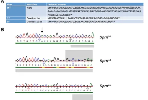
The ZFN target site is located at the start of the Sprn ORF (Fig. 1). ZFN mRNAwas injected into 1 cell-fertilized FVB/NJ mouse eggs that were transferred into pseudo-pregnant mice. Tail-DNA analysis of the 29 resulting pups by PCR amplification of a ~400 bp genomic region surrounding the ZFN-target site and sequencing analysis allowed identifying 9 transgenic founder mice, carrying small deletions ranging from 1 to 13 bp. All founder mice were heterozygous for the mutated allele.
Offspring from two founders (17 and 05) were used to generate Sprn0/0 mice. These founders were chosen as
they transmitted a mutated allele (deletion of 1 nucleotide associated with the mutation of an adjacent nucleotide and deletion of 10 nucleotides, respectively), inducing a frameshift leading to an early premature stop codon. Translation of the corresponding mRNA is predicted to produce truncated nonsense proteins (Fig. 2) lacking all Shadoo domains (Fig. 1).
Intercrosses of Sprn0/+ mice produced Sprn0/0 mice (Table 1). Knockout mice appear normal and fertile, as
previously observed12. However, the percentage of Sprn0/0 mice was significantly lower than expected (Table 1),
suggesting embryonic lethality. Intercrosses of Sprn0/0 mice led to litters with an average size of 6.44 (174 pups/27
litters; 54/8 for line 05, 120/19 for line 17). This litter size was significantly smaller compared to FVB/NJ mice (8.6 pups/litter; 215/25, t-test p < 0.05). To assess the fertility of Sprn0/0 males and females, crosses between Sprn0/0
and WT mice were studied. The average litter sizes were of 9.7 (88/9) and 9.8 (59/6) for Sprn0/0 males and females,
respectively. These results were similar to those of FVB/NJ mice, suggesting that Sprn knockout is not associated with sub-fertile sex-related phenotype or inability of Sprn0/0 females to sustain normal gestations. Of note, such
a decrease in litter size was reported for Prnp-overexpressing mice14. As both Sprn0/0 lines behaved similarly
(average litter sizes, transmission rate), line 17 was chosen for further detailed analyses. It is referred to as Sprn0/0
thereafter.
Sprn expression analysis was investigated in Sprn0/0 mouse brains. Sprn-transcript levels seemed unaffected,
as judged by semi-quantitative RT-PCR experiments (data not shown). This observation was expected since the ORF is located within the last exon of the gene and the mutated transcript should thus escape nonsense mediated decay15. Microarray transcriptomic analysis of other tissues led to similar observations as Sprn was not detected
as differentially expressed between Sprn0/0 and FVB/NJ mice (see below). The entire ORF was reverse-transcribed
from brain RNAs of WT and Sprn0/0 mice. No alternative splicing was detected and Sprn ORF from Sprn0/0 mouse Figure 1. Schematic representation of the Sprn gene and location of the ZFN target site. (A) Schematic
representation of the Sprn gene. Bar: flanking and intronic sequences. Grey boxes: exonic untranslated regions. Black box: Open Reading Frame (ORF) sequence. Location of the ZFN target site is indicated on the top. The scheme is not at scale. (B) Representation of the Shadoo protein. (C) Location of the ZFN target site. Blue sequence: partial Sprn exon 2. Red sequence: ZFN target site. Green sequences: oligonucleotides used for genotyping analysis. Bold, underlined nucleotides: start of Sprn ORF (reverse orientation).
www.nature.com/scientificreports
www.nature.com/scientificreports/
mRNAs carried the expected nonsense mutation (Fig. S1A). Of note is the lack of downstream in frame ATG codon close to the nonsense mutation, avoiding translation re-initiation (Fig. 2). Brain western blots analyses did not allow detecting Shadoo in WT or Sprn0/0 brains, despite using several commercially (Abgent # AP4754b,
PA5 24688, ab175070) or laboratory homemade (1909 S0, S4 et S5, J74 et J740 from CEA, France and 06sh1 from University Alberta, Canada) antibodies and detection techniques (ECL, IF, data not shown). Nevertheless, RNA investigations clearly demonstrate the knockout genetic status of these mice.
increased lethality of Sprn
0/0embryos.
We next investigated whether the reduced litter size in Sprn0/0mice was associated with increased embryonic lethality. Pregnant females from Sprn0/0 × Sprn0/0 or WT × WT
crossings were analyzed at G7.5 and G14.5. At G7.5, no resorption was observed in FVB/NJ mice (0/76 implanta-tion sites (IS)). In contrast, a statistically significant 12.7% resorpimplanta-tion rate was detected in Sprn0/0 females (7/55 IS,
p < 0.05). Empty decidua suggested that resorptions occurred early after implantation. At G14.5, this significant difference remained with a resorption rate of 19.7% in Sprn0/0 mice (14/71 IS) compared to 6.2% in WT mice
(10/162 IS). Thus, the reduced litter size observed in Sprn0/0 × Sprn0/0 crossings results from an increased fragility
of the embryos at early post-implantation, a key period of mammalian fetal development.
Transcriptomic analysis of E7.5 Sprn
0/0embryos.
To get deeper insight into the biologicalconse-quences of Sprn knockout in embryonic development, comparative microarray transcriptomic analysis of WT and Sprn0/0 embryos was performed at E7.5. Following raw data normalization, differentially expressed genes
(DEG) with a p-value < 0.05 and a fold change (fc) <−2 or >2 were analyzed. Overall, 147 DEG were iden-tified, 86 upregulated and 61 downregulated, of which 29 were small nucleolar RNAs (Table S1). These small RNA genes regulate cell proliferation, migration and invasion by inducing Epithelial-Mesenchymal transition (EMT)16,17. Notably, a large set of small nucleolar RNAs with a conserved H/ACA box (snoRA) were
downreg-ulated. Such downregulation was found to be associated with cellular apoptosis17. Further, gene set enrichment
analysis (GSEA) identified downregulation of genes involved in dorsal spinal cord development (enrichment score (ESc) 3.37, false discovery rate (FDR) 0.006), mitochondrial translational termination and elongation (ESc 2.7, FDR 3.08 × 10⁻⁵), constituent of ribosomes (ESc 3.47, FDR 4.5 × 10⁻¹⁷) and proteasome core complex (ESc
2.6, FDR 4.2 × 10⁻⁵). Identification of these biological pathways revealed that Sprn0/0 E7.5 embryos suffer from
developmental defects.
Figure 2. Sequence analysis of mutant mice. (A) Sequence of Shadoo and protein sequences of mutant Sprn
genes from lines 17 and 05. *end of the protein sequence. (B) Sequence analysis of the Sprn genotypes in line 17 following PCR amplification of the mutated region.
Parents: Sprn+/0 ×
Sprn+/0 Observed pups’ genotypes Expected (Mendelian transmission)
Sprn+/+ 47 39.5
Sprn+/0 85 79
Sprn0/0 26* 39.5
Ingenuity
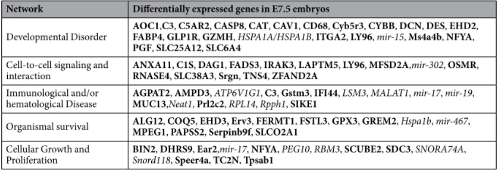
®
Pathway Analysis (IPA®
) (http://www.ingenuity.com/) of the 147 DEG identified 4 significant networks (score > 22); developmental disorder, cell-to-cell signaling and interaction, immunological and/or hematological disease, organism survival and cell growth and proliferation (Table 2). It suggested that Shadoo deficiency alters cell proliferation and differentiation resulting in abnormal early development. Potential related master regulator genes were identified in further analyses of the DEG, but at reduced fc (note S1).Collectively, these transcriptomic analyses suggest that Sprn knockout induces alteration of EMT pathways, impacting cell fate determination and inducing embryonic lethality soon after the implantation.
Potential opposite transcriptomic alterations between Sprn and Prnp knockout E7.5 embryos.
Some pathways identified above are reminiscent of those described for PrP modulation of EMT18,19. PrP regulates
embryonic stem cell pluripotency and differentiation, and Shadoo and PrP share biological properties9 although
recent experiments suggest that it could depend of the model system used20,21. As we previously performed
tran-scriptomic analysis of E7.5 Prnp0/0 embryos11, we searched for potential shared DEG between Prnp0/0 and Sprn0/0
embryos. Two master regulators of pluripotency were differentially expressed in E7.5 Prnp0/0 embryos, Oct4 and
TGFβr2. Oct4 was overexpressed and TGFβr2 downregulated in Prnp0/0 embryos11, i.e. oppositely to what is
observed in Sprn0/0 (note S1). Furthermore, out of the 147 DEG observed in Sprn0/0 embryos, 12 were also
differ-entially expressed in Prnp0/0 embryos11 but in an opposite sense, those indicated in Table 2 alongside Hspb7 and
Igsf11 (Table S1). Importantly, Prnp was not differentially expressed in Sprn0/0 embryo, excluding this observation
be an indirect consequence of Prnp overexpression.
Altogether, these observations suggest that Shadoo and PrP exert antagonist functions in early embryonic development. It fits in with the above-mentioned observation that Sprn0/0 and Prnp-overexpression14 both induces
smaller average litter sizes. However, these opposite differential gene expressions involved a limited number of genes and small variation levels, and we cannot formally exclude that this observation made across different experiments, might be coincidental.
comparative analyses of Sprn
0/0pups’ growth curves and of Sprn
0/0placentas.
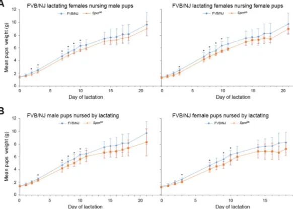
To assess whetherpups body weight is affected in Sprn0/0 mice, as previously reported12, we compared the growth curves of WT and
Sprn0/0 pups fed by WT lactating females. The body weight gain was lower for Sprn0/0 versus WT pups (statistically
significance between L3-L10, Fig. 3A). At variance with the previous data12, there was no difference according to
the pup sex. Abnormal growth could have various origins, one being placental dysfunction as in Prnp0/0 mice14.
Placental dysfunction would explain that at birth, the weight of the Sprn0/0 pups were significantly lighter (1.34
+/− 0.14 g for Sprn0/0 pups (n = 45) versus 1.4 +/− 0.16 g for WT pups (n = 97), t-test p < 0.05), suggesting
intra-uterine growth retardation. We thus analyzed whether Sprn knockout affects placental efficiency. Fetuses and placental tissues from E14.5 WT and Sprn0/0 mice were weighted. No significant difference was observed
between the two genotypes (Table 3). Histological examination of E14.5 and E17.5 placentas did not reveal struc-tural or vascularization modification (data not shown). The relative proportions of the labyrinth layer, spongio-trophoblast and decidua zones were unaffected.
We next performed comparative transcriptomic analysis on E14.5 WT and Sprn0/0 placentas. Following raw
data normalization, DEG with a p-value < 0.05 and a fc < −2 or > 2 were selected. 133 genes were identified, of which 17 were downregulated (Table S2), including 12 small nuclear RNAs. Seven DEG were common between placentas and embryos, all of them behaved similarly between the two tissues; 4 small nuclear RNAs alongside
Glp1r, LY96 and SLCO4C1 genes. This small number of shared DEG was expected due to transcriptomic
diver-gences between organs besides early stages of development22. Validation of the transcriptional regulation of five
genes, TBPA, Gli3, GC, PSG23, SerpinA1 (α1—antitrypsin), was tested by RT-qPCR. All results were concordant (Table S3). Furthermore, immunoblot analyses were performed to examine the expression levels of two proteins, SerpinA1 and Fibrinogen. The results confirmed the tendency observed at the transcriptomic levels, with on average a higher expression level of the two proteins in the knockout animals, which did not reach statistical significance (Fig. S2).
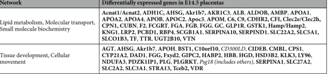
IPA
®
of the DEG identified two major associated networks (score > 35); lipid metabolism, molecular trans-port, small molecule biochemistry and tissue development, cellular movement (Table 4). Global gene activationNetwork Differentially expressed genes in E7.5 embryos
Developmental Disorder AOC1,C3, C5AR2, CASP8, CAT, CAV1, CD68, Cyb5r3, CYBB, DCN, DES, EHD2, FABP4, GLP1R, GZMH, HSPA1A/HSPA1B, ITGA2, LY96, mir-15, Ms4a4b, NFYA, PGF, SLC25A12, SLC6A4
Cell-to-cell signaling and
interaction ANXA11, C1S, DAG1, FADS3, IRAK3, LAPTM5, LY96, MFSD2A,mir-302, OSMR, RNASE4, SLC38A3, Srgn, TNS4, ZFAND2A
Immunological and/or
hematological Disease AGPAT2, AMPD3, ATP6V1G1, C3, Gstm3, IFI44, LSM3, MALAT1, mir-17, mir-19, MUC13,Neat1, Prl2c2, RPL14, Rpph1, SIKE1
Organismal survival ALG12, COQ5, EHD3, Erv3, FERMT1, FSTL3, GPX3, GREM2, Hspa1b, mir-467, MPEG1, PAPSS2, Serpinb9f, SLCO2A1 Cellular Growth and
Proliferation BIN2, DHRS9, Ear2,mir-17, NFYA, PEG10, RBM3, SCUBE2, SDC3, SNORA74A, Snord118, Speer4a, TC2N, Tpsab1
Table 2. Identified networks with differentially expressed genes in E7.5 embryos. Bold-faced genes: positively
differentially expressed genes in Sprn0/0 embryos. Underlined genes: negatively differentially expressed genes in
Sprn0/0 embryos. Yellow colored genes: genes also found differentially expressed in Prnp0/0 E7.5 embryos11. Of
www.nature.com/scientificreports
www.nature.com/scientificreports/
within these networks led to the overexpression of various protein families, including apo-lipoproteins and fibrin-ogens. Both gene families exert protective effects against stresses, supporting placentation and embryonic devel-opment23,24. Potential related master regulator genes were identified in further analyses of DEG, but at reduced
fc (note S2).
Altogether, these data suggest that at E14.5, Sprn0/0 placentas underwent an adaptive biological process,
involving LXR/RXR and FXR/RXR activation highlighted by the apo-lipoproteins and serpinA1 upregulations, to compensate earlier developmental dysfunctions.
Sprn-invalidation affects mouse lactation.
Since Sprn appears to be involved in embryonic EMT and in cellular proliferation and tissue development and since PrPC is involved in the self-renewal of mammary adultstem cells25, we assessed whether Sprn knockout affects lactation. We compared the growth curves of WT pups
fed by WT or Sprn0/0 lactating females. Their weight gain was significantly lower when fed by Sprn0/0 females
between L2 and L10 (Fig. 3B). No significant difference was observed between WT and Sprn0/0 milk protein
con-centrations at L7.5 (Table S4). Consistently, SDS-PAGE and western analyses did not reveal modification of the milk protein profiles (Fig. S3). Iron, zinc and copper concentrations were also similar between Sprn0/0 and WT
milks (data not shown). These data thus suggest that Sprn invalidation affects mouse lactation.
Figure 3. Analysis of pup’s growth curves. (A) Analysis of potential growth differences according to the
genotype and sex of the pups. Male or females pups from FVB/NJ (blue) or Sprn0/0 (orange) genotype were
fed by FVB/NJ females. Statistically different mean weights between pups according to their genotypes (t test p < 0.05) are indicated by *. At least 6 pups of each genotypes for both sexes were weighted per stage (see Material and Method section) (B) Analysis of potential lactating differences according to the genotype of the females. FVB/NJ male or female pups were fed by either FVB/NJ (blue) or Sprn0/0 (orange) females. Statistically
different mean weights between pups fed by the two types of lactating female (t test p < 0.05) are indicated by *. At least 6 pups of each genotypes for both sexes were weighted per stage (see Material and Methods).
Mouse genotype FVB/NJ Sprn0/0
Placenta Weight E14,5 (average +/− SEM) 0.064 ± 0.008 0.065 ± 0.012
Fetal Weight E14,5 (average +/− SEM) 0.236 ± 0.029 0.230 ± 0.034
Placental efficiency 3.754 ± 0.526 3.585 ± 0.580
Number of analyzed Pups 66 63
Number of analyzed Litters 7 11
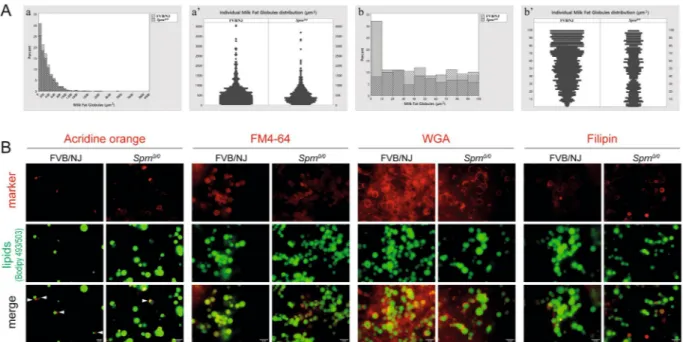
Sprn-invalidation affects milk fat globules volumes and morphology.
Analysis of milk fat globules (MFGs) revealed a significant reduction of the medians volume of Sprn0/0 MFGs (n = 3166) compared to that ofFVB/NJ MGFs (n = 5643) (Fig. 4A, kruskal-Wallis chi-squared = 22.114, p-value = 2.57 10−06). Analysis of the
size distribution of the MFGs showed that the majority of MFGs were smaller in Sprn0/0 milk with a significant
increase (up to 30% of total MFGs) of very small MFGs (size between 0 and 100 µm3, Fig. 4Aa). Intriguingly, the
detailed analysis of this size range (Fig. 4A,b,b’) showed that more than 30% of the very small MFGs in Sprn0/0
milk were <10 µm3 (Fig. 4Ab). Moreover, while size distribution of MFGs from FVB/NJ mice between 0 and 100
µm3 was in favor of the largest ones, those from Sprn0/0 mice were evenly distributed (Fig. 4Ab’). To what extend
this difference in MGF size distribution affects milk digestibility and pups’ growth remain unknown. Nevertheless, these observations suggest that the growth of intracellular lipid droplets (CLDs), potentially linked to their apical transport and secretory mechanisms, could be altered in the absence of Shadoo. We thus investigated whether Shadoo absence affects MFGs morphological properties. MFGs were co-labeled with Bodipy 493/503 for neutral lipids and either acridine orange for cytoplasmic crescents, FM4-64 for membranes, wheat germ agglutinin for glycoproteins and filipin for free cholesterol, before analysis by immunofluorescence microscopy (Fig. 4B). While MFGs from Sprn0/0 and FVB/NJ mice exhibited similar proportions of cytoplasmic crescents, those from Sprn0/0
mice were rather labeled at their periphery, suggesting a more acidic environment (Fig. 4B, AO). MFGs membra-nous morphology and surface glycoproteins distribution was similar in both genotypes (Fig. 4B, FM4-64; WGA). However, MFGs from Sprn0/0 mice appeared enriched in free cholesterol, a hallmark of membrane microdomains
called rafts (Fig. 4B, filipin). Collectively, these results point to subtle modifications of MFGs membranes from
Sprn0/0 mice, which may impact their size distribution26,27.
Sprn invalidation affects mouse mammary epithelial tissue development.
As Sprn0/0 MGFsmorphological properties were altered, we investigated whether differences could be observed at the MG level. Confocal imaging on tissue sections were performed at three MG developmental stages: mid-gestation (G10) and full lactation (L10) in the presence or absence of pups. Key features such as lipid droplets morphology, apical polarity and organization of the myoepithelial cells were analyzed (Fig. 5). At these stages, the lipid droplets, while correctly associated with the raft specific marker GM1 ganglioside, were generally smaller in Sprn0/0 mammary
epithelial cells with a more disperse distribution. The myoepithelial cells morphology and general organization around forming or active alveoli was markedly disturbed in Sprn0/0 mice, as assessed by glycoproteins (WGA) and
actin (PHA) staining. These data thus suggest that the lack of shadoo disturbs the establishment of the apico-basal polarity of mammary epithelial cells and alveoli formation.
Sprn-invalidation affects mouse mammary proliferation.
We next investigated the potential inci-dence of Sprn0/0 invalidation on mammary gland development by whole mount analyses. The selected picturesshowed in Fig. 6 are representative of the mammary morphology of at least 3 animals of both genotypes. In virgin, G7.5 and lactating MGs, some mammary primary canals were enlarged in Sprn0/0 tissues. In gestating and
lactat-ing tissues, Sprn0/0 MGs had less and only primary side branching compared to their wild-type counterparts and
were overall less dense. This suggests that Sprn-knockout is associated with developmental defects.
To consolidate these observations, we explored whether Shadoo invalidation impacts proliferation and/or apoptosis of mammary epithelial cells. MG sections of each genotype were labeled for Ki67, active caspase3 at mid-gestation and full lactation (Fig. S4). At mid-gestation both proliferating and apoptotic cells were detected in developing alveoli from FVB/NJ MG. Double labeling with TUNEL indicated that aCasp3-positive apoptotic cells differed from Ki67-positive proliferating cells. In Sprn0/0 MGs, there were fewer proliferating Ki67-positive cells
while apoptotic cells were found in similar or higher proportions than in WT MG. While apoptotic cells mostly localized near the forming lumen of alveoli in FVB/NJ, they appeared more dispersed in Sprn0/0 MG. During
lac-tation, few proliferating and apoptotic cells were observed at the periphery of the alveoli in the mammary tissue of both FVB/NJ and Sprn0/0 mice. These results suggest that the absence of Shadoo generates an imbalance in
favor of apoptosis in the growing mammary epithelial tissue, associated with a defect/delay in lumen formation.
Sprn-invalidation affects mouse mammary transcriptomic.
Comparative transcriptomic analysis was investigated on G7.5 WT and Sprn0/0 MGs. Fifty-three DEG were identified with a p-value < 0.05 and fc < −2or >2, 21 downregulated and 32 upregulated (Table S5), including 8 downregulated small nuclear RNAs. Five differentially expressed genes were common between the placentas and the MGs (SLC27A2, Snord13, Snord45b,
Network Differentially expressed genes in E14.5 placentas
Lipid metabolism, Molecular transport, Small molecule biochemistry
Acnat1/Acnat2, ADH1C, AHSG, Akr1b7, AKR1C3, ALB, ALDOB, AMBP, APOA1, APOA2, APOA4, APOB, APOC2, Apoc3, APOM, C6, C9, CDHR2, CFI, Clec2e/Clec2h, CPN1, CUBN, F2, FCGRT, FGA, FGB, FGG, GC, GLP1R, GSTK1, Hamp/Hamp2, KNG1, LRP2, PCBD1, RBP4, SCGB1A1, SERPINA10, SERPIND1, SLC22A2, SLC5A1, SLCO1B3, TF, TTR, UGT2B10, VTN
Tissue development, Cellular movement
AGT, AHSG, Akr1b7, APOH, BST1, C10orf10, CD300LD, CIDEB, CMBL, CPS1, CYP21A2, DAD1, FGG, Fxyd2, GIPC2, HABP2, HBB, HGD, HSD3B2, KLK3, LY96, NDUFA3, PDZK1IP1, PLG, PLGRKT, Psg18 (includes others), SERPINA1, SLC27A2, SLC2A2, SLC3A1, STRA13, Tceb2, VDR
Table 4. Identified networks with differentially expressed genes in E14.5 placentas. Bold-faced genes: positively
differentially expressed genes in Sprn0/0 placentas. Italized genes: negatively differentially expressed genes in
www.nature.com/scientificreports
www.nature.com/scientificreports/
Rnu3a, Snora73a) and 9 between the embryos and the MGs (AF357399, Hist2H2BF, MRP536, Ms4a4b, Snora23, Snora74a, Snora81, Rnu3a and Snora73a), all of them behaving similarly between the different tissues, suggestive
of shared but not yet identified pathways.
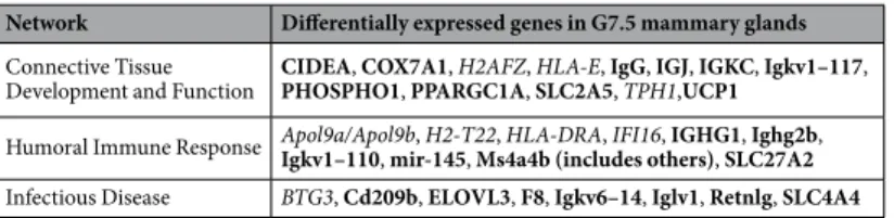
Sprn-knockout induces in the MG an immune response with the upregulation of 14 immunoglobulin genes,
of CD209b and the downregulation of 2 MHC genes and 1 interferon inducible gene. IPA identified 3 networks
Figure 4. Effects of Sprn invalidation on Milk fat globules volume and morphology. (A) (a,a’) Comparative
representations of the milk fat globule volumes distribution between FVB/NJ and Sprn0/0 milks. At least 3
individual milks were analyzed per genotype, as described in the Material and Methods section. (A) (b,b’) Comparative representations of 0 to 100 µm3 milk fat globule distribution between FVB/NJ and Sprn0/0 milks.
At least 3 individual milks were analyzed per genotype, as described in the Material and Methods section. (B) Immunofluorescence imaging of MFGs from FVB/NJ or Sprn0/0 mice at day 10 of lactation. MFGs were
co-stained for for neutral lipids (green) with Bodipy 493/503 and (red) for either cytoplasmic crescents (arrowheads) with acridine orange, membranes with FM4–64, glycoproteins with wheat germ agglutinin (WGA) or free cholesterol with filipin. Bar, 10 µm.
Figure 5. Mammary gland morphology and mammary epithelial cell architecture. Confocal microscopy
imaging of mammary gland sections from FVB/NJ and Sprn0/0 mice at mid-gestation (G10) and full lactation
(L10, in the presence or absence (±pups) co-stained for neutral lipids (Bodipy 493/503 (bod), green), rafts microdomains (cholera toxin B subunit (GM1), red), glycoproteins (wheat germ agglutinin (WGA), green), and/or actin (Phalloïdin (Pha), red). Nuclei were counterstained with nuclear marker 4′,6-diamidino-2-phenylindole (DAPI, blue). The Asterisks indicate the lumen. Bar, 10 µm.
(score > 17); Connective Tissue Development and Function, Humoral Immune Response, Infectious Disease (Table 5). It suggests that in Sprn0/0, gestating mammary epithelial cells undergo, associated with an immune
reaction, a growth inhibition correlating with the whole mount observations. Search for deregulation of known
Figure 6. Mammary gland development analysis. Representative whole mount analysis of mammary gland
structure from WT (FVB/NJ; a,c,e,g,i) or mutant (Sprn0/0, b,d,f,h,j) mice. It highlights defects in duct tree
formation in 6-week-old virgin, in duct side-branching during early gestation (G7,5) and consequently in the density and organization of the mammary epithelium during lactation (L2: day 2 of lactation, 10: day 10 of lactation; +pups: in the presence of pups, −pups: in the absence of pups). Bar = 0.5 mm. Enlarged views of each developmental stage show the differences in mammary epithelial tissue organization between WT and Sprn0/0
www.nature.com/scientificreports
www.nature.com/scientificreports/
regulators of mammary growth and branching morphogenesis revealed miR-145 overexpression in Sprn0/0 MGs.
MiR-145 acts as an antitumor miRNA that targets Wnt5B whose overexpression is associated with mammary tumors28. Wnt5B was indeed downregulated in Sprn0/0 MGs but at a reduced fold-change (fc −1.4, p < 0.01).
CIDEA, another potential regulator contributing to the control of mammary epithelial growth, is also upregulated (Table 6, Table S5). Transcriptional alteration of these genes is likely to contribute to epithelial growth inhibition. Furthermore, upregulation of both CIDEA and PPARGC1A (Table S5) may contribute to the observed difference in milk fat globules’ size distribution. GSEA analysis confirmed an abnormal proliferation of Sprn0/0 mammary
tissue, highlighting a downregulation of genes related to protein localization to kinetochore (ESc4.593, FDR 2.75 10−6) and to DNA replication (ESc3.4, FDR 4.01 10−8).
Overall, transcriptomic analysis of Sprn0/0 gestating MG revealed the potential deregulations of pathways
cor-roborating phenotypic and histologic analyses of this tissue. These results highlight a yet undescribed role of Shadoo in lactation.
Discussion
The biological role of the members of the prion family remains mostly unknown, despite intensive researches focusing on PrP due to its major implication in TSE and potential involvement in other more prevalent neurode-generative diseases such as Alzheimer’s disease. Less attention focused on the related Shadoo protein, probably resulting from its recent discovery and potentially limited implication in TSE29,30. The previously reported
knock-out of Sprn did not induce overt phenotypes beside a moderate, transient and sex-related, growth deficiency that could originate from a role of Shadoo in the control of feeding behavior, in association with its expression in hypothalamic neurons12.
A role of PrP in mouse embryonic development, around the implantation stage and the transition from anaerobic to aerobic metabolism, was suggested as well as in the development of the nervous system and of extra-embryonic tissues. Shadoo shares with PrP some biological properties9, although other biological roles
are specific to this protein and/or even antagonist to PrP20,21. A biological redundancy between PrP and Shadoo
was evocated following the observation that the knockdown of Sprn induces early embryonic lethality in Prnp0/0
mouse transgenic lines10,11. This hypothesis was weakened by the absence of phenotype of double Prnp/Sprn
knockout mice12. The present report reinvestigates the role of Shadoo in FVB/NJ embryonic development
through the generation of knockout mice using a ZFN approach, allowing its assessment in a non-mixed genetic background. This study highlights that Sprn0/0 mice suffer from subtle, sometimes transient, but significant
alter-ations of developmental and differentiation processes that affect both embryonic and adult tissues (Table 6).
Sprn0/0 mice were viable and fertile, but analysis of the transmission rate in heterozygous intercrosses and of
the average litter size in knockout homozygous crossings revealed a non-Mendelian distribution of the genotypes and a reduced number of pups, respectively. Further investigations highlighted an abnormal lethality rate of
Sprn0/0 embryos at an early post implantation developmental stage, between E4.5 and E6.5. This suggested that
the absence of Shadoo is not detrimental for embryonic implantation but weakens the survival potential of the embryos early post-implantation.
Absence of Shadoo transiently also affects the growth rate of the pups in this genetic background but inde-pendently from their sexes. This slightly differs from previous observations12. The difference could originate from
the genetic backgrounds used in relation to the invalidation strategies. Although not formally exclusive with an abnormal feeding behavior, this transient phenotype could correlate with an abnormal biology of the pla-centas, as judged by our transcriptomic studies (Table 6). Invalidation of Sprn led to placental defects, recalling observations made following Prnp-knockout14. However, in Sprn0/0 mice, no histological placental defect were
evidenced and the few genes described as DEG in Prnp0/0 placentas were not transcriptionally affected in Sprn0/0.
Our transcriptomic analysis rather suggests that Sprn0/0 extra-embryonic tissues might undergo early
develop-mental defects, highlighted by transcriptomic alteration in E7.5 embryos of genes affecting the formation of mouse extra-embryonic tissues such as Smad1. These early potential defects seem at least partially corrected dur-ing development, as exemplified by the activation of pathways at G14.5 supportdur-ing placentation and embryonic development. Studies of earlier developmental stages to assess placenta development should help validating this hypothesis.
We also evidenced an impact of the absence of Shadoo on MG physiology, objectivized by (i) growth curve analyses of FVB/NJ pups fed by Sprn0/0 females, (ii) transcriptomic alterations of G7.5 mammary tissue, (iii)
mor-phological development defects and alteration of milk fat globules synthesis that correlate with transcriptomic alterations, (iv) mammary cell/membrane alterations. Implication of the Prion protein family in the physiology of the MG per se has received little attention. Implication of PrP in mammosphere formation and repopulating activity was documented25, but its relationship with lactation performances remains controversial. Of note is the
Network Differentially expressed genes in G7.5 mammary glands
Connective Tissue
Development and Function CIDEA, COX7A1, H2AFZ, HLA-E, IgG, IGJ, IGKC, Igkv1–117, PHOSPHO1, PPARGC1A, SLC2A5, TPH1,UCP1
Humoral Immune Response Apol9a/Apol9b, H2-T22, HLA-DRA, IFI16, IGHG1, Ighg2b, Igkv1–110, mir-145, Ms4a4b (includes others), SLC27A2
Infectious Disease BTG3, Cd209b, ELOVL3, F8, Igkv6–14, Iglv1, Retnlg, SLC4A4
Table 5. Identified networks with differentially expressed genes in G7.5 mammary glands. Bold-faced genes:
positively differentially expressed genes in Sprn0/0 mammary glands. Italized genes: negatively differentially expressed genes in Sprn0/0 mammary glands.
absence of any mammary phenotype described so far for Prnp0/0 mammals. In the present study, invalidation of
Shadoo appears to induce various gestational/lactation mammary tissue and cellular defects that results in tran-sient lactation deficit.
This phenotype exemplified Shadoo implication in the control of cellular proliferation and differentiation, also revealed at early post-implantation embryonic stages. Such an involvement in cell proliferation and dif-ferentiation was highlighted for PrP31. Implication of these two related proteins in similar biological functions
alongside their expression in developing mammalian fetuses could substantiate the hypothesis of their biolog-ical redundancy. Surprisingly, our data rather suggest that Sprn and Prnp exert opposite effects on expression of master regulators of pluripotency and/or embryonic development, at least at E7.5. Thus, one could expect their co-invalidation to be neutral, as documented12. However, comparison of the data described here and for
Sprn-knockdown/Prnp0/0 embryos10,11 highlights a developmental delay in the embryonic lethality between
Sprn0/0 and Sprn-knockdown/Prnp0/0 embryos. Lethality of Sprn0/0 embryos occurs early after implantation while
that of Sprn-knockdown/Prnp0/0 embryos was evidenced only at E10.510, with E7.5 decidua carrying abnormal
embryos11. It could suggest that Sprn-knockdown induces a fragility of the embryos at implantation that becomes
detrimental to the further development of the Prnp0/0 ones, between E8.5 and E9, when upturn of PrP RNA
expres-sion should occur32. Absence of such a scenario in Prnp0/0/Sprn0/0 mice might result from genetic adaptation13.
In conclusion, this study provides evidence for a yet controversial role of the Prion-related protein Shadoo during mouse embryogenesis and more generally in the control of cellular proliferation and differentiation of various tissues, including adult ones such as the mammary epithelium. It also highlights a potential embryonic antagonist action of PrP and Shadoo, revisiting alongside recent papers the current view of a potential biological redundancy between these two proteins.
Methods
ethics statement.
All animal experiments were carried out in strict accordance with the recommendations in the guidelines of the Code for Methods and Welfare Considerations in Behavioral Research with Animals (Directive 2016/63/UE). All efforts were made to minimize suffering. Experiments were approved by the INRAE local animal experiment ethics committee of Jouy-en-Josas (Comethea, Permit Number 02532.01). All animal manipulations were done according to the recommendations of the French Haut Conseil aux Biotechnologies, HCB (Permit Number N°6461 and DUO N°5468).Generation and establishment of Sprn-knockout mice.
A preparation of mRNA encoding a ZFN targeting the start of the Sprn ORF was purchased from Sigma-Aldrich Corp (CompoZrTM ZFN design). The ZFN target site was 5′-ctgtgacagctgttccgccangggcggccgcggaggcg-3′. ZFN mRNA was injected at 2 ng/ μl into 1 cell-fertilized FVB/NJ mouse eggs. Surviving injected eggs were transferred into pseudo-pregnant recipient mice. Tail-DNA analysis of the 29 resulting live pups was performed by PCR using oligonucleotides 5′-gccggccttacgcgtactcttga-3′ and 5′-gcctattcccaagccctgatac-3′, using the Promega GoTaq G2 Flexi DNA poly-merase kit. PCR conditions were made of 40 amplifications cycles; 94 °C-30 s, 60 °C-30 s and 72 °C-30 s. The amplified 400 bp long DNA genomic region surrounding the ZFN target site was sequenced to search for putative mutations.Transgenic founder mice were crossed with FVB/NJ mice to establish transgenic lines. Intercrosses between heterozygous mice were used to derive transgenic knockout lines.
Reproductive performance analyses.
All examined females were on their first pregnancy or lactation. Intercrosses between heterozygous mice and between homozygous mice were performed and the number and genotype of pups recorded. To assess embryonic lethality, pregnant FVB/NJ or Sprn0/0 females, followingmat-tings with males of the same genotype, were euthanized at different gestation stages. Implantation points were recorded. Development and vitality of the embryos were observed under binocular microscope.
Comparative analysis of growth curves of FVB/NJ and Sprn0/0 pups was performed, using FVB/NJ lactating
females as foster mothers. Lactation performance was indirectly assessed by analysis of growth curves of FVB/ NJ pups fed either by FVB/NJ or Sprn0/0 females. Six pups were placed under each lactating females and 3 females
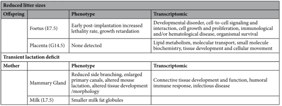
Reduced litter sizes
Offspring Phenotype Transcriptomic
Foetus (E7.5) Early post-implantation increased lethality rate, growth retardation Developmental disorder, cell-to-cell signaling and interaction, cell growth and proliferation, immunological and/or hematological disease, organismal survival Placenta (G14.5) None detected Lipid metabolism, molecular transport, small molecule biochemistry, tissue development and cellular movement
Transient lactation deficit
Mother Phenotype Transcriptomic
Mammary Gland
Reduced side branching, enlarged primary canals, altered mouse lactation, altered tissue development /morphology
Connective tissue development and function, humoral immune response, infectious disease
Milk (L7.5) Smaller milk fat globules
www.nature.com/scientificreports
www.nature.com/scientificreports/
in their first lactation of each genotype analyzed. Milk samples were collected at L7.5 on other lactating females following pups removal for 3 hours and 300 µl −10 UI intra-peritoneal injection of oxytocin.
RNA purifications and RT-(q)PCR.
Total RNA was isolated from (i) E7.5 mouse embryos, (ii) E14.5 pla-centas and (iii) G7.5 MGs (first pregnancy). Six gestating females of each genotype were used at each analyzed stages. Individual RNA extractions were performed using the RNeasy Lipid Tissue Mini kit (Qiagen cat # 75842). RNA concentration was calculated by electro-spectrophotometry and the RNA integrity checked with the Agilent Bioanalyser (Waldbroom, Germany). All extracted RNA samples had a RIN value > 8.5.Reverse transcription was performed on 5μg of total RNA, using Invitrogen SuperScriptTMIII reverse
tran-scriptase kit (18080051) or RTVilo (11754050) and random primers, according to the manufacturer’s instructions. Amplification of the mouse Sprn cDNA ORF was performed using oligonucleotides 5′-taggcttgtgaccaattcttgcc-3′ and 5′-ggagtttagcctggtctaaggc-3′, that are located on the two separate Sprn exons to avoid amplification of poten-tial contaminating genomic DNA. PCR conditions were made of 40 amplifications cycles; 94 °C-30 s, 60 °C-60 s and 72 °C-30 s, using GC-rich adapted PCR amplification kit (KAPA2G Robust PCR kit, SigmaAldrich). Two WT and two Sprn0/0 mouse brains were analyzed.
For RT-qPCR, quantification was performed on triplicates, using 3 different RNA samples for each gen-otype, using the ABsolute Blue QPCR SYBR Green ROX Mix (Thermo Scientific) and standard PCR condi-tions. Primers were designed on separate exons, using Primer 3 software (http://primer3.ut.er) to produce 100-bp amplicons, with a Tm of 60 °C. The TBP and SDHA genes were used for normalization, using prim-ers 5′-ttcgtgcaagaaatgctgaa-3′ and 5′-tcctgtgccacaccatttttc-3′ for TBP and 5′-aaggacctggcatcaagaga-3′ and 5′-tgatctttctcagggccaca-3′ for SDHA. Tested genes were α1-antitrypsin, using primers 5′-ctccggaatcacagaggaaa-3′ and 5′-gcttctgttcctgtctcatcg-3′, GC using primers 5′-gcaaagacctctgtggtcag-3′ and 5′-agggttggctccagaacttt-3′, Gli3 using primers 5′-tcccacgagaacagatgtca-3′ and 5′-tgaggctgcatatgtattgc-3′, TBPA using primers 5′-tgaagagctgaaccactgga-3′ and 5′-ccaggcataggatgactagga-3′ and PSG23 using primers 5′tcacctggaaagacactgga-3′ and 5′-ggcagagtcaagagggtcac-3′.
Western blot analysis.
Following mouse cervical dislocation, FVB/NJ and Sprn0/0 mouse brains werecrushed in 0.32 M Sucrose solution containing cocktail of protease inhibitors (Roche) and enriched in subcellular membranes by ultra-centrifugation (150 000 × g) as developed in Westaway et al. (2011). Proteins were extracted from membrane pellets by N-PER Neuronal Protein reagent (Thermo). Amounts of 50 μg sample proteins were separated on a home-cast 14% polyacrylamide gel SDS-PAGE and were transferred on PVDF membrane by the Trans-blot Turbo (Bio-Rad). Non-specific reactivity was blocked by incubation for 1 h with a 5% skimmed milk in 0.1% TBS-Tween 20 [TBS-T]. Blot was incubated with a serum from a rabbit immunized against mouse Shadoo, obtained from Covalab (https://www.covalab.com/), at 1/750e dilution in 0.1% TBS-T for two hours at room temperature or with other antibodies directed against Shadoo that are listed in the text at recommended dilutions. Horseradish peroxidase-conjugated anti-IgG from various origins according to the primary antibody were used as secondary antibody with 1 h incubation. Excess first and second antibodies were removed by wash-ing 3 × 10 min in TBS-T buffer. Detection was accomplished with a chemiluminescence system (ECL Prime, GE Healthcare) and exposure in ChemiDoc Touch device (Bio-Rad).
Fibrinogen and α1-antitrypsin protein ratio between wild type and Sprn0/0 mouse were estimated in placenta
samples with Image Lab software 5.2 (BioRad). They were crushed with ultra-turrax in N-PER Neuronal Protein reagent containing cocktail protease inhibitors (Roche). The experimental protocol was followed as described above according recommended factor dilutions, using Abcam ab189490 and Abcam ab231093 antibodies, respectively.
Milk collect and analysis.
Milk was collected from L7.5 Sprn0/0 and FVB/NJ females on their first lactation.Milk protein concentration was estimated using Bradford’s procedure and fractionation of its proteins was per-formed on home-cast SDS-PAGE 12.5% polyacrylamide followed by coomassie blue staining. A western blot was performed as described above with a special antibody designed against all mouse milk proteins (009 RAM/MSP Nordic Immunology).
Milk fat globules (MFGs) labeling, image acquisition and analysis.
Experiments involved analyses of at least 3 mice per genotype and condition. Whole mouse milk (L10) was diluted 1:4 in sterile water and fixed with PBS containing 4% paraformaldehyde (PFA) for 10 minutes at 4 °C, as described26. Milk fat globules (MFGs)were then labeled with acridine orange (AO, 0.1%), Alexa594-conjugated wheat germ agglutinin (WGA, 1 mg/ ml), or filipin (1 mg/ml) diluted in PBS for 30 min at 4 °C. Neutral lipids were concomitantly counterstained with BODIPY 493/503 (3 µg/ml) or Nile Red (1 µg/ml). 50 µl of each stained sample was mixed with 50 µl of 1% low melt agarose solution, placed on a glass slide and topped with a coverslip. Fluorescence microscopy was per-formed with an Axio observer Z1 microscope (Zeiss) equipped with standard filters for FITC, Rhodamine and DAPI emissions, using a 40x objective and a Greyscale CCD camera CoolSnap HQ2 camera (Photometrics) cou-pled to the Axiovision imaging system software. About thirty pictures of each condition were randomly acquired for each condition and both the number and the perimeter (µm) of the MFGs were estimated using ImageJ (1.47q software 2012, http://rsb.info.nih.gov/ij/). The volume of the MFGs (µm3) were calculated and a Kruskall-Wallis
rank test was performed using the version 3.5.2 of the R Software (R core Team 2018).
placenta histological analysis.
Immediately after dams’ sacrifice by cervical dislocation at G14.5 or G17.5, the uterus were removed and the placentas collected. Intact placentas (6 per genotype and stage from 3 different dams) were fixed in formalin (48 hours). After fixation, each placenta was bisected adjacent to theumbilical vessels. Both halves were embedded in paraffin. Section (3–5 µM) from each placenta were processed for Hematoxylin-Eosin coloration and examined by a qualified veterinary pathologist.
MG whole mount and immunohistological analyses.
Experiments involved analyses of at least 3 mice per genotype and condition. Inguinal MGs (4th, left) were excised, spread on glass slides and fixed in Carnoy’sfixative (100% EtOH, chloroform, glacial acetic acid; 6∶3∶1) for 4 h at room temperature, washed in 70% EtOH for 15 min, rehydrated in water, and stained in carmine alum (2% carmine and 5% aluminum potassium sulfate in water) overnight. Tissues were then gradually dehydrated through serial ethanol baths and cleared in xylene overnight. MGs were kept in Permount Mounting Media (Fisher Scientific) until images were captured.
For immunohistological analyses, mouse MGs were collected at different physiological stages and processed as described26. Tissue sections were labeled using Alexa488-conjugated WGA, rhodamine-conjugated phalloidin
(Thermo Fisher), or filipin (Sigma-Aldrich) diluted in PBS for 30 min at 4 °C. Neutral lipids and nuclei were coun-terstained with BODIPY 493/503 or Nile Red and DAPI (4′-6-diamidino-2-phenylindole), respectively. Slides were mounted with Vectashield (Vector Laboratories LTD, United Kingdom) and stored at 4 °C until observation. For some experiments, Tissue sections or MFGs were also labeled for actin with rhodamine-conjugated phalloi-din (Molecular Probes), GM1 ganglioside with Alexa 594–conjugated CTxB (Invitrogen), or free CH using filipin (Sigma-Aldrich). Each experiment was performed at least twice and included negative control without primary antibody. At least 20 images were randomly acquired per condition tested and further analyzed. All images were analyzed using ImageJ 1.47q software (http://rsb.info.nih.gov/ij/).
transcriptomic analyses.
Transcriptomic analyses were performed using Affymetrix MouseGene 2,OSt microarray on the Institute Cochin Genom’ic platform. Each analyzed embryonic RNA sample derived from the equal mixing of 6 individual total RNA preparations from E7.5 embryos. Different embryonic samples were used for each mixture. Similarly, each analyzed placenta RNA sample derived from the equal mixing of 6 indi-vidual total RNA preparations from E14.5 placentas. Different indiindi-vidual samples were used for each mixture. Each MG RNA sample derived from the equal mixing of 3 individual total RNA preparations from G7.5 MGs. Different individual samples were used for each mixture. Three biological replicates per tissue and genotype were performed. Differentially expressed genes between FVB/NJ and FVB/NJ Sprn-knockout tissues were identified following normalization of the raw data, with fold chance ratios and p values determined as described in the results section. Data were analyzed by Gene set enrichment analysis (GSEA, http://software.broadinstitute.org/ gsea/index.jsp). Differentially expressed genes were clustered and classified in pathways and networks by using Ingenuity (http://www.ingenuity.com/).Received: 17 June 2019; Accepted: 3 April 2020; Published: xx xx xxxx
References
1. Prusiner, S. B. Early evidence that a protease-resistant protein is an active component of the infectious prion. Cell. 116(2 Suppl), S109 (2004).
2. Büeler, H. et al. Normal development and behaviour of mice lacking the neuronal cell-surface PrP protein. Nature. 356(6370), 577–582 (1992).
3. Richt, J. A. et al. Production of cattle lacking prion protein. Nat. Biotechnol. 25(1), 132–138 (2007). 4. Yu, G. et al. Generation of goats lacking prion protein. Mol. Reprod. Dev. 76(1), 3 (2009).
5. Benestad, S. L., Austbø, L., Tranulis, M. A., Espenes, A. & Olsaker, I. Healthy goats naturally devoid of prion protein. Vet. Res. 43, 87 (2012).
6. Leighton, P. L. A., Kanyo, R., Neil, G. J., Pollock, N. M. & Allison, W. T. Prion gene paralogs are dispensable for early zebrafish development but have non-additive roles in seizure susceptibility. J Biol Chem. Jun 14. pii: jbc.RA117.001171. (2018).
7. Westaway, D., Daude, N., Wohlgemuth, S. & Harrison, P. The PrP-like proteins Shadoo and Doppel. Top. Curr. Chem. 305, 225–256 (2011).
8. Paisley, D. et al. Male infertility and DNA damage in Doppel knockout and prion protein/Doppel double-knockout mice. Am. J.
Pathol. 4(6), 2279–2288 (2004).
9. Daude, N. & Westaway, D. Biological properties of the PrP-like Shadoo protein. Front. Biosci. 16, 1505–1516 (2011).
10. Young, R. et al. The prion or the related Shadoo protein is required for early mouse embryogenesis. FEBS Lett. 583(19), 3296–3300 (2009).
11. Passet, B. et al. Prion protein and Shadoo are involved in overlapping embryonic pathways and trophoblastic development. PLoS
One. 7(7), e41959 (2012).
12. Daude, N. et al. Knockout of the prion protein (PrP)-like Sprn gene does not produce embryonic lethality in combination with PrP(C)-deficiency. Proc. Natl Acad. Sci. USA 109(23), 9035–9040 (2012).
13. Rossi, A. et al. Genetic compensation induced by deleterious mutations but not gene knockdowns. Nature. 524(7564), 230–233 (2015).
14. Alfaidy, N. et al. Prion protein expression and functional importance in developmental angiogenesis: role in oxidative stress and copper homeostasis. Antioxid. Redox Signal. 18(4), 400–411 (2013).
15. Kishor, A., Fritz, S. E. & Hogg, J. R. Nonsense-mediated mRNA decay: The challenge of telling right from wrong in a complexe transcriptome. Wiley Interdiscip. Rev. RNA e1548, 1–22 (2019).
16. Wu, L., Zheng, J., Chen, P., Liu, Q. & Yuan, Y. Small nucleolar RNA ACA11 promotes proliferation, migration and invasion in hepatocellular carcinoma by targeting the PI3K/AKT signaling pathway. Biomed. Pharmacother. 90, 705–712 (2017).
17. Li, G. et al. Small nucleolar RNA 47 promotes tumorigenesis by regulating EMT markers in hepatocellular carcinoma. Minerva Med. 108, 396–404 (2017).
18. Mehrabian, M., Ehsani, S. & Schmitt-Ulms, G. An emerging role of the cellular prion protein as a modulator of a morphogenetic program underlying epithelial-to-mesenchymal transition. Front. Cell Dev. Biol. 18(2), 53 (2014).
19. Ghodrati, F. et al. The prion protein is embedded in a molecular environment that modulates transforming growth factor β and integrin signaling. Sci. Rep. 8(1), 8654 (2018).
20. Daude, N., Gapeshina, H., Dong, B., Winship, I. & Westaway, D. Neuroprotective properties of the PrP-like Shadoo glycoprotein assessed in the middle cerebral artery occlusion model of ischemia. Prion. 9(5), 376–393 (2015).
www.nature.com/scientificreports
www.nature.com/scientificreports/
21. Nyeste, A. et al. Expression of the Prion Protein Family Member Shadoo Causes Drug Hypersensitivity That Is Diminished by the Coexpression of the Wild Type Prion Protein. J. Biol. Chem. 291(9), 4473–4486 (2016).
22. Cardoso-Moreira, M. et al. Gene expression across mammalian organ development. Nature 571(7766), 505–509 (2019).
23. Iwaki, T. et al. Fibrinogen stabilizes placental-maternal attachment during embryonic development in the mouse. Am. J. Pathol. 160(3), 1021–1034 (2002).
24. Charlton, F. et al. The protective effect of apolipoprotein in models of trophoblast invasion and preeclampsia. Am. J. Physiol. Regul.
Integr. Comp. Physiol. 312(1), R40–R48 (2017).
25. Liao, M. J. et al. Enrichment of a population of mammary gland cells that form mammospheres and have in vivo repopulating activity. Cancer Res. 67(17), 8131–8138 (2007).
26. Honvo-Houéto, E. et al. The endoplasmic reticulum and casein-containing vesicles contribute to milk fat globule membrane. Mol.
Biol. Cell. 27(19), 2946–64 (2016).
27. Mesilati-Stahy, R., Mida, K. & Argov-Argaman, N. Size-dependent lipid content of bovine milk fat globule and membrane phospholipids. J. Agric. Food Chem. 59(13), 7427–35 (2011).
28. Yamada, Y. et al. Impact of novel oncogenic pathways regulated by antitumor miR-451a in renal cell carcinoma. Cancer Sci. 109(4), 1239–1253 (2018).
29. Watts, J. C. et al. Protease-resistant prions selectively decrease Shadoo protein. PLoS Pathog. 7(11), e1002382 (2011).
30. Wang, H. et al. Overexpression of Shadoo protein in transgenic mice does not impact the pathogenesis of scrapie. Neurosci. Lett. 496(1), 1–4 (2011).
31. Miranda, A., Pericuesta, E., Ramírez, M. Á. & Gutierrez-Adan, A. Prion protein expression regulates embryonic stem cell pluripotency and differentiation. PLoS One. 6(4), e18422 (2011).
32. Miele, G. et al. Embryonic activation and developmental expression of the murine prion protein gene. Gene Expr. 11(1), 1–12 (2003).
Acknowledgements
We are grateful to Drs. Nathalie Daude and David Westaway, University of Alberta, Edmonton, Canada, for the kind gift of the anti-Shadoo 06sh1 antibody and advices for Western analyses.
Author contributions
Conception of the Work: V.B., A.D., K.M.G., P.C., B.P. and J.L.V. Design of the work: B.P., S.M., J.C., O.A., A.D., K.M.G., J.L.V. Acquisition, analysis and interpretation of the data: B.P., J.C., S.M., S.T., A.V., S.F., C.B., A.L.G., L.H., Ma.B., O.A., D.V., M.V., S.M.R., P.C., M.M., N.D.C., D.L., Mo.B. Drafted the work: V.B., J.L.V., D.V., S.M.R., A.D., K.M.G.
competing interests
The authors declare no competing interests.
Additional information
Supplementary information is available for this paper at https://doi.org/10.1038/s41598-020-63805-y.
Correspondence and requests for materials should be addressed to J.-L.V. Reprints and permissions information is available at www.nature.com/reprints.
Publisher’s note Springer Nature remains neutral with regard to jurisdictional claims in published maps and
institutional affiliations.
Open Access This article is licensed under a Creative Commons Attribution 4.0 International
License, which permits use, sharing, adaptation, distribution and reproduction in any medium or format, as long as you give appropriate credit to the original author(s) and the source, provide a link to the Cre-ative Commons license, and indicate if changes were made. The images or other third party material in this article are included in the article’s Creative Commons license, unless indicated otherwise in a credit line to the material. If material is not included in the article’s Creative Commons license and your intended use is not per-mitted by statutory regulation or exceeds the perper-mitted use, you will need to obtain permission directly from the copyright holder. To view a copy of this license, visit http://creativecommons.org/licenses/by/4.0/.