HAL Id: tel-00692346
https://tel.archives-ouvertes.fr/tel-00692346
Submitted on 30 Apr 2012HAL is a multi-disciplinary open access archive for the deposit and dissemination of sci-entific research documents, whether they are pub-lished or not. The documents may come from teaching and research institutions in France or abroad, or from public or private research centers.
L’archive ouverte pluridisciplinaire HAL, est destinée au dépôt et à la diffusion de documents scientifiques de niveau recherche, publiés ou non, émanant des établissements d’enseignement et de recherche français ou étrangers, des laboratoires publics ou privés.
integrated with RF receiver coil for high resolution
MRI-controlled thermal therapy
Mihaela Rata
To cite this version:
Mihaela Rata. Endocavitary applicator of therapeutic ultrasound integrated with RF receiver coil for high resolution MRI-controlled thermal therapy. Human health and pathology. Université Claude Bernard - Lyon I, 2009. English. �NNT : 2009LYO10282�. �tel-00692346�
THESE DE L‘UNIVERSITE DE LYON
Délivrée par
L’UNIVERSITE CLAUDE BERNARD LYON 1 ECOLE DOCTORALE
EDISS
DIPLOME DE DOCTORAT (arrêté du 7 août 2006)
soutenue publiquement le 15 décembre 2009
par
Mlle. Mihaela RATA
TITRE :
Endocavitary applicator of therapeutic ultrasound integrated with
RF receiver coil for high resolution MRI-controlled thermal therapy
Directeur de thèse :
M. Rares Salomir
JURY:
M. Hervé Saint-Jalmes, Rapporteur M. Ian Rivens, Rapporteur M. François Cotton
M. Philippe Douek M. Mihai Todica M. Rares Salomir
UNIVERSITE CLAUDE BERNARD - LYON 1
Président de l’Université
Vice-président du Conseil Scientifique Vice-président du Conseil d’Administration
Vice-président du Conseil des Etudes et de la Vie Universitaire Secrétaire Général M. le Professeur L. Collet M. le Professeur J-F. Mornex M. le Professeur G. Annat M. le Professeur D. Simon M. G. Gay
COMPOSANTES SANTE
Faculté de Médecine Lyon Est – Claude Bernard Faculté de Médecine Lyon Sud – Charles Mérieux UFR d’Odontologie
Institut des Sciences Pharmaceutiques et Biologiques Institut des Sciences et Techniques de Réadaptation
Département de Formation et Centre de Recherche en Biologie Humaine
Directeur : M. le Professeur J. Etienne Directeur : M. le Professeur F-N. Gilly Directeur : M. le Professeur D. Bourgeois Directeur : M. le Professeur F. Locher Directeur : M. le Professeur Y. Matillon Directeur : M. le Professeur P. Farge
COMPOSANTES SCIENCES ET TECHNOLOGIE
Faculté des Sciences et Technologies
UFR Sciences et Techniques des Activités Physiques et Sportives Observatoire de Lyon
Institut des Sciences et des Techniques de l’Ingénieur de Lyon Institut Universitaire de Technologie A
Institut Universitaire de Technologie B Institut de Science Financière et d'Assurance Institut Universitaire de Formation des Maîtres
Directeur : M. Le Professeur F. Gieres Directeur : M. C. Collignon
Directeur : M. B. Guiderdoni Directeur : M. le Professeur J. Lieto Directeur : M. le Professeur C. Coulet Directeur : M. le Professeur R. Lamartine Directeur : M. le Professeur J-C. Augros Directeur : M R. Bernard
Remerciements
Heureusement, ces lignes me permettent de laisser une trace de ma gratitude à tous ceux qui, scientifiquement ou humainement, ont aidé au bon déroulement de cette thèse.
Premièrement, je tiens à remercier mon directeur de thèse, monsieur Rares Salomir pour son aide à découvrir des choses intéressantes et nouvelles, pour sa disponibilité et ses conseils bienvenus. Son énergie et son enthousiasme nous ont permis de surmonter les obstacles qui se sont présentés.
J’exprime ma gratitude à messieurs Hervé Saint-Jalmes et Ian Rivens d’avoir accepté la fonction de rapporteurs de cette thèse, ainsi qu’à messieurs François Cotton, Philippe Douek et Mihai Todica pour l’intérêt qu’ils ont montré à ce travail en acceptant d’être membres du jury. Je remercie également M. François Cotton du Service de Médecine Nucléaire et Radiologie du CHU Lyon-Sud pour ses conseils et la mise à disposition du temps machine sur l’imageur clinique RMN 1.5 T corps entier.
Je remercie monsieur Jean-Yves Chapelon, directeur de l’unité 556 de l’Inserm, pour m’avoir accueilli au sein du laboratoire. Je remercie plus particulièrement messieurs Dominique Cathignol et Cyril Lafon, ancien et actuel directeurs de l’équipe 1, d’avoir facilité mon intégration au sein de l’équipe.
Je remercie chaleureusement M. Onuc Cozar de l’Université Babes-Bolyai, Cluj-Napoca, Roumanie de m’avoir offert l’opportunité d’effectuer un stage à Lyon dans le cadre du programme "Socrates/Erasmus". Je remercie aussi M. André Briguet de l’Université Claude Bernard Lyon 1 de m’avoir accueilli dans cette Université dans le cadre de la même mobilité "Socrates/Erasmus".
Merci encore à messieurs Michael Bock, Jürgen Jenne et Reiner Umathum de DKFZ, Heidelberg, Allemagne pour la fructueuse collaboration au sujet des antennes RF. Egalement, un grand merci à M. Christian Paquet de l’Ecole Vétérinaire de Lyon pour sa disponibilité et son énergie nocturne pendant les expérimentations in-vivo. Je remercie aussi M. Adrien Matias pour tous les montages réalisés ensemble et M. Alain Birer pour son savoir-faire au sujet des transducteurs.
J'adresse mes remerciements sincères à l'ensemble du personnel de l'unité pour leur gentillesse et leur soutien. Je pense en particulièrement à Isabelle Besançon, Christophe Béra, Remy Souchon, Françoise Chavrier, Bernard Lavandier, David Melodelima.
Je suis reconnaissante d’avoir connu des gens extraordinaires pendant ces années de thèse avec lesquels j’ai partagé des moments merveilleux au laboratoire, mais aussi en dehors. Merci pour votre amitié: Adriana, Guillaume, Lorena, Apoutou, Izella, Vincent, Jhonny, Lucie, Jérémy et tous les autres.
Dernièrement, mais les premiers dans mon âme, merci à mes proches, à Cédric, à ma famille, à mon frère et à mes amis depuis toujours, pour leur soutien inconditionnel durant toute cette thèse. J’espère que vous serez content de moi.
Contents
Remerciements...3
Chapter I. Introduction
I.1 Cancer in general context...
.8
I.1.1 Esophageal cancer.....9
Statistics...9
Characteristics...10
Classical treatments...12
New treatments...12
I.1.2 Rectal cancer...13
Statistics...13
Characteristics...14
Classical treatments...15
New treatments...17
I.1.3 Ultrasound for digestive cancer treatment.....17
I.2 Therapeutic ultrasound……….
.19
I.2.1 Ultrasound basics.....19
Definition...19
Generation...19
Acoustic field...20
Characteristics...21
I.2.2 Biological effects of ultrasound...22
Thermal effects...23
Non-thermal effects...26
I.2.3 Applications.....27
I.3 MRI introduction...
.29
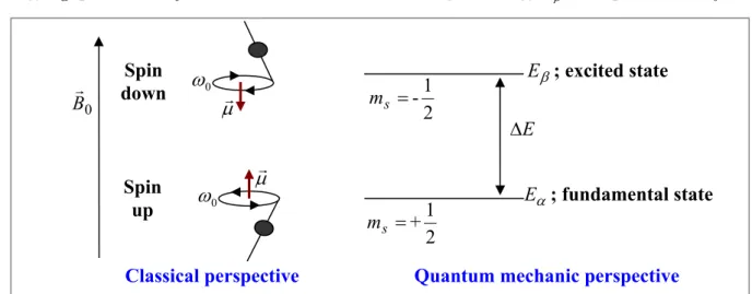
I.3.1 MRI basics....29
Physical principle...29
Relaxation phenomena...31
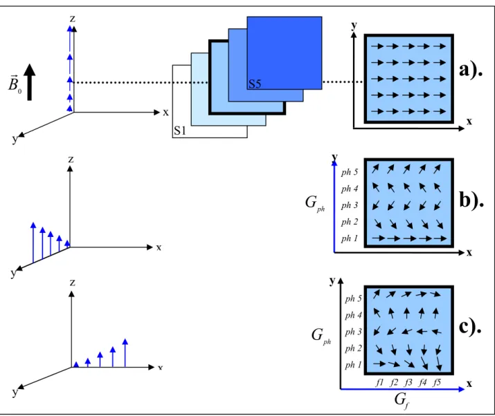
Signal and image acquisition...32
Image quality...35
I.3.2 MR thermometry and tissue heating control....38
Temperature dependence of water proton resonance frequency...39
Pulse sequences for PRFS thermometry...42
Thermotherapy with closed-loop feedback control...43
I.3.3 RF receive-only coils.....45
Chapter II. Experimental ex-vivo validation of a RF coil integrated with a
planar ultrasound transducer for esophageal applications
II.1 Introduction...48
II.2 Material and methods...48
II.2.1 Ultrasound device and experimental setup....48
II.2.2 Coil design and RF parameters...50
II.2.3 MR data acquisition....52
II.3.1 Image acquisition.....55
II.3.2 Comparative study: miniature versus standard coil.....57
II.3.3 Ex-vivo esophagus study....59
II.3.4 Temperature monitoring and therapy control...60
II.4 Discussion...
.62
II.5 Conclusion...
.66
Chapter III. In-vivo evaluation of high resolution MR-guided thermotherapy
for rectal applications: RF coil integrated with phased-array transducer
III.1 Introduction...
.68
III.2 Material, methods and preliminary tests...
.68
III.2.1 Rectal ultrasound transducer & electronics......68
Phased-array ultrasound transducer and its cooling circuit...68
Principle of the beam generation: focused or plane wave...69
Matching of the electrical impedance...71
Calibration of the electrical driving circuit...71
Characterization of the transducer by the radiation force technique...73
III.2.2 Opposed-solenoid RF coil.....75
Coil design: wiring and insulation...75
Coil prototype: 1 turn versus 5 turns...76
Influence of the transducer on the coil...77
III.2.3 Automatic temperature feedback control.....77
Physical model for temperature estimation...78
Implementation of the PID controller...79
III.2.4 MR data acquisition and processing........81
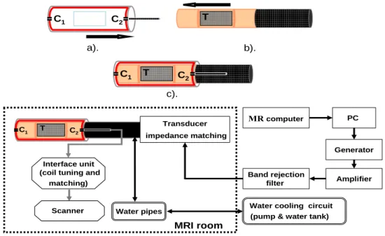
MR system and experimental setup...81
Ex-vivo experiments...82
In-vivo experiments...85
III.3 Experimental results...
.85
III.3.1 Phased-array structure of the transducer...85
Beam type...85
Multiple beams...86
III.3.2 PID controller stability....88
III.3.3 Comparative studies of the coils....89
1 versus 5 turns...89
Transducer influence...90
Internal versus external coil...91
III.3.4 In-vivo results....92
Internal/external coil...92
Thermotherapy with automatic temperature control...93
Evaluation of tissue destruction...95
III.4 Discussion...96
III.5 Conclusion...99
Chapter IV. Optimization of high resolution MR-guided contact
thermotherapy for digestive applications: treatment planning and embedded
integration of the RF coil
IV.1 Introduction...
.101
Matching of the electrical impedance...103
Electrical calibration of the transducer...104
Acoustic calibration of the transducer...106
Cooling balloon attenuation measurement...107
IV.2.2 Opposed-solenoid RF coil....107
Coil prototype: 1 turn versus 3 turns...107
RF coil integrated within the phased-array transducer...110
IV.2.3 Planning of the thermotherapy treatment....110
Algorithm description...110
Simulation results...113
IV.2.4 MR data acquisition and processing...115
MR system and experimental setup...115
Ex-vivo experiments...116
IV.3 Experimental results...
.117
MR compatibility of the integrated device...117
IV.3.1 Comparative study of the coils...118
Comparative study of the coils...118
Integrated coil performance...120
IV.3.2 Thermotherapy with automatic temperature control......121
IV.3.3 Feasibility of the treatment planning...122
IV.4 Discussion...124
IV.5 Conclusion...128
Chapter V. Design of a new generation of therapeutic ultrasound applicator,
general discussion and conclusions of the thesis
V.1 Introduction...130
V.2 Acoustic simulation for a new ultrasound transducer...130
V.2.1 Design of the ultrasound transducer......130
V.2.2 Multiple configurations investigation....132
V.2.3 Results and discussion....135
V.3 General discussion and conclusion of this thesis...138
V.3.1 RF receive-only coils...138
V.3.2 Active temperature control...142
V.3.3 Ultrasound transducer efficiency and treatment planning...143
V.4 Perspectives...144
References………...145
Appendix……….155
Publications………
.156
Résumé………158
Chapter I. Introduction
I.1 Cancer in general context
I.1.1 Esophageal cancer
StatisticsCharacteristics Classical treatments New treatments
I.1.2 Rectal cancer
Statistics Characteristics Classical treatments New treatments
I.1.3 Ultrasound for digestive cancer treatment
I.2 Therapeutic ultrasound
I.2.1 Ultrasound basics
DefinitionGeneration Acoustic field Characteristics
I.2.2
Biological effects of ultrasound
Thermal effectsNon-thermal effects
I.2.3
Applications
I.3 MRI introduction
I.3.1 MRI basics
Physical principles Relaxation phenomena Signal and image acquisition Image quality
I.3.2 MR thermometry and tissue heating control
Temperature dependence of water proton resonance frequency Pulse sequences for PRFS thermometry
Thermotherapy with closed-loop feedback control
I.3.3 RF receive-only coils
I.1 Cancer in general context
Although the possibilities to cure cancer reach more than 50%, with some cancer completely cured (as testicular cancer, for example), and despite the incessant progress made in terms of treatment, detection, and prevention, the disease remains feared by the majority of patients. At global level, cancer data are always collected and compiled sometimes after the events to which they relate, so that the most recent statistics available are always "old". Moreover, because the sources of data are continuously improving in quality and extent, estimates may not be truly comparable along time. The most recent worldwide cancer
statistics available is GLOBOCAN 2002 database [Ferlay et al, 2004]. Figure I.1 is derived from this source and presents the incidence and mortality of cancer, for men and women, in 2002. Lung cancer was the first cancer in term of incidence, showing also a high mortality rate. At that time, 10.9 million people worldwide were annually diagnosed with cancer and 6.7 million people died. These deaths represented around 12% of deaths worldwide. The same source (GLOBOCAN 2002) estimated that 24.6 million people worldwide were diagnosed with a cancer in the last five years and around half of these people lived in Europe and North America.
Figure I.1. Age-standardized incidence/mortality rates, all cancers, world, 2002 estimates.
At national level, cancer data are more recent and cancer trend may be observed. In France, cancer becomes, in 2004, the first cause of death [Aouba et al, 2007]. Also, cancer remains the most lethal disease for people between 45 and 64 years. One of two men and one of three women are concerned. According to the latest data from French National Institute of Health Monitoring [Belot et al, 2008], cancer incidence doubled from 1980 to 2005, but the mortality risk declined with 25%. This divergence may be explained by a diminution of aggressive cancers (such as esophagus or stomach) and an increase of favorable prognosis cancers (breast, prostate). In 2005,
at national level, 320 000 new cases of cancer (180 000 for men and 140 000 for women) were diagnosed and 146 000 deaths were registered (please see Table I.1 for further details). Contrary to the worldwide behavior observed in 2002 (GLOBOCAN 2002), in France, the most common cancer was breast cancer, followed by colon and rectum, lung and prostate. This classification of cancer changed [Belot et al, 2008], especially due to the rising incidence of prostate cancer (for men) and of lung cancer (for women). Therefore, the prostate cancer becomes the most frequent cancer, for both sexes. For men, the three most common cancers are prostate, lung and colon/rectum cancers, while women are mostly affected by breast, colon/rectum and lung cancers.
In the work described in this thesis, regions of the digestive tract containing a natural cavity, i.e. esophagus, and colon & rectum, are considered. Table I.1 [Belot et al, 2008] summarizes statistics data trends for esophageal and colon/rectum cancer in French population for a period of 25 years. Data for all cancers are also presented for comparative purpose.
Incidence 1980 1985 1990 1995 2000 2005 Esophagus 494 5 591 5 542 5 381 5 091 4 721 Colon-rectum 23 804 26 470 29 187 31 990 34 623 37 413 All cancers 168 850 188 742 211 776 239 792 273 518 319 380 a). Mortality 1980 1985 1990 1995 2000 2005 Esophagus 5 378 5 147 4 875 4 568 4 222 3 850 Colon-rectum 15 153 15 238 15 446 15 778 16 141 16 865 All cancers 129 274 136 335 141 751 145 378 146 165 145 762 b).
Table I.1. a), b). Incidence (a) and mortality (b) cases
for esophageal and colon/rectum cancer, over 1980-2005, in France.
I.1.1 Esophageal cancer
Statistics.
Esophagus cancer shows, in 2002, a worldwide incidence of about 462 000 cases annually (the eighth most common cancer, corresponding to 4.2% of the total) and causes 386 000 deaths (the sixth most common cause of death from cancer, i.e. 5.7% of the total) [Parkin et al, 2005]. The majority of cases (80-85%) are diagnosed in developing countries where it is the fourth most common cancer in men [Cancer Research UK, 2005]. The area with the highest reported incidence is the so-called Asian "esophageal cancer belt", from Turkey to China, but restrained region from south/south-east Africa, South America or parts of Europe were also reported.Within European countries, the GLOBOCAN 2002 data shows that French men had the highest incidence rates, followed by men from the UK and Hungary, while UK women had the highest reported incidence of female esophageal cancer. In 2005, esophageal cancer was the 9th
cancer in term of incidence in UK (7 823 new cases with a male/female ratio of 1.8) [Office for National Statistics, 2008]. With 7 405 deaths in 2006, esophageal cancer is responsible for around 5% of all cancer deaths, making it the 5th most lethal cancer. In France, esophageal cancer incidence represents 1.5% of all cancers (see Table I.1.a), year 2005), i.e. the 15th most frequent cancer, men and women together. With a sex-ratio of 5.3, this cancer is one of the highest men-oriented diseases. A mortality rate of 2.6% of all cancers (the 11th rank of cancer in terms of mortality), closely related to that of incidence, reflect the lethal character of this type of cancer, unfortunately worldwide valid.
Characteristics.
Esophagus (gullet) extends from the pharynx to the stomach and in adults is approximately 26 cm in length and 2 cm wide [Cancer Research UK, 2005]. The normal wall thickness varies in function of its state (contracted/dilated) and also for the different segments of esophagus (cervical, thoracic, retrocardiac and intra-abdominal). When contracted, the average thickness varies between 4.05-5.68 mm (the intra-abdominal segment has the highest value). In dilated state, the thickness varies from 1.87 to 2.7 mm (with cervical esophagus the thickest part) [Xia et al, 2008]. A cross-section of esophageal tissue shows 4 layers, from the lumen to the periphery: a) mucosa containing stratified squamous epithelial cells, glands and muscularis mucosa; b) submucosa – adipose, fibrous connective tissue and blood vessels; c) muscularisexterna (propria) – circular and longitudinal muscle layers; and d) serosa (adventitia).
Esophageal cancer has two main histological causes. Sixty percents of esophageal cancers appear in the cells lining the esophageal tube (squamous cell carcinoma-SCC), usually in the upper two-thirds of the esophagus. The remaining 40% is represented by adenocarcinomas (AC-malignant cells developed in the glandular tissue), and generally this type of cancer is localized in the lower esophagus. Before an adenocarcinoma can develop, glandular cells (which normally do not cover the esophagus) must replace an area of squamous cells, as in the case of Barrett’s esophagus. Over the past several years, the incidence of adenocarcinomas was found increasing in Western populations, but the underlying reasons for this are unclear. If these trends continue, AC will become the dominant histology; this has already happened for white men in the USA and UK [Wild et al, 2003].
The principal risk factors are age, sex, alcohol, tobacco, other type of irritation of the esophagus and medical history. In Europe and Northern America most esophageal cancers are caused by tobacco and alcohol, but in South America the consumption of hot beverages (maté) are thought to be important. Nutritional deficiencies (zinc) may underlie the high rates in Central Asia and China [Cancer Research UK, 2005]. There are well established risk factors for both types of esophageal tumor. American data [Engel et al, 2003] suggest that smoking, higher body mass index, low intake of fresh fruit and vegetables, and gastro-esophageal reflux are responsible for an
estimated 79% of AC; while tobacco consumption, excessive alcohol consumption, and low fruit and vegetable intake are estimated to cause 89% of SCC. Tobacco and/or alcohol remain the main risk factors, whose effect is directly related to quantity and duration of consumption. For example, smoking cessation induces an immediate esophageal risk decline, but a value equal to that of a non-smoker will be obtained only 10 or more years after giving up. Risk of esophageal cancer is increased in people who have had radiotherapy to the mediastinum for another primary cancer: breast cancer [Roychoudhuri et al, 2004] or Hodgkin’s disease [Dores et al, 2002].
The most common symptom is difficulty in swallowing (obstructive dysphagia). It can also be associated with weight loss and sometimes pain or discomfort behind the breast bone or in the back. However, these are not regarded as certain signs of cancer. Esophageal cancer is usually diagnosed with an endoscopic examination of the esophagus (esophagoscopy) or barium swallow X-ray. Generally, biopsy is performed during endoscopy in order to establish the histological diagnosis: SCC or AC. More detailed examinations (endoscopic ultrasound, bronchoscopy, thoracoscopy/laparoscopy, CT-computer tomography, PET-positron emission tomography or MRI -magnetic resonance imaging) are essential to delimit the cancer position/size and to detect the lymph nodes state or possible distant metastases. Generally, cancer staging (TNM system) describes important data about primary tumor (T), lymph nodes (N) and metastases (M). Tumor stage is classified from T0 to T4 by the degree of advancement of tumoral cells: T0 (carcinoma in situ): the cancer is localized only in the epithelium of the esophagus (the first layer of the mucosa); T1: the mucosa or submucosa are affected; T2: the tumor has invaded the muscularis externa, the third layer of esophageal wall; T3: the tumor has advanced through the entire esophageal wall, including adventitia; T4: the nearby tissues of esophagus are affected. Note that, when stenosis is observed, the tumor stage is regarded as T3. Concerning the stage of lymph nodes N and metastasis M, a value of 0 indicates no extension, while 1 corresponds to cancer spreading to lymph nodes or distant organs. Finally, all data are combined to assign a complete cancer stage:
stage 0 (T0, N0, M0): epithelium, no lymph nodes, no metastases;
stage I (T1, N0, M0): only first or the second layer of esophagus, no lymph nodes, no metastases;
stage II with IIA (less aggressive) and IIB (more aggressive):
stage IIA (T2 or T3, N0, M0): to the third layer or all esophagus, no lymph nodes, no metastases; stage IIB (T1 or T2, N1, M0): any of the three inner layers of esophagus (but not adventitia) & nearby lymph nodes, no metastases.
stage III (T3, N1, M0 or T4, any N, M0): all esophagus & nearby lymph nodes or nearby organs with or without nearby lymph nodes, no distant nodes or organs;
Classical treatments.
A complete and correct staging of the cancer will help the medical staff to decide on an adequate treatment. Unfortunately, esophageal cancer has a high instance of mortality with a prognosis of less than 10% survival rate at five years after diagnosis, all stages included (0% if metastasis, 10% if lymph nods are involved, 25% if there is no adenopathy or metastasis) [Simon et al, 2003].Currently, the single option for curative treatment is surgery (with lymph nodes removal), but only for stage I and II. Metastatic cancer or tumor invasion of surrounding structures (e.g. trachea, bronchi, aorta, pericardium) is not considered for surgery. Also, clavicular or celiac lymphadenopathy is counted for metastasis. However, regardless tumor staging, the patient general condition (age, weight loss, cardiovascular or pulmonary problems, liver) may be a contraindication to surgery. Only about a third of cancer patients are candidates for esophageal surgery. Moreover, after a surgery with curative resection (R0), only 30% of patients shown a 5-year survival rate [Bouvier et al, 2006]. After surgery, most patients are able to resume normal activities within 2 months. Side effects of the procedure include pain and tenderness, which usually can be controlled with medication. Complications may be severe (bleeding, cardiac or pulmonary complication, infection at the incision site, nerve injury) and occur in about 40% of patients.
For advanced stages of disease (i.e. tumor invading adjacent structures such as mediastinal extension), surgery must be associated with chemotherapy (ChT) and radiotherapy (RT) [Mariette
et al, 2007]. The single association of chemotherapy [Hwang, 2007], [Urschel et al, 2002] or
radiotherapy [Lordick, 2005], [Mariette et al, 2007] with surgery does not offer good results. A study investigating the role of chemotherapy and radiation therapy [Graham et al, 2007] showed that neoadjuvant (before surgery) radiochemotherapy (RCT) appears to be correlated with the best survival rates and the largest improvement in quality of life, but definitive proof is not currently available. The role of surgery, radiotherapy, and chemotherapy, alone or combined, in the optimal management of esophageal cancer is still under intensive research.
However, for inoperable patients, the combination of radiotherapy (dose limit: 50 Gy) and chemotherapy (reference drugs: 5FU-fluouracil and cisplatin) remains the standard curative treatment. These techniques are also widely used for palliation. Nevertheless, important side effects must be considered: fatigue, reddening of the skin (radiotherapy), and nausea, alopecia, skin rash and itching, mouth and lip sores (chemotherapy). Generally, the outcome is improved when patients are managed by multidisciplinary teams at specialized units.
New treatments.
Numerous endoscopic therapy are proposed as an alternative to classical treatment: endoscopic mucosal resection (EMR) [Ell et al, 2000], photodynamic therapy (PDT) (Photofrin®, 5-aminolevulinic acid) [Lightdale et al, 1995], laser therapy (Nd:YAG – neodymium:yttrium-aluminum-garnet, KTP – potassium-titanyl-phosphate)[Gossner et al, 1999], intraluminal stents [Siersema, 2006], endoscopic radiofrequency (RF) ablation [Sharma et al, 2007], argon plasma coagulation (APC) [May et al, 1999], or, recently reported, cryospray ablation [Cash et al, 2007].
Because of their small penetration depth –APC, PDT and laser therapy– are generally indicated for the treatment of early stage cancers, when only the mucosal layer (typically 2 mm width) is affected. However, even if such endoscopic ablative techniques have high success rates in removing Barrett's epithelium or other superficial cancers, their effect at the genetic level is unclear and the remaining tissue may still contain genetic alterations associated with malignant progression to cancer [Hage et al, 2006]. Among the currently available endoscopic treatments of the superficial cancers in esophagus, only endoscopic mucosal resection offers the advantage of histological analysis of the removed sample [Gossner, 2006] and may therefore represent a truly curative treatment. Moreover, EMR is indicated for cancer size of less than 2 cm in diameter.
Endoscopic palliation is the most suitable approach for metastatic patients with poor performance status (not capable to support radiochemotherapy) or with esophago-tracheal fistula. Palliation aims to improve the patient quality of life, and especially, to relief the dysphagia. Two modalities are generally used: stenting the stenose (expandable metal/plastic stent) or reducing the
tumor mass using thermoablation (methods cited above). The risks associated with these strategies cannot be neglected (esophageal stricture, hemorrhage, stent migration, wall perforation, temporary light sensitivity). Also, single dose brachytherapy (12 Gy) may be an effective option for palliation.
I.1.2 Rectal cancer
Generally, the colon (especially the sigmoid colon) and rectum cancers are widely grouped together because their similarities (risk factors, histological pathology). We have preserved this classification only for the following paragraph (statistics). Furthermore, we refer exclusively to rectal cancer, since this was the object of interest of a part of the work described in this thesis.
Statistics.
Colon and rectum cancers account for about 1 million new casesin 2002 (9.4% of the world total) and 529 000 deaths [Parkin et al, 2005]. In termsof incidence, colorectal cancers rank fourth in frequency in men and third in women, showing very little difference between sexes (men/women ratio of 1.2:1). Contrary to the esophageal cancer, colorectal cancer has a relatively good prognosis, with mortality to incidence ratio of about 50%. Therefore, the worldwide prevalence (in 2002) of colorectal cancer is second after breast cancer, with an estimate of 2.8 million people alive with colorectal cancer within 5 years following diagnosis. Geographic variation in occurrence of colorectal cancers appears at the level of world area. A 25-fold variationis observed between high-risk region (North America, Australia/New Zealand, Western Europe, and, in men especially, Japan) and low-risk region (Africa and Asia, parts of South America). Moreover, in high-risk populations, the ratio of colon to rectal cancerincidence is 2:1 [Parkin et al, 2005].
American data from National Cancer Institute/ Surveillance Epidemiology and End Results [Ries et al, 2008] estimates 148 810 new cases of colorectal cancer and 49 960 deaths, in 2008. However, American population has the highest survival rate at 5 years after diagnosis: 65%. This rate may be explained by a descending trend in terms of incidence and mortality rates. Unfortunately, this trend is not the case of France. From 1980 to 2005, according to Table I.1, the cases of colorectal cancer (both terms of incidence and mortality) continue to increase in French population. In 2005, 37 413 new cases were diagnosed (11.7% of all cancers) and 16 865 deaths (11.6% of all) were noted. In France, the colorectal cancer is the third most common cancer (incidence) and the second cancer in term of mortality.
Characteristics.
The normal rectum is 11 to 15 cm long and has a maximum diameter of 4 cm [Santoro et al, 2006]. It is continuous with the sigmoid colon superiorly and the anal canal inferiorly. The mean thickness of normal rectal wall is estimated at 2.57 mm with individual variation thickness from 2.2 to 3.6 mm [Huh et al, 2003]. However, a maximal thickness of 4 mm for the entire wall is still considered normal. Moreover, maximal values of 1 mm for mucosa and 2 mm for muscularis propria are considered normal [Van Outryve et al, 1993]. Component of digestive system like the esophagus, the rectum wall has also 4 layers (see Fig. I.2): mucosa,submucosa, muscularis propria, and serosa with perirectal fat.
Rectal cancer is, with an overwhelming majority (90%), an adenocarcinoma-type [Gasowski et al, 2003]. Other rarer types include lymphoma and squamous cell carcinoma. Generally, it develops from adenomatous polyps (adenomas); it was observed that an adenoma may develop a cancer over an average period of 9 years. The number of these polyps increases with age, but only large polyps (size >1 cm) are potentially dangerous. However, people with some hereditary condition like familial adenomatous polyposis (FAP) or hereditary nonpolyposis colon cancer (HNPCC or Lynch syndrome) must be periodically screened. Also, people who had
inflammatory bowel disease (Crohn's disease, ulcerative colitis) or cancer history (colon, ovary, and uterus) must be under surveillance. Important risk factors –that may be controlled– are diet (read meat), smoking, alcohol, obesity and physical inactivity.
Symptoms for rectal cancer are not specific and generally appear for advanced stage: weight loss, blood in the stools, abdominal/pelvic pain, changes in bowel activity. Diagnosis is based on endoscopy (sigmoidoscopy - for rectum and sigmoid colon visualization; colonoscopy when entire colon is investigated) or the less accurate DRE (digital rectal examination). After
cancer confirmation by endoscopy, staging techniques (endosonography, MRI, CT) are used in order to determine how far the cancer has spread. The TNM system (substitute of an ancient system known as Dukes’) is similar to the description of the esophageal system. The T and M stages are the same, while the N stage is subdivided into N0 (no lymph node involvement), N1
Figure I.2. Cancer staging (0 to IV) and normal wall structure (4 layers) for the rectum. Source: site of Siteman Cancer Center /Barnes-Jewish Hospital /Washington Univ. School of Medicine-www.siteman.wustl.edu
(extension to 1 to 3 nodes), and N2 (extension to more than 3 nodes). Metastases are generally observed in liver, lungs, colon or pelvis. The external iliac nodes are also considered as metastasis. Finally, the stage grouping is slightly different. Figure I.2 summarizes this classification: stage 0 (T0, N0, M0), stage I (T1 or T2, N0, M0), stage II (T3 or T4, N0, M0), stage III (any T, N1-2, M0), and stage IV (any T, any N, M1).
The most suitable staging technique is endosonography (endorectal ultrasonography ERUS) since it allows exact differentiation of the rectal wall layers [Beynon, 1989], and tumor staging (up to stage 3) with median accuracy of 89% [Maier et al, 2003]. But, lymph nodes staging by ERUS is less accurate (80%) [Glaser et al, 1990]. When compared to endosonography, MRI has less accuracy (85%) in tumor staging [Maier et al, 2003], but it permits the mesorectum and the mesorectal fascia evaluation [Beets-Tan et al, 2001]. This is a very important feature concerning the cancer resectability and the risk of recurrence. Furthermore, MRI can be employed in cases of stenosing tumors and also for the detection of metastases located at different sites within the body. However, important improvement in technologies for all imagistic methods (contrast agent or higher field for MRI, combination PET-CT) may lead to more accurate cancer staging and that it is only the benefit of the patient.
Classical treatments.
Clinical decision in treatment of rectal carcinoma depends on several factors: tumor location, penetration of bowel wall, lymph node status, and metastasis. Hence, an exact preoperative staging of the cancer will help to decide the best treatment. For example, a standard curative surgery must contain a total excision of the mesorectum*. Therefore the cancer resectability is decided –before the surgery– on the basis of MR image of the mesorectal fascia (the external limit of the mesorectum). A safety lateral margin of minimum 1 mm is demanded for a curative resection (R0). Also, the localization of the tumor (upper, middle or lower rectum) with respect to the anal sphincter is an important criterion for the preservation of the anal function andfor choosing the type of exeresis. A distal margin of resection (distance between inferior limit of the tumor and the distal limit of rectum) of at least 1 cm may be an appropriate clearance of cancer, still preserving the anal sphincter [Shirouzu et al, 1995].
Like in the case of esophageal cancer, surgery remains the standard curative treatment. This time, the prognosis is considerably better. The survival rate at five years after tumor resection is 100% (stage 0), 85-100% (stage I), 65-75% (stage II), 50% (stage III, with 1-3 nodes involved), 25% (stage III, more than 3 nodes) and 0% (stage IV) [Gasowski et al, 2003]. The two main types of rectum surgery are the low anterior resection (LAR) and the abdominal perianal resection (APR). The LAR technique consists in partial/total rectum resection with anastomosis to the rest of rectum (colorectal) or the anus (coloanal), together with total mesorectal excision (TME) and at least 8 lymph nodes removal. The introduction of TME [Heald et al, 1982] as a standard procedure represented a large improvement in recurrence decrease and overall survival. Unfortunately, the APR technique is performed for patients without possible preservation of anal sphincter. The morbidity (fistula, abscess) rate for rectum surgery is less than 15%. However, surgery alone is curative only for stage I (tumors T1 or T2, N0).
For stages II or III, surgical resection (with TME) may still be curative, but only associated to (chemo)radiotherapy. The association of radiotherapy prior to surgery reduces the recurrence rate, when compared to surgery alone, but no differences were observed regarding the overall survival (at 2 years after intervention) [Kapiteijn et al, 2001]. The radiotherapy (RT) –long therapy (45 Gy in 5 weeks) or more recently, short therapy (25 Gy in 5 days)– can be delivered pre-, intra-, or post-operatively, but preoperative variant is preferred for his efficiency and reduced toxicity. The potential advantage of preoperative radiation is tumor down-staging, hence an enlarged resectability with possible sphincter-spare procedure. Also, the radiation therapy works better in well-oxygenated tissues prior to surgery and that conducts to a decrease in tumor viability, which results in less local recurrence. The recurrence rate may be further reduced by the addition of chemotherapy to RT, and once again the preoperative radiochemotherapy (RCT) option is preferred [Sauer et al, 2004]. The chemotherapy (ChT) is based on 5FU (fluouracil) associated with folinic acid (FUFOL) or more recent drugs (oxaliplatin, irinotecan), and may be adjuvant (for tumors with nodes involvement) or neoadjuvant, enhancing the RT effects. Overall, there is still debate (European countries, USA, Japan) regarding the best association of RT or/and ChT to surgery for rectal cancer, especially in terms of survival. The difficultly of a final conclusion on benefits of RCT to surgery may also arise from the leak of standardized surgery (with or without TME) in some trials. However, a worldwide consensus considers the multimodal therapy involving a multidisciplinary team of cancer specialists (gastroenterologists, surgeons, medical and radiation oncologists, radiologists and pathologists) as the best approach to the management of
rectal cancer. Moreover, RT and ChT are the standard methods used to fight cancer when surgical resection is not possible.
New treatments.
Alternative endoscopic techniques are suitable for patients who are not candidates to standard methods (surgery or RCT), or may be used as complementary approaches. Also, alternative methods are more widely used for recurrent cancer treatment since, generally, patient condition is poor after a primary rectal cancer treatment. But, their use is mainly limited to palliation. Three main types of treatment can be counted: local excision (transanal endoscopic microsurgery), ablative techniques and stent application. The utilization for curative purpose of the less invasive transanal endoscopic microsurgery versus the radical surgery is still controversial, but its role in T1 and possibly T2 tumor treatment is growing in acceptance [Zieren et al, 2007]. The ablative technique includes various tools: Nd-YAG laser ablation [Rao et al, 2005], photodynamic therapy (PDT) [Ross et al, 2006], Radiofrequency ablation (RF) [Green et al, 2008], argon plasma coagulation (APC) [Dees et al, 2006] or the almost abandoned techniques of electrocoagulation [Eisenstat et al, 1992] and cryotherapy [Meijer et al, 1999]. Their applications vary from palliation (laser ablation), recurrent cancer treatment (neoadjuvant PDT to surgery, RF alone) to treatment of a specific complication of pelvic radiation therapy (hemorrhagic radiation proctitis) manifesting by bleeding (APC). More recently, the self-expanding metal stents [Sebastian et al, 2004] become widely used to palliate rectal cancer. Overall, although the endoscopic techniques may offers solutions for bleeding, tenesmus (difficulty to defecate) or obstructions, they show limited efficacy in pain management or distant metastasis. Consequently, a multidisciplinary approach including radiotherapy and chemotherapy should be considered in incurable patients with a reasonable life expectancy.I.1.3 Ultrasound for digestive cancer treatment
This paragraph discusses the potential use of ultrasound for the treatment of digestive cancers (esophagus and rectum) among all the possibilities used to fight cancer.
Currently, an ideal treatment to cure cancer regardless of the type of cancer staging doesn’t exist. We have seen that for both types of cancer discussed here and it is widely valid for another cancer. Standard techniques are currently used to cure cancer, but they are limited to a specific stage (surgery alone for early stages and in association with chemo-, and radiotherapy for more advanced stage). Unfortunately, the existence of distant metastasis gives no hope for curative treatment and in these cases the objective is rapidly reduced from "cure" to "palliation". On the other hand, in the case of superficial cancer (carcinoma in situ or early stage I), alternative endoscopic techniques for curative purpose are investigated [Saurin, 2000]. These curative ablative techniques aim to limit the invasiveness of surgery or the toxicity of radiochemotherapy. However,
these methods (and every other except surgery) lack a real, histological evidence of cured cancer. Nevertheless, even a clear curative surgery (respecting all standard procedures) may be unsuccessfully, and especially when fighting highly recurrent cancer, like rectal cancer. The necessity of constant surveillance of the patient after a curative treatment of cancer is then obvious. Only a sufficient period of time like the actual 5-year period may permit to consider that a cancer is cured. Further clinical studies are needed to determine equivalence or superiority between these endoscopic ablatives technologies with respect to success rates and complications.
When considering palliative purpose, endoscopic procedures are proposed for patients with contraindication to surgery or RCT, and also for recurrence cases. Specific drawbacks of these techniques are known: inaccurate monitoring for RF heat delivery [Viallon et al, 2010], necessity of multiple treatment sessions for laser ablation [Norberto et al, 2005], [Rao et al, 2005], side effects like 6 weeks of hypersensitivity to light after a PDT with Photofrin® [Saurin, 2000] and, overall, possible complications such as bleeding or perforation. The small number of randomized trials cannot conclude with an ideal palliation. Therefore these procedures are primarily used only for patients with very advanced disease and short life expectancy, or as an adjuvant therapy. However, the optimal treatment must be decided by a multidisciplinary team for each case.
In this context, a minimal invasive endoscopic method using therapeutic ultrasound has been initially proposed by Lafon et al [2000] and further applied to esophagus by Melodelima et al [2005]. The extracorporeal focused ultrasound approach is not indicated because of the anatomical position of esophagus and rectum, i.e. the vicinity of important organ such as trachea, spinal column or genito-urinary organs. Therefore, on the basis of the natural cavity of these structures, the high intensity contact ultrasound (HICU) was considered as an adequate method to treat esophageal/rectal cancer. HICU, under MRI guidance, may offer some advantages over other endoscopic methods. These advantages include increased penetration depth (versus laser therapy or PDT), accurate control of heating (versus RF ablation, electrocoagulation, cryotherapy) and absence of side effects (like the case of PDT). The intents of the MRI-guided HICU method are both palliative and curative (depending on tumor staging). The curative purpose is motivated by the increasing number of early diagnosed cancer due to the introduction of nationwide screening of digestive cancer. A limit in palliation –with HICU– is the total obstruction of the lumen, induced by very large tumors. In these cases, there is no physical space to position the transducer inside the lumen, in direct contact with tumor. Hence, possible indications for HICU procedure are palliation of tumor causing only partial stenosis, recurrent cancers, and superficial cancer cure (stage I, II, without lymph node involvement), without complications. The goal of this thesis was to develop an ablative technique (MRI-guided HICU) for esophagus or rectum cancer having the potential for safe, effective and long term outcome.
I.2 Therapeutic ultrasound
This section is meant to be a brief review of the physics underlying the clinical application of therapeutic ultrasound. For further details the reader is kindly referred to "Biomedical ultrasonics" by P.N.T Wells [1977], "Ultrasound Physics and Instrumentation" by Hedrick et al [1995] or Duck’s "Physical properties of tissue" [1990].
I.2.1 Ultrasound basics
Definition.
Ultrasound is a form of vibrational energy propagating as a mechanical wave, with a frequency superior to 20 kHz, the limit of human hearing. The mechanical wave is a periodic motion of particles* within a medium, transmitting its energy successively from one particle to the next. Contrary to light, which, as an electromagnetic wave can propagate in vacuum, the ultrasound needs a medium to support its propagation. When propagating through a medium, the wave entails local vibrations of the particles constituting the medium. If the displacement of the particles and the propagation of the wave have the same direction, then the wave is said longitudinal. The wave is said to be transversal when vibrations are normal to the direction of wave propagation. In medical applications (frequency range 1-20 MHz), ultrasound is a longitudinal wave, unless some particular conditions exist, like soft tissue-bone interface [Fujii etal, 1999], when both types of wave are observed. The result of such longitudinal vibrations is the
creation of compression and rarefaction regions along the wave propagation axis, i.e. high and low pressure regions. Therefore, a longitudinal acoustic wave propagating as repeating patterns of pressure through a medium is also named a pressure wave.
Generation.
Ultrasound may be generated, in a large range of frequency (from 50 kHz to 20 MHz) by various methods: mechanical (whistles, sirens), electric (piezoelectric materials) or magnetic (magnetostrictive materials). Since the achievable frequency is strongly related to the modality of ultrasound generation, piezoelectric material (with a frequency on the order of MHz) became the most indicated for medical transducers. The reverse piezoelectric effect demonstrated in 1881 by the Curie brothers consists in generation of a mechanical deformation of a piezoelectric material when applying an electric field over a direction of polarization.Some natural piezoelectric crystals like quartz, Rochelle salt or tourmaline were initially used, but synthetic ceramics like lead zirconate titanate (PZT) introduced by Jaffe in 1955 rapidly demonstrated their superiority in converting energy. PZTs are still widely used today due to their good piezoelectric properties and their ease of manufacture into a variety of shapes and sizes. They
* A particle is a volume element which is large enough to contain millions of molecules, so that it is continuous with its surroundings; but it is so small that quantities variable within the medium (like displacement amplitude) are constant within the particle.
also operate at low voltage and are usable up to about 300oC. Currently, the newly developed generation of piezoelectric materials is represented by piezocomposite: large number of piezoceramic rods embedded into a matrix of polymer [Fleury et al, 2002]. Using piezocomposite materials, Imasonic S.A. (Besancon, France) has developed two technologies named HI-1 (air backing) and HI-2 (solid backing). The "HI-1" transducers working at frequencies up to 10 MHz have an efficiency of 60-70% and a maximal acoustic intensity of 10 W/cm². The "HI-2" generation, limited at a frequency of 5 MHz, was developed to deliver a higher intensity (up to 30 W/cm²), but with a reduced efficiency (40-60%). The resonance frequency of a piezoelectric transducer depends on the intrinsic characteristics of the piezoelectric material, and also on its form and thickness. Therefore, high frequency transducers must be used with caution since the active element layer is very thin.
Acoustic field.
Medical transducers may be flat (plane wave) or focused (convergent wave). The pressure field generated with a typical flat transducer of diameter D excited by a sinusoidal signal of frequency F is shown in Fig. I.3. Note that the acoustic beam originates from the entire surface of the piezoelectric element, and not from a point. Therefore, the pattern of the acoustic beam is affected by constructive and destructive wave interference. Two different regions are distinguished: a near field (or Fresnel zone) with important fluctuation in the pressure field and aFigure I.3. Pressure acoustic field for a flat transducer. Adapted image from Acoustique & Techniques [Lefebvre et al, 2004]
far field (or Fraunhofer zone) characterized by a relatively uniform field. The transition from the near to the far field occurs at a distance L from transducer: λ 4 2 D L= Eq. I.1, where λ is the wavelength. The pressure amplitude has, at this limit-point, a maximal strength A0 and
begins to spread out from the acoustic axis (z) of the transducer as it propagates through the medium.
The divergence angle γ of the beam is related to the transducer diameter D and to the wavelength λ by the following equation:
D
λ γ 1.22
This deviation from the central axis of the transducer (also called beam divergence or acoustic diffraction) causes a smooth decline of the pressure amplitude and the apparition of secondary acoustic lobes (see Fig. I.3). The same phenomena appear when using a focused transducer. However, in this case, the pressure field has more strength than a flat transducer and the distance L (now named focal distance) is nearer to the transducer [Lefebvre et al, 2004].
Characteristics.
The characteristics of the acoustic wave are determined by the source of disturbance, i.e. the transducer in our case, and the properties of the medium through which the wave travels. The parameters used to characterize ultrasound are: frequency F (in Hz, or practical unit MHz), wavelength λ (in m, or commonly mm), period τ (in s), acoustic velocity c and particle velocity v (in m/s), acoustic impedance Z (in kg/(m²·s) = Rayl), amplitude A, acoustic pressure p (in N/m² = Pa), acoustic intensity I (in W/m², usually expressed in W/cm²), acoustic energy E (in W). The acoustic velocity c depends on the elastic properties of the medium (compressibility χ and mass density ρ), varying from 330 m/s in air to 1480 m/s in water [Wells, 1977] and 1540 m/s (average value) in soft tissue. Different values of c in biological media (biological fluids, soft tissue, bone and teeth, and even some pathological tissue) are available thanks to Duck’s review work. Note that acoustic velocity has a constant value for a specific tissue, but it may slightly vary with temperature and frequency inside that tissue. However, important* variation in acoustic velocity with frequency was found only for lung and bones [Duck, 1990]. The ultrasound wavelength in soft tissue is approximately 1.5 mm, at 1 MHz, and about 0.3 mm at 5 MHz.Another intrinsic characteristic for a specific tissue is the acoustic impedance Z, which is defined by the following relation (in the particular case of plane wave):
Z =ρ⋅c Eq. I.3. Similarly to the definition of the electrical impedance, Z represents the resistance of the tissue to ultrasound propagation and therefore an "Ohm’s law applied to acoustics" may be expressed as follows: p=Z⋅v Eq. I.4, where p is the acoustic pressure (the local pressure deviation from the equilibrium pressure, caused by the traveling ultrasound), and v is the particle velocity (the speed at which the particles vibrate with respect to their rest positions). The values of acoustic impedance Z are important when referring to an interface between two media. An ultrasound incident wave is totally transmitted to a second medium if the acoustic impedances of these two media are equal. However, this assumption is idealistic and, in practice, the impedance mismatches at the interface between two media causing both a reflection and a reduced transmission. Hence, the incident wave is partially reflected (reflection angle identical to the incidence angle) and partially transmitted (transmission
angle governed by the Snell-Descartes’s law). Note that this type of reflection appears at smooth interfaces larger than the wavelength (e.g. diaphragm, heart valves or air bubble) and it is usually named specular reflection (speculum (lat.) = mirror). On the contrary, when the objects are smaller than λ (like red blood cells for example), the wave is scattered in all directions (Rayleigh scattering). The interference of these scattered waves causes a fluctuation of the (backscattered) signal and, hence, brightness nonuniformities called speckle, in ultrasound imaging of a homogeneous object.
An important parameter in therapeutic ultrasound is its intensity. The acoustic intensity I is defined as the acoustic power Pac per unit area S. For a continuous wave, the time-averaged
intensity over a period may be expressed as in Eq. I.5:
c p S Pac I ⋅ = = ρ 2 2
max Eq. I.5,
where pmaxis the maximal pressure amplitude.
I.2.2 Biological effects of ultrasound
When an ultrasound beam travels through a real medium, its intensity is reduced as a function of distance: I(z)=I0⋅exp−α⋅z Eq. I.6, whereI is the acoustic intensity at the origin (z = 0), 0 I(z) is the acoustic intensity at a distance z from the transducer and α is the (intensity) attenuation coefficient (in Nepers/m). The coefficient α is often expressed in dB/m and, for that reason, a multiplication by a factor 8.686 must be used. Since the intensity and the amplitude of a mechanical wave are linked by a square function, then: α = 2 µ Eq. I.7, with µ the (amplitude) attenuation coefficient.
For a specific tissue, the attenuation coefficient is affected by temperature and frequency. For example, at a frequency of 1 MHz, the ultrasonic attenuation in soft tissue is approximately 0.7 dB/cm, whereas at 2 MHz, it is 1.4 dB/cm [O'Brien, 2007]. Numerous studies reported measurements of attenuation coefficients under different conditions (temperature, frequency) and for various tissues. The results of these studies were collected by [Duck, 1990] in a helpful database of attenuation values. Moreover, the attenuation coefficient, as a result of frequency dependency, influences the ultrasound penetration into the tissue. Hence, when the frequency is increased, the attenuation is increased, and the penetration is reduced. Also, although isotropic for most tissues, the attenuation is anisotropic for skeletal muscle, nerve or tendon. The longitudinal attenuation (along the fibers) measured in muscle is greater than the across attenuation by a factor 2 to 3 [Duck, 1990].
This wave attenuation is explained by several factors: deviation from a parallel beam (divergence); mode conversion (two or more wave traveling at different velocities and in different directions); scattering (non-specular reflection) by objects that are of the size of the wavelength or smaller (i.e. blood cells), and absorption (conversion of acoustic energy into heat). The most important contribution to wave attenuation arises from two factors: absorption and scattering. Hence, the attenuation coefficient is often defined as the sum of these two components. The absorption accounts for 60-80% of total attenuation [ter Haar, 1999].
Thermal effects.
An ultrasound wave traveling through a tissue is partially absorbed by the tissue, causing the heating of the medium (thermal effect of ultrasound). Initially, the ultrasound absorption was explained only by frictional forces (due to the viscosity of the medium), which oppose the periodic motion induced by the ultrasound wave. According to this classical theory, the absorption coefficient would be proportional to the square of the frequency (see calculations based on viscous forces in [Rayleigh, 1945]). Yet, the quadratic relationship was confirmed only for pure water (at frequency higher than 3 MHz) [Duck, 1990], whereas in tissue, the absorption coefficienta
α shows experimentally a near-linearly relationship with frequency: αa =α0⋅Fm Eq. I.8. The α0 (absorption coefficient at 1 MHz) and m parameters are both intrinsic characteristics of the tissue. For soft tissue, m varies from 1 to 1.2 [Goss et al, 1979]. A linear relationship between absorption and frequency is governed by relaxation mechanisms of the medium. Therefore, the absorption mechanism is currently explained by the relaxation theory, in addition to the viscosity theory. During the acoustic wave propagation through the medium, the structures of the medium endure a cyclic motion of compression and decompression. Ideally, in the case of a perfectly elastic medium, this motion is associated with a reversible energy transfer, i.e. the energy transferred from the acoustic wave to the system during the compression part will be entirely returned during the decompression part. But, since the tissues are not totally elastic, a part of the transferred energy during compression will remain stored into the tissular structure. This is a result of relaxation phenomena of the system, which try to redistribute the surplus of energy brought by the acoustic wave in order to guaranty the stability of the system. This energy redistribution causes the absorption of ultrasound and, hence, the increase in tissue temperature. Implicitly, the acoustic wave is attenuated, loosing at every cycle a constant quantity of energy (as a hysteresis-type mechanism). Concurrently, relaxation mechanisms may explain the observed velocity variation with frequency (dispersion phenomena).
The absorbed acoustic energy determines the heating of the medium in the case of a rate of heat production greater than the rate of heat removal. For a small control volume, the balance of thermal energy can be stated as:
W Q
Q
Qgain = storage + loss + Eq. I.9, whereQgain: heat rate gained by absorption process and from the surrounding control volumes,
storage
Q : heat rate stored by the tissue, Q : heat rate lost through the boundary of the considered loss
volume and W: work performed by the tissue and metabolic heating. Without considering the
boundary conditions, the two mechanisms of heat flowing inside a tissue are: conduction (diffusion, i.e. the temperature gradient) and convection (the perfusing blood). The heat conduction flux (conducted heat per unit area per unit time) qcond is governed by the Fourier law:
T k
qcond =− ∇ Eq. I.10, where k is the heat conductivity in W/(m·K) and ∇T is the temperature gradient (variation of temperature with distance) in K/m. The heat convection term, introduced by Pennes [Pennes, 1948]
describes the thermal interaction between tissue and perfused blood. The temperature distribution in the tissue is then expressed by the bio heat transfer equation (BHTE), as a summation of different mechanisms (diffusion, convection, metabolism, and external source of heating):
Q Q T T C q t T C =−∇⋅ cond + b⋅ b⋅ b a − + m + ∂ ∂ ⋅ ω ρ ( ) ρ Eq. I.11,
whereρ: tissue density in kg/m3, C: tissue specific heat in J/(kg·K); t T ∂ ∂ temporal derivative of tissue temperature T in K/s;
∇⋅qcond is the divergence operator applied to heat conduction flux vector in W/m3;
b
ρ : blood density, C : blood specific heat, b ωb: blood perfusion rate in m3 of blood /(m3 of tissue ·s) and T : arterial blood temperature in K; a
Q : rate of metabolism heat generation per unit volume in W/mm 3;
Q : rate of heat generation per unit volume in W/m3.
The last factor (Q) is the external source of heating for the tissue. Different methods (ultrasound,
laser, microwave or radiofrequency) may be used in order to induce controlled hyperthermia of a tissue. In the case of ultrasound, Q is defined as the product of (intensity) absorption coefficient,
and acoustic intensity I calculated from Eq. I.5. Therefore, for an ultrasound heating source, the
BHTE becomes: I Q T T C T k t T C = ∇ + b⋅ b⋅ b a − + m+ a⋅ ∂ ∂ ⋅ ω ρ α ρ 2 ( ) Eq. I.12,
where )∆T =∇2T =∇⋅(∇T is the Laplacian operator applied to tissue temperature T. The BHTE remains the most widely used thermal model of living tissues.
The temperature elevation in tissue may lead to cell death, depending on both temperature and duration of applied ultrasound energy (see Fig. I.4). Two possibilities of tumor treatment are available: hyperthermia and thermal ablation (ultrasound surgery). Hyperthermia consists of applying of a relatively low temperature (43-45°C) during a long period (around 60 minutes) [ter
becomes impossible to neglect and therefore inhomogeneous tissue heating may appear. Quite differently, thermal ablation (generally based on focused transducers) relies on a high temperature (above 55°C) being applied for a very short time (several seconds) causing irreversible cell death through tissue coagulative necrosis*. For example, Chapelon et al [1992] achieved kidney necrosis, by keeping the tissular temperature at 80°C for 5 s (focus intensity on the order of 1000 W/cm²). Contrary to hyperthermia, in the case of high intensity focused ultrasound (HIFU), the perfusion may be neglected, but cavitation effects are susceptible to appear [Kennedy et al, 2003].
Quantification of thermal damage induced by ultrasound may be expressed by the thermal dose TD (in minutes) or the thermal index TI. The TI is defined by the AIUM (American Institute of Ultrasound in Medicine) standards as the ratio of the in situ acoustic power to the acoustic power required to raise tissue temperature by 1°C, under specific conditions. Three thermal indices corresponding to soft tissue (TIS) [O’Brien and Ellis, 1999], [Karagoz and Kartal, 2006], bone (TIB), and cranial bone (TIC) have been developed for different examinations.
The thermal dose concept relates any time and temperature data combination to cell death. The relationship between the rate of cell killing, and time & temperature has been found to be strongly non-linear. In order to compare different heating regimes, the time-at-temperature data must be normalized to a common unit. As proposed by Sapareto and Dewey in a classical paper [1984], an empirical formula introduces the isoeffective thermal dose:
∑
∆ = t R −T t t 0 43 43 where ⎪⎩ ⎪ ⎨ ⎧ ° > ° ≤ = C T for C T for R 43 , 5 . 0 43 , 25 . 0 Eq. I.13. Here t is the thermal dose in equivalent minutes at 43°C (EM43 43°C), t is the heating exposure timeand T is the average temperature during the time interval∆t. Thus, the thermal dose represents the
* Swelling of the cells and their organelles leading to disruption of the cell membrane [de Zwart, 2000].
Figure I.4. Thermal damages on biological tissue, function of temperature and duration. Adapted from [Lele, 1977]
Haar, 1999]. This modality of energy delivery is based on the sensitivity of malignant cells to temperature rise. More sensitive to temperature than the normal cells [Li and Liu, 1997], the tumoral cells dies while the normal cells suffer only reversible damages [Hahn, 1984]. Since the process is relatively slow, blood perfusion
necessary time –at temperature T– to accumulate the same thermal damage (isoeffect) as the damage that would occur at a temperature of 43°C. No particular reason can justify the choice of 43°C as reference, except that this value is near the break point for some cell lines [Dewhirst et al, 2003]. The constant R is derived from experimental studies for different biological systems and endpoints. Theoretical considerations based on reaction kinetics (thermodynamic Arrhenius analyses) led to the prediction that the temperature dependence of the rate of protein denaturation is determined primarily by the activation energy. Hence, R is an expression of the relative increase in reaction rate for a 1°C increase in temperature [O'Brien, 2007]. Note that Eq. I.13 assumes an accumulation of thermal dose only during heating, neglecting any contribution from the cool-off period. Moreover, this equation does not imply that all cells exhibit the same sensitivity to heat, but only that a consistent relationship (for many types of cells) between time and temperature may correlate to cellular damage. An equivalent time of 240 minutes at 43°C is often regarded as a threshold for tissue necrosis [Sapareto and Dewey, 1984], [Daum and Hynynen, 1998] and 250-300 EM43°C permit already visible coagulation [Damianou et al, 1995], [Diederich et al, 1999].
Non-thermal effects.
Non-thermal mechanisms such as cavitation, radiation force, acoustic streaming, microjets and strain may occur in ultrasound exposed tissue [O'Brien, 2007]. Cavitation refers to the creation, oscillation and collapse of gas bubbles within a medium under the action of an ultrasound wave. The existence of nucleation sites within the medium is primordial to initiate cavitation. These nuclei can be, for example, particles in suspension, interfaces or microbubbles. The onset of cavitation depends also on several factors, such as acoustic intensity, frequency of the wave, static temperature and pressure, viscosity or superficial tension, making the control of cavitation difficult. Note that, for identical conditions, the cavitation threshold increases with the frequency of the ultrasound. Thus, by operating at a higher frequency, cavitation may be avoided.In stable cavitation, microbubbles expand and contract during each cycle in response to the applied pressure oscillations. The bubbles grow, as far as dissolved gas leaves the medium during the negative-pressure phase, a process called rectified diffusion. Each bubble oscillates for many cycles without collapsing completely. These oscillations can produce high shearing forces in the nearby surrounding areas and may give birth to microstreaming. These phenomena can fragment biomolecules or cellular membranes.
Transient (inertial) cavitation is a more violent form of microbubbles dynamics in which short-lifetime bubbles undergo large cycle changes over a few acoustic cycles before completely collapsing. Contrary to stable cavitation, inertial cavitation appears at intensities typically above 700 W/cm²/MHz in in-vivo tissues [Hynynen, 1991]. Transient cavitation shows a threshold that depends on the peak negative pressure p- of the acoustic wave and on frequency F, exhibiting a


![[PDF] Eléments de programmation en Delphi](data:image/gif;base64,R0lGODlhAQABAIAAAP///wAAACH5BAEAAAAALAAAAAABAAEAAAICRAEAOw==)