Contributions of Distinct Interneuron Types
to Neocortical Dynamics
by
Ulf Knoblich
Dipl.-Inform., Saarland University, 2004
Submitted to the Department of Brain & Cognitive Sciences
in Partial Fulfillment of the Requirements for the Degree of
DOCTOR OF PHILOSOPHY IN NEUROSCIENCE
AT THE
MASSACHUSETTS INSTITUTE OF TECHNOLOGY
FEBRUARY 2011
©2010 Massachusetts Institute of Technology. All rights reserved.
Author . . .
Department of Brain & Cognitive Sciences
December 7, 2010
Certified by . . .
Christopher I. Moore
Associate Professor of Neuroscience
Thesis Supervisor
Accepted by . . .
Earl K. Miller
Picower Professor of Neuroscience
Director, BCS Graduate Program
Contributions of Distinct Interneuron Types
to Neocortical Dynamics
by
Ulf Knoblich
Submitted to the Department of Brain & Cognitive Sciences
on December 7, 2010 in Partial Fulfillment of the Requirements
for the Degree of Doctor of Philosophy in Neuroscience
ABSTRACT
Inhibitory interneurons are thought to play a crucial role in several features of neocortical processing, including dynamics on the timescale of milliseconds. Their anatomical and physiological characteristics are diverse, suggesting that different types regulate distinct aspects of neocortical dynamics. Interneurons expressing parvalbumin (PV) and somatostatin (SOM) form two non-overlapping populations. Here, I describe computational, correlational (neurophysiological) and causal (optogenetic) studies testing the role of PV and SOM neurons in dynamic regulation of sensory processing.
First, by combining extra- and intracellular recordings with optogenetic and sensory stimulation and pharmacology, we have shown that PV cells play a key role in the generation of neocortical gamma oscillations, confirming the predictions of prior theoretical and correlative studies. Following this experimental study, we used a biophysically plausible model, simulating thousands of neurons, to explore mechanisms by which these gamma oscillations shape sensory responses, and how such transformations impact signal relay to downstream neocortical areas. We found that the local increase in spike synchrony of sensory-driven responses, which occurs without decreasing spike rate, can be explained by pre- and post-stimulus inhibition acting on pyramidal and PV cells. This transformation led to increased activity downstream, constituting an increase in gain between the two regions. This putative benefit of PV-mediated inhibition for signal transmission is only realized if the strength and timing of inhibition in the downstream area is matched to the upstream source.
Second, we tested the hypothesis that SOM cells impact a distinct form of dynamics, sensory adaptation, using intracellular recordings, optogenetics and sensory stimulation. In resting neocortex, we found that SOM cell activation generated inhibition in pyramidal neurons that matched that seen in in vitro studies. Optical SOM cell activation also transformed sensory-driven responses, decreasing evoked activity. In adapted responses, optical SOM cell inactivation relieved the impact of sustained sensory input, leading to increased membrane potential and spike rate. In contrast, SOM cell inactivation had minimal impact on sensory responses in a non-adapted neocortex, supporting the prediction that this class of interneurons is only recruited when the network is in an activated state. These findings present a previously unappreciated mechanism controlling sensory adaptation.
Thesis Supervisor: Christopher I. Moore Title: Associate Professor of Neuroscience
Acknowledgments
R
o
ng
Mr
ig
anka
SriniRobA
lexis
Ila
nL
a
m
p
lLab
M
c
G
o
v
e
rn
In
st
it
u
t
e
To
m
m
y Po
g
g
io
L
a
b
Sa
nm
a
y
Kathleen
Jo
el Jim
Ga
di
Thoma
s
To
n
y
C
h
ri
sM
o
o
re
L
ab
T
yl
e
rD
o
m
Ja
s
on
Ro
sa
Br yan
Kate J
osh
D
e
n
is
e
Di
eterMaria
La
rr
y
Ita m
a r MarieD
in
os
C
ar
o
lin
e
Jess
Chris
tof
Table of Contents
Preface: Unifying the Dichotomy: Models and Experiments ...9
Chapter 1: Introduction ...11
1.1 Neocortical Dynamics... 12 1.2 Interneuron Diversity... 12 1.3 Parvalbumin-positive interneurons ... 14 1.4 Somatostatin-positive interneurons... 16 1.5 Outline... 18 1.6 References... 18Chapter 2: Driving fast-spiking cells induces gamma rhythm and
controls sensory responses...25
2.1 Abstract... 26 2.2 Main Text... 26 2.3 Methods... 31 2.4 Author Contributions ... 35 2.5 References... 35 2.6 Figures... 38 2.7 Supplementary Figures ... 45 2.8 Supplementary Methods ... 62
Chapter 3: What do we gain from gamma? Local dynamic gain
modulation drives enhanced efficacy and efficiency of signal
transmission ...65
3.1 Abstract... 66
3.2 Introduction... 66
3.3 Materials and Methods... 68
3.4 Results... 75
3.5 Discussion... 81
3.6 References... 84
3.7 Figures... 89
Chapter 4: Somatostatin-positive interneurons regulate network
activity and contribute to sensory adaptation in vivo ...95
4.1 Abstract... 96 4.2 Introduction... 96 4.3 Results... 99 4.4 Discussion... 102 4.5 Methods... 105 4.6 References... 107 4.7 Figures... 111
Chapter 5: Conclusion ...115
Preface
:
Unifying the Dichotomy: Of Models and Experiments
Most neuroscientists can unambiguously identify as either theorists or experimentalists. My world is not that simple. While training in computer science my specialization was artificial intelligence, so studying natural intelligence on a computational level seemed to be a natural next step. Soon I realized that most conclusions in computational neuroscience suffer from a high level of conditionality. Results from modeling studies, no matter how sophisticated, can only tell us how the brain could work if all the assumptions are correct, not how it actually does. In other words, the outcome of these studies might more appropriately be called predictions rather than conclusions. Computational modeling does not work in a proverbial vacuum, in fact it is highly dependent on experimental data in at least two ways. First, models need to be constrained by data to provide a set of basic assumptions and limit the space of possible models. A model that does not capture the relevant aspects of the available data on a particular phenomenon in systems neuroscience is unlikely to provide any insight into the underlying neural mechanisms. Second, model predictions need to be tested experimentally to evaluate whether the model assumptions were correct, ideally converting a possibility into an assertion. The data collected in the new experiment will provide further constraints for the next version of the model and start a new iteration of this cycle of scientific progress. Since models are that depend on experiments, one could ask why we need any computational models, at all. The wealth and complexity of experimental data available today has grown far beyond what can be captured with simple mental models. Computational simulations provide the possibility of integrating a wide range of information at different levels, from channel biophysics to behavioral states, and require rigorous quantification. An important result of this need for specification is guaranteed internal consistency. Many mental “box and arrow” models sound very reasonable, but the lack of explicit quantification makes them more vulnerable to inconsistencies that are difficult to detect on a qualitative level. As Tommy Poggio pointed out to me, all models are wrong. They key is to know what about them is wrong and which are less wrong than others, and experiments are the only way to determine that. This document describes some of my attempts to complete the cycle of progress and bridge the divide between computational and experimental neuroscience.
Chapter 1:
1.1 Neocortical Dynamics
Perception, action, and cognition in higher vertebrates all depend crucially on the neocortex. Reflecting these various behavioral demands, neocortical neurons are selective for many different kinds of features and stimuli. Sensory neocortical neurons, for example, can respond preferentially to a specific face, to a specific auditory tone, or to taps on a fingertip. This tuning is robust across a variety of stimulus conditions. The same neuron can respond to the same face presented as a line drawing or in a naturalistic form (Tsao et al., 2006). This sustainability of tuning is believed to be a key to perceptual constancy. We can recognize our grandmother on a rainy day in Illinois, on a sunny day in Arizona, and in a faded grainy photograph.
Neocortical neurons also demonstrate modulation of their sensitivity on the timescale of milliseconds to seconds. These dynamics can be driven by external or internal changes in context. A classic example is the adaptation generated by recurring sensory stimulation. The same neuron gives a much smaller response to the repeated presentation of a stimulus, compared with the initial presentation that occurred only milliseconds earlier. Neuronal sensitivity may also shift to reflect internal changes. For example, during tasks that require focused attention, neurons can show an enhanced response to the attended stimulus. This flexibility of neocortical circuits is thought to underlie our ability to adjust and process information optimally under a wide variety of situations. To attain such flexibility, it seems likely that a variety of processes operating on different time scales is needed. Learning, probably mediated by long-term synaptic plasticity, is a good candidate for changes occurring on the order of minutes and longer, up to years. However, even faster processes exhibit different time scales from milliseconds to seconds.
1.2 Interneuron Diversity
While inhibitory interneurons only comprise approximately 20% of all neurons in neocortex, their morphological, molecular and physiological characteristics are far more diverse than those of the prevalent excitatory (pyramidal and stellate) cells (Cauli et al., 1997; Markram et al., 2004; Burkhalter, 2008), leading to the conjecture that they are more likely to mediate the rich functional diversity found in higher vertebrates (Moore et al., 2010).
Several features have been identified for each of these properties of interneurons, and in most cases there does not seem to be any simple one-to-one mapping between them, making it difficult to find an objective and general framework to identify distinct types of interneurons
(Ascoli et al., 2008). This heterogeneity complicates the ongoing effort to elucidate the functional role of these different interneuron types, in particular because it is difficult to compare and integrate studies using different features for their classification. The predominant method for functional studies has been electrophysiology, and thus it is not surprising that the classification used in these studies was most often based on the spiking pattern of the neurons under investigation. However, with the advent of optogenetics and the growing repertoire of transgenic mouse lines, molecular features have been increasingly used in functional studies utilizing electrophysiology, two-photon imaging or a combination thereof (Cardin et al., 2009; hua Liu et al., 2009).
The molecular markers most commonly used for classification are the calcium-binding proteins calbindin, calretinin and parvalbumin (PV) as well as the neuropeptides cholecystokinin, neuropeptide Y, somatostatin (SOM) and vasoactive intestinal peptide, and most interneurons express more than one of these markers. However, PV and SOM containing cells seem to be two non-overlapping populations across species (Gonchar et al., 2007; Xu et al., 2010), a classification which is supported by other morphological and physiological properties. Most PV containing cells are morphologically classified as basket or chandelier cells and make strong synapses mostly on the soma and axon hillock of their target neurons, while SOM interneurons tend to innervate the dendrites. In addition, most PV cells are characterized as “fast spiking”, in contrast to “regular spiking” pyramidal cells and “low-threshold spiking” SOM interneurons. All these differences seem to point to a network architecture in which soma-targeting PV neurons with their strong peri-somatic synapses preferentially act on the output of a neuron, influencing the generation of a spike and modulating its precise timing, whereas dendrite-targeting non-PV interneurons including SOM cells provide more subtle inhibition on its inputs before they are integrated at the soma, shaping the spatiotemporal integration of multiple post-synaptic potentials in the dendrites.
Neurons containing PV are by far the most numerous, accounting for approximately 40% of GABAergic interneurons in mouse neocortex, followed by neurons expressing SOM or CR with 10-15% each (Xu et al., 2010), making these populations an obvious first choice for the investigation of the functional role of different subtypes of interneurons.
1.3 Parvalbumin-positive interneurons
In part due to their relative abundance and the ability to (putatively) identify fast-spiking neurons based on extracellular spike shape, PV expressing interneurons are the most studied interneuron type. One widely occurring cortical motif featuring these fast-spiking PV interneurons is disynaptic feed-forward inhibition: The incoming axons of excitatory cells synapse onto excitatory and inhibitory cells in a given target area, and the inhibitory cells provide inhibition to the excitatory cells (Swadlow, 2003; Gabernet et al., 2005; Inoue and Imoto, 2006). In this architecture, inputs to an area will elicit excitatory and inhibitory post-synaptic potentials. This parity in sensory drive is a key determinant in generating balanced excitation and inhibition in the neocortex (Moore and Nelson, 1998; Wehr and Zador, 2003; Vogels and Abbott, 2009; Stimberg et al., 2009; Tan and Wehr, 2009; Sun et al., 2010). Because of their synaptic and cell-intrinsic properties, PV+ interneurons spike rapidly, usually before nearby pyramidal cells (Pouille et al., 2009), such that the inhibitory post-synaptic potential they trigger in the excitatory cells interferes with the EPSP, creating a narrow time window in which the cells can fire, often termed “window of integration” or “window of opportunity” (Pinto et al., 2000; Wehr and Zador, 2003; Hasenstaub et al., 2005; Wilent and Contreras, 2005).
PV cells also play an important role in another form of precise temporal gating, as they are crucial to the genesis and impact of oscillations in the gamma range (30-80 Hz). Studies performed in vitro, in vivo, and in silico point to a unifying mechanism for these oscillations: volleys of alternating inhibition and excitation between PV fast-spiking interneurons and pyramidal cells (Freeman, 1968; Wang and Buzsáki, 1996; Fisahn et al., 1998; Whittington et al., 2000; Traub et al., 2005; Bartos et al., 2007; Börgers et al., 2008; Börgers and Kopell, 2008; Atallah and Scanziani, 2009; Cardin et al., 2009; Paik et al., 2009)
Cortical oscillations in the gamma range have been observed in numerous brain regions in a variety of species (Gray and Singer, 1989; Engel et al., 1991; Ribary et al., 1991; Maldonado et al., 2000; Nase et al., 2003), and during a wide range of behavioral states, from attentive wakefulness to REM sleep (Maloney et al., 1997; Gruber et al., 1999). The appearance of gamma at specific times relative to task performance implicates these rhythms in sensory processing, perceptual binding, memory formation, and conscious experience (Tallon-Baudry et al., 1997; Tallon-Baudry and Bertrand, 1999; Fries et al., 2001; Womelsdorf et al., 2006; Jensen et al., 2007; Fries, 2009; Gregoriou et al., 2009). However, the correlation between gamma expression
and enhanced processing is nevertheless a debated issue, particularly in primary sensory neocortex (Chalk et al., 2010).
To move beyond assertions based in correlation and directly test the hypothesis that the precise spike timing brought about by gamma oscillations enhances intracortical communication, it is necessary to bring this oscillation under experimental control. Enforcing temporal precision within a local network will require interventions that are somewhat artificial, but which are essential for understanding the benefits of gamma. In a recent study, we used optical stimulation to drive PV neurons in the gamma frequency range, inducing network effects that mimic physiological gamma. When punctate sensory stimuli (brief vibrissa deflections) were presented, the precise timing of inhibition relative to sensory input altered the evoked neural response. For certain delays, the overall number of spikes was reduced, indicating gamma can change the input gain of a region. For other delays, rhythmic inhibition did not decrease the total number of spikes, but did cause spiking to occur in a more compressed temporal window, increasing the synchrony of the evoked response, in alignment with several previous experimental and computational studies reporting similar effects under conditions of natural, intrinsically generated gamma oscillations in vitro, in vivo and in silico (Burchell et al., 1998; Pouille and Scanziani, 2001; Fries et al., 2001; Börgers et al., 2005; Womelsdorf et al., 2006; Fries et al., 2008).
The computational benefit of gamma has been described as synchronizing spikes within a local population without changing the overall number of spikes, effectively creating a sequence of impactful spike packets interspersed with brief periods of relative silence, in contrast to a continuous stream of spikes without temporal structure. This view implies that each gamma cycle might be viewed as a separate “window of opportunity”, similar to the mechanisms controlling the transient imbalance of excitation and inhibition in response to a brief sensory stimulus. Mechanistic explanations of gamma-related redistribution of spikes have focused on the effect of the rhythmic inhibitory post-synaptic potentials in pyramidal cells suppressing spikes or delaying spiking in response to a sustained stimulus, leading to a compression of spike times into a shorter window and thus increased synchrony (Whittington et al., 2000; Börgers and Kopell, 2003; Tiesinga and Sejnowski, 2009).
In considering dynamics in rate coding, a fundamental question is the value that an action potential (or a fixed number of action potentials) has in generating firing in a downstream area.
Central to this question of gain modulation is whether the same number of spikes in a local area can generate a greater number of spikes in a target area, enhancing the efficacy of signal transmission. Synchrony is often cited as a potential mechanism for increasing the value of a given spike rate in a local area (König et al., 1996; Azouz and Gray, 2000; Pinto et al., 2000; Azouz and Gray, 2003; Börgers and Kopell, 2005; Bruno and Sakmann, 2006; Wang et al., 2010), though relatively little direct experimental evidence has been offered for this idea. Taking into account the correlation between attention and gamma band activity (Fries et al., 2001; Börgers et al., 2005; Womelsdorf et al., 2006; Roy et al., 2007; Börgers et al., 2008; Fries et al., 2008), these findings support the view that attention might act by increasing synchrony among local ensembles of neurons and thus selectively enhancing their impact on a target area, effectively increasing signal-to-noise without large increases in average spike rate (Steinmetz et al., 2000; Fries et al., 2001; Buia and Tiesinga, 2006; Fries et al., 2008).
Presuming efficacy can be modulated, we can begin to explore the boundaries on this improvement in transmission. For example, it is important to know the limit beyond which firing in the local area cannot be further optimized, leading to diminishing returns when more local spikes are added. We refer to this as the efficiency of transmission, as additional, less useful spikes would reflect “wasted” effort of the pre-synaptic area.
1.4 Somatostatin-positive interneurons
Interneurons expressing SOM are found throughout layers 2-6 in rat, mouse and primate neocortex (Hendry et al., 1984; Melchitzky and Lewis, 2008; Xu et al., 2010) and preferentially target dendrites of non-GABAergic cells (Kawaguchi and Kubota, 1998; Melchitzky and Lewis, 2008). Despite these commonalities, however, SOM+ interneurons form a more heterogeneous group than PV+ cells. In rat cerebral neocortex there is no overlap between CR, PV and SOM expressing neurons (Kubota et al., 1994; Gonchar and Burkhalter, 1997; Kawaguchi and Kubota, 1997), however there is considerable overlap between SOM and CR populations in mouse neocortex, and in addition a subset of SOM+ neurons expresses calbindin or CR, while others do not (Cauli et al., 1997; Halabisky et al., 2006; Ma et al., 2006; Xu et al., 2006; Gonchar et al., 2007; Xu et al., 2010).
In terms of physiological features, there is converging evidence that the populations of low-threshold spiking (LTS) cells and SOM cells are highly overlapping (Kawaguchi and Kubota,
1996; Reyes et al., 1998; Gibson et al., 1999; Amitai et al., 2002; Beierlein et al., 2003; Ma et al., 2006). LTS interneurons in layer 4 display distinct activation dynamics in comparison to fast-spiking PV cells. While synapses from regular fast-spiking excitatory cells onto fast-fast-spiking interneurons undergo short-term synaptic depression in response to incoming spike trains, synapses onto LTS cells show strong short-term facilitation (Beierlein et al., 2003). This finding has been replicated using thalamocortical stimulation, leading to the proposal that while PV cells are the main source of inhibition early during a sensory response, this balance shifts over time due to the different synaptic short-term dynamics and LTS/SOM cells provide the majority of late inhibition (Tan et al., 2008; Moore et al., 2010). In addition, a similar circuit has been identified in hippocampus, also involving SOM cells, in this case oriens-lacunosum-molecolare interneurons, and PV cells (Pouille and Scanziani, 2004).
Morphologically, a subset of SOM cells have a main axon ascending towards and ramifying in layer 1 characteristic of Martinotti cells (Kawaguchi and Kubota, 1996; McGarry et al., 2010). Further, it has been found that all Martinotti cells express SOM (Wang et al., 2004). Functionally, SOM Martinotti cells have been shown to mediate a phenomenon termed frequency-dependent disynaptic inhibition (FDDI) between pyramidal cells in layers 2/3 (Silberberg and Markram, 2007) as well as layer 5 (Kapfer et al., 2007). Activating a single pyramidal cell at high frequencies activates Martinotti cells, which in turn inhibit other pyramidal cells (and often the activated cell, as well), and this disynaptic inhibition increases supra-linearly with the number of activated pyramidal cells. It was also found that the probability of observing this disynaptic inhibition between two pyramidal cells was twice as high as the probability of a monosynaptic excitatory connection (Kapfer et al., 2007), indicating a net divergence of inhibition (Silberberg and Markram, 2007). While these initial reports were based on recordings in primary somatosensory cortex in rats, recently FDDI has also been shown to occur in a variety of other cortical areas including primary motor cortex, secondary visual cortex, primary auditory cortex and medial prefrontal cortex of the rat (Berger et al., 2009).
Using calcium imaging of apical dendrites of layer 5 pyramidal neurons in combination with pharmacology targeted to either upper or deep layers, it has been shown that disrupting synaptic transmission in layer 5 increased the calcium signal in superficial dendrites in response to hind limb stimulations. In addition, blocking GABAergic synaptic transmission near the cortical surface drastically increased responses (Murayama et al., 2009). From these manipulations, the
authors inferred that the inhibition they observed was most likely mediated by layer 5 Martinotti cells. However, this hypothesis has not directly been tested.
1.5 Outline
In the following chapters, I describe correlational (neurophysiological), causal (optogenetic) and computational studies testing the role of PV and SOM neurons in dynamic regulation of sensory processing.
In chapter 2 I present a study I contributed to, in which, by combining extra- and intracellular recordings with optogenetic and sensory stimulation and pharmacology, we have shown that PV cells play a key role in the generation of neocortical gamma oscillations, confirming the predictions of prior theoretical and correlative studies. We have also investigated the impact of an ongoing gamma oscillation on a punctuate sensory stimulus, providing new data towards the impact of gamma on sensory processing.
Following this experimental study, we used a biophysically plausible model, simulating thousands of neurons, to explore mechanisms by which these gamma oscillations shape sensory responses, and how such transformations impact signal relay to downstream neocortical areas, which is described in chapter 3.
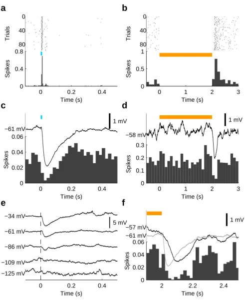
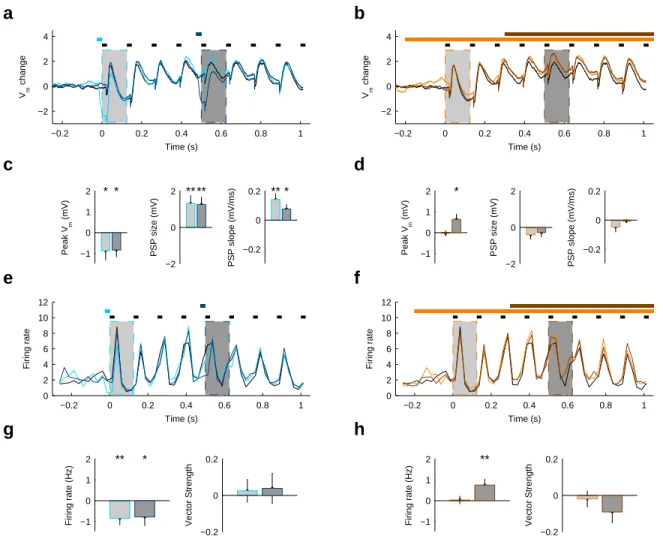
In chapter 4, we tested the hypothesis that SOM cells impact a distinct form of dynamics, sensory adaptation, using intracellular recordings, optogenetics and sensory stimulation. Even though the role of interneurons in sensory adaptation has thus far largely been dismissed, in vitro studies have shown that SOM cells are well positioned to contribute to this phenomenon. Our findings confirm the previous findings in vivo and present a previously unappreciated mechanism for controlling sensory adaptation.
1.6 References
Amitai, Y., Gibson, J. R., Beierlein, M., Patrick, S. L., Ho, A. M., Connors, B. W., Golomb, D., 2002. The spatial dimensions of electrically coupled networks of interneurons in the neocortex. J. Neurosci. 22 (10), 4142–4152.
Ascoli, G. A., Alonso-Nanclares, L., Anderson, S. A., Barrionuevo, G., Benavides-Piccione, R., Burkhalter, A., Buzsáki, G., Cauli, B., DeFelipe, J., Fairén, A., Feldmeyer, D., Fishell, G., Frégnac, Y., Freund, T. F., Gardner, D., Gardner, E. P., Goldberg, J. H., Helmstaedter, M., Hestrin, S., Karube, F., Kisvárday, Z. F., Lambolez, B., Lewis, D. A., Marin, O., Markram, H., Muñoz, A., Packer, A. M., Petersen, C. C. H., Rockland, K. S., Rossier, J., Rudy, B., Somogyi,
P., 2008. Petilla terminology: nomenclature of features of GABAergic interneurons of the cerebral cortex. Nat. Rev. Neurosci. 9 (7), 557–568.
Atallah, B. V., Scanziani, M., 2009. Instantaneous modulation of gamma oscillation frequency by balancing excitation with inhibition. Neuron 62 (4), 566–577.
Azouz, R., Gray, C. M., 2000. Dynamic spike threshold reveals a mechanism for synaptic coincidence detection in cortical neurons in vivo. Proc. Natl. Acad. Sci. U.S.A. 97 (14), 8110– 8115.
Azouz, R., Gray, C. M., 2003. Adaptive coincidence detection and dynamic gain control in visual cortical neurons in vivo. Neuron 37 (3), 513–523.
Börgers, C., Epstein, S., Kopell, N. J., 2005. Background gamma rhythmicity and attention in cortical local circuits: a computational study. Proc. Natl. Acad. Sci. U.S.A. 102 (19), 7002–7007. Börgers, C., Epstein, S., Kopell, N. J., 2008. Gamma oscillations mediate stimulus competition and attentional selection in a cortical network model. Proc. Natl. Acad. Sci. U.S.A. 105 (46), 18023–18028.
Börgers, C., Kopell, N. J., 2003. Synchronization in networks of excitatory and inhibitory neurons with sparse, random connectivity. Neural Comput. 15 (3), 509–538.
Börgers, C., Kopell, N. J., 2005. Effects of noisy drive on rhythms in networks of excitatory and inhibitory neurons. Neural Comput. 17 (3), 557–608.
Börgers, C., Kopell, N. J., 2008. Gamma oscillations and stimulus selection. Neural Comput. 20 (2), 383–414.
Bartos, M., Vida, I., Jonas, P., 2007. Synaptic mechanisms of synchronized gamma oscillations in inhibitory interneuron networks. Nat. Rev. Neurosci. 8 (1), 45–56.
Beierlein, M., Gibson, J. R., Connors, B. W., 2003. Two dynamically distinct inhibitory networks in layer 4 of the neocortex. J. Neurophysiol. 90 (5), 2987–3000.
Berger, T. K., Perin, R., Silberberg, G., Markram, H., 2009. Frequency-dependent disynaptic inhibition in the pyramidal network: a ubiquitous pathway in the developing rat neocortex. J. Physiol. (Lond.) 587 (22), 5411–5425.
Bruno, R. M., Sakmann, B., 2006. Cortex is driven by weak but synchronously active thalamocortical synapses. Science 312 (5780), 1622–1627.
Buia, C. I., Tiesinga, P. H., 2006. Attentional modulation of firing rate and synchrony in a model cortical network. J. Comput. Neurosci. 20 (3), 247–264.
Burchell, T. R., Faulkner, H. J., Whittington, M. A., 1998. Gamma frequency oscillations gate temporally coded afferent inputs in the rat hippocampal slice. Neurosci. Lett. 255 (3), 151–154. Burkhalter, A., 2008. Many specialists for suppressing cortical excitation. Front Neurosci. 2 (2), 155–167.
Cardin, J. A., Carlén, M., Meletis, K., Knoblich, U., Zhang, F., Deisseroth, K., Tsai, L.-H., Moore, C. I., 2009. Driving fast-spiking cells induces gamma rhythm and controls sensory responses. Nature 459 (7247), 663–667.
Cauli, B., Audinat, E., Lambolez, B., Angulo, M. C., Ropert, N., Tsuzuki, K., Hestrin, S., Rossier, J., 1997. Molecular and physiological diversity of cortical nonpyramidal cells. J. Neurosci. 17 (10), 3894–3906.
Chalk, M., Herrero, J. L., Gieselmann, M. A., Delicato, L. S., Gotthardt, S., Thiele, A., 2010. Attention reduces stimulus-driven gamma frequency oscillations and spike field coherence in V1. Neuron 66 (1), 114–125.
Engel, A. K., König, P., Kreiter, A. K., Singer, W., 1991. Interhemispheric synchronization of oscillatory neuronal responses in cat visual cortex. Science 252 (5010), 1177–1179.
Fisahn, A., Pike, F. G., Buhl, E. H., Paulsen, O., 1998. Cholinergic induction of network oscillations at 40 Hz in the hippocampus in vitro. Nature 394 (6689), 186–189.
Freeman, W. J., 1968. Relations between unit activity and evoked potentials in prepyriform cortex of cats. J. Neurophysiol. 31 (3), 337–348.
Fries, P., 2009. Neuronal gamma-band synchronization as a fundamental process in cortical computation. Annu. Rev. Neurosci. 32, 209–224.
Fries, P., Reynolds, J. H., Rorie, A. E., Desimone, R., 2001. Modulation of oscillatory neuronal synchronization by selective visual attention. Science 291 (5508), 1560–1563.
Fries, P., Womelsdorf, T., Oostenveld, R., Desimone, R., 2008. The effects of visual stimulation and selective visual attention on rhythmic neuronal synchronization in macaque area V4. J. Neurosci. 28 (18), 4823–4835.
Gabernet, L., Jadhav, S. P., Feldman, D. E., Carandini, M., Scanziani, M., 2005. Somatosensory integration controlled by dynamic thalamocortical feed-forward inhibition. Neuron 48 (2), 315– 327.
Gibson, J. R., Beierlein, M., Connors, B. W., 1999. Two networks of electrically coupled inhibitory neurons in neocortex. Nature 402 (6757), 75–79.
Gonchar, Y., Burkhalter, A., 1997. Three distinct families of GABAergic neurons in rat visual cortex. Cereb. Cortex 7 (4), 347–358.
Gonchar, Y., Wang, Q., Burkhalter, A., 2007. Multiple distinct subtypes of GABAergic neurons in mouse visual cortex identified by triple immunostaining. Front. Neuroanat. 1.
Gray, C. M., Singer, W., 1989. Stimulus-specific neuronal oscillations in orientation columns of cat visual cortex. Proc. Natl. Acad. Sci. U.S.A. 86 (5), 1698–1702.
Gregoriou, G. G., Gotts, S. J., Zhou, H.-H., Desimone, R., 2009. High-frequency, long-range coupling between prefrontal and visual cortex during attention. Science 324 (5931), 1207–1210. Gruber, T., Müller, M. M., Keil, A., Elbert, T., 1999. Selective visual-spatial attention alters induced gamma band responses in the human EEG. Clin Neurophysiol 110 (12), 2074–2085. Halabisky, B., Shen, F., Huguenard, J. R., Prince, D. A., 2006. Electrophysiological classification of somatostatin-positive interneurons in mouse sensorimotor cortex. J. Neurophysiol. 96 (2), 834–845.
Hasenstaub, A. R., Shu, Y., Haider, B., Kraushaar, U., Duque, A., McCormick, D. A., 2005. Inhibitory postsynaptic potentials carry synchronized frequency information in active cortical networks. Neuron 47 (3), 423–435.
Hendry, S. H., Jones, E. G., Emson, P. C., 1984. Morphology, distribution, and synaptic relations of somatostatin- and neuropeptide Y-immunoreactive neurons in rat and monkey neocortex. J. Neurosci. 4 (10), 2497–2517.
hua Liu, B., Li, P., tang Li, Y., Sun, Y. J., Yanagawa, Y., Obata, K., Zhang, L. I., Tao, H. W., 2009. Visual receptive field structure of cortical inhibitory neurons revealed by two-photon imaging guided recording. J. Neurosci. 29 (34), 10520–10532.
Inoue, T., Imoto, K., 2006. Feedforward inhibitory connections from multiple thalamic cells to multiple regular-spiking cells in layer 4 of the somatosensory cortex. J. Neurophysiol. 96 (4), 1746–1754.
Jensen, O., Kaiser, J., Lachaux, J.-P., 2007. Human gamma-frequency oscillations associated with attention and memory. Trends Neurosci. 30 (7), 317–324.
König, P., Engel, A. K., Singer, W., 1996. Integrator or coincidence detector? The role of the cortical neuron revisited. Trends Neurosci. 19 (4), 130–137.
Kapfer, C., Glickfeld, L. L., Atallah, B. V., Scanziani, M., 2007. Supralinear increase of recurrent inhibition during sparse activity in the somatosensory cortex. Nat. Neurosci. 10 (6), 743–753.
Kawaguchi, Y., Kubota, Y., 1996. Physiological and morphological identification of somatostatin- or vasoactive intestinal polypeptide-containing cells among GABAergic cell subtypes in rat frontal cortex. J. Neurosci. 16 (8), 2701–2715.
Kawaguchi, Y., Kubota, Y., 1997. GABAergic cell subtypes and their synaptic connections in rat frontal cortex. Cereb. Cortex 7 (6), 476–486.
Kawaguchi, Y., Kubota, Y., 1998. Neurochemical features and synaptic connections of large physiologically-identified GABAergic cells in the rat frontal cortex. Neuroscience 85 (3), 677– 701.
Kubota, Y., Hattori, R., Yui, Y., 1994. Three distinct subpopulations of GABAergic neurons in rat frontal agranular cortex. Brain Res. 649 (1), 159–173.
Ma, Y., Hu, H., Berrebi, A. S., Mathers, P. H., Agmon, A., 2006. Distinct subtypes of somatostatin-containing neocortical interneurons revealed in transgenic mice. J. Neurosci. 26 (19), 5069–5082.
Maldonado, P. E., Friedman-Hill, S., Gray, C. M., 2000. Dynamics of striate cortical activity in the alert macaque: II. Fast time scale synchronization. Cereb. Cortex 10 (11), 1117–1131.
Maloney, K. J., Cape, E. G., Gotman, J., Jones, B. E., 1997. High-frequency gamma electroencephalogram activity in association with sleep-wake states and spontaneous behaviors in the rat. Neuroscience 76 (2), 541–555.
Markram, H., Toledo-Rodriguez, M., Wang, Y., Gupta, A., Silberberg, G., Wu, C., 2004. Interneurons of the neocortical inhibitory system. Nat. Rev. Neurosci. 5 (10), 793–807.
McGarry, L. M., Packer, A. M., Fino, E., Nikolenko, V., Sippy, T., Yuste, R., 2010. Quantitative classification of somatostatin-positive neocortical interneurons identifies three interneuron subtypes. Front Neural Circuits 4.
Melchitzky, D. S., Lewis, D. A., 2008. Dendritic-targeting GABA neurons in monkey prefrontal cortex: comparison of somatostatin- and calretinin-immunoreactive axon terminals. Synapse 62 (6), 456–465.
Moore, C. I., Carlén, M., Knoblich, U., Cardin, J. A., 2010. Neocortical interneurons: from diversity, strength. Cell 142 (2), 189–193.
Moore, C. I., Nelson, S. B., 1998. Spatio-temporal subthreshold receptive fields in the vibrissa representation of rat primary somatosensory cortex. J. Neurophysiol. 80 (6), 2882–2892.
Murayama, M., Pérez-Garci, E., Nevian, T., Bock, T., Senn, W., Larkum, M. E., 2009. Dendritic encoding of sensory stimuli controlled by deep cortical interneurons. Nature 457 (7233), 1137– 1141.
Nase, G., Singer, W., Monyer, H., Engel, A. K., 2003. Features of neuronal synchrony in mouse visual cortex. J. Neurophysiol. 90 (2), 1115–1123.
Paik, S.-B., Kumar, T., Glaser, D. A., 2009. Spontaneous local gamma oscillation selectively enhances neural network responsiveness. PLoS Comput. Biol. 5 (3).
Pinto, D. J., Brumberg, J. C., Simons, D. J., 2000. Circuit dynamics and coding strategies in rodent somatosensory cortex. J. Neurophysiol. 83 (3), 1158–1166.
Pouille, F., Marin-Burgin, A., Adesnik, H., Atallah, B. V., Scanziani, M., 2009. Input normalization by global feedforward inhibition expands cortical dynamic range. Nat. Neurosci. 12 (12), 1577–1585.
Pouille, F., Scanziani, M., 2001. Enforcement of temporal fidelity in pyramidal cells by somatic feed-forward inhibition. Science 293 (5532), 1159–1163.
Pouille, F., Scanziani, M., 2004. Routing of spike series by dynamic circuits in the hippocampus. Nature 429 (6993), 717–723.
Reyes, A. D., Lujan, R., Rozov, A., Burnashev, N., Somogyi, P., Sakmann, B., 1998. Target-cell-specific facilitation and depression in neocortical circuits. Nat. Neurosci. 1 (4), 279–285.
Ribary, U., Ioannides, A. A., Singh, K. D., Hasson, R., Bolton, J. P., Lado, F., Mogilner, A., Llinas, R. R., 1991. Magnetic field tomography of coherent thalamocortical 40-Hz oscillations in humans. Proc. Natl. Acad. Sci. U.S.A. 88 (24), 11037–11041.
Roy, A., Steinmetz, P. N., Hsiao, S. S., Johnson, K. O., Niebur, E., 2007. Synchrony: a neural correlate of somatosensory attention. J. Neurophysiol. 98 (3), 1645–1661.
Silberberg, G., Markram, H., 2007. Disynaptic inhibition between neocortical pyramidal cells mediated by Martinotti cells. Neuron 53 (5), 735–746.
Steinmetz, P. N., Roy, A., Fitzgerald, P. J., Hsiao, S. S., Johnson, K. O., Niebur, E., 2000. Attention modulates synchronized neuronal firing in primate somatosensory cortex. Nature 404 (6774), 187–190.
Stimberg, M., Wimmer, K., Martin, R., Schwabe, L., Mariño, J., Schummers, J., Lyon, D. C., Sur, M., Obermayer, K., 2009. The Operating Regime of Local Computations in Primary Visual Cortex. Cereb. Cortex.
Sun, Y. J., Wu, G. K., hua Liu, B., Li, P., Zhou, M., Xiao, Z., Tao, H. W., Zhang, L. I., 2010. Fine-tuning of pre-balanced excitation and inhibition during auditory cortical development. Nature 465 (7300), 927–931.
Swadlow, H. A., 2003. Fast-spike interneurons and feedforward inhibition in awake sensory neocortex. Cereb. Cortex 13 (1), 25–32.
Tallon-Baudry, C., Bertrand, O., 1999. Oscillatory gamma activity in humans and its role in object representation. Trends Cogn. Sci. (Regul. Ed.) 3 (4), 151–162.
Tallon-Baudry, C., Bertrand, O., Delpuech, C., Permier, J., 1997. Oscillatory gamma-band (30-70 Hz) activity induced by a visual search task in humans. J. Neurosci. 17 (2), 722–734.
Tan, A. Y. Y., Wehr, M., 2009. Balanced tone-evoked synaptic excitation and inhibition in mouse auditory cortex. Neuroscience 163 (4), 1302–1315.
Tan, Z., Hu, H., Huang, Z. J., Agmon, A., 2008. Robust but delayed thalamocortical activation of dendritic-targeting inhibitory interneurons. Proc. Natl. Acad. Sci. U.S.A. 105 (6), 2187–2192. Tiesinga, P. H., Sejnowski, T. J., 2009. Cortical enlightenment: are attentional gamma oscillations driven by ING or PING. Neuron 63 (6), 727–732.
Traub, R. D., Contreras, D., Cunningham, M. O., Murray, H., LeBeau, F. E. N., Roopun, A. K., Bibbig, A., Wilent, W. B., Higley, M. J., Whittington, M. A., 2005. Single-column thalamocortical network model exhibiting gamma oscillations, sleep spindles, and epileptogenic bursts. J. Neurophysiol. 93 (4), 2194–2232.
Tsao, D. Y., Freiwald, W. A., Tootell, R. B. H., Livingstone, M. S., 2006. A cortical region consisting entirely of face-selective cells. Science 311 (5761), 670–674.
Vogels, T. P., Abbott, L. F., 2009. Gating multiple signals through detailed balance of excitation and inhibition in spiking networks. Nat. Neurosci. 12 (4), 483–491.
Wang, H.-P., Spencer, D., Fellous, J.-M., Sejnowski, T. J., 2010. Synchrony of thalamocortical inputs maximizes cortical reliability. Science 328 (5974), 106–109.
Wang, X.-J., Buzsáki, G., 1996. Gamma oscillation by synaptic inhibition in a hippocampal interneuronal network model. J. Neurosci. 16 (20), 6402–6413.
Wang, Y., Toledo-Rodriguez, M., Gupta, A., Wu, C., Silberberg, G., Luo, J., Markram, H., 2004. Anatomical, physiological and molecular properties of Martinotti cells in the somatosensory cortex of the juvenile rat. J. Physiol. (Lond.) 561 (1), 65–90.
Wehr, M., Zador, A. M., 2003. Balanced inhibition underlies tuning and sharpens spike timing in auditory cortex. Nature 426 (6965), 442–446.
Whittington, M. A., Traub, R. D., Kopell, N. J., Ermentrout, G. B., Buhl, E. H., 2000. Inhibition-based rhythms: experimental and mathematical observations on network dynamics. Int J Psychophysiol 38 (3), 315–336.
Wilent, W. B., Contreras, D., 2005. Dynamics of excitation and inhibition underlying stimulus selectivity in rat somatosensory cortex. Nat. Neurosci. 8 (10), 1364–1370.
Womelsdorf, T., Fries, P., Mitra, P. P., Desimone, R., 2006. Gamma-band synchronization in visual cortex predicts speed of change detection. Nature 439 (7077), 733–736.
Xu, X., Roby, K. D., Callaway, E. M., 2006. Mouse cortical inhibitory neuron type that coexpresses somatostatin and calretinin. J. Comp. Neurol. 499 (1), 144–160.
Xu, X., Roby, K. D., Callaway, E. M., 2010. Immunochemical characterization of inhibitory mouse cortical neurons: three chemically distinct classes of inhibitory cells. J. Comp. Neurol. 518 (3), 389–404.
Chapter 2:
Driving fast-spiking cells induces
gamma rhythm and controls sensory
responses
This chapter has previously been published as:
Jessica A. Cardin*, Marie Carlén*, Konstantinos Meletis, Ulf Knoblich, Feng Zhang, Karl Deisseroth, Li-Huei Tsai and Christopher I. Moore (2009). Driving fast-spiking cells induces gamma rhythm and controls sensory responses. Nature 459 (7247), 663–667.
I performed and analyzed the intracellular in vivo recordings demonstrating the subthreshold effect in pyramidal neurons of optogenetic activation of PV cells in primary somatosensory cortex, confirming perisomatic inhibitory post-synaptic potentials most likely mediated by GABAA. These results are shown in Figure 2b and 2c of the manuscript.
2.1 Abstract
Gamma oscillations at 20-80Hz in the cortex predict increased attention1 and are thought to bind sensory representations into coherent percepts2. Failed regulation of these oscillations is a hallmark of neurological and psychiatric diseases, including schizophrenia3-5. Current theory predicts that gamma oscillations are generated by synchronous activity of fast-spiking (FS) inhibitory interneurons6-9, with the time course of FS-evoked inhibition leading to selective expression of 20-80 Hz activity in the local network. This synchronous inhibition is further predicted to create a narrow window for effective excitation, creating synchrony in neural ensembles. We causally tested these hypotheses in the in vivo barrel cortex (SI) by targeting optogenetic manipulation selectively to FS interneurons. By pulsing light across a broad range of frequencies (8-200 Hz), we directly linked activation of fast spiking inhibition and gamma oscillations (20-80 Hz). In contrast, activation of pyramidal neurons using a parallel approach amplified only lower frequency bands (<24 Hz). This cell type-specific double dissociation provides direct in vivo evidence that synchronous FS inhibition generates cortical gamma oscillations. This approach further allowed us to test the prediction that gamma oscillations temporally regulate sensory transmission. In agreement with this hypothesis, we find that the fine timing of sensory input relative to a cycle of gamma determines the amplitude, timing and precision of evoked sensory activity in SI. Our data provide the first causal evidence that a network activity state can be induced in vivo by cell-type specific activation, and directly support the FS-gamma hypothesis.
2.2 Main Text
Brain states characterized by rhythmic electrophysiological activity have been studied intensively for over 80 years10,11. Because these brain rhythms are believed to be essential to information processing, many theories have been proposed to explain their origin, with several emphasizing the activity of neuronal sub-types. One of the strongest cases yet made for the importance of a specific cell type in rhythm induction is the suggested role of FS interneurons in gamma (oscillations8,12 Networks of FS cells connected by gap junctions13,14 provide large,
synchronous inhibitory postsynaptic potentials (IPSPs) to local excitatory neurons15. Computational modeling suggests that this synchronous activity is sufficient to induce 20-80 Hz
oscillations that are stabilized and regulated by fast excitatory feedback from pyramidal neurons16,17. Cortical recordings in vivo show sensory-evoked oscillations in the local field potential (LFP) and phase-locked firing of excitatory pyramidal cells, suggesting entrainment of excitatory neurons to rhythmic inhibitory activity15,18. Despite considerable study of cortical oscillations, and the importance of understanding their origins, induction of a given network state by stimulation of specific neural cell types in vivo has not previously been possible.
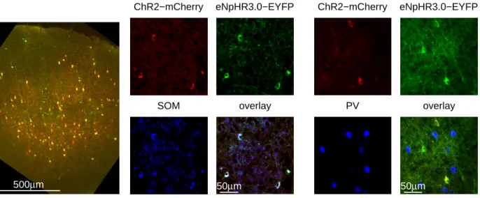
To directly test the hypothesis that FS interneuron activity in an in vivo cortical circuit is sufficient to induce oscillations, we used the light-sensitive bacteriorhodopsin Chlamydomonas reinhardtii Channelrhodopsin-2 (ChR2), a cation channel activated by ~ 470 nm blue light19,20. We targeted expression of ChR2 specifically to parvalbumin-positive fast-spiking (FS-PV+) interneurons by injecting the adeno-associated viral vector AAV Double-floxed Inverted Open reading frame-ChR2-mCherry (AAV DIO ChR2-mCherry), with Cre-dependent expression of ChR2, into PV-Cre knock-in mice (Fig. 1a; Supplementary Fig. 1-2 and Methods)21,22. Six days after virus injection into barrel cortex of adult PV-Cre mice, ChR2-mCherry expression covered an anterioposterior distance up to 1740 m (1695 57.4 m, mean SD, n = 3), resulting in robust labeling of PV+ interneurons across cortical layers (Fig. 1b). The labeling efficiency of AAV DIO ChR2-mCherry varied over distance from the injection site; close to the center of the injection, >97% of the PV+ interneurons expressed ChR2-mCherry. Immunohistochemistry confirmed that 96.7 1.0% (mean SD, n = 4234 ChR2-mCherry+ neurons, 4 animals) of the
ChR2-mCherry+ neurons expressed PV (Fig. 1d-e, Supplementary Fig. 2), and almost all expressed the inhibitory neurotransmitter gamma-aminobutyric acid (GABA) (Supplementary Fig. 3)23,24. Expression of mCherry was not induced after injection of AAV DIO ChR2-mCherry into wild-type mice (data not shown) or in vitro in the absence of Cre (see Supplementary Methods; data not shown).
In experiments targeting excitatory neurons, AAV DIO ChR2-mCherry was injected into the barrel cortex of adult CW2 mice25 that express Cre from the CamKII promoter (‘CamKII-Cre mice’), inducing recombination in excitatory neurons in cortex25. Robust ChR2-mCherry expression was observed in excitatory neurons in a laminar profile corresponding to the Cre expression pattern25 (Fig. 1c, Supplementary Fig. 4). At least 50% of the CamKII+ neurons in layer 2/3 expressed ChR2-mCherry (913 of 1638 cells in a total area of 8.4 x 106 µm3) close to the injection site, covering an anterioposterior distance of 1560 154.9 m (mean SD, n = 3).
Immunohistochemical analysis revealed that 100 0% (mean SD, n = 4024 ChR2-mCherry+
neurons, 4 animals) of the ChR2-mCherry expressing neurons were immuno-negative for PV (Fig. 1f-g, Supplementary Fig. 2), and 100 0% expressed the neuronal marker NeuN (data not shown).
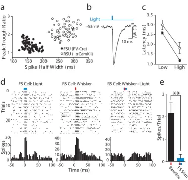
We recorded light-activated FS and regular spiking (RS) single units in layers 2/3 and 4 of barrel cortex (SI) in PV-Cre (n = 64 FS in 15 animals) and CamKII-Cre (n = 56 RS in 7 animals) mice. We did not observe light activation of layer 5 FS cells (n = 12 sites in 7 animals). Barrel cortex, which processes information from the rodent vibrissae (whiskers), was targeted as a well-defined model of basic sensory cortical function. In agreement with the immunohistological results, the action potential shapes of the neurons activated by light pulses were differentiated into two discrete populations based on mouse type, PV-Cre/FS and CamKII-Cre/RS (p < 0.01; Fig. 2a).
To confirm the activation of inhibitory interneurons and their postsynaptic impact on excitatory neurons, we performed in vivo intracellular recordings of RS cells in barrel cortex in PV-Cre mice (n = 5). We found that a 1 ms light pulse was sufficient to evoke large, fast IPSPs, confirming direct synaptic inhibition of RS cells by light-activated FS cells (Fig. 2b). The latencies of the presynaptic light-evoked FS spikes agreed well with the onset times of the postsynaptic IPSPs, with FS spikes preceding IPSP onset by 0.5 to 0.75 ms (Fig. 2c). Both the time to peak and the peak timing variability of the evoked IPSPs decreased with increasing light pulse power (Fig. 2c). Mean IPSP peak amplitude at membrane potentials of -55 to –60 mV was 2.7 1.0 mV. The mean reversal potential of the evoked IPSPs (see Supplementary Methods) was –67.6 1.9 mV, indicating a GABAA-mediated Cl- conductance characteristic of FS
synapses. Consistent with IPSP induction, activation of FS cells blocked vibrissa-evoked responses in neighboring RS cells (Figure 2d-e; n = 6 sites in 5 PV-Cre mice).
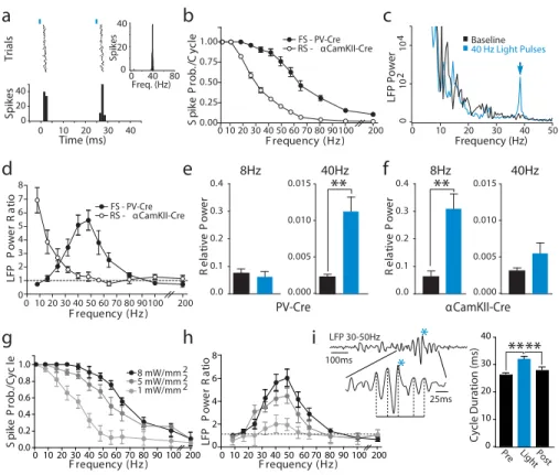
A strong prediction of the FS-hypothesis is that synchronously active FS cells are sufficient for induction. This hypothesis predicts that light pulses presented at a broad range of frequencies should reveal a selective peak in enhancement of the LFP, a measure of synchronous local network activity26, when FS cells are driven in the range.
To test this hypothesis, we drove cortical FS cell spiking in virus-transduced PV-Cre mice at a range of frequencies (8 to 200 Hz) with 1 ms light pulses. Light pulses in the range (40 Hz)
resulted in reliable action potential output at 25 ms intervals (Fig. 3a). Across the population, FS and RS cells were driven with equally high reliability by light pulses at low frequencies (Fig. 3b). At higher frequencies, spike probability on each light cycle remained high for FS cells but decreased for RS cells.
Driving FS cells at 40 Hz caused a specific increase in the 35-40 Hz frequency band in the LFP (Fig. 3c, Supplementary Fig. 5-6). We found that activation of FS cells in the 20-80 Hz range resulted in significant amplification of LFP power at those frequencies (n = 14 sites in 6 animals; Fig. 3d). However, activation of FS cells at lower frequencies did not affect LFP power, despite robust evoked FS firing on every light cycle. In contrast, 8-24 Hz light activation of RS cells in CamKII-Cre mice induced increased LFP power at these frequencies, but RS activation at higher frequencies did not affect LFP power (n = 13 sites in 5 mice; Fig. 3d, Suppl. Fig. 5). Light stimulation in the untransduced contralateral barrel cortex did not affect LFP power at any frequency (n = 6 PV-Cre and 5 CamKII-Cre animals; Supplementary Fig. 6).
This double dissociation of cell-type specific state induction (by FS and lower frequencies by RS) directly supports the prediction that FS-PV+ interneuron activation is sufficient and specific for induction of oscillations. To highlight this distinction, we compared the effects of stimulating the two cell types at 8 and 40 Hz. Stimulation of FS cells at 8 Hz in the PV-Cre mice had no effect on LFP power at 8Hz, but FS stimulation at 40 Hz caused a significant increase in 40 Hz LFP power (paired t-test; p < 0.001; Fig. 3e). In contrast, stimulation of RS cells at 8 Hz in the CamKII-Cre mice caused a significant elevation of LFP power at 8 Hz (p < 0.001), whereas RS stimulation at 40 Hz caused only a small, nonsignificant increase in 40 Hz LFP power (Fig. 3f).
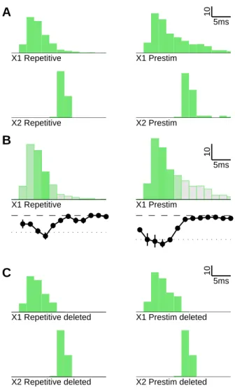
One possible explanation for these results is that increased FS firing recruits resonant range activity in the surrounding local network as a function of the synaptic and biophysical properties of the cortical circuit. Alternatively, the increase in activity may result from the specific level of evoked FS spiking, and changing spiking probability would shift the frequency of the enhanced LFP band. To discriminate between these possibilities, we stimulated FS cells at varying levels of light intensity. We found that FS spike probability changed with light intensity such that the spike probability curve shifted laterally (Fig. 3g). While drive impacted the amplitude of enhancement, LFP power was selectively amplified within the range regardless of light intensity or spike probability (Fig. 3h), indicating that the oscillations evoked by FS
activity are a resonant circuit property. In addition, randomly patterned light stimulation of FS cells with frequencies evenly distributed across a broad range evoked a significant increase in LFP power specific to the range (n = 7 sites in 4 animals; p < 0.05; Supplementary Fig. 7), further indicating that FS-evoked oscillations are an emergent property of the circuit, and do not require exclusive drive in the range.
To test whether intrinsically occurring oscillations show a similar dependence on FS activity, we gave single light pulses during epochs of natural . We found that brief FS activation shifted the phase of both spontaneously occurring oscillations (n = 26 trials, 4 animals; Kruskal-Wallis test with Dunn’s post-test; p < 0.01; Fig. 3i) and those evoked by midbrain reticular formation stimulation (n = 18 trials, 2 animals; p< 0.05; Supplementary Fig. 8). Further, light-induced oscillations were largely eliminated by blocking AMPA and NMDA receptors, despite high levels of evoked FS firing (n = 4 sites in 4 animals; p < 0.01; Supplementary Fig. 9). These results indicate that induced oscillations dependent on rhythmic excitatory synaptic activity, as predicted by computational models of natural oscillations and prior experiments6,15-17,27. In
further agreement, spontaneous RS activity was entrained by 40 Hz FS stimulation, resulting in RS firing during the decay phase of the IPSP and preceding subsequent evoked FS spiking (Supplementary Fig. 10).
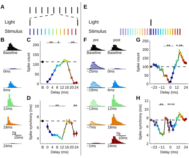
Gamma oscillations are thought to have a functional impact on cortical information processing by synchronizing the output of excitatory neurons. This synchrony selects cell assemblies involved in a common task, such as encoding a sensory stimulus, and enhances their impact on downstream targets2. The cyclical FS inhibition underlying oscillations is believed to cause this synchrony by rhythmically gating synaptic inputs2,28. Synaptic inputs arriving at the peak of inhibition should therefore produce a diminished response, but those arriving at the opposite phase in the cycle should evoke a large response.
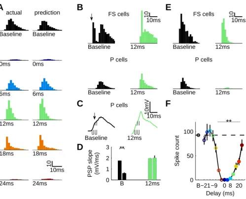
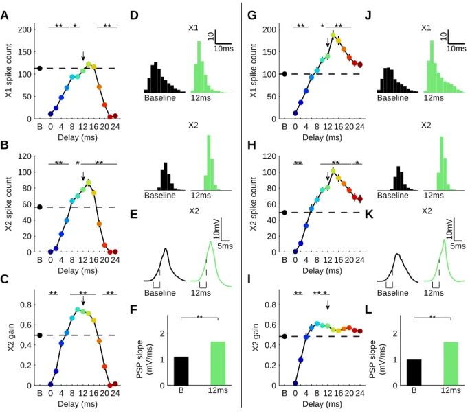
To directly test this hypothesis, we stimulated FS cells at 40 Hz with light pulses to establish oscillations, and recorded the responses of RS cells to a single vibrissa deflection presented at one of five phases relative to a single cycle (n = 20 cells in 3 animals; Fig. 4a). The timing of vibrissa-induced RS action potentials relative to light-evoked inhibition and the cycle had a significant impact on the amplitude, timing and precision of the sensory-evoked responses of RS cells (Fig. 4b-c). The presence of oscillations significantly decreased the amplitude of the RS
sensory response at three phase points, consistent with the enhanced level of overall inhibition in this state (p < 0.05; 1-way ANOVA with Dunnett’s post-test; Fig. 4d)29. Gamma phase also modulated the overall timing of the sensory response (p < 0.01; Fig. 4e), with spike latency delayed at phases 1-3 and unaffected at phases 4-529. The precision of sensory-evoked spikes was significantly enhanced in a phase-dependent manner (p < 0.01; Fig. 4f). Our results suggest that the rhythmic, FS-induced IPSP restricts sensory transmission during its peak, and permits transmission after its decay, leading to a temporal sharpening of cortical sensory responses (Fig. 4g).
Our results provide the first causal demonstration of cortical oscillations induced by cell type-specific activation. Synchronous FS-PV+ interneuron activity driven by periodic stimulation of light-activated channels generated oscillations in a cortical network, and these gated sensory processing in a temporally specific manner. These findings also demonstrate a unique application of optogenetic engineering in the in vivo brain for the study of discrete neuronal cell types under active network conditions. Future use of these techniques will allow direct testing of the impact of brain states on information processing in the behaving animal30, and potentially the rescue of functional states in models of brain disease3,4.
2.3 Methods
AAV DIO ChR2-mCherry was injected into barrel cortex in adult PV-Cre21 and CW225 mice. Six days after injection, a subset of mice was perfused and tissue sectioned for immunohistochemistry to confirm location, transduction efficiency, and cell type specificity of ChR2 expression. Spread and immunohistochemical analysis of ChR2-mCherry expression was scored by hand through examination of every 30 µm coronal section for the presence of mCherry fluorescence. For electrophysiology, mice were anesthetized with isoflurane, and extracellular single-unit and local field potential recordings were made in layers 2/3 and 4 of barrel cortex with tetrodes or stereotrodes. Intracellular recordings were made in the whole-cell configuration. Light pulses were given via an optical fiber positioned at the cortical surface directly above the recording site. For experiments using a broad range of light stimulation frequencies, we stimulated in bouts of 3 sec at each frequency in random order. For each stimulation frequency, we measured relative power in an 8 Hz band centered on that frequency. Relative power was calculated by measuring the ratio of power within the band of interest to total power in the power
spectrum. To illustrate the impact of stimulation on the LFP, we also measured the ratio of power in a band with light stimulation to power in the band under baseline conditions. Vibrissae were stimulated by computer-controlled movements of piezoelectric wafers and consisted of single high-velocity deflections in the dorsal direction. For phase experiments, we gave a series of trials each consisting of a 1 sec series of 1 ms light pulses at 40 Hz, with a single whisker deflection after the 30th light pulse. The precise timing of the whisker deflection relative to the light pulses was varied across five phase points. All numbers are given as mean SEM, except where otherwise noted.
2.3.1 Animals
All procedures were conducted in accordance with the National Institutes of Health guidelines and with the approval of the Committee on Animal Care at MIT. PV-Cre (n = 21) and CW2 (n = 7) mice were 6-12 weeks of age at time of virus injections. Electrophysiological recordings and immunohistochemical analyses were performed 1-3 weeks after viral injections.
2.3.2 AAV vectors
ChR2 fused to the fluorescent protein mCherry was cloned in antisense direction into pAAV-MCS (Stratagene) to create AAV Double-floxed Inverted Open reading frame-ChR2-mCherry (AAV DIO ChR2-mCherry) (Fig.1a and Supplementary Fig. 1; for vector outline and sequence see www.optogentics.org). ChR2-mCherry was flanked by a pair of canonical loxP sites and a pair of mutated loxP2272 sites. A woodchuck hepatatis B virus post-transcriptional element (WPRE) was placed in sense direction 5’ of the poly-A. Adeno-associated viral particles of serotype 2 were produced by the Vector Core Facility at The University of North Carolina at Chapel Hill.
2.3.3 Virus injections
Adult PV-Cre21 or CW225 mice were anesthetized with an intraperitoneal injection of a mixture of ketamine (1.1 mg/kg) and xylazine (0.16 mg/kg). A small craniotomy was made 1.5 mm posterior to bregma and 3.0 mm lateral to the midline. Virus was delivered through a small durotomy by a glass micropipette attached to a Quintessential Stereotaxic Injector (Stoelting). The glass micropipette was lowered to 0.4 mm below the cortical surface. A bolus of 0.5 l of
virus (AAV DIO ChR2-mCherry; 2x10¹² viral molecules/ml) was injected into barrel cortex at 0.1 µl/min. The pipette was then retracted to a depth of 250 microns below the surface and an additional 0.5 µl virus was injected at the same rate. The pipette was held in place for 5 minutes after the injection before being retracted from the brain. The scalp incision was sutured, and post-injection analgesics were given to aid recovery (0.1 mg/kg Buprenex).
2.3.4 Immunohistochemistry
Mice were transcardially perfused with 100 mM PBS followed by 4% formaldehyde in PBS, and brains were post-fixed overnight at 4°C. Free floating sections (30 µm) were cut using a vibratome (Leica VT100) and incubated with blocking solution (10% donkey serum in PBS with 0.3% Triton-X 100) for 1h at room temperature, then incubated at room temperature over night with primary antibody diluted in blocking solution. The following primary antibodies were used: NeuN (Chemicon; 1:1000), Parvalbumin PVG-214 (Swant; 1:2000), GABA (Sigma; 1:4000), and CamKII (Epitomics 1:500). After washing, antibody staining was revealed using species-specific fluorophore-conjugated secondary antibodies (Cy5 from Jackson, Alexa 488 from Molecular Probes). GABA was detected with biotinylated secondary antibodies (Jackson Laboratories) and revealed using a combination of ABC kit (Vector Laboratories) and TSA fluorescent amplification kit (Perkin-Elmer). Sections were mounted on glass slides with Vectashield (Vector) and coverslipped.
Spread and labeling efficiency were scored by hand through examination of every 30 µm coronal section (n = 3 animals per genotype) for the presence of mCherry fluorescence using a Zeiss LSM510 confocal microscope. For quantification of co-labeling of ChR2-mCherry and PV (n = 4 animals per genotype) confocal images were acquired and individual cells were identified independently for each of the two fluorescent channels. Scans from each channel were collected in multi-track mode to avoid cross-talk between channels.
2.3.5 Electrophysiology
Mice were anesthetized with isoflurane and held in place with a head post cemented to the skull. All incisions were infiltrated with lidocaine. A small craniotomy was made over barrel cortex approx 200 m anterior to the virus injection site. Extracellular single-unit and local field potential recordings were made with tetrodes or stereotrodes. Intracellular recordings were
conducted by whole cell in vivo recording in current clamp mode. Stimulus control and data acquisition was performed using software custom written in LabView (National Instruments, Austin TX) and Matlab (The Mathworks, Natick MA) by U.K. Further electrophysiology methods and a description of the reversal potential calculation are given in Supplementary Methods.
Light stimulation was generated by a 473nm laser (Shanghai Dream Lasers, Shanghai, China) controlled by Grass stimulator (Grass Technologies, West Warwick, RI) or computer. Light pulses were given via a 200m diameter, unjacketed optical fiber (Ocean Optics, Dunedin FL) positioned at the cortical surface directly above the recording site. For experiments using the broad range of light stimulation frequencies (8, 16, 24, 32, 40, 48, 80, 100, and 200 Hz), we stimulated in bouts of 3 sec of 1 ms pulses at 4 mW/mm2 at each frequency in random order. In a subset of these experiments, we stimulated at 1, 4 and 8 mW/mm2.
Vibrissae were stimulated by computer-controlled movements of piezoelectric wafers (Piezo Systems). Vibrissa stimulations were single high-velocity deflections in the dorsal then ventral direction (~6 ms duration). In most cases, adjacent vibrissae that yielded indistinguishable amplitude responses during hand mapping were deflected simultaneously. Vibrissa stimulations evoked RS spike responses with an onset latency of 9.1 .08 ms. For RS cell response suppression experiments, light pulses were given on randomly interleaved trials. For phase experiments, we gave a series of trials each consisting of a 1 sec series of 1 ms light pulses at 40 Hz, with a single whisker deflection after the 30th light pulse. The precise timing of the whisker deflection relative to the light pulses was varied across five phase points. Each of the five phase points was included in random order across a minimum of 250 total trials.
Unit and local field potential analysis used software custom written in Igor Pro (Wavemetrics, Portland OR) by J.A.C. For each stimulation frequency, we measured relative power in an 8 Hz band centered on that frequency. For each recording site, we measured power from 5-10 LFP traces under each condition. Example power spectra are averages of the power spectra from 5-10 traces of unfiltered LFPs from individual experiments. Relative power was calculated by measuring the ratio of power within the band of interest to total power in the power spectrum of the unfiltered LFP. We also measured the power ratio Plight/ Pbaseline where Plight is the relative
band in the absence of light stimulation. All numbers are given as mean SEM, except where otherwise noted.
2.4 Author Contributions
J.A.C., M.C., K.M., L.-H.T., and C.I.M. designed the experiments. F.Z. and K.D. designed and cloned the AAV DIO ChR2-mCherry vector. M.C. and K.M. characterized the virus in vitro and in vivo and injected the animals. M.C performed histological statistical analyses. J.A.C. performed and analyzed the extracellular recordings. U.K. and J.A.C. performed the intracellular recordings. U.K. analyzed the intracellular data. J.A.C., M.C., K.M., U.K., L.-H.T., and C.I.M. wrote the manuscript.
2.5 References
1. Fries, P., Reynolds, J. H., Rorie, A. E. & Desimone, R. Modulation of oscillatory neuronal synchronization by selective visual attention. Science 291, 1560-3 (2001).
2. Engel, A. K. & Singer, W. Temporal binding and the neural correlates of sensory awareness. Trends Cogn Sci 5, 16-25 (2001).
3. Orekhova, E. V. et al. Excess of high frequency electroencephalogram oscillations in boys with autism. Biol Psychiatry 62, 1022-9 (2007).
4. Spencer, K. M., Niznikiewicz, M. A., Shenton, M. E. & McCarley, R. W. Sensory-evoked gamma oscillations in chronic schizophrenia. Biol Psychiatry 63, 744-7 (2008).
5. Uhlhaas, P. J., Haenschel, C., Nikolic, D. & Singer, W. The role of oscillations and synchrony in cortical networks and their putative relevance for the pathophysiology of schizophrenia. Schizophr Bull 34, 927-43 (2008).
6. Traub, R. D., Jefferys, J. G. & Whittington, M. A. Simulation of gamma rhythms in networks of interneurons and pyramidal cells. J Comput Neurosci 4, 141-50 (1997).
7. Traub, R. D., Whittington, M. A., Stanford, I. M. & Jefferys, J. G. A mechanism for generation of long-range synchronous fast oscillations in the cortex. Nature 383, 621-4 (1996). 8. Whittington, M. A., Traub, R. D. & Jefferys, J. G. Synchronized oscillations in interneuron networks driven by metabotropic glutamate receptor activation. Nature 373, 612-5 (1995).
9. Wang, X. J. & Buzsaki, G. Gamma oscillation by synaptic inhibition in a hippocampal interneuronal network model. J Neurosci 16, 6402-13 (1996).
10. Steriade, M. Grouping of brain rhythms in corticothalamic systems. Neuroscience 137, 1087-106 (2006).
11. Berger, H. On the electroencephalogram of man. Electroencephalogr Clin Neurophysiol, Suppl 28:37+ (1969).
12. Whittington, M. A., Faulkner, H. J., Doheny, H. C. & Traub, R. D. Neuronal fast oscillations as a target site for psychoactive drugs. Pharmacol Ther 86, 171-90 (2000).
13. Deans, M. R., Gibson, J. R., Sellitto, C., Connors, B. W. & Paul, D. L. Synchronous activity of inhibitory networks in neocortex requires electrical synapses containing connexin36. Neuron 31, 477-85 (2001).
14. Galarreta, M. & Hestrin, S. A network of fast-spiking cells in the neocortex connected by electrical synapses. Nature 402, 72-5 (1999).
15. Hasenstaub, A. et al. Inhibitory postsynaptic potentials carry synchronized frequency information in active cortical networks. Neuron 47, 423-35 (2005).
16. Whittington, M. A., Traub, R. D., Faulkner, H. J., Stanford, I. M. & Jefferys, J. G. Recurrent excitatory postsynaptic potentials induced by synchronized fast cortical oscillations. Proc Natl Acad Sci U S A 94, 12198-203 (1997).
17. Borgers, C., Epstein, S. & Kopell, N. J. Background gamma rhythmicity and attention in cortical local circuits: a computational study. Proc Natl Acad Sci U S A 102, 7002-7 (2005). 18. Gray, C. M. & Singer, W. Stimulus-specific neuronal oscillations in orientation columns of cat visual cortex. Proc Natl Acad Sci U S A 86, 1698-702 (1989).
19. Boyden, E. S., Zhang, F., Bamberg, E., Nagel, G. & Deisseroth, K. Millisecond-timescale, genetically targeted optical control of neural activity. Nat Neurosci 8, 1263-8 (2005). 20. Deisseroth, K. et al. Next-generation optical technologies for illuminating genetically targeted brain circuits. J Neurosci 26, 10380-6 (2006).
21. Hippenmeyer, S. et al. A developmental switch in the response of DRG neurons to ETS transcription factor signaling. PLoS Biol 3, e159 (2005).
22. Kuhlman, S. J. & Huang, Z. J. High-resolution labeling and functional manipulation of specific neuron types in mouse brain by Cre-activated viral gene expression. PLoS ONE 3, e2005 (2008).
23. Ren, J. Q., Aika, Y., Heizmann, C. W. & Kosaka, T. Quantitative analysis of neurons and glial cells in the rat somatosensory cortex, with special reference to GABAergic neurons and parvalbumin-containing neurons. Exp Brain Res 92, 1-14 (1992).
24. Ascoli, G. A. et al. Petilla terminology: nomenclature of features of GABAergic interneurons of the cerebral cortex. Nat Rev Neurosci 9, 557-68 (2008).
25. Zeng, H. et al. Forebrain-specific calcineurin knockout selectively impairs bidirectional synaptic plasticity and working/episodic-like memory. Cell 107, 617-29 (2001).
26. Hubbard, J. I., Llinas, R. & Quastel, D. M. J. Electrophysiological analysis of synaptic transmission. (The Camelot Press Ltd., London, 1969).
27. Borgers, C. & Kopell, N. Effects of noisy drive on rhythms in networks of excitatory and inhibitory neurons. Neural Comput 17, 557-608 (2005).
28. Burchell, T. R., Faulkner, H. J. & Whittington, M. A. Gamma frequency oscillations gate temporally coded afferent inputs in the rat hippocampal slice. Neurosci Lett 255, 151-4 (1998).