Phage Lytic Enzyme Cpl-1 for
Antibacterial Therapy in Experimental
Pneumococcal Meningitis
Denis Grandgirard,1Jutta M. Loeffler,2Vincent A. Fischetti,2and Stephen L. Leib1
1Institute for Infectious Diseases, University of Bern, Switzerland;2Laboratory of
Bacterial Pathogenesis, The Rockefeller University, New York, New York
Treatment of bacterial meningitis caused by Streptococcus pneumoniae is increasingly difficult, because of emerging re-sistance to antibiotics. Recombinant Cpl-1, a phage lysin spe-cific for S. pneumoniae, was evaluated for antimicrobial ther-apy in experimental pneumococcal meningitis using infant Wistar rats. A single intracisternal injection (20 mg/kg) of Cpl-1 resulted in a rapid (within 30 min) decrease in pneumo-cocci in cerebrospinal fluid (CSF) by 3 orders of magnitude lasting for 2 h. Intraperitoneal administration of Cpl-1 (200 mg/kg) led to an antibacterial effect in CSF of 2 orders of mag-nitude for 3 h. Cpl-1 may hold promise as an alternative treat-ment option in pneumococcal meningitis.
Streptococcus pneumoniae causes severe infectious diseases, in-cluding pneumonia, sinusitis, otitis media, bronchitis, septic ar-thritis, endocarditis, and sepsis. It has also been shown to be the leading cause of bacterial meningitis (47%), in a survey study conducted in the United States [1]. Despite emerging vaccina-tion strategies and the development of new antibiotics, pneumo-coccal meningitis (PM) is still associated with a high mortality rate, ranging from 19% to 26% [1]. Prompt and adequate anti-microbial therapy reduces fatality rates to⬍10% in infants and children [2].
Antibiotic-resistant strains have emerged because of the ge-netic plasticity of S. pneumoniae and the high exposure to
anti-biotic environments. Replacement by nonvaccine serotypes un-der vaccination pressure has been demonstrated [3].
Phage lytic enzymes have recently been proposed for the re-duction of nasopharyngeal carriage of S. pneumoniae [4 – 6]. Cpl-1, a lytic enzyme from the phage Cp-1 [7, 8], has been shown to kill in vitro all tested serotypes of S. pneumoniae. By virtue of its choline-binding domain, it displays an exquisite specificity toward S. pneumoniae and can act synergistically with penicillin and/or gentamicin in vitro [9]. Furthermore, Cpl-1 has success-fully been tested as a therapy in rodent models of pneumococcal sepsis [4, 6] and endocarditis [10]. It can also prevent otitis me-dia caused by colonizing S. pneumoniae [11]. Here, we report on the efficiency of Cpl-1 as an antimicrobial agent in an infant-rat model of PM.
Methods. An established model of PM in infant rats was used [12, 13]. The animal studies were approved by the Animal Care and Experimentation Committee of Canton Bern, Switzer-land, and National Institutes of Health guidelines for the perfor-mance of animal experimentation were followed. Wistar rats (Charles River Laboratories) were infected on postnatal day 11 by intracisternal (ic) injection of 10 L of a log10 5.7⫾ 0.3
cfu/mL suspension of a penicillin-susceptible clinical isolate of S. pneumoniae (serogroup 3) [12, 13] in 0.85% NaCl saline solu-tion.
To produce recombinant Cpl-1, this enzyme, a muramidase of the S. pneumoniae–specific lytic phage Cp-1, was expressed in Escherichia coli DH5␣(pJML6), purified to homogeneity using DEAE column chromatography, and suspended in enzyme buffer (50 mmol/L phosphate buffer [pH 7.0] with 1 mmol/L EDTA and 1 mmol/L dithiothreitol). The specific activity, de-fined elsewhere [5], of a freshly produced and purified batch was ⬃1 U/g. Endotoxin was removed with ActiClean Etox (Stero-gene Bioseparations).
The treatment regimen was as follows. Eighteen hours after infection, cerebrospinal fluid (CSF) was harvested by ic punc-ture and culpunc-tured quantitatively to document meningitis. Im-mediately afterward, Cpl-1 was injected ic (600 U in 30L [i.e., 20 mg/kg of body weight] n⫽ 30) or intraperitoneally (ip; 6000 U in 300L [i.e., 200 mg/kg of body weight]; n ⫽ 24). Un-treated infected animals (n⫽ 12) were injected ic with 30L of sterile, pyrogen-free saline as control rats. Animals were killed at 30 min (n⫽ 8 for ic and n ⫽ 4 for ip) , 1 h (n ⫽ 7 for ic and n⫽ 5 for ip), 2 h (n ⫽ 4 for ic and n ⫽ 5 for ip), 3 h (n ⫽ 4 for ic and n⫽ 5 for ip), and 4 h (n ⫽ 7 for ic and n ⫽ 5 for ip), and samples of CSF and plasma were collected for determination of bacterial titers by serial dilution and plating on sheep blood agar Received 20 August 2007; accepted 17 December 2007; electronically published 7 April
2008.
Potential conflicts of interest: none reported.
Presented in part: 44th Interscience Conference on Antimicrobial Agents and Chemotherapy, Washington, DC, 30 October–2 November 2004 (abstract B-940).
Financial support: Swiss National Science Foundation (grant 31- 116257); US Public Health Service (grant AI-057472 VAF).
Reprints or correspondence: Dr. Stephen L. Leib, Institute for Infectious Diseases, Fried-buehlstrasse 51, CH-3010 Bern, Switzerland ([email protected]).
The Journal of Infectious Diseases 2008; 197:1519 –22
© 2008 by the Infectious Diseases Society of America. All rights reserved. 0022-1899/2008/19711-0007$15.00
DOI: 10.1086/587942
B R I E F R E P O R T
plates. Subsequently, the concentration of Cpl-1 was determined in the samples after centrifugation at 4°C for 5 min at 10,000 g. The volume of CSF that can be obtained from infant rats is sub-ject to variation because of the individual clinical course of dis-ease. Therefore, we were unable to both determine titers and conduct Western blot analysis at all time points. When the health status of an animal worsened rapidly because the biolog-ical activity of Cpl-1 waned and the bacteria reestablished infec-tion or because multiple ic injecinfec-tions caused clinical deteriora-tion, it was euthanized for ethical reasons.
The lytic activity of Cpl-1 against the pneumococcal isolate was tested in vitro by incubating 1⫻ 107cfu of S. pneumoniae
with 30 U of Cpl-1 at room temperature in a total volume of 300 L. At 30 s and at 10 min after the start of the incubation, 10 L was taken and immediately diluted 1:100 in ice-cold saline. Fur-ther decimal dilutions were also performed with ice-cold saline. The amount of Cpl-1 in CSF at different time points after injection was assessed by Western blotting and spot densitome-try. Briefly, proteins in 15L of CSF or 1 L of plasma were separated on a 12.5% SDS-polyacrylamide gel. Cpl-1 in serial dilutions was added as standards. After transfer of the pro-teins on a polyvinylidene fluoride membrane (Immobilon-P; Pierce), Cpl-1 was detected as a 39-kDa band with a rabbit poly-clonal antibody and an enhanced chemiluminescence detection kit (Supersignal West Pico; Pierce). CSF or plasma from at least 2 animals was analyzed at each time point during the first 8 h after Cpl-1 administration. A 1-phase exponential decay curve was fitted to calculate the half-life, by means of GraphPad Prism (version 4.03; GraphPad Software).
All statistical analyses were performed using GraphPad Prism. For the difference in bacterial titers between different treatment regimens, 2-way analysis of variance was performed. Compari-son between groups was then performed with Bonferroni post-tests. Because titers at 3 h were not determined for ic control rats, this time point was excluded from the statistical analyses.
Results. The ability of Cpl-1 to kill the S. pneumoniae strain used in this study was tested in vitro. Incubation of 1⫻ 107cfu
of S. pneumoniae with 30 U of Cpl-1 for 30 s and 10 min, respec-tively, decreased bacterial titers to below the detection limit (⬍1 ⫻ 103cfu/mL) for both time points tested. This is in
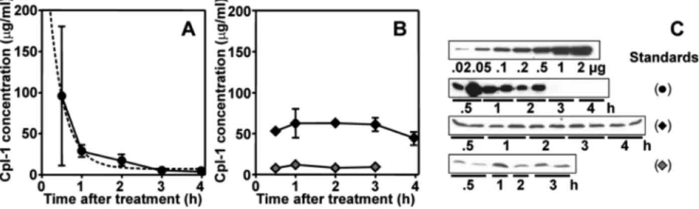
agree-ment with the killing rate observed for other strains [6]. Cpl-1 was then evaluated as a candidate for the treatment of experimental PM. After a single ic injection of 600 U (600g) of Cpl-1, bacterial titers quickly decreased over the first 4 h, whereas treatment with buffer had no effect (figure 1). Starting at 30 min and for the next 2 h, CSF titers in rats treated with Cpl-1 were at or below the detection limit. Soon after, pneumo-cocci started to multiply again in CSF (figure 1). The amount of Cpl-1 in each sample was calculated by densitometric scanning of Western blots, using a standard curve of defined amounts of Cpl-1 (figure 2). Cpl-1 was consistently detected in CSF during the first 2 h after ic injection of 600 U in 2 independent
experi-ments, (figure 2C). The half-life of Cpl-1 in CSF was estimated to be⬃16 min (figure 2A). Thus, the regrowth of bacteria 4 h after ic Cpl-1 administration was due to the short biological half-life of Cpl-1 in CSF.
In a second approach that more closely modeled an envi-sioned clinical application, Cpl-1 was administrated by ip injec-tion (200 mg/kg of body weight). Western blot analysis of plasma samples at different time points after infection demon-strated that Cpl-1 was continuously released in the bloodstream over the first 4 h after injection, and concentrations remained stable between 50 and 60g/mL (figure 2B and 2C). During the same time, the corresponding bacterial titers in blood were be-low the detection limit (1⫻ 103 cfu/mL). Importantly, a
marked reduction in bacterial titer in CSF was observed after ip injection of Cpl-1 (figure 1, gray circles). After 2 h, titers de-creased from a mean⫾ SD of 6.99 ⫾ 0.11 to 5.23 ⫾ 0.6 log10
cfu/mL, representing a drop of 98%. This antibacterial effect was detectable in CSF for as long as 4 h after initiation of ip therapy
Figure 1. Effect of intracisternal (ic) and intraperitoneal (ip)
adminis-tration of Cpl-1 on cerebrospinal fluid (CSF) bacterial titers. CSF bacterial titers were unaffected by ic application of saline (white circles). Injection of Cpl-1 ic (600g; black circles) resulted in a rapid (within 30 min) decrease in bacterial titer by 3 orders of magnitude. Injection of Cpl-1 ip (6 mg; gray circles) led to the clearance of pneumococci from the CSF by 2 orders of magnitude until 3 h after administration. The limit of detection for the determination of CSF bacterial titers was 1⫻ 104 cfu/mL of undiluted CSF (dashed line). Two-way analysis of variance revealed that both treatment and time after treatment were significant sources of variation (P⬍ .0001). Bonferroni posttests showed significant differ-ences between treatment groups (P⬍ .05) for the 0.5-, 1-, 2-, and 4-h time points for all except control ic vs. Cpl-1 ic at 0.5 h and Cpl-1 ic vs. Cpl-1 ip at 4 h.
(figure 1, gray circles). The reduction of bacterial titers in CSF is concomitant to the presence of Cpl-1 in the same compartment. In contrast to CSF concentrations after ic injection, Cpl-1 showed a prolonged presence for up to 3 h after ip injection of 200 mg/kg, ranging from 7 to 12g/mL of CSF (figure 2B and 2C).
Discussion. Here, we demonstrated the ability of Cpl-1, a phage lytic enzyme, to rapidly kill S. pneumoniae in an infant rat model of bacterial meningitis when injected ic or ip. A single ic injection of Cpl-1 decreased CSF bacterial counts to below the detection limit as early as 30 min after injection. This effect was evident for up to 3 h after administration, after which time bac-teria in CSF were detected again. The antibacbac-terial effect of Cpl-1 is temporally defined by the 16-min half-life in CSF. Increasing the bioavailability of Cpl-1 in CSF by multiple ic injections was attempted, but these repetitive injections proved to be exces-sively harmful to the animals (data not shown).
Cpl-1 has been investigated previously for in vivo therapeutic use in 2 murine sepsis models. In one model, mice were infected and Cpl-1 was injected intravenously [6]. In the other model, both the infection and Cpl-1 administration were performed by ip injection [4]. In both models, Cpl-1 was able to rapidly reduce blood titers. Furthermore, the half-life of Cpl-1 in plasma was estimated to be⬃20 min [4], a value similar to what we observed in the CSF of infected animals. In contrast to these previous sepsis experiments, in which bacterial eradication was achieved only when Cpl-1 was administered within 4 h after infection, we initiated therapy only after clinical signs of PM had developed (i.e., at 18 h after infection) [6]. In the present study, the ob-served bacterial regrowth in the CSF after 4 h may have been due to S. pneumoniae transiently invading epithelial or endothelial cells [14] and thus being out of the reach of Cpl-1. Another possibility is that a single dose of Cpl-1 is not sufficient to contact
all the pneumococci in the CSF and that additional doses may be necessary for complete eradication.
Administration of Cpl-1 ip resulted in prolonged systemic re-lease, as documented by a constant Cpl-1 level in plasma and CSF for a period of⬎4 h after injection. The amount of Cpl-1 in the CSF correlated with CSF total protein concentration (data not shown), suggesting that changes in the blood-brain barrier may have facilitated the diffusion of Cpl-1 from the blood. Ad-ministration of Cpl-1 ip (250 mg/kg) led to a Cpl-1 concentra-tion that was sufficient to decrease CSF bacterial titers by⬃98% over the first 4 h after administration. We speculate that the sustained release of biologically active Cpl-1 from the peritoneal cavity, in conjunction with the breakdown of the blood-brain barrier during meningitis, explains the extended presence of Cpl-1 in the CSF.
The data presented in this study identify Cpl-1 as a prom-ising candidate for antibacterial therapy for several reasons. The presence of biologically active Cpl-1 in the CSF after ip application has not been demonstrated to date. This finding opens new doors for future investigations into the use of Cpl-1 in the therapy of invasive pneumococcal infections. It has been shown that Cpl-1 can act synergistically with Pal, another phage lytic enzyme [15], and (more recently) with penicillin or gentamicin [9]. Of special interest, Cpl-1 and penicillin showed synergy against a highly penicillin-resistant strain in vitro. With respect to emerging resistance, a com-bined treatment of Cpl-1 with conventional antibiotic ther-apy could therefore be beneficial. Furthermore, rapid bacte-rial clearance of the CSF by Cpl-1 in synergy with antibiotics may prove beneficial, because rapid CSF sterilization has been associated with an improved outcome of BM [2]. To our knowledge, the use of phage lysins to control bacterial infec-tions of the central nervous system has never been demon-strated.
Figure 2. Assessment of Cpl-1 in cerebrospinal fluid (CSF) and plasma by Western blot. A, Decrease in Cpl-1 concentration in the CSF after
intracisternal (ic) administration of 600g (black circles). The half-life was determined to be ⬃16 min by fitting a 1-phase exponential decay curve (dashed line). B, Concentration of Cpl-1 in serum (black diamonds) and CSF (gray diamonds) after intraperitoneal (ip) application of 6 mg. Cpl-1 concentrations remained stable for the time period tested. C, Anti–Cpl-1 Western blots of standards and samples used to perform the densitometric determination of Cpl-1 concentrations shown in panels A and B.
Given the emergence of antibiotic-resistant strains, consid-erable attention is given to alternative antibiotic therapy. In this context, phage lysins could represent alternatives for both prophylaxis (e.g., by selective reduction or eradication of carriage) and treatment by offering rapidity, safety, and lack of resistance.
Acknowledgment
We thank Jürg Kummer (Institute for Infectious Diseases, University of Bern) for excellent technical support.
References
1. Schuchat A, Robinson K, Wenger JD, et al. Bacterial meningitis in the United States in 1995. Active Surveillance Team. N Engl J Med 1997;
337:970 – 6.
2. Lebel MH, McCracken GH Jr. Delayed cerebrospinal fluid sterilization and adverse outcome of bacterial meningitis in infants and children. Pediatrics 1989; 83:161–7.
3. Singleton RJ, Hennessy TW, Bulkow LR, et al. Invasive pneumococcal disease caused by nonvaccine serotypes among Alaska Native children with high levels of 7-valent pneumococcal conjugate vaccine coverage. JAMA 2007; 297:1784 –92.
4. Jado I, Lopez R, Garcia E, Fenoll A, Casal J, Garcia P. Phage lytic enzymes as therapy for antibiotic-resistant Streptococcus pneumoniae infection in a murine sepsis model. J Antimicrob Chemother 2003; 52:967–73. 5. Loeffler JM, Fischetti VA. Synergistic lethal effect of a combination of
phage lytic enzymes with different activities on penicillin-sensitive and -resistant Streptococcus pneumoniae strains. Antimicrob Agents Chemo-ther 2003; 47:375–7.
6. Loeffler JM, Djurkovic S, Fischetti VA. Phage lytic enzyme Cpl-1 as a novel antimicrobial for pneumococcal bacteremia. Infect Immun 2003;
71:6199 –204.
7. Ronda C, Lopez R, Garcia E. Isolation and characterization of a new bacteriophage, Cp-1, infecting Streptococcus pneumoniae. J Virol 1981;
40:551–9.
8. Garcia JL, Garcia E, Arraras A, Garcia P, Ronda C, Lopez R. Cloning, purification, and biochemical characterization of the pneumococcal bacteriophage Cp-1 lysin. J Virol 1987; 61:2573– 80.
9. Djurkovic S, Loeffler JM, Fischetti VA. Synergistic killing of
Streptococ-cus pneumoniae with the bacteriophage lytic enzyme Cpl-1 and
penicil-lin or gentamicin depends on the level of penicilpenicil-lin resistance. Antimi-crob Agents Chemother 2005; 49:1225– 8.
10. Entenza JM, Loeffler JM, Grandgirard D, Fischetti VA, Moreillon P. Therapeutic effects of bacteriophage Cpl-1 lysin against Streptococcus
pneumoniae endocarditis in rats. Antimicrob Agents Chemother 2005;
49:4789 –92.
11. McCullers JA, Karlstrom A, Iverson AR, Loeffler JM, Fischetti VA. Novel strategy to prevent otitis media caused by colonizing Streptococcus
pneu-moniae. PLoS Pathog 2007; 3:e28.
12. Grandgirard D, Schurch C, Cottagnoud P, Leib SL. Prevention of brain injury by the nonbacteriolytic antibiotic daptomycin in experimen-tal pneumococcal meningitis. Antimicrob Agents Chemother 2007; 51: 2173– 8.
13. Bifrare YD, Kummer J, Joss P, Täuber MG, Leib SL. Brain-derived neu-rotrophic factor protects against multiple forms of brain injury in bac-terial meningitis. J Infect Dis 2005; 191:40 –5.
14. Cundell DR, Gerard NP, Gerard C, Idanpaan-Heikkila I, Tuomanen EI.
Streptococcus pneumoniae anchor to activated human cells by the
recep-tor for platelet-activating facrecep-tor. Nature 1995; 377:435– 8.
15. Loeffler JM, Nelson D, Fischetti VA. Rapid killing of Streptococcus
pneumoniae with a bacteriophage cell wall hydrolase. Science 2001; 294:
2170 –2.