Printed in Great Britain
Fundamentals of drug design from a
biophysical viewpoint
WILFRED F. VAN GUNSTEREN, PAUL M. KINGf,
ALAN E. MARK
Laboratory of Physical Chemistry, Swiss Federal Institute of Technology Zurich, ETH Zentrum, CH-8og2 Zurich, Switzerland
•[Current address: Department of Chemistry, Birkbeck College, University of London, Gordon House, 2g Gordon Square, London WCiH oPP, England
1. INTRODUCTION 436
2. DRUG DESIGN BASED ON LEAD COMPOUNDS 437
2.1 Classical quantitative structure activity relationship {QSAR) approaches 437
2.2 Empirical methods incorporating spatial information 439
2.3 Empirical methods based on the calculation and comparison of molecular
quantum mechanical and electrostatic properties 442
2.3.1 Conformational energy 442 2.3.2 Electron density 443 2.3.3 Electrostatic potential 446 2.3.4 Electric field 447
3. DRUG DESIGN BASED ON RECEPTOR STRUCTURE 448
3.1 Site identification 448 3.2 Shape based docking 449 3.3 Fragment build-up 450
4. DRUG DESIGN BASED ON RECEPTOR-LIGAND INTERACTIONS 452
4.1 Structure and energy calculations using flexible, jD-structure-based models
456
4.1.1 Flexibility of ligand and receptor 456 4.1.2 Solvent effects 457
4.1.3 Incorporation of experimental data in a simulation 459
4.2 Free energy calculations using flexible, ^D-structure-based models 461 4.2.1 Free energy differences by thermodynamic integration 463 4.2.2 Thermodynamic cycles 466
4.2.3 Use of restraints or constraints in a free energy calculation 467 4.2.4 Reliability and test of computed free energy differences 468 4.2.5 Free energy decomposition 470
4.2.6 Free energy changes by extrapolation 471
5. OUTLOOK 473 6. REFERENCES 4 7 4
I. INTRODUCTION
Drug design means many things to many people. Commercially the aim is the
development of compounds that can be patented and meet a variety of regulatory
standards. In drug design, for medical purposes, toxicity and bio-availability are
major considerations. Synthetically, questions related to ease of synthesis and
chemical stability may dominate. From a physical perspective drug design is seen
primarily as the process of optimizing specific (bio)mplecular interactions. The
biological activity of any compound can essentially be considered as a series of
independent binding, transport and processing events. These events begin when
the compound enters the body and end when it is either metabolized or excreted.
Thermodynamically and kinetically each of these events may be expressed in
terms of changes in free energy. The difference in free energy will determine how
the compound partitions between different environments or between reactants
and products and the intervening free energy barrier will determine the rate of
such partitioning. Therefore, from a theoretical biophysical viewpoint drug
design is concerned primarily with the estimation of the change in free energy for
compounds in different environments. Most frequently it will be the difference
between the free energy of a compound in water compared to that of the same
compound bound to a specific protein receptor site that is of interest.
Alternatively, it might be the difference in free energy of a compound bound to a
bacterial or a mammalian form of the same enzyme. It may, however, correspond
to the difference in free energy of a compound in an oxidizing or reducing
intra-cellular environment where no specific macromolecular receptor can be identified.
An example of such a case may be anti-cancer drugs designed to accumulate
preferentially in rapidly metabolizing cells.
Free energy is a global property of a system and it is this that gives rise to the
essential computational difficulty in drug design. Quantum mechanically, the
Helmholtz free energy, F, of a system of N particles in a volume F a t a
temperature T, in terms of the canonical partition function, Q, is given by,
= -k
BTlnQ(N,V,T)
E i N V ) / l T ( i )
where the energy of a quantum mechanical state, j , of the system is given by
Ej(N, V), and k
Bis Boltzmann's constant. The total free energy of a system is thus
dependent on all possible electronic and nuclear degrees of freedom.
For any realistic system the absolute free energy cannot be calculated. It is only
ever possible to estimate the change in free energy between two systems and then
only by evoking a large number of often very crude assumptions or empirical
Abbreviations. CNDO, Complete Neglect of Differential Overlap; COMFA, Comparative Molecular
Field Analysis; DNA, deoxyribose nucleic acid; HOMO, Highest Occupied Molecular Orbital; LFER, Linear Free Energy Relationship; LUMO, Lowest Unoccupied Molecular Orbital; MC, Monte Carlo; MD, molecular dynamics; MEF, Molecular Electric Field; MEP, Molecular Electrostatic Potential; QSAR, Quantitative Structure Activity Relationship; SAR, Structure Activity Relationship.
approximations. The relevant question for any given drug design problem is, for which sets of assumptions or approximations will the estimate of the change in free energy be useful ? The answer will depend on the nature of the system, the amount of structural information available, the relative importance of quantum mechanical effects, the degrees of freedom in the system that can be safely ignored and the nature of the biological data against which the results can be compared. This review aims to provide a broad overview of the range of methodologies that can be used to estimate relative free energies in drug design. The focus is not on applications, but on the physical bases of the methodologies, the assumptions on which they are based and the conditions under which such assumptions are valid. The primary purpose is to enable the reader to assess the applicability of a given class of methods to tackle a specific problem. Specific methods are only discussed by way of example. We do not aim to be encyclopaedic. Despite the fact that empirical methods currently dominate drug design the review is also intentionally biased toward the use of explicit free energy calculations based on detailed structural information. This bias merely reflects the fact that as the cost of computational resources falls the trend toward potentially more accurate explicit methods will be inevitable.
2. DRUG DESIGN BASED ON LEAD COMPOUNDS
All structure based drug design methods aim to derive a relationship between the topology or properties of a given molecular structure and a specific biological activity. Where the precise target or action of a lead compound is not known, such structure activity relationships (SAR's) must be derived empirically. The aim is to obtain a set of mathematical relationships between the properties of a set of related structures which form a given training set and some measure of biological activity that can later be used in a quantitative fashion to predict the activity of novel compounds (QSAR). In principle any physico-chemical or derived property that varies systematically in the series of test compounds can form the basis of a QSAR study.
The literature relating to the use of empirical methods in specific drug design studies is immense. A number of excellent general reviews exist (Martin, 1978; Franke, 1984; Martin, 1991; Silverman, 1992; Kubinyi, 1993) and it is not our intention to repeat the exercise here. Instead we wish to address the question of why certain molecular properties frequently show simple correlations to a variety of biological activities and under what conditions the assumptions that are evoked to account for such correlations might hold.
2.1 Classical quantitative structure activity relationship (QSAR) approaches A century ago it was recognized that the narcotic effect of a series of neutral compounds appeared to be a function of their oil :water partition coefficient, P. Subsequently, the biological activity within many series of related compounds was shown to depend in a simple manner on the free energy of transfer from water to
438 W. F. van Gunsteren et al.
a variety of organic phases given by the logarithm of the partition coefficient, log
P. The basic assumption inspired by such studies was that the organic phase
mimicked the interaction of the compounds with their site of action in biological
membranes (Martin, 1978).
Modern QSAR studies stem, however, from work on the effects of sterically
remote substituents on the rates of organic reactions which led Hammett
(Hammett, 1940) to propose SAR's based on an empirical electronic parameter, cr,
defined as
k
xlog-r- = p<r
x(2)
where k
0is the rate or equilibrium constant of a reference compound, k
xthe
constant for the compound containing the substituent X and p is a proportionality
constant characteristic of the sensitivity of the reference compound to substitution
at a specific site. Taft (1956) extended the work of Hammett to include an
additional parameter, E
s, defined in an analogous manner to the electronic
parameter cr, to account for steric effects such that
log-£ = pa
x+ 8E. . (3)
where 8 is again a system dependent scaling parameter analogous to p. The
approach illustrated by the work of Hammett and Taft was generalized by Hansch
and coworkers (Hansch et al., 1963; Fujita et al., 1964; Hansch & Fujita, 1964)
who proposed a group hydrophobicity parameter, n, defined in an analogous
manner to tr and E
sbased on octanol: water partition coefficients and introduced
the assumption that the effects of the steric, electronic and hydrophobic properties
of a given molecule on biological activity were independent and additive. This led
to the general principle of linear free energy relationships (LFER's) where the
activity, A, or the inverse of the effective concentration, i/C, could be expressed
as
log A = f
h(x
h) +/,(*,) +/,(*,) + constant .(4)
in which the log of the activity (a free energy) is assumed to be a linear
combination of independent functions describing the hydrophobic, h, electronic,
e, and steric, s, properties of a given compound. The functions,/(#), are frequently
assumed to be linear but may in principle take any form. Also, it should be noted
that although originally expressed in terms of n, <r and E
sa myriad of other
physical and derived parameters have been used in different QSAR studies
(Franke, 1984).
LFER's and QSAR studies have proven to be very useful. From a physical
perspective, however, it is not clear why LFER's should hold or what can be
inferred in regard to the particular system when they do hold. The simplest type
of biological activity is the direct binding of a test compound to an isolated
receptor. In this case log A is directly proportional to the free energy of binding.
Free energy is a global property of a system and cannot formally be separated into
a sum of components or group contributions unless the interactions on which the separation is based are either uncorrelated or purely enthalpic (van Gunsteren et al. 1993). For the interaction of specific substituents with specific residues of a receptor protein this will most likely not be the case. In contrast, in whole body or cellular assays the measured activity may reflect a sequence of uncorrelated transport and recognition events. The value of log Poctanoi.water might then reflect
the rate of transport through the membrane and the oxidation potential of the compound its reactivity within the cell. In this case the application of (2) may well be valid.
The difficulty in assigning group free energies for use in QSAR studies can be illustrated in relation to the group hydrophobicity parameters of Hansch. Hansch defined the hydrophobicity nx of a given substituent X as
TT x = \ogPx-log PH (5)
where Px and PH are the octanol: water partition coefficients of a compound
containing the substituent X and a hydrogen atom respectively. Octanol is often chosen as a reference solvent as, due to the presence of hydroxyl groups, it is assumed to approximate the hydrogen bonding environment of a biological membrane or protein. In the series of substituted aromatic compounds initially investigated by Hansch and coworkers specific n values were, in the absence of strong electronic effects, shown to be constant and additive. Hydrophobicity is, nevertheless, an essentially entropic phenomenon (Tanford, 1973). In this respect the additivity of n values is surprising. Both phases are, however, liquid. The nature of the local solvent environment in which the substituent is inserted, and hence the associated change in entropy, will depend primarily on the nearest neighbour atoms. For a series of aromatic parent compounds the nature and spatial arrangement of the neighbouring atoms is essentially constant. As expected, different hydrophobicity parameters or the use of correction terms are required for the same substituents attached to aliphatic chains or in close proximity to groups that perturb the local solvent environment. Using a variety of approaches log P values can be empirically predicted with high accuracy (Suzuki & Kudo, 1990). It is to be expected, however, that the use of group hydrophobicity parameters is more appropriate in liquid-like rather than highly structured environments.
2.2 Empirical methods incorporating spatial information
The interaction of a given compound with a specific receptor site will depend not only on the physical properties of the isolated substituents but also on their spatial arrangement. Crude spatial indices such as the steric parameters of Verloop et al. (1976), can be used to describe the volume and shape of a given substituent but such measures do not explicitly incorporate conformational information. To include explicit spatial information assumptions in regard to the active conformation and the mutual alignment of the test compounds must be made. In the distance geometry approach of Crippen (Crippen & Havel, 1988) such model
dependencies are minimized by considering only distances between atoms or interaction sites. Sets of distance bounds describing the conformational flexibility of each of the test compounds are generated. A comparison of the test compounds is then made based on the assumption that there is a single active conformation or pharmacophore. Sets of interaction sites are defined and geometrically allowed binding modes for each of the test compounds are evaluated. A simple scoring function based on favourable and unfavourable interactions with these sites can then be used to correlate structural features with some measure of biological activity (Ghose & Crippen, 1985). Alternatively, the ensemble of test compound configurations weighted by some measure of biological activity can be used to extract a set of common distances between potential interaction sites in order to define a potential pharmacophore or pseudo binding site (Sheridan et at. 1986). Although defining such a consensus binding site may be helpful in proposing alternate test compounds, it does not necessarily bear any relationship to a physical binding site with which the compounds under investigation might interact. This is especially true if the biological data with which it is correlated has not been derived from binding studies using an isolated receptor. Distance geometry and other 3D-QSAR methods depend strongly on the assumption that chemically related structures bind in a similar conformation and in the same orientation to a given receptor. While for simple rigid molecules this generally may be a very good assumption, it is certainly not always the case, as illustrated by three closely related elastase inhibitors which not only show different binding modes, but interact with different subsites (Mattos et al. 1994).
Distances between interaction sites is only one of a number of measures of molecular shape or molecular similarity that have been used to incorporate conformational information into QSAR studies. Other indices include steric volume overlap, charge matching and atom pair matching (Hopfinger & Burke, 1990). Similarity indices based on quantum mechanical calculations are discussed in the next section. Similarity indices discriminate on the basis of molecular conformation. Thus, results from such comparisons will depend on the model used to generate the three-dimensional structure of the test compounds. Structures can be generated using knowledge based approaches such as the programs CONCORD or WIZARD (Rusinko III et al. 1988; Leach et al. 1990), or minimum energy configurations from quantum mechanical or empirical force field calculations can be used. Similarity indices have also been derived as trajectory averages from molecular dynamics simulations. As with distance geometry methods it is assumed that there is a single active conformation and test compounds are superimposed on a given reference compound before comparison. This inevitably leads to a dependence on the choice of reference compound and the superposition criteria.
In the comparative molecular field analysis (CoMFA) method of Cramer III et al. (1988) molecules in a given test series are again aligned on a chosen parent structure. The potential energy with respect to a given force field is then sampled in the space surrounding each molecule using a regularly spaced grid and correlated at each point with a measure of biological activity. The correlation is
performed using the partial least squares method developed by Wold and co-workers (Wold et al. 1984) with cross-validation to give some measure of the predictive ability of the potential energy at each grid point. The result can then be expressed as a 3-dimensional contour surface reflecting the relationship between the molecular field and a specific biological activity.
CoMFA analysis is in general performed using the non-bonded terms from classical molecular mechanics force fields. The implementation in the molecular modelling package SYBIL standardly uses a 6-12 van der Waals and a Coulomb potential energy function, the latter with a distance dependent dielectric. The potential energy is calculated with respect to a probe atom. Although Cramer et al. (1988) initially used parameters for an sp3 hybridised carbon atom carrying a charge of + 1, the choice of probe atom and charge is essentially arbitrary and may be varied in order to optimize the correlation to a given set of experimental data. The use of such a pseudo physical force field has, however, led to ambiguities in the manner in which results from such studies should be interpreted. Specifically, there are questions remaining about whether the potential energy surface generated in the analysis reflects the structure of a specific receptor and whether the use of such a potential limits the analysis to purely enthalpic effects. Interaction energies obtained from static modelling with classical molecular mechanics force fields can at best only indicate enthalpic contributions to binding. It has been argued, therefore, that a CoMFA study using a potential energy function expressed purely in terms of van der Waals and Coulomb interactions cannot be expected to correlate with entropically driven phenomena or atom type specific interactions such as hydrogen bonding without the inclusion of special terms. In practice, inclusion of hydrophobic potentials (Kellogg et al. 10.91) and/or an explicit hydrogen bonding term using for example the GRID potential energy function (Goodford, 1985; Kim, 1991) does not necessarily improve the overall correlation (Folkers et al. 1993; Kim et al. 1993). As an empirical method CoMFA is not limited to the use molecular mechanics force fields (Waller & Marshall, 1993). Conversely, the use of such force fields to extract correlations should not be confused with calculation of interaction energies based on the structure of the ligand-receptor complex. This was illustrated in a recent study of Klebe & Abraham (1993). Using crystallographic data to align a series of endothiapepsin inhibitors they demonstrated a significantly better correlation to enthalpic changes on binding than to entropy or free energy changes using a van der Waals and Coulomb potential energy function. The alignment correctly represented the binding to the receptor. Since the enthalpic changes were large and as enthalpic changes can be both formally separated into atomic contributions and equated to interaction energies, this result is not surprising. Fitted to free energies, however, the correlations generated by CoMFA can neither be used to infer interaction energies nor be expected to reflect details of the actual receptor. In the same study Klebe and Abraham observed for inhibitors of thermolysin that alignment based on crystallographic data yielded substantially inferior correlations to free energy data than two alternative alignment procedures. Where the activity data relates not to receptor studies but to cell or organ assays any
44 W. F. van Gunsteren et al.
inferences drawn in regard to a specific receptor site from the generated correlations are even less reliable. In summary, although the potential energy terms commonly used in CoMFA have been derived in relation to detailed 3-dimensional structural information they are used in such studies simply as parameters. In this way the combination of potential energy terms from different sources, truncation or scaling of specific interactions, and fitting to non-linear functions can all be justified. In doing so, however, the biophysical basis of the force field is largely lost.
2.3 Empirical methods based on the calculation and comparison of molecular quantum mechanical and electrostatic properties
Quantum mechanical methods permit the calculation of a large number of molecular properties that can be empirically related to the action of a drug molecule. Quantum mechanical calculations can indicate preferred conformations, the distribution of charge within a molecule, possible tautomeric states, and potentially reactive functional groups (Richards, 1983). Such calculations are nowadays fairly routine with semi-empirical and ab initio quantum mechanical packages (for example Frisch et al. 1992; Stewart, 1990) and are very often the first step in understanding the behaviour and action of a potential drug molecule. Due to computational limitations and the fact that the relevant environment in which the molecule acts or the receptor to which it binds is often unknown many approximations must be made. Most commonly the calculations are performed in vacuo or alternatively in a dielectric continuum whose permittivity reflects that of the proposed environment. Alternatively a supermolecule calculation can be performed in which a few atoms of the environment (solvent molecules, amino acid residues etc.) are placed to mimic, at least approximately, the perturbing effect of the surroundings. The accuracy of quantum mechanical calculations is also limited by the number of electrons in the molecule which itself places limitations on the size of basis set that can be used (Hehre et al. 1986). Given that the relationship between the drug molecule's behaviour in isolation and at a receptor is uncertain, the use of the highest levels of theory incorporating large basis sets and electron correlation is not usually warranted. In cases where a whole range of molecules is systematically being studied such extensive calculations would in any case be prohibitively expensive. Calculations based on the quantum mechanical or electrostatic properties of molecules tend to ignore the entropic component of drug action, that is, the underlying assumption is that enthalpic terms dominate the activity. Thus drug design efforts are frequently aimed at maximizing binding capacity, particularly through complementarity of properties for the ligand and host believed to be involved in drug action (Dean, 1987).
2.3.1 Conformational energy
One of the most useful quantities that can be derived from quantum calculations is the energy of the molecule and how this changes with conformation. This can
give insights into the low energy conformations which the molecule might adopt at the receptor or alternatively a range of conformations which may not have minimal energy but which may be active and stabilized at the receptor. Such calculations are usually performed by systematically changing all the dihedral angles within the molecule, optimizing each structure and calculating the energy for each conformation. In cases where there are too many such dihedral angles, portions of the molecule are independently optimized and held rigid while rotation about a few critical angles is performed. The resulting information, namely the conformational energy, is usually contoured as a function of two dihedral angles. Conformational free energies can also be determined, which involve the additional calculation of vibrational frequencies for use in determining the vibrational partition function. A reaction mechanism can be postulated and the structure and energetics along a proposed reaction coordinate can be determined, including identification of the transition state. This may be relevant to drug action in cases where an enzyme is believed to stabilize the transition state of a reaction and one is interested in designing a transition state mimic to block the reaction. The energies of simple reactions in vacuum such as protonation and tautomerism are straightforward to determine using quantum mechanical calculations. Such calculations may indicate whether alternative structures or protonation states are energetically feasible. The reactivity of a molecule can also be investigated by visualizing the frontier orbital electron density (Fukui et al. 1952). Electron density in the HOMO (highest occupied molecular orbital) will, in principle, indicate the position of electrophilic attack while for nucleophilic attack the LUMO (lowest unoccupied molecular orbital) is of importance. In order to compare frontier orbital electron density on different molecules the concept of superdelocalizability has been introduced. Here the contribution to the electron density from each molecular orbital is weighted by the energy of the orbital.
2.3.2 Electron density
Given a lead compound one usually wishes to compare it with a potential drug molecule so that the efficacy of the latter may be estimated. Perhaps the most natural choice for a quantum mechanical property with which to compare molecules is the electron density, p(r). This is the number of electrons per unit volume and can be written in terms of the total molecular wavefunction V as follows:
p(r) = N f... f I V(*i, *2 *Ar)lsd*i d*,.. • dxN. (6)
Here N is the total number of electrons of the system and x( = (s,,r,) are the spin
and space coordinates of electron i. Integration of p{r) over the whole space yields the total number of electrons in the system. The electron density lies at the heart of the currently fashionable density functional approaches to quantum chemistry (Parr & Yang, 1989) which follow from the realization that p{r) determines the ground state wavefunction of the system and thus all its electronic properties. The electron density can also be expected to represent the underlying nuclear
framework of the molecule since its overall electron density can essentially be considered as the sum of spherical atomic electron densities with slight deformations. Electron density is a useful concept with which to work since it can be directly determined by experimental methods such as X-ray diffraction. The electron density is straightforward to compute using both ab initio and semi-empirical quantum mechanical programs. There are, however, limitations on the size of molecule and the number of basis functions used in the calculation. Semi-empirical methods can be used for a few hundreds of atoms while the highest accuracy ab initio methods are restricted to an order of magnitude fewer atoms. The easiest, and crudest, way of indicating the charge distribution within a molecule is to calculate atom-centred partial charges. This is the net charge, expressed in number of electrons, residing on an atom within a molecule and can be calculated from quantum mechanical or more empirical methods. Because the partial charge on an atom is not a physical quantity, i.e. it cannot be measured experimentally and there is no associated quantum mechanical operator, many methods of calculating it exist and assignment of charge is somewhat arbitrary. However, the partial atomic charges can indicate (i) whether a given atom has increased or decreased charge density relative to the unbound state and (ii) the relative size of build-up or depletion of charge on different atoms within the molecule. Inspection of a point charge distribution can indicate possible positions of nucleophilic or electrophilic attack.
Visualization of electron density can enable qualitative comparisons between molecules to be made. Very often two-dimensional contour plots are used to represent electron density in various planes of the molecule. Alternatively isodensity surfaces can be drawn around the molecules. These are sets of points in three-dimensional space at which the electron density attains a certain pre-set value, and are usually represented as a dot-surface, a triangulated mesh or an interpolated smooth surface. This can be used as a descriptor of the shape or surface of the molecule and can serve as a property to compare a given series of molecules or to map out the shape of an unknown receptor. Conversely one can define a certain molecular surface, such as the van der Waals or solvent-accessible surface, and evaluate the electron density at points evenly scattered over the area. The values of the electron density at these points can then be colour-coded to ease visualization. One might also analyze the difference density using similar methods. This is the difference between the molecular electron density and the density obtained by superposition of unperturbed atomic densities. Isodensity surfaces of the difference density clearly show how the electron distribution changes on formation of the molecule and can thus highlight regions of enhanced or diminished electron density. These may well be indicative of sites for electrophilic or nucleophilic attack, and one might envisage that such regions will occur in similar locations for molecules which bind to the same receptor and react by the same mechanism.
While qualitatively the display of electron density for a series of molecules can be instructive, to quantify the similarity between two molecular charge distributions some form of simple index is required. Similarity indices can serve
= \[p
A(r)-p
B(r)Tdr = J p »
2d r + jp
B(r)
2dr-
2Jp»p
B(r)dr.
both as a criterion for the superposition of the molecules to aid in the mapping of an unknown receptor and as a descriptor for QSAR studies. One such similarity index was introduced by Carbo (Carbo et al. 1980). Given two molecules A and B, with respective electron densities pA{r) and pB(r), then a measure of the
difference between the two densities is given by
J (7)
If both molecules remain rigid then only the last term of (7) varies as the relative position of the molecules changes. Thus superimposing the molecules by minimizing the difference of the charge densities corresponds to determining a maximum for the integral jpA(r) pB(r) dr. Carbo proceeded to define a normalized
measure of similarity, rAB, given by
(8)
where rAB lies in the interval [0,1]. Molecules with complete similarity, pA{r) =
PBC*"). will have rAB = 1 while complete dissimilarity will be indicated by rAB = o.
This was originally implemented within a semi-empirical CNDO framework although an improved ab initio formulation has been presented (Bowen-Jenkins et al. 1985). The less accurate quantum mechanical methods tend to be less discriminating than the more accurate ones, while the latter tend to be too expensive for optimization of the similarity. This similarity measure is also very dependent on the manner in which the molecules are superimposed. In the presence of heavy atoms the similarity is dominated by their large electron density close to the nucleus and small misalignment of such atoms can give rise to unrealistically low similarity values. To overcome this problem use of only the valence electron density has been proposed and found to give results more in tune with chemical intuition.
The Carbo index of electron density similarity is not unique and while rAB is
sensitive to the shape of the electron density it is not sensitive to its magnitude. For example, when pA(r) = npB(r) then rAB is still equal to unity indicating full
similarity. An alternative index, albeit in a different context, has been proposed by Hodgkin (Hodgkin & Richards, 1987):
2\pA(r)pB(r)dr
* = (9)
sAB compares the shape of the electron density and also its magnitude. Given the
condition pA[r) = npB(r) then sAB = zn/(n2+ 1) and can thus distinguish between
sensitive to the positions of the nuclei than to the long-range valence electron density. An innovative attempt to overcome this limitation was made by using densities in momentum space, p(J>), rather than position space (Cooper & Allen, 1989). The alternative similarity index
2\p
np
A(P)p
BiP)dp
SAB(n) = i j , (10)
was introduced, in which p = \p\. This measure of similarity has a number of advantages. Firstly, it is independent of the distance between the molecules in position-space and so.many of the problems associated with the superposition of the two molecules will be avoided. Secondly, p(p) is dominated by the valence electrons, which have low p values, whereas p(r) is dominated by core-electrons and thus the position of the nuclei. Furthermore, use of SAB(n) for n = —1,0,1,2
can be used to measure similarity between different regions of the electron density.
2.3.3 Electrostatic potential
A quantity that has perhaps received more attention than the electron density in studies of molecules and in QSAR is the electrostatic potential, V(r), defined as follows
The first term represents the contribution from the N atomic nuclei of charge {Z(}
situated at {Rf}, while the second term is the contribution from the electronic
charge density. V(r), often called the molecular electrostatic potential (MEP), represents the energy of interaction between the molecule and a proton situated at r, or if a point-charge of value q is situated at r the energy of interaction will be given by qV(r). It should be stressed that the electrostatic potential is usually calculated for an isolated molecule and so using qV(r) to evaluate an energy of interaction is a first-order approximation, neglecting cooperative effects such as polarization and charge transfer. However the MEP can be used to give a very clear indication of the areas of three-dimensional space where groups of a given charge will be attracted or repelled. One can thus use the MEP to map out the surface of a pseudo receptor based on the assumption that the receptor will have a complementary electrostatic potential to that of the ligand. Furthermore, by comparing the MEP's for a number of ligands one can propose areas of the molecules associated with binding to a receptor or possible areas for electrophilic or nucleophilic attack. The electrostatic potential can be calculated in most ab initio or semi-empirical quantum chemistry programs, although once again there are limitations on the size of molecule that can be treated. Very often when working with a large number of molecules or with very large molecules, such as
proteins or DNA fragments, where a fully quantum mechanical treatment is not possible, a point charge approximation is made and the following expression used
^(r) = — S i - % 7 (12)
l R l
where {gj are the net atomic charges situated on the nuclei.
As with electron density one can introduce a similarity index with which to compare molecular electrostatic potentials (Hodgkin & Richards, 1987). This can be of the same form as expressions (8) or (9) with the electrostatic potential at position r, V(r), replacing the electron density p(r). In their implementation Hodgkin and Richards used a point charge model and evaluated the integrals required for the calculation of the similarity index numerically on a grid. Generally reasonable results could be achieved with a grid which extends r o nm beyond the molecule and has a mesh of o-i nm. To avoid singularities associated with the evaluation of the MEP at the nuclear sites the ' inside' of the molecule was excluded from the calculation. Details associated with the grid extent and fineness, the method used to optimize molecular geometry and obtain atomic charges have been addressed (Burt et al. 1990) as too has the introduction of flexible fitting for optimization of the similarity index (Burt & Richards, 1990). The use of a 2- or 3-Gaussian expansion for i/r (Good et al. 1992 a) allows the grid-based determination of the electrostatic potential to be replaced by analytic evaluation which makes the calculation two orders of magnitude faster. Furthermore more robust methods of optimization can be used so that the fitting of molecules is less likely to become stuck in local minima, and because there is no singularity at the nucleus there is no need to exclude the molecular volume from the similarity calculation. This method has been widely applied to the screening of results from 3D-database searches (Good et al. 19926) and to the calculation of similarity matrices for use in QSAR studies (Good et al. 1993).
2.3.4 Electric field
A further molecular property of use in analysing and comparing structures is the molecular electric field (MEF), defined as follows
*(r) = - V F ( r ) . (13) The electric field is thus the negative of the gradient of the electrostatic potential and as such is a vector quantity. The molecular electric field is usually considered in two ways. Firstly, the scalar product #(r)-(i, where \i is a permanent dipole moment of a second molecule, gives the energy of interaction of the dipole with the field of the original molecule. Such interactions can be important in the binding of a ligand to a receptor. Alternatively one can study the field vector itself which indicates the force on a proton placed at r. The MEF of a receptor may indicate the route by which a ligand is guided towards the binding pocket. Being a vector quantity the field is rather more difficult to visualize than the electrostatic potential and electron density, and hence it has probably been less used in drug design studies. However, it is possible to visualize the electric field, either by
displaying the vector orientations at points on a regular lattice or by plotting field lines which indicate the motion of a freely translating proton in the region of the molecule. The MEF has also been used in quantitative studies of molecular similarity (Hodgkin & Richards, 1987).
3. DRUG DESIGN BASED ON RECEPTOR STRUCTURE
If the three dimensional structure of a drug target receptor is known the design problem is substantially different from that based on lead compounds. In this case direct methods can be used to estimate differences in binding free energy for different compounds as opposed to the use of purely empirical correlations. The design process itself is, therefore, often referred to as being de novo. The primary concern in de novo drug design is computational efficiency. The increased accuracy of predictions based on higher level theory must be offset against a smaller range of compounds that can be investigated for the same cost. The choice of method is also governed by the quality of the structural data and which degrees of freedom in the system can be safely neglected.
In cases where the structure of a biological target is known, e.g. the crystal structure of an enzyme critical to viral reproduction, but few or no potential inhibitors exist, one is faced both with the problem of determining where an active molecule may bind and proposing potential agonists or antagonists. In such a case it is common to fix the conformation of the receptor. This neglects the dynamics of the receptor and implicitly assumes that entropic or enthalpic changes associated with the accommodation of the receptor to a specific ligand are negligible. Although this is a big approximation and may result in significant artifacts from a computational perspective, it is frequently the only option that yields a tractable starting point for further investigations.
3.1 Site identification
Before an agonist or antagonist can be proposed potential binding sites must be identified. Although these often can be inferred from mutation studies or by inspection of the structure for cavities within the molecule or clefts lying on the surface, in large systems containing many such sites it can be difficult to definitively identify by inspection for example the enzymically active site. Calculation and visualization of the electrostatic potential and electric field, as described previously in regard to empirical approaches, can be used to aid this process. As the host molecule consists very often of many thousands of atoms, one usually assigns point charges to atoms and calculates the various properties classically. The molecular electrostatic potential (MEP) can indicate regions where positive or negative charge will bind favourably. The MEP of the active site is expected to be complementary to that of ligands which bind - this is one criterion for identification of the site. The M E F can indicate the force on a charge and thus the vector field can be particularly instructive in showing how a charged ligand is directed towards the binding site, thus helping to identify the site.
One step beyond a simple electrostatic view of the host molecule is to incorporate other force-field terms to represent additional intermolecular forces. A widely used and conceptually appealing method is that of Goodford and co-workers called GRID (Goodford, 1985). The interaction of a probe, e.g. a functional group typically found on a ligand, such as methyl (-CH3), amino
(-NH2), carbonyl ( = O), and hydroxy (-OH), or a water molecule, is calculated with
the fixed macromolecule. The energy of interaction of the probe is calculated at the points of a three dimensional grid superimposed over the macromolecule using an empirical potential energy function. The resulting energy values are contoured in three dimensions and displayed graphically along with the macromolecule. Contours at negative energy are assumed to indicate attractive regions for the probe and should occur in the binding pocket for probes found on known ligands. The most negative regions indicate the most favourable binding locations. The potential energy function used by GRID consists of three terms: (i) a van der Waals 6—12 function, which can be considered as defining the shape of the probe, (ii) an electrostatic interaction, which is a Coulombic potential containing a distance-dependent dielectric permittivity related to the environment of the interacting atoms, and (iii) a hydrogen bonding term which is angle dependent and allows for some mobility of the hydrogen atoms and lone-pairs (Boobbyer et al. 1989; Wade et al. 1993; Wade & Goodford, 1993). There are a number of simplifications present in this approach, which also occur in some other force field methods. Entropy is totally neglected, pairwise additivity of terms is assumed, no polarization or redistribution of charge is permitted and the macromolecule is treated as a static entity. The method is widely used because the information that results is simple to visualize and straightforward to interpret. The most negative contour regions show where most favourable enthalpic binding of a functional group will occur. Contours at less negative values indicate the amount of movement a probe may undergo and still have favourable interactions. The closeness of the contours can indicate what forces the probe would be under and in what direction these act. Together these factors often allow one to identify a binding site and further help in the design of potential ligands that will bind to the site.
3.2 Shape based docking
The DOCK algorithm (Kuntz et al. 1982; Desjarlais et al. 1986; Shoichet et al. 1992) attempts to generate geometrically feasible alignments of ligands within a receptor site of known structure based on a detailed matching of molecular surfaces. The volume of the binding site and that of potential ligands are first represented as a series of spheres which fit into their respective solvent accessible surfaces (Connolly, 1983). The ligands are treated either as completely rigid or as a series of rigid sub-fragments. Inter-sphere distances are then scanned for matches within a given tolerance and the structures superimposed based on matched distances. This generates a large number of potential alignments that are ranked in accordance to a given scoring function. The default scoring function has
varied in each implementation. In the most recent implementation a lattice is placed covering the receptor site and scores are accumulated for each lattice point that lies within a given cutoff distance of a ligand atom (Shoichet et al. 1992). Large negative scores are recorded for atoms which overlap with the lattice points. Different cutoff distances can be chosen for different atom types to account for close contacts of atoms involved in hydrogen bonding. Other scoring functions including a mixture of hard sphere and hydrogen bonding terms, a continuous scoring function based on inter-atom distances or interaction energies based on the AMBER force field have been used with mixed success.
Shape complementarity algorithms such as DOCK discriminate on a detailed matching of molecular surfaces. Using conformations of the receptor and ligand derived from crystallographic studies of the complex such algorithms routinely reproduce the experimental structure. However, such methods are sensitive to changes in the geometry of either the ligand or the receptor. Using DOCK to scan a data base for possible inhibitors of thymidylate synthase 3 of the 25 best scoring compounds inhibited the enzyme at sub millimolar concentrations. Crystallographic studies of one of the resulting complexes revealed that the bound inhibitor was shifted by o-6-o-9 nm, binding to a different region of the active site (Shoichet et al. 1993). In a more recent study on non-peptide inhibitors of HIV-protease a proposed inhibitor bound 048 nm away and rotated by 790 from the predicted location in a different conformation than the docked structure (Rutenber et al. 1993). In that these and other investigations have led to novel inhibitors in different systems the method has been an undoubtable success. In regard to the prediction of the location of the binding site, the orientation of the ligand within a given site, or specific hydrogen bonding contacts, purposes for which the method was designed, the results are so far discouraging.
3.3 Fragment build-up
An alternative to molecular docking algorithms are fragment build-up procedures in which potential ligand molecules are constructed from simple precursors. Such schemes have the advantage that completely novel compounds can be suggested. However, as with docking algorithms such as DOCK, the accuracy of these methods is determined primarily by the form of the scoring function or force field calculation that must be used to discriminate between potential ligands and binding modes. Because the methods generate a very large number of alternatives, the discriminating function is in general crude and factors such as potential flexibility in the receptor site are not considered.
The types of compounds that will be suggested and the sophistication of the discriminating functions that can be used depend to a large degree on the implementation of the method. The build-up procedure GROW (Moon & Howe,
1991) constructs peptide models from a user selected starting position by piecing together amino acid fragments in conformations that will interact most favourably with surrounding atoms in the proposed receptor site. GROW operates by sequentially attempting to add all fragments from a preconstructed library to a given seed. To score each compound an interaction energy between the receptor
and the test compound is determined based on the AMBER molecular mechanics force field and a correction term for ligand desolvation. A manageable number of test compounds is maintained by only propagating the ten lowest energy conformations each round. The combination of assuming a rigid receptor, fixed ligand geometries and scoring on the basis of an interaction energy that includes a highly non-linear 6-12 van der Waals term means that, like DOCK, GROW discriminates primarily on a detailed matching of molecular surfaces. For this reason GROW, as expected, faithfully reproduces the sequence and configuration of peptides co-crystallized with a given receptor. The method is, however, extremely sensitive to the choice of starting position which must either be selected by the user or obtained using an algorithm such as DOCK. For this reason GROW is primarily applicable to the extension of a pre-existing ligand or in proposing alternate amino acid residues for a pre-existing ligand.
An example of an alternative implementation of a build-up procedure is the program LUDI (Bohm, 1992 a, b). LUDI aims to position small molecules into clefts or cavities in a protein structure such that protein hydrogen bond donors or acceptors and possible hydrophobic contacts are satisfied. This is done by first defining a series of interaction sites. In the later implementation a rule based procedure is preferred to define sites, but methods based on hydrogen bond and hydrophobic distributions extracted from structural databases or a probe atom procedure such as GRID are also described. Distances between interaction sites are then used to select and dock molecules from a rigid fragment library. To generate the final compounds bridging groups are used to join fragments. Again this procedure will correctly suggest a ligand that has been co-crystallized with a given receptor but is later deleted from the structure. The advantage of a LUDI type build-up procedure over DOCK or GROW is that the initial placing of unbridged fragments is performed independently, and a degree of tolerance in the positioning of the fragments is inherent in the method. Thus, the method is more tolerant, but less discriminating on the basis of surface matching. LUDI uses a rule based scoring function to discriminate between potential ligands based on hydrogen bond geometries, volume overlap and buried surface area. Such rule based functions are essentially empirical and necessarily hold only for the range of compounds against which they are fitted.
Buildup procedures illustrated by GROW and LUDI in general attempt to grow or join molecular fragments based on known structures. Recently, however, a number of atom based methods have also been proposed (Nishibata & Itai, 1991; Rotstein & Murcko, 1993; Pearlman & Murcko, 1993). The most novel of these methods is that of Pearlman and Murcko, which attempts to dynamically allocate atom types and bonding topologies during a molecular dynamics simulation of a number of initially unconnected atoms, the motions of which are restrained to the volume of a proposed binding site. The atoms interact with the receptor via the AMBER molecular mechanics force field and Monte Carlo type moves are used to change atom types and bonded interactions. Although conceptually interesting, the computational cost of this method probably cannot be justified in terms of its ability to suggest and discriminate between potential inhibitors. Atom based build-up procedures can also lead to chemically unreasonable structures.
A. Model building
B. Energy calculation
• packing considerations • matching of hydrogen b o n d
d o n o r s and acceptors
• matching of opposite charges • electric field evaluation
r
- systematic search (SS) • heuristic search (HS) • energy minimization (EM) • Monte Carlo simulation (MC) • molecular dynamics (MD) • stochastic dynamics (SD) >
„ _ it- f - solven
C. Free energy calculation • { . • . • \ — bindi
- solvent effect 1
binding constant) Fig. i. Different levels of sophistication of molecular modelling.
RIGID
FLEXIBLE
FLEXIBLE ENTROPY
4. DRUG DESIGN BASED ON RECEPTOR-LI GAND INTERACTIONS
When both the spatial structure of receptor and ligand and their relative position and orientation are approximately known, one may attempt to calculate a binding constant based on an evaluation of the atomic interactions between receptor and ligand atoms, both in the bound and unbound states. In such a calculation a variety of choices is to be made. Which (atomic) interaction sites are included in the (free) energy calculation ? Which degrees of freedom (atomic, electronic) are explicitly used as variables in the calculation. For example, is a rigid model or a flexible model used? T o which extent is the environment (solvent, ions, membrane) taken into account in the calculation, and in which manner, e.g. as a mean interaction or explicitly? Is only energy calculated or are entropic contributions to the free energy also included ?
Different levels of sophistication with respect to the modelling of receptor-ligand interactions are distinguished in Fig. 1. The simplest level is indicated by the term model building, and involves an evaluation of the binding capacity of receptor and ligand based on spatial packing considerations, the matching of hydrogen bond donors and acceptors, of opposite electric charges or on an electric field evaluation, all using a rigid receptor-ligand structural model. For example, the electrostatic component of a binding energy may be estimated by calculating the electric field at the binding site due to charges on the receptor and then evaluating the energy of the ligand charges in this field. In such a calculation the precise conformations of receptor and ligand are not expected to play a significant role, since the electrostatic interaction is a slowly varying function of the distance between charges. It is the absence or presence of a charge which influences the field and energy, not its precise location at a distance from the binding site. However, when considering interactions such as the van der Waals, covalent bond-length and bond-angle interactions, which possess a much larger sensitivity to the distance between interaction sites, the interaction energy obtained will be sensitive to small changes in receptor and ligand structure. In this case the relative
energies obtained for different rigid receptor-ligand structures are not likely to correspond to the relative binding energies of the relaxed structures of the complexes.
The next level of sophistication of molecular modelling (Fig. 1) involves the incorporation of molecular flexibility as illustrated in Fig. 2. Flexibility can be taken into account by using an energy function or interaction potential Uphys{r),
which contains general physical information on molecular structure and flexibility. For example (van Gunsteren & Berendsen, 1990), an interaction function for biomolecular systems is
bonds n angles n improper dihedralsn + S *£• [1+cos (mB & , -dihedrals n < , J / y 4 ,0e r r < , ) ] . ( 1 4 ) pairs (i, j)
It describes the energy of a molecular system as a function of the atomic coordinates of the JVatoms of the system, indicated generally by r. Expression (14) uses internal coordinates such as bond lengths bn, bond angles 6n, improper
dihedral angles £n and (proper) dihedral or torsional angles <j)n . The last term in
(14) representing the nonbonded interaction is expressed in terms of the distance rti = [(r{ — Tj) • (rt — r3-)p between atoms i and j . In the numerical practice the
internal coordinates bn, 6n, £n and <pn are also expressed in terms of the cartesian
coordinates (rltr2,... ,rN) = r of the N atoms. The functional form and the
parameters (bon,kbn,Bon,kbna,^n, k%,mn,8n,kf,Bt}^li},qi,eT) = sof Uphyi{r;s) contain the
general physical information on biomolecular systems: ideal bond lengths b°n, the
variation of which is controlled by the size of kbn, partial atomic charges qt , van
der Waals parameters Ay and Btj , etc. They are chosen such that the function
Uphv5(r;s) represents, as well as possible, the energy of a particular type of
molecular system as a function of molecular configuration r.
Molecular modelling using an energy function such as (14) involves three basic choices.
1. Which degrees of freedom are explicitly treated as variables, e.g. rlt r2,..., rN
in Uphys(r;s), and which are implicitly taken into account through the use of an
effective interaction or potential of mean force for the explicit degrees of freedom ? Such an effective interaction should contain the mean effect of the degrees of freedom which are not explicitly treated as variables in the molecular model, but which are averaged or treated as parameters. For example, the nuclear coordinates are often treated as parameters in a quantum mechanical
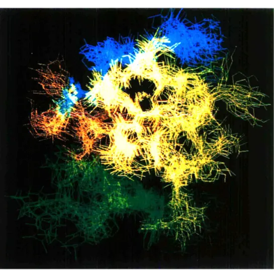
Fig. 2. An overlay of twenty configurations after a best fit superposition of all C atoms from a loops simulation of porcine insulin in aqueous solution illustrating the range of configurational space accessible to the protein on a subanosecond time scale.
calculation of molecular properties using (part of) the electronic degrees of freedom as variables. Or, the parameters (s) of empirical classical interaction functions such as (14) are generally determined such that the interaction energy Uphys includes the mean effect of the (omitted) electronic degrees of freedom.
A biomolecular system such as a receptor-ligand complex has in general more degrees of freedom (electronic, atomic) than can be reasonably treated as variables. Therefore, one has to select those degrees of freedom as variables in the molecular model, that are essential to a proper representation of the quantity or phenomenon one is interested in. With respect to the calculation of binding constants all degrees of freedom that are expected to give significant contributions to the energy or entropy of the complex compared to the individual receptor and ligand molecules, should be explicitly treated.
How is the interaction function for these degrees of freedom denned ? This can be done at different levels of sophistication. Interaction functions of the
simplest type that are generally used in crystallographic modelling contain only terms describing covalent bond lengths, bond angles and chirality centers, and van der Waals repulsion between atoms. General empirical energy functions usually also contain terms representing dispersion energy and Coulomb energy (Gelin, 1993). A higher level of sophistication, without explicitly entering the realm of quantum mechanics, is reached by the inclusion of terms describing the electronic polarisability of atoms in the energy function. With respect to the calculation of binding constants such polarisation terms will be particularly important when a charged ligand is bound to a receptor. A mean treatment of electronic polarisation as in (14) may not be sufficient to obtain accurate binding constants in such a case.
How are the explicitly treated degrees of freedom sampled ? Biomolecular complexes constitute microscopic systems, the properties of which are governed by the laws of statistical mechanics. This means that the probability of occurrence of a molecular (receptor-ligand) configuration r with energy
Uphys{r;s) is proportional to its Boltzmann factor
P(r)ccexp[-Uphys(r;s)/kBT]. (15)
A set of structures or configurations, in which each particular structure r occurs with relative probability P(r) according to (15) is called a Boltzmann ensemble or distribution of configurations. The third basic choice of molecular modelling using energy functions involves the method by which a set of molecular configurations is generated (Fig. 1). For molecular complexes with a few explicit degrees of freedom these may be systematically varied (or searched :SS) to find the configurations with the lowest energy Uphys(r;s), which will have the
highest probability of occurrence P(r). If the number of degrees of freedom grows, a systematic search of the vast configurational space of the molecular complex becomes impossible. Then, heuristic search (HS) methods, which visit a tiny, but hopefully representative set of configurations, are the only way to sample the Boltzmann ensemble. Though a simple method, energy minimisation (EM) is a very poor searching and sampling method, since it produces only one configuration which is a local minimum close to the initial structure. Much more powerful are methods such as Monte Carlo (MC) (Frenkel, 1993), molecular dynamics (MD) or stochastic dynamics (SD) (van Gunsteren, 1993) simulation, or combinations of these, that directly generate a Boltzmann ensemble. Since each configuration r in such an ensemble occurs with probability (15), averages and higher moments of the distributions can directly be calculated over the ensemble using a weight factor 1. A variety of even more powerful searching and sampling methods are known (van Schaik et
al. 1992, 1993; Scheraga, 1993; Huber et al. 1994), which generally do not
produce a Boltzmann ensemble of configurations. When taking averages of physical quantities over such a (non-Boltzmann) set of configurations, each configuration should be given the weight (15) in order to obtain a Boltzmann average. The calculation of binding constants should always be based on a Boltzmann average.
The next level of sophistication of molecular modelling involves the explicit evaluation of entropy or free energy (Fig. 1). From a Boltzmann ensemble the statistical equilibrium averages can be obtained for any desired property of the molecular system for which a value can be computed for each configuration of the ensemble. Examples of such properties are the potential or kinetic energy of (parts of) the system, structural properties, electric fields, etc. A number of thermodynamic properties can be derived from such averages. However, two important thermodynamic quantities, the entropy and the (Gibbs or Helmholtz) free energy, generally cannot be calculated using a statistical average. They are global properties of the molecular system that depend on the extent of configuration (or phase) space accessible to the system. Therefore, computation of the absolute free energy of a molecular system is virtually impossible. Yet, quantities important to drug design, such as binding constants and solubilities, are directly related to the free energy. Fortunately, over the past decade statistical mechanical procedures have evolved for evaluating relative free energy differences. They are rather demanding as far as computing power is concerned, but are very applicable in drug design based on receptor-ligand interactions.
4.1 Structure and energy calculations using flexible jD-structure-based models Molecular modelling of receptor-ligand complexes based on the matching of receptor and ligand properties or using rigid 3D-structures of receptor and ligand will produce a first estimate of the binding constant. Whether such an estimate bears any relation to reality will depend on the characteristics of the molecular system. For the binding of a rigid, positively charged receptor to a relatively rigid, negatively charged receptor (protein) it may be a reasonable estimate. However, for highly flexible molecules such as antibodies and antigens such an estimate is likely to be useless. In such cases flexibility of receptor, ligand and solvent has to be accounted for. Fig. 2 gives an impression of the flexibility of the protein insulin. 4.1.1 Flexibility of ligand and receptor
By using an energy function such as (14) for the atoms of the receptor and ligand, molecular flexibility can be accounted for in the drug design procedure. However, it depends on the technique used to search and sample molecular configurations, whether the potential molecular flexibility inherent to the energy function will be reflected in the energies and structures of the receptor-ligand complex that are obtained. The most commonly used search and sampling techniques are energy minimization, which only relaxes local strain in a molecular structure, and MC or M D simulation combined with high temperature annealing to extend the search (Goodsell & Olson, 1990; Kuntz, 1992; Stoddard & Koshland, 1992; Hodgkin et al. 1993).
Although the introduction of flexibility will in general improve the molecular model, it will only do so if the assumption of molecular rigidity is the accuracy limiting factor. If this is not the case the use of e.g. MC sampling techniques to sample conformations of flexible protein sidechains will not produce better
agreement with experimental data than simple modelling based on geometrical properties of amino acid sidechains (van Gunsteren & Mark, 19926).
4.1.2 Solvent effects
An obvious way to limit the number of explicitly treated degrees of freedom in a biomolecular system is to omit all or almost .all solvent degrees of freedom. Due to the abundance of solvent degrees of freedom for a biomolecule in solution, omission of these in an energy calculation easily reduces the required computing power by a factor of 10 to 50. Therefore, most drug design studies are carried out for molecules in vacua. The complete neglect of solvent effects will limit the accuracy of the calculated properties, such as binding constants. So, what is the role of solvent molecules in a biomolecular system ? The structure and stability of a molecular complex may depend on the type of solvent. Individual water molecules may play a structural role in e.g. protein-ligand or inhibitor binding. Polar solvents exert a dielectric screening effect on interactions between charges on the receptor and ligand, and the viscosity of the solvent will influence the dynamics of the atoms of the molecular complex, and may also influence the kinetics of the binding process.
Due to the many different low energy configurations occurring in a liquid, a solvent cannot be characterized by one or a few molecular configurations, but a (Boltzmann) ensemble of solvent configurations should be generated. So, when solvent degrees of freedom are included in the energy function (14), simulation techniques such as MC or MD are to be used, and systematic search (SS) and energy minimization (EM) are useless.
When simulating a microscopic system of finite size, the boundary of the system should be treated such as to minimize edge effects. The standard procedure is to use periodic boundary conditions. The solute or molecular complex and the surrounding solvent molecules are put into a periodic space-filling box, which is treated as if it is surrounded by identical translated images of itself. In this way basically an infinite periodic system is simulated. The periodic box should be taken large enough to avoid interactions between molecules and their periodic images, as illustrated in Fig. 3. This condition leads to sizeable amounts of solvent molecules, typically a few hundreds to thousands, to solvate the receptor, ligand or the complex, which makes such simulations expensive (van Gunsteren & Mark,
1992a).
An alternative to the explicit treatment of solvent molecules in a biomolecular simulation is an implicit one: the influence of the solvent on the solute degrees of freedom is incorporated in the energy function of the latter in an average manner. A potential of mean (solvent) force is used for the solute. Different mean solvation models are in use (van Gunsteren et al. 19946).
1. Accessible surface area type models
In this type of mean solvation model the local solvent contribution to the potential of mean force for solute atoms is taken to be proportional to the area of the solute atom or group of atoms that is accessible to solvent molecules (Ooi et al. 1987; Still et al. 1990; Schiffer et al. 1992; Wesson & Eisenberg, 1992).
Fig. 3. The central or parent simulation cell from a simulation of hen egg white lysozyme (green) solvated by 5300 SPC water molecules with spherical (truncated octahedron) periodic boundary conditions illustrating the amount of solvent required to approximate infinite dilution in a periodic system.
Other local quantities, such as the hydration volume (Kang et al. 1988; Vila et al. 1991), or the number of solute-solvent contacts (Stouten et al. 1993) can be used too. The expression for the accessible surface area of a solute atom generally depends on the coordinates of the solute atom itself and those of its nearest neighbour solute atoms. Thus, a mean force potential based on an accessible area model becomes a many-body interaction which generally involves non-negligible computational effort. Currently, it is not clear whether this increased effort is offset by the limited accuracy of such mean solvation models in mimicking solvent effects.
2. Simple pairwise solvation force models
The computation of the mean force of solvation would be considerably simplified and sped up, if the mean force could be formulated as a sum of pairwise (two-body) interactions. In fact, the mean force potential in the solvent contact or occupancy model (Stouten et al. 1993) can be expressed as a sum of two-body terms of the form expi — r^/za2), where rtj is the distance
by van Gunsteren et al. (1994^). Although the use of a simple pairwise solvation force induces only a minor increase of the required computing effort, it remains to be investigated whether its accuracy is comparable to that of a simulation including a (thick) layer of solvent molecules.
3. Dielectric screening models
Different approximate models for treating the long-range solute-solvent interactions due to dielectric screening and polarization effects are available too (Still et al. 1990; Solmajer & Mehler, 1991; Sharp, 1991, 1993; Gilson & Honig, 1991). Still et al. (1990) use an expression involving only one-body and two-body terms, which is based on the continuum approximation of a dielectric medium. A simple approach is to make the relative dielectric permittivity, e, distance dependent, for example using a linear (Pickersgill, 1988) or sigmoidal function (Solmajer & Mehler, 1991) of the atom-atom distance. A more complicated way to incorporate long-range electrostatic effects using a continuum representation of the solvent is based on solving the Poisson-Boltzmann equation on a 3-dimensional grid (Sharp, 1991), from which a simple two-body interaction term is derived that accounts for charge-solvent interactions in an average way (Gilson & Honig, 1991). For reviews on the treatment of long-range electrostatic interactions in molecular simulations we refer to (Sharp, 1993; Berendsen, 1993; Smith & van Gunsteren, 1993). Generally, the structure and stability of a molecular complex in aqueous solution will depend on the ionic strength of the solution. This implies that the presence of ions in the solvent should either be explicitly simulated (de Vlieg et al. 1989) or accounted for in an average manner by e.g. a Poisson-Boltzmann continuum model (Smith & van Gunsteren, 1993). Although the long-ranged interactions between ions in aqueous solution can be adequately approximated in a simulation, the explicit inclusion of ions in a simulation of a receptor-ligand complex in aqueous solution may not yield reliable results, since the relaxation time of a ionic distribution is likely to be longer than the simulation period. This slow relaxation is caused by the slow diffusion of hydrated ions in solution. In a biomolecular simulation including water and counterions, the simulation averages may then be easily based on non-equilibrated ion distributions, causing sizeable deviations from the mean effect of the ions. Therefore, the mean influence of the ionic solution might be better approximated by a simulation which only includes solvating water molecules.
4.1.3 Incorporation of experimental data in a simulation
If the molecular model and energy function Uphys(r;s) are perfect and if a
simulation can be carried on for an infinitely long time, the generated ensemble of molecular configurations will exactly represent the real molecular system: the structural and energetic properties derived from such an ensemble will be correct. In practice, neither condition is fulfilled. Atomic interaction functions, such as (14), are not infinitely accurate due to various approximations with respect to electronic degrees of freedom, quantum mechanical effects, many-body interactions, etc. that are made in their derivation. Second, an MD or MC