Spatiotemporal brain dynamics underlying attentional bias modi
fications
Etienne Sallard
a,⁎, Lea Hartmann
a, Radek Ptak
b,c, Lucas Spierer
a,⁎aNeurology Unit, Medicine Section, Faculty of Sciences and Medicine, University of Fribourg, Switzerland
bLaboratory of Cognitive Neurorehabilitation, Faculty of Medicine, University of Geneva, Switzerland
cFaculty of Psychology and Educational Sciences, University of Geneva, Switzerland
Keywords:
attentional bias modification inhibitory control ERP
source estimations
Exaggerated attentional biases toward specific elements of the environment contribute to the maintenance of several psychiatric conditions, such as biases to threatening faces in social anxiety. Although recent literature indicates that attentional bias modification may constitute an effective approach for psychiatric remediation, the underlying neurophysiological mechanisms remain unclear. We addressed this question by recording EEG in 24 healthy participants performing a modified dot-probe task in which pairs of neutral cues (colored shapes) were replaced by probe stimuli requiring a discrimination judgment. To induce an attentional bias toward or away from the cues, the probes were systematically presented either at the same or at the opposite position of a specific cue color. This paradigm enabled participants to spontaneously develop biases to initially unbiased, neutral cues, as measured by the response speed to the probe presented after the cues. Behavioral result in-dicated that the ABM procedure induced approach and avoidance biases. The influence of ABM on inhibitory control was assessed in a separated Go/NoGo task: changes in AB did not influence participants' capacity to inhibit their responses to the cues. Attentional bias modification was associated with a topographic modulation of event-related potentials already 50–84 ms following the onset of the cues. Statistical analyses of distributed electrical source estimations revealed that the development of attentional biases was associated with decreased activity in the left temporo-parieto-occipital junction. These findings suggest that attentional bias modification affects early sensory processing phases related to the extraction of information based on stimulus saliency.
1. Introduction
Whether an object attracts attention depends on its relevance to the current goals (i.e. attentional set) and its physical features (i.e. stimulus saliencyKoch and Ullman, 1985;Wolfe, 1994;Wolfe et al., 1989). For example, people dressed in blue will attract attention when one is looking for a friend with a blue t-shirt, while an unexpected, loud sound may capture attention independently of one's current behavioral goals. “Attentional bias” (AB) refers to the tendency to allocate more atten-tional resources to specific objects, such as toward food when one is hungry. ABs often have an obvious adaptive value (e.g. a bias toward food items may facilitate foraging), but may become maladaptive when expanding beyond the normal range or to irrelevant objects. For ex-ample, exaggerated biases toward emotionally negative stimuli could participates in intensifying anxiety (e.g.Amir et al., 2008;Britton et al., 2015; Heeren et al., 2015). Based on the evidence that abnormal AB contributes to the development and maintenance of many psychiatric disorders, important efforts have been invested into the development of behavioral interventions aiming to reduce ABs (Hakamata et al., 2010;
Lopes et al., 2015; MacLeod and Clarke, 2014). However, while at-tentional bias modification (ABM) procedures have shown promising behavioral effects, their underlying neurocognitive mechanisms remain unclear. The present study addresses this question by investigating the spatiotemporal brain dynamics of ABM to initially neutral stimuli in healthy adults.
ABM procedures typically involve practicing so-called “modified dot-probe tasks” (MacLeod et al., 1986;MacLeod and Mathews, 2012). In such tasks, pairs of visual cues differing along a given dimension (e.g. emotionally positive vs. negative faces) are briefly presented at one of two positions on the screen. Participants are asked to make a visual discrimination judgment of a probe stimulus appearing at the location of the cue or at the opposite location. ABs are expressed as faster re-sponses to the probes appearing at the location of cues attracting at-tention (e.g. an angry face) compared to the alternative location. Cri-tically, ABs can be modified by modulating the probability of the association between the location of a given cue and the location of the probe: if the probe is systematically presented on the same side as a specific type of cue, attention will progressively become attracted by
⁎Corresponding authors at: Neurology Unit, Medicine Section, Faculty of Science and Medicine, University of Fribourg, PER 09, Chemin du Musée 5, CH-1700 Fribourg, Switzerland.
E-mail addresses:[email protected](E. Sallard),[email protected](L. Spierer).
http://doc.rero.ch
Published in "International Journal of Psychophysiology 130 (): 29–39, 2018"
which should be cited to refer to this work.
this cue. Conversely, if the probe is always presented opposite to a given cue, subjects will develop an attentional bias away from the cue.
While many studies investigated ABMs using dot-probe tasks in healthy (e.g.Amir et al., 2008;Suway et al., 2013) and clinical popu-lations (e.g. Attwood et al., 2008; Eldar and Bar-Haim, 2010; Field et al., 2009; Lopes et al., 2014; Schoenmakers et al., 2010; Shafran et al., 2008), only few examined the neural underpinnings of ABM (Britton et al., 2015;Browning et al., 2010;Eldar and Bar-Haim, 2010; Li et al., 2016;Nelson et al., 2015;O'Toole and Dennis, 2012;Osinsky et al., 2014;Suway et al., 2013).
This literature suggests that ABM procedures may influence dif-ferent functional processing stages by showing effects at both late-la-tency high-order top-down control mechanisms, and at the level of early latency in brain areas involved in low-level processing.
Functional magnetic resonance imaging (fMRI) studies for instance showed associations between the modification of ABs toward emotional stimuli (threat-related vs. neutral or positive stimuli) and changes in activity within right lateral prefrontal cortices (rlPFC;Browning et al., 2010), middle frontal gyri (rMFG) and anterior insula (rAI; Li et al., 2016). Based on previous evidence for associations between these re-gions and voluntary control of attention, ABM procedures have been advanced to influence behavioral responses to the biased stimuli by modifying top-down attentional set. In contrast, other neuroimaging studies have suggested that the effects of ABM may also be influenced by low-level subcortical structures. Britton et al., 2015 for instance showed an increased activity in bilateral amygdala following an ABM training away from threatening faces in adults with high social anxiety symptoms.
In line with these fMRI literature, event-related potential (ERP) studies indicate that ABM training influences both late latency atten-tional control and error-related frontal N2 and conflict resolution P3 components (Eldar and Bar-Haim, 2010;Nelson et al., 2015;O'Toole and Dennis, 2012;Suway et al., 2013). Attentional allocation is typi-cally indexed by the N2pc ERP component, an occipital negativity contralateral to the side of an attended stimulus and manifesting be-tween 180 and 300 ms post stimulus onset (Eimer and Kiss, 2008; Holmes et al., 2014;Kappenman et al., 2015;Kappenman et al., 2014; Osinsky et al., 2014;Reutter et al., 2017;Weymar et al., 2011). The N2pc is notably assumed to reflect the attentional selection of a target stimulus among distractors. Further ERP studies revealed effects of ABM on even earlier latency parieto-occipital P1 sensory components, from 100 ms onwards.O'Toole and Dennis (2012)for instance showed that an ABM training toward or away from threat pictures modifies P1 amplitude to the emotional faces cues of the dot-probe task, suggesting that the procedure influenced early spatial attention.
Critically, the hypothesis for ABM influencing prefrontal control and lower-level bottom-up mechanisms are most likely complementary; top-down influence has indeed repeatedly been shown to alter long-term activity of low-level structure, with e.g. frontal cortices modulating amygdala responses and in turn attentional biases (Britton et al., 2015; Taylor et al., 2013; seeGilbert and Li, 2013for review).
A limitation of these previous investigations of ABM is that they fo-cused on emotional stimuli and were thus potentially confounded by individual variations in the initial biases to the stimuli. Pre-existing ‘natural’ biases to emotional stimuli may likewise influence the effects of ABM, limiting the generalizability of studies based on non-neutral sti-muli. In addition, previous ABM paradigms were designed to have single-direction effects by either focusing on developing approach or avoidance biases (e.g.Amir et al., 2008;Britton et al., 2015;Browning et al., 2010; Osinsky et al., 2014), preventing the comparison between the neuro-physiological mechanisms supporting the developments of approach vs. avoidance biases. For these reasons, it is important to study ABM with initially neutral stimuli that only obtain significance through practice, leaving open the possibility to spontaneously develop approach or avoidance biases. On this basis, the neural underpinnings of the devel-opment of approach vs. avoidance AB could then be compared.
To further characterize the nature of the biases induced by ABM procedures we investigated whether the development of biases influ-enced the executive control of the responses to the biased stimuli. Dual-process models posit that behavioral outcomes depend on interactions between bottom-up, implicit motivational responses to stimuli (as those putatively manipulated by ABM procedures), and top-down controlled, effortful actions such as response inhibition (e.g.Strack and Deutsch, 2004). While growing evidence indicate that maladaptive behaviors indeed depend on the relative strength of automatic approach biases and inhibitory control capacity (e.g.Kakoschke et al., 2015), to our knowledge no study so far directly tested whether modifying atten-tional biases actually modulates inhibitory control performance. Results for an influence of the ABM procedure on inhibitory control perfor-mance would enable linking the processes involved in each system. For instance, if the development of an approach bias to a given stimulus with the ABM procedure leads to more inhibition failures to this sti-mulus, it would suggest that at least part of the cognitive processes inhibited during the control task are those modified when an atten-tional bias develops (Kakoschke et al., 2015;Meule and Platte, 2016). To address these questions, we examined the spatiotemporal brain mechanisms underlying attentional bias modification toward and away from initially neutral stimuli using a modified dot-probe task. We in-vestigated when and where in the brain the ABM training modulates the processing of task-relevant stimuli by comparing ERPs to the cues measured at the beginning vs. the end of the ABM training. We ana-lyzed the ERPs within the electrical neuroimaging framework, in which modulations in the strength and the topography of the electricfield at the scalp are analyzed with robust randomization statistics and com-bined with intracranial source estimations (Michel and Murray, 2012; Murray et al., 2008a). Because changes in ERP topography necessarily follow from changes in the configuration of the underlying neural generators and changes in the globalfield power index modulation in the strength of the generator activity, our analyses could help disen-tangling the neurophysiological mechanisms underlying ABM (Murray et al., 2008a;Tzovara et al., 2012). Following the ERP analyses, dis-tributed source estimations were statistically analyzed over the periods showing strength and/or topographic ERP modulations. Since this ap-proach is data-driven and applied to the whole ERP epoch, it further overcomes the limitations of classical local ERP analyses focusing only on a priori determined electrodes and periods of interest, and allows identifying both when and where in the brain modifications of cortical processing are associated with the development of AB. This later methodological advantage is particularly important in the study of ABM because it may help better characterizing the interaction between late frontal and early low-level processing stage thought to underlie this phenomenon.
We hypothesized that ABM would affect both early stages of elec-trocortical processing reflecting gating mechanisms (latencies between 50–100 ms) or bottom-up capture of attention (50 and 200 ms, notably on the P1 parieto-occipital and N2pc ERP component;Fellrath et al., 2014;Hickey et al., 2006) and later, top-down attentional mechanisms (latencies after 150 ms within parietal and frontal sites corresponding to the N2 (Eldar and Bar-Haim, 2010;Osinsky et al., 2014), P2 (Eldar and Bar-Haim, 2010;Suway et al., 2013) and/or P3 components (Eldar and Bar-Haim, 2010)). We further assessed whether modulating at-tentional biases influenced inhibitory control to the biased stimuli by testing participants in a Go/NoGo task in which they had to withhold responses to the cues biased during ABM training. We predicted that increases in approach biases would decrease the ability to withhold responses to the cues (and the reverse effect with avoidance biases). 2. Material and methods
2.1. Participants
Thirty-two right-handed volunteers participated in this study.
Participants gave written informed consent; the study has been ap-proved by our local ethics committee and have therefore been per-formed in accordance with the ethical standards laid down in the 1964 Declaration of Helsinki. All participants had normal or corrected to normal vision, and no history of psychiatric or neurologic disorders. Four subjects were excluded because they responded only to probe stimuli displayed at the top of the screen (n = 1) or had errors rates > 2.5 standard deviations from the group mean (n = 2) or did not complete the task to the end (n = 1). Four additional participants were excluded because they already exhibited an attentional bias at the be-ginning of the training (see the Results section for details on how at-tentional bias was determined). Thefinal sample consisted of 24 par-ticipants (16 females) aged 23.9 ± 3.8 years.
2.2. Procedure and tasks
Participants first completed questionnaires and then the experi-mental tasks. A debriefing questionnaire was filled out at the end of the experiment.
2.2.1. Questionnaires
Psychological questionnaires assessed traits of anxiety and depres-sion (Hospital Anxiety and Depresdepres-sion Scale, HADS; Zigmond and Snaith, 1983) and impulsivity (Barratt Impulsiveness Scale;Bayle et al., 2000). Additional measures of state of anxiety, mood and stress were assessed using 10 cm long visual analog scales (VAS) with a cross mark at the center of the line. The scales had the terminal labels“very re-laxed” and “very anxious” for the anxiety state, “very happy” and “very unhappy” for the mood state and “very calm” and “very stressed” for the stress state. The debriefing questionnaire consisted of 5 questions
about the participants' awareness of the spatial association between cue-color and probe, the strategy they might have used during the task, and the aim of study.
2.2.2. Dot-probe task
In the modified dot-probe task a colored shape (predictive cue) was briefly presented 8.3° above or below the central fixation, together with a white shape (neutral cue) at the opposite location. The cues were presented in a vertical axis because this configuration has been sug-gested to result in larger effect sizes than an horizontal presentation (Hakamata et al., 2010). Both cues had a diamond or circle shape. The cue predicting the probe location was orange or blue while the neutral cue was always white. Probes were two horizontally or vertically aligned small double dots (0.1°). The colored cues were systematically presented at the same (Approach condition) or at the opposite position (Avoid condition) to the probe. The white cue was not predictive of the probe position because since it was presented half of the trials with the Approach or Avoid color condition, it was not possible to predict the position of the probe only from the position of the white cue. The lo-cation of the probe could only be deduced from the (colored) cues. The combinations between the cue color and the Approach/Avoid condi-tions were randomized across participants. Variacondi-tions in colors unlikely confounded our results because i) the white cue was always presented together with a colored cue; ii) the colors used for each condition were counterbalanced across participants; and iii) we focused on the Group∗ Condition ∗ Session interaction (and not a main effect of the factor Condition). The presentation of horizontal or vertical probes was equiprobable after each type of cue and throughout the experiment.
Participants were comfortably seated in a quiet room at 110 cm distance from a 19-inch LCD screen. Each trial started with a black
Fig. 1. Experimental paradigms. A. Modified Dot-probe task used to develop attentional biases: Pairs of colored shapes (the cue) were replaced by horizontally or vertically aligned double dots (the probes). The probes were systematically presented at the same (Approach condition) or at the opposite position (Avoid condition) of a given color. Participants had to respond as fast as possible to the orientation of the double dots during the probe presentation. B. Go/NoGo task. The cue stimuli whose color was biased during the attentional bias modification dot-probe task were used as stimuli during the Go/NoGo task. Participants had to respond as fast as possible to stimuli with a specific shape while withholding their responses to stimuli with another shape, independently of the stimuli's color. Since attention was biased to the color and the Go/NoGo task was based on the shape of the stimuli, we could use all types of biased cues (approach, avoidance and neutral colors) as NoGo stimuli to assess the effect of attentional bias modification on inhibition performance.
fixation cross presented on neutral gray background for 500 ms, fol-lowed by the presentation of the cues (Fig. 1A). During this time period, participants were instructed tofixate the cross at the center of the screen. After 1000 ms, the cue display was replaced by the probe dis-play (300 ms). The cues were presented during 1000 ms to facilitate the learning of the cue-probe association. Simultaneously with the probe, a response window opened, and terminated when the participant sponded or after 1200 ms. A 1000 ms wait period followed the re-sponse, which was replaced by a short feedback about performance (500 ms). In order to motivate participants to react as fast and accu-rately as possible, each response that was incorrect or slower than the individual reaction time (RT) threshold (see details below) was fol-lowed by an additional‘punishment’ wait period of 2500 ms. Individual trials were separated by inter-trial intervals of 500–800 ms. Participants were asked to respond as fast as possible by pressing a button with the right indexfinger when double dots were aligned vertically and another button with the right middlefinger when the double dots were aligned horizontally.
Participants performed 10 experimental blocks of 64 trials (32 Approach; 32 Avoid). Each block was preceded by a calibration phase of 8 trials (4 Approach; 4 Avoid) used to compute an individual threshold RT (average RT of all calibration trials). Feedback given to the participant in the subsequent experimental block depended on a comparison between the trial RT and the RT threshold: “V” if RT < threshold;“Too late” if RT > threshold; “X and too late” for RT > threshold and incorrect response;“X” for RT < threshold and incorrect response; and “no response” for omission. Because the threshold RT was calculated before each block and for each participant separately, and because response times above the threshold were ‘punished’ with a wait period, a constant time pressure, and thus the task difficulty, were kept constant across participants and blocks. This procedure further allowed encouraging the participants to pay attention to the cue because it could help them improving their performance: since the cue predicted the location of the probe, paying attention to the cue enabled responding faster to the probe and thus avoiding the wait periods.
The experiment lasted about 1 h with 1–2 min breaks between each experimental block. Participants were naive regarding the aim of the study. Stimulus presentation and response recording were controlled using E-Prime 2.0 (Psychology Software Tools, Inc., Sharpsburg, PA).
2.2.3. Go/NoGo task
The Go/NoGo task was performed before and after the ABM training and consisted in a calibration phase of 12 trials (6 Go; 6 NoGo) followed by the experimental phase of 60 trials (30 Go; 30 NoGo). The calibra-tion phase was used to determine the reaccalibra-tion time threshold (RTt; calculated as 90% of the mean RT to Go trials). As detailed below, a feedback on response speed based on the RTt was presented to parti-cipants during the experimental phase to maintain an individually ad-justed time pressure (for similar procedure, seeHartmann et al., 2016; Manuel et al., 2010;Pourtois et al., 2010).
Stimuli were colored shapes (orange, blue and white circles and diamonds) presented at the center of the screen (Fig. 1B). Trials started with the presentation of afixation cross during 1000–1300 ms, followed by the stimulus (500 ms) and a response window (1500 ms, terminating when the participant responded, but with a minimal duration of 250 ms). A wait period of 750 ms was then presented and participants received a feedback on their performance during 500 ms: a“V” for Hits (response after a Go trial) with a RT < RTt and for correct rejections (no response after a NoGo stimulus); a “Too late!” for Hits with a RT > RTt; a“X” after misses (no response after a Go stimulus) and false alarms (response after a NoGo stimulus).
Participants were instructed to respond as fast as possible to a Go stimulus represented by a given shape (e.g. circle) and withhold the response to a NoGo stimulus defined by another given shape (e.g. diamond) independently of its color. The NoGo stimuli (circle or
diamond shape) were counterbalanced across participants. Note that in the ABM training, attention was biased toward or away from specific colors independently of cue shape, while in the Go/NoGo task, the type of response was determined by stimulus shape. We could therefore orthogonally vary the color of the Go/NoGo stimuli and examine the effect of ABM training on reaction time and false alarm rate in-dependently. For participants trained toward orange and away from blue during the ABM dot-probe task, the Go/NoGo conditions were “Approach” for the orange Go or NoGo shapes, “Avoid” for the blue shapes and“Neutral” for the white shapes; for participants trained to-ward the blue and away from the orange during the ABM dot-probe task, the Go/NoGo conditions were “Approach” for the blue shape, “Avoid” for the orange shape and “Neutral” for the white shape. 2.3. Behavioral analyses
2.3.1. Attentional bias modification Dot-probe task
Scores on questionnaires, error rates and RTs in the dot-probe task were analyzed as dependent variables. For each participant separately, individual trials with a RT < 100 ms and/or > mean RT ± 2 SD were excluded. Then, to ensure that no attentional bias was present at the beginning of the task (i.e. that the stimuli were indeed initially neutral to the participant), an unpaired t-test was performed for each partici-pant separately between the Approach vs. Avoid condition of the 3first blocks (beginning: BEG). Participants with a significant (p < 0.05) difference in RT between the two conditions at the beginning of the task were excluded (n = 4). Finally, participants with an error rate greater than ± 2.5 SD from the group mean in Approach and/or Avoid condi-tion at the beginning and/or the end (3 last blocks: END) of the ABM task were excluded (n = 2).
The attentional bias in the dot-probe task was calculated by sub-tracting the mean RT (correct trials only) to the probe between the Approach vs. Avoid condition and then between the BEG vs. the END of the task: ΔABM = BEG(RT(Approach) − RT(Avoid)) − END(RT-(Approach)− RT(Avoid)).
A negativeΔABM value (i.e. participants included in the Toward group) indicated that training led to the development of an approach bias, while a positive value (i.e. participants included in the Away group) indicated an avoidance bias. Since we were interested in neu-rophysiological correlates of the training outcome, participants were split in those with a greater relative bias in favor of the Approach cue (henceforth termed“Toward” group), and those with a greater relative bias away from the Avoid cue (“Away” group). The RT were then compared between the beginning and end of the task in Approach and Avoid conditions and between each group using a Training (BEG; END)∗ Cue (Approach; Avoid) ∗ Group (Toward; Away) three-way ANOVA.
2.3.2. Go/NoGo task
We analyzed the Go/NoGo task using the inverse efficiency score (Townsend, 1975;Townsend and Ashby, 1979), corresponding to the mean RT (RT < 100 ms and/or > mean RT ± 2 SD were excluded) divided by the percent correct rejection (100− percent false alarms). The advantage of this index is that it accounts for effects of potential speed-accuracy trade-off. The efficiency index was then compared be-fore vs. after the ABM training in each condition and between the two groups using a Training (Pre-ABM; Post-ABM)∗ Stimuli (Approach; Avoid; Neutral)∗ Group (Toward; Away) three-way ANOVA.
2.4. Electrophysiological recording and analysis
2.4.1. EEG recording
Continuous electroencephalogram (EEG) was recorded with a sampling rate of 1024 Hz through a 64-channel 10–20 Biosemi Active-Two system (Biosemi, Amsterdam, Netherlands), referenced to a ground circuitry (common mode sense/driven right leg ground or
CMS–DRL, placed on each side of POz). This circuitry consists of a feedback loop driving the average potential across the montage as close as possible to the amplifier zero (cf. the Biosemi website:http://www. biosemi.com/pics/zero_ref1_big.giffor a diagram).
Offline ERP processing was performed with the Cartool software (Brunet et al., 2011), and statistical analyses were performed with the RAGU (Koenig and Melie-Garcia, 2009; Koenig et al., 2011) and the STEN toolbox developed by Jean-François Knebel and Michael Notter (http://doi.org/10.5281/zenodo.1164038). Raw EEG was first band-passfiltered (second order Butterworth with −12 db/octave roll-off; 0.1 Hz high-pass; 40 Hz low-pass; 50 Hz notch). Then, epochs from -100 pre- to 500 ms post-cue onset were extracted. Epochs containing at least one-time frame at ± 80μV in at least one electrode were rejected. The remaining artifact-free epochs were then averaged in each participant separately for the Approach and Avoid condition for the beginning (BEG; 3first blocks) and the end (END; 3 last blocks) of the task and re-referenced to the average reference. We focused on the threefirst and three last blocks as it allowed reaching a reliable signal-to-noise ratio in the ERP while keeping the duration of the training time separating these blocks as long as possible. As a result of the procedure described above, averaged ERPs were based on mean ± SD 85 ± 17 (Approach) and 84 ± 16 epochs (Avoid) in the BEG condition and 86 ± 13 (Ap-proach) and 85 ± 15 epochs (Avoid) in the END condition. These numbers did not differ (p > 0.05) between conditions, notably for our statistical interaction of interest (2 × 2 × 2 ANOVA with Group (To-ward; Away) as between-subject factor and Cue (Approach; Avoid) and Training (BEG; END) as within-subject factors). Once the ERP were computed, waveforms at electrodes still showing artifacted signal of each participant were interpolated using a 3D-spline approach (Perrin et al., 1987); mean 5 ± 2% electrodes were interpolated.
2.5. General analysis strategy
ERPs were analyzed with a three-way ANOVA with Group (Toward; Away) as between-subject factor, and Cue (Approach; Avoid) and Training (BEG; END) as within-subject factors. Because the main effects provide no information about training-induced modifications of atten-tional bias, and to reduce the number of tests performed, we focused our analysis only on interaction terms.
Wefirst conducted electrophysiological analyses both on local and global measures of the electricfield at the scalp. Local analyses refer to the comparison between the experimental condition at the level of the ERP waveform at each electrode separately. In contrast, global analyses of the ERP focus on the strength and topography of the whole electric field at the scalp. These analyses have the advantage of being in-dependent on the choice of the reference and of enabling to differ-entiate effects following from modulations in the strength of responses of statistically indistinguishable brain generators from alterations in the configuration of these generators (viz. the topography of the electric field at the scalp). These methods have been shown to be useful for analyzing EEG data from larger electrode sensor arrays and have been extensively detailed elsewhere (Murray et al., 2008a; Tzovara et al., 2012for a method tutorial).
2.6. Local event-related potentials (ERP) analyses
As afirst step, we conducted local electrode analyses: ERP voltage waveforms were submitted to the Group∗ Cue ∗ Training three-way ANOVA described above at each time frame and for each electrode separately. This analysis of the ERP voltage waveform data at the single electrode level are presented here only to help comparing the result of the present study with previous literature using canonical ERP analysis approaches, namely a comparison of voltage amplitudes for specific ERP components of interest (i.e. the ERP voltage at a given electrode and latency). For example, our time-wise analysis over the whole electrode montage could reveal an effect on the classical N2 component
by showing an interaction on the typical electrode and latency of the N2: Frontocentral/anterior electrodes between 200 and 250 ms. While this approach is highly sensitive to detect ERP modulations, it entails a large number of statistical tests and is thus prone to false positive. To (partially) correct for multiple comparisons and for temporal and spa-tial auto-correlation, we considered in this analysis only the periods showing the interaction of interest lasting longer than 20 ms on > 10% of the electrodes (e.g.Guthrie and Buchwald, 1991; see e.g.Hartmann et al., 2016;Murray et al., 2008bfor corresponding approaches). Cri-tically, another limitation of the local electrode analyses is that the location and timing of their results are dependent on the choice of the reference electrode and thus subject to experimenter biases (Murray et al., 2008a;Tzovara et al., 2012).
To circumvent these problems, we did not interpret the results of this analysis in the present study, but rather based our conclusions on reference-independent global measures of the strength and topography of the electricfield at the scalp (e.g.Michel and Murray, 2012;Tzovara et al., 2012for reviews on this approach).
2.7. Global ERP analyses
Modulations of the strength of the electricfield at the scalp were analyzed using the globalfield power index (GFPKoenig et al., 2011; Koenig and Melie-Garcia, 2010;Lehmann and Skrandies, 1980). GFP is calculated as the spatial standard deviation of the electricfield (i.e. the root mean square of the difference between two normalized vectors computed across the entire electrode set). Larger GFP amplitudes in-dicate stronger electric fields; GFP peaks thus indicate highly syn-chronized neural sources underlying the scalp-recorded activity (Michel and Murray, 2012).
Modulations of the topography of the electricfield at the scalp were analyzed using the global map dissimilarity index (GMDLehmann and Skrandies, 1980). GMD indexes differences in the configuration be-tween two electricfield and is calculated as the root mean square of the difference between the potentials measured at each electrode for the different experimental conditions normalized by instantaneous GFP. Because changes in topography forcibly follow from changes in the configuration of the underlying active sources (Lehmann, 1987), to-pographic modulations reveal when distinct brain networks are acti-vated across experimental conditions.
Since the GFP is insensitive to spatial (i.e. topographic) changes in the ERP, and that GMD is calculated on GFP-normalized data, the GFP and GMD are orthogonal measures and can thus be interpreted sepa-rately.
GFP and GMD were compared across experimental conditions at each time frame using non-parametric randomization statistics (Monte Carlo bootstrapping): the differences in GFP and GMD between the experimental conditions were compared with a distribution of the dif-ferences derived from permuting 5000 times the conditions' label of the data for each participant (Koenig et al., 2011;Murray et al., 2008a). The probability of obtaining a GMD and GFP value from the permuta-tions higher than the measured value was then determined. The threshold for statistical significance was set at p < 0.05, and to correct for multiple comparison and temporal autocorrelation, differences were only accepted as significant if they were present for > 21 continuous time-frames (i.e. 20 ms for our sampling rate at 1024 Hz;Guthrie and Buchwald, 1991). This analysis allowed to identify the period of in-terest showing sustained Group∗ Cue ∗ Training interactions at the ERP level. These periods were then used to determine the periods over which the analysis of source estimations was conducted.
As a further control for the confounds related to splitting the par-ticipants in two groups, we conducted the GMD analyses with the be-tween factor Group as a continuous variable (see e.g. Koenig et al., 2011for details on the topographic ANCOVA method; Koenig et al., 2008;Pedroni et al., 2011). The topographic ANCOVA revealed a sig-nificant Group ∗ Cue ∗ Training interaction from 46 to 72 ms
(supplementary Fig. 1), confirming the first analyses with the factor group dichotomized between the participants developing a relative approach vs. avoidance bias.
2.8. Electrical source estimations
Brain sources of ERP modulations were estimated using a dis-tributed linear inverse solution model (a minimum norm inverse solu-tion) combined with the local autoregressive average (LAURA) reg-ularization approach, which describes the spatial gradient across neighboring solution points (Grave de Peralta Menendez et al., 2004; Grave de Peralta Menendez et al., 2001). LAURA enables investigating multiple simultaneously active sources and selects the configuration of active brain networks which better mimics biophysical behavior of neuralfields. In LAURA's approach, the strength of the potentials at a given location depends on the activity of its neighbor nodes according to electromagnetic laws derived from the quasi-static Maxwell's equa-tions stating that the strength of a source falls off with the inverse of the squared distance for potentialfields (cubic distance for vector fields; Grave de Peralta Menendez et al., 2004,Grave de Peralta Menendez et al., 2001;Grave de Peralta Menendez and Gonzalez Andino, 2002; for a review see Michel et al., 2004). LAURA uses a realistic head model, and the solution space included 3005 nodes, selected from a grid equally distributed within the gray matter of the Montreal Neurological Institute's average brain (gray matter segmentation courtesy of Grave de Peralta Menendez and Gonzalez Andino; http://www.electrical-neuroimaging.ch). The head model and leadfield matrix were gener-ated with the Spherical Model with Anatomical Constraints (SMAC; Spinelli et al., 2000). Fundamental and clinical research has assessed the spatial accuracy of this inverse solution (e.g.Gonzalez Andino et al., 2005a, 2005b;Grave de Peralta Menendez et al., 2001;Michel et al., 2004). As an output, LAURA provides current density measures; their scalar values were evaluated at each node. The ERP were averaged for the period of interest determined by the global ERP analyses, their source calculated and then submitted to a 2 × 2 × 2 ANOVA with be-tween-subject factor Group (Toward; Away) and within-subject factors Cue (Approach; Avoid) and Training (BEG; END). To correct for mul-tiple testing and spatial auto-correlation, we applied a spatial-extent correction (Ke) of≥16 contiguous nodes with a p-value < 0.05. This spatial criterion was determined using the AlphaSim program (avail-able athttp://afni.nimh.nih.gov) and assuming a spatial smoothing of 6 mm FWHM. This program applies a cluster randomization approach. The 10,000 Monte Carlo permutations performed on our leadfield matrix revealed a false positive probability of < 0.005 for a cluster > 16 nodes. Given the spatial resolution of the LAURA source estimation (ca. 1 cm with our 64 channel EEG set-up; seeMichel et al., 2004), we interpret our results with the resolution of the AAL atlas ( Tzourio-Mazoyer et al., 2002).
3. Results
3.1. Behavioral results
3.1.1. Modified Dot-probe task
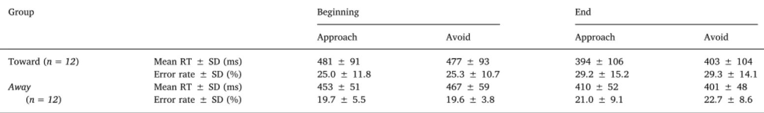
The mean percentage of error and reaction time (RT ± SD) are reported inTable 1. We did not analyze the change in RT between the groups because these behavioral data were used as the criteria to split the participants between the Toward and Away group; the analysis would thus have been circular (Fig. 2A).
3.1.2. Go/NoGo task
We analyzed the inverse efficiency index (IEI) with the same three-way Training∗ Group ∗ Cue design as for the ABM dot-probe task (Fig. 2B). There was a main effect of factor Training driven by better performance (lower IEI) after than before the ABM dot-probe task (F(1; 22) = 12.87, p < 0.01;η2P = 0.37), most likely resulting from a retest
effect since it was stimulus- and group- independent. There was also a main effect of factor Stimulus (F(1; 22) = 3.71, p < 0.05; η2P = 0.26) and a Group∗ Stimulus interaction (F(1; 21) = 5.44, p < 0.05; η2P = 0.34). We did not conduct follow-up tests on these effects be-cause our primary interest was on the three-way interaction. Critically, there was no Training∗ Group ∗ Stimulus interaction (F(2; 21) = 0.34, p = 0.7; η2P = 0.03 Fig. 2B), and thus no evidence that the ABM training changed the participants' ability to inhibit their responses to the biased stimuli.
3.1.3. Questionnaires
Scores to the questionnaires (VAS of anxiety, mood and stress; BIS11 and HADS anxiety and depression) did not differ between the Toward and Away group (all p-values > 0.05;Table 2). There was no correla-tion between ΔABM and the scores to the questionnaires (all p-va-lues > 0.05;Table 3). The debriefing questionnaire revealed that only 2 out of the 24 participants (9%) were aware of the association between cue color and location of the probe. Both indicated having identified this association during the last block of the ABM dot-probe task.
3.2. EEG results
3.2.1. Event-related potentials
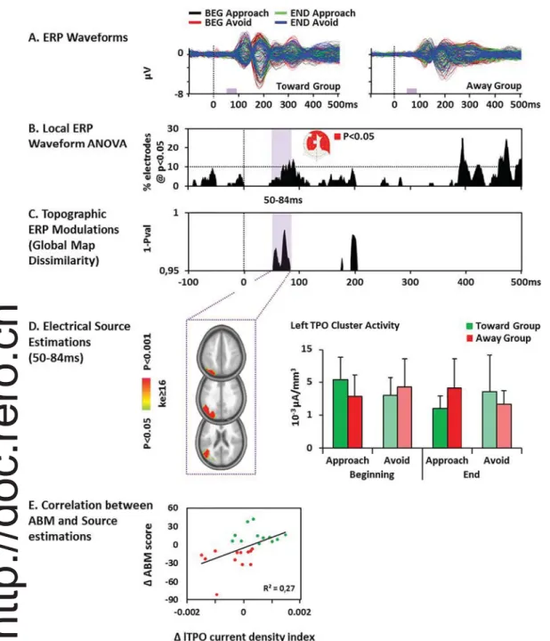
The analysis of the ERPs at electrode level showed a significant Group∗ Cue ∗ Training interaction over three time periods: 54–115 ms, 377–432 ms and 440–500 ms (Fig. 3A). While the analysis of GFP re-vealed no significant effect (Fig. 3B), there was a significant Group∗ Cue ∗ Training interaction 50–84 ms after the onset of the cue in the analysis of GMD (p < 0.05, > 20 time frames; seeFig. 3C), in-dicating topographic differences within this interval related to training effects.
3.2.2. Distributed source estimations
Source estimations were analyzed within the period of interest showing significant topographic modulations (i.e., 50–84 ms). The re-sults revealed a Group∗ Cue ∗ Training interaction in the left temporo-parieto-occipital (TPO) junction (F(1; 22) = 12.28, p < 0.01; η2P = 0.36) including the angular and supramarginal gyrus (i.e. in-ferior parietal lobule), the middle and superior temporal gyrus as well as the precuneus, cuneus and superior occipital gyrus (Fig. 3D). Follow-up Session∗ Condition ANOVAs were then conducted separately for the Toward and the Away group. These analyses revealed a Ses-sion∗ Condition interaction in the Toward group (F(1; 11) = 8.44, p < 0.05;η2P = 0.43), driven by a higher activity of the TPO cluster in the BEG Approach compared to END Approach condition (t(11) = 4.51, p = 0.001; using anα = 0.0063 Bonferroni adjusted threshold).
We further calculated on the whole group of participants the corre-lation between theΔABM score (i.e. [(BEG (Approach − Avoid) − END (Approach− Avoid)] index) calculated on the response time and on the source estimation current densities (Inverse Solution index:ΔIS). This brain/behavioral Pearson analysis revealed a relationship between the development of the AB and the related modification in the TPO cluster activity (r(22) = 0.51, p = 0.01; seeFig.3E), suggesting that the more the TPO activity increased, the more the participant developed a relative approach bias.
4. Discussion
We identified the spatiotemporal brain dynamics underlying atten-tional bias modification (ABM) to initially neutral stimuli in healthy participants. Behaviorally, half of the participants developed a bias in favor of the cue whose spatial location matched with the location of the stimulus of interest (approach bias:“Toward” group), and the other half in favor of the cue at the opposite location to the probe stimuli (avoidance bias: “Away” group). Electrical neuroimaging results in-dicated that the development of ABs was associated with decreases in
response of the left temporo-parieto-occipital (TPO) junction to the stimuli biased by the ABM training (the cue at the same or opposite position to the probe in the Toward and Away group, respectively). Finally, we found that the more the TPO activity increased, the more the participant developed a relative approach bias, further supporting the functional relevance of the change brain activity associated with the attentional biases developments.
Our results show that participants developed ABs to initially neutral stimuli. Contrasting with studies in which participants were assigned to an ABM procedure favoring the development of an approach or an avoidance bias (Amir et al., 2008;Browning et al., 2010;Chen et al., 2015;O'Toole and Dennis, 2012), participants in our study were pre-sented with a situation in which they could develop spontaneously both types of biases. This approach revealed inter-individual differences in the sensitivity to the two types of probabilistic associations between cues and task-relevant information (same or opposite location). Al-though negative results should be interpreted with caution, the analyses of correlations between the development of a specific bias and ques-tionnaire scores suggest that the individual inclination to develop an approach or an avoidance bias was not associated with traits or states of stress, anxiety, depression or impulsivity. Non-significant initial ten-dencies for biases toward or away from specific colors might have been amplified by the procedure and resulted in the observed pattern (7/12 participants in the Toward group and 12/12 participants in the Away group showed afinal bias in the same direction as the tendency they showed before the ABM). This result suggests that measures of small, initial biases could potentially be used to predict the development of abnormal, clinically relevant biases.
We would further note that the tendency to develop approach vs. avoidance attentional strategies might also be modulated by the mod-ality or complexity of the stimuli used as the cues, as well as by the
task-at-hand (e.g.Giard and Peronnet, 1999;Stevenson and Wallace, 2013; Thelen et al., 2014). The effect of these factors should be further ex-amined to draw general conclusions on the general relationship be-tween the development of specific AB and functional activity.
We found no effect of modifying AB on the capacity of participants to inhibit their responses to the biased stimuli. Since the effect of our ABM were in the same order of magnitude as the effect of previous studies (e.g.Suway et al., 2013), and we used a well-established ap-proach to measure inhibitory control, it is unlikely that methodological limitation account for the negative results. Our results question pre-dictions from 2-systems models for a direct relationship between mo-tivational biases and inhibitory control performance (Field and Cox, 2008;Strack and Deutsch, 2004), and rather suggest that i) modi fica-tions in the ability to withhold responses to incentive-motivational stimuli only manifest in situations of extreme biases and/or abnormally low inhibition (Dawe et al., 2004) ii) ABM might modify a type of bias that do not directly influence control capacities (e.g. attentional vs. approach bias; Sharbanee et al., 2013) or iii) that decision-related “cognitive” impulsivity which would interact with AB, should be dis-tinguished from the“motor” impulsivity measured in our study (de Wit and Richards, 2004;Olmstead, 2006;Reynolds et al., 2006). Our ne-gative results however call for emphasizing that automatic and con-trolled systems in dual process models might be independent from each other. They further suggest that clinical interventions aiming at redu-cing ABs or at improving the capacity to inhibit unwanted responses would unlikely benefit from targeting the other type of process.
EEG analyses revealed a topographic ERP modulation 50–84 ms post-cue onset associated with training-related differences between participants developing an approach or an avoidance bias. Previous ERP studies have related brain activity in the 50–100 ms latency range to the gating of stimuli for further processing and/or process related to
Table 1
Mean RT ± SD (ms) and error rate ± SD (%) during the dot-probe task.
Group Beginning End
Approach Avoid Approach Avoid
Toward (n = 12) Mean RT ± SD (ms) 481 ± 91 477 ± 93 394 ± 106 403 ± 104 Error rate ± SD (%) 25.0 ± 11.8 25.3 ± 10.7 29.2 ± 15.2 29.3 ± 14.1 Away (n = 12) Mean RT ± SD (ms) 453 ± 51 467 ± 59 410 ± 52 401 ± 48 Error rate ± SD (%) 19.7 ± 5.5 19.6 ± 3.8 21.0 ± 9.1 22.7 ± 8.6
Fig. 2. Behavioral results. A. Modifications of the attentional biases (ABs, in millisecond). The difference in response time to the probes in the ‘Approach’ minus the ‘Avoid’ condition, between the beginning and the end of the attentional bias modification training is depicted for each participant. The Toward group (positive ΔABM score), who developed a larger AB to the Approach cue than to the Avoid cue is in green and the Away group (positiveΔABM score), who developed an AB to the Avoid cue condition is in red. B. Box plot for the inverse efficiency index (RT/%correct rejection) during the Go/NoGo task. The median (horizontal line), the mean (cross), and the minimal and maximal values (whiskers) are represented. There was no effect of the ABM training on the Go/NoGo performance to the biased cues. (For interpretation of the references to color in thisfigure legend, the reader is referred to the web version of this article.)
the bottom-up capture of visual attention by salient stimuli (Clark and Hillyard, 1996;Egeth and Yantis, 1997;Eimer and Kiss, 2008;Fellrath et al., 2014;Fichtenholtz et al., 2007;Hillyard and Anllo-Vento, 1998; Luck et al., 1990;Mangun, 1995;Mangun and Buck, 1998). Three main ERP components manifest over this time period, the C1, P1 and N1. The C1 occipito-parietal component peaks 50–100 ms, and while it is mostly sensitive to low-level stimulus features (spatial position, size, etc.; Rauss et al., 2011), this component is also sensitive to stimuli-related internal states, notably including top-down attention (Gilbert and Li, 2013; Kelly et al., 2008) or motivational value (Rossi et al., 2017; Stolarova et al., 2006). The posterior P1 and N1 ERP components peak at 60–190 ms and are sensitive to observer's focus of attention (Fellrath et al., 2014;Mangun, 1995;Mangun and Buck, 1998;Vogel and Luck, 2000; though see e.g.Ding et al., 2014), a process that is also modu-lated in a bottom-up fashion by the saliency of the stimuli, even if saliency is irrelevant to the task (Fellrath et al., 2014). The P1 com-ponent has also been interpreted as indexing stimulus gating, notably based on evidence for its sensitivity to the attended location (Clark and Hillyard, 1996;Egeth and Yantis, 1997;Eimer and Kiss, 2008;Fellrath et al., 2014;Fichtenholtz et al., 2007;Hillyard and Anllo-Vento, 1998; Luck et al., 2000;Luck et al., 1990;Mangun, 1995;Mangun and Buck, 1998).
With regard to this literature, the early ERP modulation observed in our study thus suggests that ABM procedures impact on stimulus gating and on how the stimuli capture attention. This conclusion is further supported by our result that the training modulated the left TPO junction response to the cue. This region was previously shown to be recruited when salient information must be ignored and when low-saliency stimuli must be selected by attention (Gillebert et al., 2011; Mevorach et al., 2009;Mevorach et al., 2006). The left lateralization of the TPO junction modulation observed here echoes findings from Mevorach et al. (2009), who report a left lateralized saliency-based selection process involving the left angular gyrus, superior parietal lo-bule and superior medial gyrus when participants were under condi-tions of high distractor saliency. InMevorach et al. (2009), repeated stimulation of the left posterior parietal cortex with repeated tran-scranial magnetic stimulation decreased the ability to bias selection away from salient stimuli. Finally,Gillebert et al. (2011)reported at-tentional deficit in patient with left intra-parietal lesion.
Importantly, the positive correlation between the modification of the AB and the TPO junction modulation support the behavioral re-levance of this change in functional activity. Covariations between changes in ABs and underlying electrophysiological activity had al-ready been reported but only for later-latency ERP components (e.g. P2 and N170 inO'Toole and Dennis, 2012).
While the localization of the effects of ABM in previous functional neuroimaging studies overlap with our effects, current literature tends to report ABM-related changes in brain activity within more anterior areas (e.g. the rlPFC) inBrowning et al., 2010; and the rAI, rMFG and bilateral inferior frontal gyrus (IFG) inLi et al., 2016). For example, Browning et al. (2010)showed that an approach- or avoid- threatening-face ABM training modulated lPFC activation to these emotional sti-muli, with increases in activity associated to attention directed contrary to the trained association (e.g. the avoid-threat group showed increased lPFC when looking toward threatening faces). In the fMRI study byLi et al. (2016), women with depression who received an ABM training toward positive items during 4 weeks showed reduced amplitude of low frequency resting statefluctuations of brain activity within the rAI and rMFG when compared to a placebo group, and within the rAI, rMFG, and bilateral IFG when compared to their state before the ABM training. The partial discrepancies between our and previous studies might follow from the following differences: because electrical neuroimaging has a much higher temporal resolution than fMRI, it was likely more sensitive to the fast early latency modulations of posterior responses to the cues found in the present study. In addition, we used simple stimuli, whose discrimination likely involved earlier, lower-level processing phases than e.g. faces, and which did not involve other perceptual systems as those supporting face recognition or comparisons with pre-existing representations of valence. The absence of initial biases to the stimuli and the fact they were not emotionally laden could further ex-plain the absence of modulation in subcortical limbic areas (Britton et al., 2015). Additional modulations within higher-order associative areas or emotion-/reward- related networks would probably manifest if more complex stimuli involving these systems were used. Of note, most of previous studies used horizontal alignment for the cues and probes, a parameter shown to influence the amplitude of attention-related ERP components (e.g.Eldar and Bar-Haim, 2010;Li et al., 2008;Lopes et al., 2014). The long cue duration in our study might also have influenced this aspect by modulating anticipatory processes. Further studies are needed to identify whether and how cue durations interact with the latency and location of the brain modulation supporting ABM.
We showed that independently on whether the AB developed away from or toward the cues, it was associated with a decrease in early-latency responses of the TPO junction activity. In contrast with previous studies which focused on modulating pre-existing biases (either because the participants had abnormal biases or because the cues used in ABM had an emotional value), we examined ABM to initially neutral stimuli in healthy participants. Since the ABM dot-probe task increased the incentive value of the cues, our results indicate that the ABs resulting from such processes actually depend on early sensori-perceptive stages of stimuli processing. We suggest that the bias induced by the ABM training modifies the capture of attention by the stimuli. This process would then required less functional resources (Kelly and Garavan, 2005), which manifest as a decrease in the amplitude of the response of TPO junction to the biased cues. Although they would act more as purely cortical‘preconscious priming’, such effects of attentional bias on low-level, bottom-up processing phases echoe findings for fast pathway of emotional stimuli processing (Pessoa and Adolphs, 2010), and suggest that corresponding mechanisms might be triggered by
Table 2
Scores ± SD and between-group comparisons for the questionnaires.
Visual analog scale (/10 cm)
BIS11 (/120) HADS
(/28)
Group Anxiety Sadness Stress Impulsivity Anxiety Depression
Toward (n = 12) 35 ± 2 33 ± 2 42 ± 2 62 ± 10 7 ± 4 3 ± 2 Away (n = 12) 34 ± 2 42 ± 2 38 ± 3 62 ± 10 7 ± 3 4 ± 2 t-test p-val 0.88 0.24 0.62 0.95 0.86 0.47 Table 3
Correlations between the questionnaires and theΔABM scores (n = 24).
visual analogic scale BIS11 HADS
Test Anxiety Sadness Stress Impulsivity Anxiety Depression
Pearson r −0.13 −0.19 0.08 −0.30 −0.15 0.32
P-val 0.55 0.37 0.71 0.16 0.49 0.13
stimuli's incentive value.
In spite of its limitations, our study expands current models of ABM by demonstrating that AB to initially neutral stimuli can be manipu-lated experimentally. Furthermore, our results point out a prominent involvement of early sensory processing in the development of AB via a bottom-up saliency processing mechanism, indicating a key role of these processing steps in integrating stimuli's motivational value.
Funding
This work was supported by grants from the Swiss National Science Foundation [Grants #32003B_156854 and #316030_144998 to L.S. and #320030_152689 to R.P.].
Acknowledgments
We thank Dr. Michael Mouthon for his help with data collection.
The Cartool software ( https://sites.google.com/site/cartoolcom-munity/) has been programmed by Denis Brunet, from the Functional Brain Mapping Laboratory, Geneva, Switzerland, and is supported by the Center for Biomedical Imaging (CIBM) of Geneva and Lausanne. The STEN toolbox (http://doi.org/10.5281/zenodo.1164038) has been programmed by Jean-François Knebel and Michael Notter, from the Laboratory for Investigative Neurophysiology (the LINE), Lausanne, Switzerland, and is supported by the Center for Biomedical Imaging (CIBM) of Geneva and Lausanne and by National Center of Competence in Research project“SYNAPSY – The Synaptic Bases of Mental Disease”; project no. 51AU40_125759.
Appendix A. Supplementary data
Supplementary data to this article can be found online athttps:// doi.org/10.1016/j.ijpsycho.2018.06.001.
Fig. 3. Electrical Neuroimaging Results: interaction term of the 3-way ANOVA with the factors Group (Toward; Away)∗ Cue (Approach; Avoid)∗ Training (Beginning; End). A. Superimposed group-averaged event-re-lated potentials (ERPs) at each electrode for the four experimental conditions in each group. The time period of the Group∗ Cue ∗ Training interaction is de-picted in lilac (p < 0.05) along the x-axis. B. Results of Group∗ Cue ∗ Training interaction for the ERP at each electrode and each time point represented as the percentage of electrode showing a sig-nificant interaction. There was a sus-tained significant (> 20 ms; p < 0.05) ERP modulation around 60 ms after the onset of the cue. The electrodes showing the interaction over the 50–84 ms period are represented in red on aflattened EEG cap (nasion upwards). C. Topographic ERP analyses revealed a significant sus-tained (> 20 ms; p < 0.05) Group∗ Cue∗ Training interaction 50 to 84 ms after the onset of the cue. D. The analyses of the distributed source estimations over the 50–84 ms period of interest showed a significant (ke ≥ 16; p < 0.05) Group ∗ Cue∗ Training interaction within left temporo-parieto-occipital regions. The bar graph depicts the mean current density ( ± SD) at the cluster's minimal p-value, and revealed less activity at the end of the training in the Approach condition for the Toward group and in the Avoid condition for the Away group. E. The correlation between the relative bias development score calculated on the response time (i.e. the ABM score, Y-axis) and the related change in the activity of the TPO cluster activity (X-axis) for the Away (red) and the Toward group (green). (For interpretation of the refer-ences to color in thisfigure legend, the reader is referred to the web version of this article.)
References
Amir, N., Weber, G., Beard, C., Bomyea, J., Taylor, C.T., 2008. The effect of a single-session attention modification program on response to a public-speaking challenge in
socially anxious individuals. J. Abnorm. Psychol. 117, 860–868.http://dx.doi.org/
10.1037/a0013445.
Attwood, A.S., O'Sullivan, H., Leonards, U., Mackintosh, B., Munafo, M.R., 2008. Attentional bias training and cue reactivity in cigarette smokers. Addiction 103,
1875–1882.http://dx.doi.org/10.1111/j.1360-0443.2008.02335.x.
Bayle, F.J., Bourdel, M.C., Caci, H., Gorwood, P., Chignon, J.M., Ades, J., Loo, H., 2000. Factor analysis of French translation of the Barratt impulsivity scale (BIS-10). Can. J. Psychiatr. 45, 156–165.
Britton, J.C., Suway, J.G., Clementi, M.A., Fox, N.A., Pine, D.S., Bar-Haim, Y., 2015. Neural changes with attention bias modification for anxiety: a randomized trial. Soc.
Cogn. Affect. Neurosci. 10, 913–920.http://dx.doi.org/10.1093/scan/nsu141.
Browning, M., Holmes, E.A., Murphy, S.E., Goodwin, G.M., Harmer, C.J., 2010. Lateral prefrontal cortex mediates the cognitive modification of attentional bias. Biol.
Psychiatry 67, 919–925.http://dx.doi.org/10.1016/j.biopsych.2009.10.031.
Brunet, D., Murray, M.M., Michel, C.M., 2011. Spatiotemporal analysis of multichannel
EEG: CARTOOL. Comput Intell Neurosci 2011, 813870.http://dx.doi.org/10.1155/
2011/813870.
Chen, N.T., Clarke, P.J., Watson, T.L., MacLeod, C., Guastella, A.J., 2015. Attentional bias modification facilitates attentional control mechanisms: evidence from eye tracking.
Biol. Psychol. 104, 139–146.http://dx.doi.org/10.1016/j.biopsycho.2014.12.002.
Clark, V.P., Hillyard, S.A., 1996. Spatial selective attention affects early extrastriate but not striate components of the visual evoked potential. J. Cogn. Neurosci. 8, 387–402.
http://dx.doi.org/10.1162/jocn.1996.8.5.387.
Dawe, S., Gullo, M.J., Loxton, N.J., 2004. Reward drive and rash impulsiveness as di-mensions of impulsivity: implications for substance misuse. Addict. Behav. 29,
1389–1405.http://dx.doi.org/10.1016/j.addbeh.2004.06.004.
Ding, Y., Martinez, A., Qu, Z., Hillyard, S.A., 2014. Earliest stages of visual cortical processing are not modified by attentional load. Hum. Brain Mapp. 35, 3008–3024. Egeth, H.E., Yantis, S., 1997. Visual attention: control, representation, and time course.
Annu. Rev. Psychol. 48, 269–297.http://dx.doi.org/10.1146/annurev.psych.48.1.
269.
Eimer, M., Kiss, M., 2008. Involuntary attentional capture is determined by task set: evidence from event-related brain potentials. J. Cogn. Neurosci. 20, 1423–1433.
http://dx.doi.org/10.1162/jocn.2008.20099.
Eldar, S., Bar-Haim, Y., 2010. Neural plasticity in response to attention training in
an-xiety. Psychol. Med. 40, 667–677.http://dx.doi.org/10.1017/S0033291709990766.
Fellrath, J., Manuel, A.L., Ptak, R., 2014. Task relevance effects in electrophysiological
brain activity: early, but notfirst. NeuroImage 101, 68–75.http://dx.doi.org/10.
1016/j.neuroimage.2014.06.059.
Fichtenholtz, H.M., Hopfinger, J.B., Graham, R., Detwiler, J.M., LaBar, K.S., 2007. Happy and fearful emotion in cues and targets modulate event-related potential indices of
gaze-directed attentional orienting. Soc. Cogn. Affect. Neurosci. 2, 323–333.http://
dx.doi.org/10.1093/scan/nsm026.
Field, M., Cox, W.M., 2008. Attentional bias in addictive behaviors: a review of its
de-velopment, causes, and consequences. Drug Alcohol Depend. 97, 1–20.http://dx.doi.
org/10.1016/j.drugalcdep.2008.03.030.
Field, M., Duka, T., Tyler, E., Schoenmakers, T., 2009. Attentional bias modification in
tobacco smokers. Nicotine Tob. Res. 11, 812–822.http://dx.doi.org/10.1093/ntr/
ntp067.
Giard, M.H., Peronnet, F., 1999. Auditory-visual integration during multimodal object recognition in humans: a behavioral and electrophysiological study. J. Cogn.
Neurosci. 11, 473–490.http://dx.doi.org/10.1162/089892999563544.
Gilbert, C.D., Li, W., 2013. Top-down influences on visual processing. Nat. Rev. Neurosci.
14, 350–363.http://dx.doi.org/10.1038/nrn3476.
Gillebert, C.R., Mantini, D., Thijs, V., Sunaert, S., Dupont, P., Vandenberghe, R., 2011. Lesion evidence for the critical role of the intraparietal sulcus in spatial attention.
Brain 134, 1694–1709.http://dx.doi.org/10.1093/brain/awr085.
Gonzalez Andino, S.L., Michel, C.M., Thut, G., Landis, T., Grave de Peralta, R., 2005a. Prediction of response speed by anticipatory high-frequency (gamma band)
oscilla-tions in the human brain. Hum. Brain Mapp. 24, 50–58.http://dx.doi.org/10.1002/
hbm.20056.
Gonzalez Andino, S.L., Murray, M.M., Foxe, J.J., de Peralta Menendez, R.G., 2005b. How single-trial electrical neuroimaging contributes to multisensory research. Exp. Brain
Res. 166, 298–304.http://dx.doi.org/10.1007/s00221-005-2371-1.
Grave de Peralta Menendez, R., Gonzalez Andino, S.L., 2002. Comparison of algorithms for the localization of focal sources: evaluation with simulated data and analysis of experimental data. Int. J. Bioelectromagn. 4.
Grave de Peralta Menendez, R., Gonzalez Andino, S., Lantz, G., Michel, C.M., Landis, T., 2001. Noninvasive localization of electromagnetic epileptic activity. I. Method de-scriptions and simulations. Brain Topogr. 14, 131–137.
Grave de Peralta Menendez, R., Murray, M.M., Michel, C.M., Martuzzi, R., Gonzalez Andino, S.L., 2004. Electrical neuroimaging based on biophysical constraints.
NeuroImage 21, 527–539.http://dx.doi.org/10.1016/j.neuroimage.2003.09.051.
Guthrie, D., Buchwald, J.S., 1991. Significance testing of difference potentials. Psychophysiology 28, 240–244.
Hakamata, Y., Lissek, S., Bar-Haim, Y., Britton, J.C., Fox, N.A., Leibenluft, E., Ernst, M., Pine, D.S., 2010. Attention bias modification treatment: a meta-analysis toward the
establishment of novel treatment for anxiety. Biol. Psychiatry 68, 982–990.http://dx.
doi.org/10.1016/j.biopsych.2010.07.021.
Hartmann, L., Sallard, E., Spierer, L., 2016. Enhancing frontal top-down inhibitory
con-trol with Go/NoGo training. Brain Struct. Funct. 221, 3835–3842.http://dx.doi.org/
10.1007/s00429-015-1131-7.
Heeren, A., Philippot, P., Koster, E.H., 2015. Impact of the temporal stability of pre-existent attentional bias for threat on its alteration through attention bias
modifica-tion. J. Behav. Ther. Exp. Psychiatry 49, 69–75.http://dx.doi.org/10.1016/j.jbtep.
2014.10.012.
Hickey, C., McDonald, J.J., Theeuwes, J., 2006. Electrophysiological evidence of the
capture of visual attention. J. Cogn. Neurosci. 18, 604–613.http://dx.doi.org/10.
1162/jocn.2006.18.4.604.
Hillyard, S.A., Anllo-Vento, L., 1998. Event-related brain potentials in the study of visual selective attention. Proc. Natl. Acad. Sci. U. S. A. 95, 781–787.
Holmes, A., Mogg, K., de Fockert, J., Nielsen, M.K., Bradley, B.P., 2014.
Electrophysiological evidence for greater attention to threat when cognitive control
resources are depleted. Cogn Affect Behav Neurosci 14, 827–835.http://dx.doi.org/
10.3758/s13415-013-0212-4.
Kakoschke, N., Kemps, E., Tiggemann, M., 2015. Combined effects of cognitive bias for food cues and poor inhibitory control on unhealthy food intake. Appetite 87,
358–364.http://dx.doi.org/10.1016/j.appet.2015.01.004.
Kappenman, E.S., Farrens, J.L., Luck, S.J., Proudfit, G.H., 2014. Behavioral and ERP measures of attentional bias to threat in the dot-probe task: poor reliability and lack
of correlation with anxiety. Front. Psychol. 5 (1368).http://dx.doi.org/10.3389/
fpsyg.2014.01368.
Kappenman, E.S., MacNamara, A., Proudfit, G.H., 2015. Electrocortical evidence for rapid allocation of attention to threat in the dot-probe task. Soc. Cogn. Affect. Neurosci. 10,
577–583.http://dx.doi.org/10.1093/scan/nsu098.
Kelly, A.M., Garavan, H., 2005. Human functional neuroimaging of brain changes
asso-ciated with practice. Cereb. Cortex 15, 1089–1102.http://dx.doi.org/10.1093/
cercor/bhi005.
Kelly, S.P., Gomez-Ramirez, M., Foxe, J.J., 2008. Spatial attention modulates initial
af-ferent activity in human primary visual cortex. Cereb. Cortex 18, 2629–2636.http://
dx.doi.org/10.1093/cercor/bhn022.
Koch, C., Ullman, S., 1985. Shifts in selective visual attention: towards the underlying neural circuitry. Hum. Neurobiol. 4, 219–227.
Koenig, T., Melie-Garcia, L., Stein, M., Strik, W., Lehman, C., 2008. Establishing corre-lations of scalpfield maps withother experimental variables using covariance analysis and resampling methods. Clin. Neurophysiol. 119, 1262–1270.
Koenig, T., Melie-Garcia, L., 2009. Statistical Analyses of Multichannel Scalp Field Data. In: Michel, Koenig, Brandeis, Gianotti, Wackermann (Eds.), Electrical NeuroImaging. Cambridge University Press.
Koenig, T., Melie-Garcia, L., 2010. A method to determine the presence of averaged
event-relatedfields using randomization tests. Brain Topogr. 23, 233–242.http://dx.doi.
org/10.1007/s10548-010-0142-1.
Koenig, T., Kottlow, M., Stein, M., Melie-Garcia, L., 2011. Ragu: a free tool for the analysis
of EEG and MEG event-related scalpfield data using global randomization statistics.
Comput Intell Neurosci 2011 (938925).http://dx.doi.org/10.1155/2011/938925.
Lehmann, D., 1987. Principles of spatial analysis. In: Gevins Remond, A.A.S. (Ed.), Electroencephalography and Clinical Neurophysiology. Methods of Analysis of Brain Electrical and Magnetic Signals. Elsevier, Amsterdam, pp. 309–354.
Lehmann, D., Skrandies, W., 1980. Reference-free identification of components of checkerboard-evoked multichannel potentialfields. Electroencephalogr. Clin. Neurophysiol. 48, 609–621.
Li, S., Tan, J., Qian, M., Liu, X., 2008. Continual training of attentional bias in social
anxiety. Behav. Res. Ther. 46, 905–912.http://dx.doi.org/10.1016/j.brat.2008.04.
005.
Li, H., Wei, D., Browning, M., Du, X., Zhang, Q., Qiu, J., 2016. Attentional bias mod-ification (ABM) training induces spontaneous brain activity changes in young women with subthreshold depression: a randomized controlled trial. Psychol. Med. 46,
909–920.http://dx.doi.org/10.1017/S003329171500238X.
Lopes, F.M., Pires, A.V., Bizarro, L., 2014. Attentional bias modification in smokers trying to quit: a longitudinal study about the effects of number of sessions. J. Subst. Abus.
Treat. 47, 50–57.http://dx.doi.org/10.1016/j.jsat.2014.03.002.
Lopes, F.M., Viacava, K.R., Bizarro, L., 2015. Attentional bias modification based on vi-sual probe task: methodological issues, results and clinical relevance. Trends
Psychiatry Psychother 37, 183–193.
http://dx.doi.org/10.1590/2237-6089-2015-0011.
Luck, S.J., Heinze, H.J., Mangun, G.R., Hillyard, S.A., 1990. Visual event-related poten-tials index focused attention within bilateral stimulus arrays. II. Functional dis-sociation of P1 and N1 components. Electroencephalogr. Clin. Neurophysiol. 75, 528–542.
Luck, S.J., Woodman, G.F., Vogel, E.K., 2000. Event-related potential studies of attention. Trends Cogn. Sci. 4, 432–440.
MacLeod, C., Clarke, P.J., 2014. The attentional bias modification approach to anxiety
intervention. Clin. Psychol. Sci. 3, 58–78.http://dx.doi.org/10.1177/
2167702614560749.
MacLeod, C., Mathews, A., 2012. Cognitive bias modification approaches to anxiety.
Annu. Rev. Clin. Psychol. 8, 189–217.
http://dx.doi.org/10.1146/annurev-clinpsy-032511-143052.
MacLeod, C., Mathews, A., Tata, P., 1986. Attentional bias in emotional disorders. J. Abnorm. Psychol. 95, 15–20.
Mangun, G.R., 1995. Neural mechanisms of visual selective attention. Psychophysiology 32, 4–18.
Mangun, G.R., Buck, L.A., 1998. Sustained visual-spatial attention produces costs and benefits in response time and evoked neural activity. Neuropsychologia 36, 189–200. Manuel, A.L., Grivel, J., Bernasconi, F., Murray, M.M., Spierer, L., 2010. Brain dynamics
underlying training-induced improvement in suppressing inappropriate action. J.
Neurosci. 30, 13670–13678.http://dx.doi.org/10.1523/JNEUROSCI.2064-10.2010.
Meule, A., Platte, P., 2016. Attentional bias toward high-calorie food-cues and trait motor