DOI 10.1007/s00402-013-1795-5 ORTHOPAEDIC SURGERY
Autologous double‑barrel vascularized fibula bone graft
for arthrodesis of the shoulder after tumor resection
Karl Wieser · Kourosh Modaressi · Franziska Seeli · Bruno Fuchs
Received: 13 March 2013 / Published online: 23 June 2013 © Springer-Verlag Berlin Heidelberg 2013
extra bone volume to prevent fractures until bone hyper-trophy occurs. Additional bone and scar formation between the two struts are believed to provide a stable and long last-ing construct, as seen in our patients.
Keywords Proximal humerus tumor resection ·
Shoulder arthrodesis · Double-barrel fibula autograft · Vascularized fibula graft
Introduction
Since Marcove [1] reported in 1977 that the tumor resec-tion of the upper extremity (modified Tikhoff-Linberg pro-cedure) can be oncologically effective similar to amputa-tion, the trend towards limb-preserving procedures started off. However, due to the extreme range of motion and the minimal intrinsic stability, shoulder reconstruction surgery remains challenging. The reconstructive procedure chosen is not only influenced by the location of the tumor and the extent of resection, but also by patients’ age, demands and activity level. Besides an osteoarticular allograft, a large-segment prosthesis or an allograft-prosthetic composite reconstruction, the arthrodesis of the shoulder is a straight-forward and established alternative to reconstruct the shoul-der function after extensive tumor resection of the proximal humerus [2–5] particularly in young patients.
Enneking [6] noted that arthrodesis of the shoulder is in principle an active arthroplasty that transfers the site of motion from the glenohumeral joint to the “thoracoscapu-lar joint” and functional results after arthrodesis compare favorably with those after prosthesis or spacer reconstruc-tion after proximal humerus resecreconstruc-tion [2, 4, 7]. Patients sat-isfaction of nearly 80 % can be expected and in contrast to prosthetic and osteoarticular allograft reconstruction,
Abstract
Introduction Arthrodesis of the shoulder is a straightfor-ward and established alternative to reconstruct the shoulder function after tumor resection of the proximal humerus. In most cases, some kind of intercalary bone graft is used to bridge the bony defect. However, due to low stability of a single fibula autograft and disadvantages of exogenous graft material when performing combined allo- and auto-graft reconstruction, efforts to develop new surgical tech-niques, with the intention to lower the complication rates, are ongoing.
Materials and methods We present a detailed description of the surgical technique and the outcome of three patients with osteosarcomas of the proximal humerus, which were treated with tumor resection and autologous double-barrel vascularized fibula bone graft for arthrodesis of the shoul-der. The construct was stabilized using a 4.5-mm pelvic reconstruction plate positioned on the scapular spine and the lateral aspect of the humerus.
Results A wide surgical margin was achieved in all patients. Two of them could be reintegrated and are able to work with excellent shoulder function. In one patient, who developed metastasis, a deep infection under chemotherapy 16 months after index surgery complicated the postopera-tive course.
Conclusion The fibula’s unique dual endosteal and peri-osteal blood supply makes it effective as a double-barrel bone graft for major long bone defects, which requires
Wieser and Modaressi contributed equally to this work. K. Wieser (*) · K. Modaressi · F. Seeli · B. Fuchs Department of Orthopaedics, Balgrist University Hospital, University of Zurich, Forchstrasse 340, 8008 Zurich, Switzerland e-mail: karl.wieser@balgrist.ch
function does not deteriorate over time [8] and remains good at a mean follow up of more than 10 years [2], mak-ing the arthrodesis a preferable procedure especially for the young and active patient.
Different techniques for shoulder arthrodesis after major resection are described in the literature [9–11] and in most cases some kind of intercalary bone graft is used to bridge the bony defect. The first successful free vascularized fibula bone graft was described more than 35 years ago [12] and afterwards numerous reports of successful microvascular bony reconstructions have been published. Shoulder arthro-desis using a single fibula auto- or allograft has, however, shown high complication rates [2, 7, 10]. Despite local wound healing problems, thrombosis and deep infections, fractures of the graft, the remaining humerus or nonunion of the graft–host fusion has been reported, due to inad-equate buttress and stability [2].The use of a combined allo- and autograft reconstruction might increase primary stability, however, bacterial infection and immune reac-tion are potential risk factors and disadvantages when using exogenous graft material [13, 14]. Therefore, with compli-cation rates of up to 43 % [2], efforts to develop new surgi-cal techniques are essential.
The purpose of the present report is first to describe a surgical technique of shoulder arthrodesis using a free double-barrel vascularized fibula autograft and second, to analyse the first mid-term experience using this technique after wide resection of malignant tumors of the proximal humerus.
Surgical technique
A longitudinal deltopectoral approach is used, starting from the acromioclavicular joint towards the elbow. The biopsy track is excised, leaving an ellipse in continuity with the resected specimen. When an intra-articular resection is chosen, the following steps were performed: depending on the tumor extension the subscapularis tendon is transected medially and the capsule is opened to obtain a clear view of the joint. The remaining rotator cuff tendons are cut cir-cumferentially around the joint while avoiding the tumor. The biceps is detached, and its tendon is left in the bicipi-tal groove with the tumor. The anterior circumflex humeral vessels are ligated to develop the interval between the axillary vessels and the tumor. These axillary vessels may serve as vascular hook-up for the planned fibula graft. The musculocutaneous nerve is protected. If the tumor involves the glenohumeral joint without involvement of the glenoid, the preparation is performed medially to the coracoid pro-cess and an extra-articular resection of the glenoid is per-formed. The rotator cuff muscles are either dissected away as far as possible from their scapular origins or resected if
infiltrated by tumor. The capsule is kept intact, and the gle-noid is osteotomized medially to the capsular attachments and laterally to the coracoid process, retaining as much of the glenoid neck as possible. After finishing proximal prep-aration we perform the distal osteotomy as planned on pre-operative MRI according to tumor extension, and elevate the humerus diaphysis starting distally to dissect as much soft tissues as necessary to ensure a wide margin. Dissec-tion may include the inserDissec-tions of the latissimus dorsi, the teres major and the deltoid muscle. If free of tumor the radial nerve is preserved. The posterior circumflex vessels and the axillary nerve are ligated, if necessary.
The free fibula graft is harvested including the proximal peroneal pedicle, and the complete periosteal sleeve. The calculated length counts double the resection length of the proximal humerus. The fibula is then cut in two equal pieces with the periosteal sleeve including the vascular pedicles remaining intact, providing blood supply to both parts.
To restore a stable construct, the double-barreled fibula is placed V-shaped in situ, providing doubled buttress at the scapular, fixing the medial limb to the glenoid and the lateral to the decorticated undersurface of the acromion. Osteotomy of the autograft is performed to maximize osseous contact with the scapular bone. Standard corti-cal lag screws are used for initial fixation. The apex of the construct is fixed to the remaining part of the humerus. To avoid detachment of the deltoid of the acromion a pre-con-toured 4.5-pelvic reconstruction plate is inserted submus-cularly onto the scapular spine and fixed to the correctly positioned remaining humerus, bridging the scapular-auto-graft-humerus junction to improve stability and protect the autograft. We intend to place the arthrodesis so that the shoulder is free of pain and comfortable while using the arm. To achieve this goal and maximize range of motion and function, the arm is positioned in 15°–30° abduction, 15°–30° forward flexion, and 30°–50° internal rotation.
After fixation the peroneal pedicle is anastomosed end-to-side, or end-to-end to a branch of the axillary artery (thoracoacromial artery) or directly to the brachial artery, respectively, using the microscope. The brachio-cephalic vein can be used for anastomosis of the peroneal vein. The vascular flow to both bone segments was confirmed by bleeding of the resection ends of the fibula graft. At the end of surgery a latissimus dorsi rotational flap is option-ally performed to provide adequate soft tissue coverage and reduce risk of infection [7].
Postoperative immobilization is realized on an ana-tomically contoured abduction brace for 12 weeks post-operatively with immediate active-assisted range of motion exercises of the wrist and elbow. X-rays are per-formed immediately postoperative, after 6 weeks and 6 and 12 and 24 months. Gentle scapulothoracic passive and
active-assisted range of motion exercises are started after 3 months when signs of progressive fusion are evident on X-rays.
Materials and methods
Based on a general permit issued by the responsible state agency, our institutional review board allows retrospec-tive analysis of patient data relating to standard diagnos-tic or therapeudiagnos-tic procedures without individual informed consent.
During the study period from 2005 to 2011, at our insti-tution we performed five proximal humerus resections in patients in the growing skeleton, where vascularized fibu-lae were used for reconstruction. Three of these patients were treated using an autologous double-barrel vascular-ized fibula bone graft. One patient had trans-articular and two patients extra-articular tumor resection of the shoulder joint, with loss of the abductor function due to axillary nerve resection in all of them. A wide surgical margin was achieved in all patients. Details of these patients are sepa-rately discussed as follows.
Results
Case 1
This 14-year-old patient with the diagnosis of a chond-roblastic high-grade, non-metastatic osteosarcoma of the proximal humerus with infiltration of the deltoid muscle underwent surgery with a trans-articular resection includ-ing 16 cm of the proximal humerus (Fig. 1) after neoadju-vant chemotherapy (EURAMOS-1). The peroneal pedicle was anastomosed to the brachial artery and the concomi-tant vein and a latissimus dorsi flap was used for soft tis-sue coverage. This flap had to be removed after 3 weeks because of tissue necrosis and thrombosis of the pedicle. Nevertheless, she recovered well and could continue with chemotherapy. Despite missing scores at this time-point, the patient showed and reported a satisfying shoulder function, was free of pain and could perform swimming and horseback riding. Nine months after index surgery, the patient developed two lung metastases, which were surgi-cally resected and treated with repeated adjuvant chemo-therapy. Seven months later a salvage procedure with implant removal and implantation of an antibiotic-impreg-nated cement spacer with external fixation was necessary due to deep infection of the autograft reconstruction. The girl deceased 3 years after the index surgery due to mul-tiple lung metastases and a soft tissue metastasis of the thigh, but no local recurrence.
Case 2
This 15-year-old patient was diagnosed with a localized, non-metastatic high-grade osteoblastic osteosarcoma located in the proximal right humerus, incasing the biceps tendon, the axillar recess and the axillary nerve. After neoadjuvant chemotherapy (EURAMOS-1) the patient underwent wide local extra-articular excision (bony length 15 cm). The peroneal pedicle was anastomosed to the sub-scapular artery and the cephalic vein. The patient showed in the initial postoperative course a complete peroneal nerve palsy, which resolved completely within 1 year after sur-gery. After surgery he could be discharged approximately 3 weeks after the intervention to start with the adjuvant chemotherapy. Five years postoperatively he is still free of tumor disease with a healed arthrodesis and a good shoul-der function [(Toronto Extremity-Salvage-Score (TESS): 73 and Musculoskeletal Tumor Society-Score (MSTS): 63 %].
Case 3
This 17-year-old patient with an osteolytic lesion in the right proximal humerus (Fig. 2a) had pain in the right shoulder for 4 months. Biopsy revealed a telangiectatic osteosarcoma with infiltration of the teres major and minor as well as the axillary nerve. After neoadjuvant chemo-therapy, (EURAMOS-1) we performed an extra-articular resection (resection length: 16 cm) (Fig. 2b). The peroneal pedicle was anastomosed to the deep brachial artery and
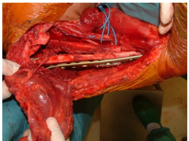
Fig. 1 This picture shows the intraoperative situs of our first patient after insertion of the free fibula bone graft and end-to-side anastomo-ses of the fibula pedicle to the brachial artery and cephalic vein. For stable fixation of the construct a pre-contoured 4.5-pelvic reconstruc-tion plate was attached along the scapular spine, the acromion and the lateral aspect of the humerus bridging the scapular-autograft-humerus junction to improve stability and to protect the allograft. A latissi-mus dorsi rotational flap was prepared to provide adequate soft tissue coverage
the cephalic vein and a latissimus dorsi flap was used for soft tissue coverage. The patient recovered quickly so the adjuvant chemotherapy could be started 4 weeks later. One year after index surgery the patient showed excellent out-come (Fig. 3) with radiographic signs of osseous integra-tion of the autologous bone graft (Fig. 2c). Two years post-operatively he presented with a fatigue fracture of the plate (Fig. 2d). However, the construct remained stable with an excellent shoulder function after a short period of immobi-lization, indicating bony fusion of the arthrodesis. Six years after the intervention, the patient is free of local or systemic recurrence and is able to work as a roadman with satisfying shoulder function (TESS: 70 and MSTS: 78 %).
Discussion
In this report, we describe a surgical technique and our first mid term experience of shoulder arthrodesis using a free double-barrel vascularized fibula autograft for the treat-ment of patients with wide resection of malignant tumors of the proximal humerus.
Several methods of reconstruction currently are being used after tumor resection around the shoulder [2–5, 7, 8, 15–17]. The status of the abductor mechanism (deltoid insertion and rotator cuff) and the integrity of the axillary nerve are important factors in the selection of the method of reconstruction. In case of a resection of the proximal humeral epi- and metaphysis, reconstruction with a large-segment prosthesis [15, 17] or an osteoarticular autograft [16] can accomplish good functional short-term results if the abductor mechanism can be restored. However, use of
the latter has become in disfavor due to nonunion, early fracture of the graft, and progressive arthritis [8, 18]. Espe-cially for young patients with strenuous activity demands and a major resection, including the glenoid, the axillary nerve and/or the rotator cuff, a glenohumeral arthrodesis has to be considered as treatment of choice [2–7, 9, 19] both for trans- and extra-articular resections.
Different techniques for shoulder arthrodesis after major resection are described in the literature [2, 7, 9–11]. In most cases some kind of intercalary bone graft is required to bridge the bone defect and achieve arthrodesis. A struc-tural bone allograft has the ability to provide excellent pri-mary stability, however, bacterial infection and immune reaction are potential risk factors and disadvantages when using exogenous graft material [13, 14]. The use of autolo-gous bone is therefore still considered the gold standard for augmentation of bone healing. Especially if the blood sup-ply can be restored (e.g. vascularized graft) the autologous bone possesses all three desirable properties of graft mate-rials: osteogenicity, osteoinductivity and osteoconductivity. Since Taylor [12] described the first free vascularized fibula graft in 1975, numerous reports of successful micro-vascular bony reconstructions have been published, and shoulder arthrodesis with a single fibula has been reported to have an acceptable outcome [9, 11]. However, as this technique may not provide adequate buttress in certain cases and has shown high complication rates [2, 7, 10], including deep infection, nonunion and graft fractures we intended to develop a surgical technique, which provides the same primary stability that might be achieved with a double strut combined allo- and autograft reconstruction, with an enlarged osseous contact between the scapula and
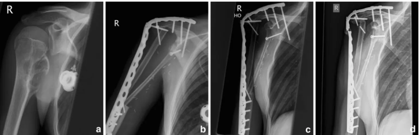
Fig. 2 Preoperative X-ray (a) of the third patient shows an osteolytic lesion at the proximal humerus with medial cortical breakthrough. After neoadjuvant chemotherapy (EURAMOS-1) we performed a wide surgical resection and a reconstruction with a V-shaped double- barreled fibula bone graft, fixing one limb to the glenoid and the coracoid process and the second to the decorticated undersurface of
the acromion (b). One year after index surgery, X-rays revealed an excellent outcome with signs of osseous integration of the autologous bone graft (c). Two years postoperatively the patient presented with a fatigue fracture of the plate (d). The patient remained, however, asymptomatic with good shoulder function and no further dislocation of the broken plate
the bone graft which is covered by vascularized perios-teum, while decreasing the potential risks of using an exog-enous bone graft. The fibula flap’s unique dual endosteal and periosteal blood supply makes it effective as a double-barrel or double strut bone flap for major long bone defects, which requires extra bone volume to prevent fractures until bone hypertrophy may occur [20–24].
Nevertheless autologous vascularized fibula bone graft-ing has still some significant disadvantages. The harvestgraft-ing process has been associated with peri- and postoperative complications and morbidity and a prolonged surgical and anaesthesiological time can cause a proportional increased risk of infection [2]. This also became obvious in our small case series, as one of our patients showed postoperatively a peroneal nerve palsy, which, however resolved sponta-neously within the first year of surgery. Despite adequate soft tissue coverage with a latissimus dorsi flap [7], in one patient, in which the local tumor situation became sys-temic, a deep infection under chemotherapy, with repeated surgical procedures complicated the postoperative course. During the revision procedures we found a strong fibrotic membrane connecting the two struts and stabilizing the
construct. This membrane was further induced by plac-ing the antibiotic-impregnated cement spacer on it. Later the cement was removed and the hole filled with cancel-lous bone graft to strengthen the connection between the struts [25]. The strength and stability of this membrane was somehow unexpected but supports our theory of increased stability due to additional bone and scar formation between the two fibula struts.
Despite, excellent long-term results of the remaining two patients, in whom tumor free survival and excellent shoulder function could be restored, one patient devel-oped a fatigue fracture of the reconstruction plate 2 years postoperatively. This might be due to the fact of using only one plate for stabilizing the reconstruction, which might raise concern about the inherent (e.g. rotational) stability of the fixation. The use of two plates on the ten-sion and compresten-sion side of the construct, as suggested by Bilgin [9], might provide additional primary fixation strength. However, in our patient the construct remained stable with an excellent shoulder function after a short period of immobilization, indicating bony fusion of the arthrodesis.
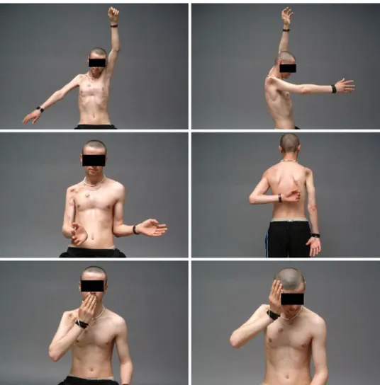
Fig. 3 Photographs of the third patient 12 months after surgery. Six years after the surgery the patient is now free of local or systemic recurrent and is able to work as a roadman with satisfy-ing shoulder function
Conclusion
For patients undergoing tumor resection at the shoulder region a careful preoperative discussion regarding poten-tial complications associated with the chosen procedure is mandatory. A better functional outcome needs to be bal-anced carefully against more extensive surgery, longer operation time, and a potentially higher complication rate. However, the described technique using a double-barrel vascularized fibula graft with additional bone and scar for-mation between the two struts has the potential to provide a stable and long lasting construct, as seen in our small series of patients.
Conflict of interest The authors have no potential conflict of interest.
References
1. Marcove RC, Lewis MM, Huvos AG (1977) En bloc upper humeral interscapulo-thoracic resection. The Tikhoff-Linberg procedure. Clin Orthop Relat Res 124:219–228
2. Fuchs B, O’Connor MI, Padgett DJ, Kaufman KR, Sim FH (2005) Arthrodesis of the shoulder after tumor resection. Clin Orthop Relat Res 436:202–207
3. O’Connor MI, Sim FH, Chao EY (1996) Limb salvage for neo-plasms of the shoulder girdle. Intermediate reconstructive and functional results. J Bone Joint Surg Am 78(12):1872–1888 4. Probyn LJ, Wunder JS, Bell RS, Griffin AM, Davis AM (1998) A
comparison of outcome of osteoarticular allograft reconstruction and shoulder arthrodesis following resection of primary tumours of the proximal humerus. Sarcoma 2(3–4):163–170
5. Kiss J, Sztrinkai G, Antal I, Kiss J, Szendro˝i M (2007) Functional results and quality of life after shoulder girdle resections in mus-culoskeletal tumors. J Shoulder Elbow Surg 16(3):273–279 6. Enneking WF (1983) Musculoskeletal Tumor Surgery. Churchill
Livingstone, New York
7. Wang J, Shen J, Dickinson IC (2011) Functional outcome of arthrodesis with a vascularized fibular graft and a rotational latis-simus dorsi flap after proximal humerus sarcoma resection. Ann Surg Oncol 18(7):1852–1859
8. Getty PJ, Peabody TD (1999) Complications and functional outcomes of reconstruction with an osteoarticular allograft after intra-articular resection of the proximal aspect of the humerus. J Bone Joint Surg Am 81(8):1138–1146
9. Bilgin SS (2012) Reconstruction of proximal humeral defects with shoulder arthrodesis using free vascularized fibular graft. J Bone Joint Surg Am 94(13):e94
10. Scalise JJ, Iannotti JP (2008) Glenohumeral arthrodesis after failed prosthetic shoulder arthroplasty. J Bone Joint Surg 90(1):70–77
11. Viehweger E, Gonzalez J-F, Launay F, Legre R, Jouve J-L, Bol-lini G (2005) Shoulder arthrodesis with vascularized fibular graft after tumor resection of the proximal humerus. Rev Chir Orthop Reparatrice Appar Mot 91(6):523–529
12. Taylor GI, Miller GD, Ham FJ (1975) The free vascularized bone graft. A clinical extension of microvascular techniques. Plast Reconstr Surg 55(5):533–544
13. Calori GM, Mazza E, Colombo M, Ripamonti C (2011) The use of bone-graft substitutes in large bone defects: any specific needs? Injury 42:56–63
14. Costain DJ, Crawford RW (2009) Fresh-frozen vs. irradiated allograft bone in orthopaedic reconstructive surgery. Injury 40(12):1260–1264
15. Manili M, Fredella N, Santori FS (2002) Shoulder prosthesis in reconstruction of the scapulohumeral girdle after wide resection to treat malignant neoformation of the proximal humerus. Chir Organi Mov 87(1):25–33
16. Wada T, Usui M, Isu K, Yamawakii S, Ishii S (1999) Reconstruc-tion and limb salvage after resecReconstruc-tion for malignant bone tumour of the proximal humerus. A sling procedure using a free vascular-ised fibular graft. J Bone Joint Surg Br 81(5):808–813
17. Wittig JC, Bickels J, Kellar-Graney KL, Kim FH, Malawer MM (2002) Osteosarcoma of the proximal humerus: long-term results with limb-sparing surgery. Clin Orthop Relat Res 397:156–176 18. Ogilvie CM, Crawford EA, Hosalkar HS, King JJ,
Lack-man RD (2009) Long-term results for limb salvage with osteoarticular allograft reconstruction. Clin Orthop Relat Res 467(10):2685–2690
19. Safran O, Iannotti JP (2006) Arthrodesis of the shoulder. J Am Acad Orthop Surg 14(3):145–153
20. Banic A, Hertel R (1993) Double vascularized fibulas for reconstruction of large tibial defects. J Reconstr Microsurg 9(6):421–428
21. Bi Z-G, Han X-G, Fu C-J, Cao Y, Yang C-L (2008) Reconstruc-tion of large limb bone defects with a double-barrel free vascular-ized fibular graft. Chin Med J 121(23):2424–2428
22. Chu C-H, Jou I-M, Shieh S-J (2009) Reconstruction of a massive femoral bone defect using a double-barreled free vascularized fib-ular bone graft after wide resection of femoral chondrosarcoma. Kaohsiung J Med Sci 25(10):552–558
23. Jones NF, Swartz WM, Mears DC, Jupiter JB, Grossman A (1998) The “double barrel” free vascularized fibular bone graft. Plast Reconstr Surg 81(3):378–385
24. Saint-Cyr M, Farkas J, Gupta A (2008) Double-barrel free fibula flap for treatment of infected non union of both forearm bones. J Reconstr Microsurg 24(08):583–587
25. Masquelet AC, Begue T (2010) The concept of induced mem-brane for reconstruction of long bone defects. Orthop Clin North Am 41(1):27–37