Chromosome Partitioning in Bacillus subtilis by
Daniel Chi-Hong Lin
Bachelor of Arts in Molecular and Cell Biology With Honors and Distinction in General Scholarship
University of California at Berkeley, 1994
Submitted to the Department of Biology
in Partial Fulfillment of the Requirements for the Degree of Doctor of Philosophy in Biology
at the
Massachusetts Institute of Technology June 1999
@ 1999 Daniel Chi-Hong Lin. All rights reserved.
The author hereby grants to MIT permission to reproduce and to distribute publicly paper and electronic copies of this thesis document in whole or in part.
Signature of A u th or...1 -.... ... .... Department of Biology May 18, 1999 Certified by...-... . .. ... Alan D. Grossman Professor of Biology Thesis Supervisor Accepted by... ... .-... Alan D. Grossman Chair, Committee on Graduate Students Department of Biology
Science
Chromosome Partitioning in Bacillus subtilis
by
Daniel Chi-Hong Lin
Submitted to the Department of Biology on May 18, 1999 in Partial Fulfillment of the Requirements for the Degree of
Doctor of Philosophy in Biology
Abstract
Chromosome partitioning is a fundamental process that ensures the stable inheritance of genetic material. I have studied this process in Bacillus subtilis, a Gram-positive bacterium that contains a single circular chromosome. The B. subtilis spoOJ gene product is required for sporulation and for normal
chromosome partitioning during vegetative growth. SpoOJ is a member of the ParB family of proteins, which are found in several bacterial species.
In this thesis, I describe experiments to characterize the function of SpoOJ in chromosome partitioning. I found that SpoOJ is a site-specific DNA binding protein, recognizing a 16 bp sequence call parS. SpoOJ binds to at least eight other parS sites, and these eight sites are all located in the origin proximal ~20% of the genome. Insertion of a single parS sequence into an unstable plasmid stabilizes that plasmid, indicating that parS can function in partitioning.
I also determined the subcellular localization of SpoOJ using
immunofluorescence and a Spo0J-GFP fusion protein. Spo0J, bound to its eight parS sites, typically localizes as two large foci, each near the 1/4 and 3/4
positions of the cell length through most of the cell cycle. This result indicates that the sister origins are separated early, and that the chromosome is in a defined orientation through most of the cell cycle. spoOl null mutants appear to have a mild defect in normal origin localization. However, SpoOJ does not appear to be involved in simply attaching parS DNA near the cell quarter positions. When I inserted multiple parS sites into the terminus region of the chromosome, I discovered that the subcellular localization of the terminus was unaffected.
Thesis Supervisor: Alan D. Grossman Title: Professor of Biology
Acknowledgments
I gratefully acknowledge my thesis advisor, Dr. Alan Grossman. Alan takes great pride in his role as a teacher. I appreciate the time, effort, and enthusiasm he has put forth in training me as a scientist. Alan has been instrumental in my achievements at MIT.
It has also been a great deal of pleasure to work with the members of the Grossman lab. I have learned a lot from the diverse pool of personalities that make up the lab. I could not have asked for a better environment to be in. I also thank members of my committee for their advice and encouragement.
I also thank the following people for reading drafts of this thesis, and for their numerous comments and suggestions: William Burkholder, Katherine Lemon, Petra Levin, Janet Lindow, John Quisel, Laura Robertson, and Tracy Tang.
I also wholeheartedly thank Tracy Tang. Since our time at Berkeley, Tracy and I have studied and grown together, encouraged each other, and have shared great memories. Thank you for not minding my long hours, my weekend shifts, and my deskworker job.
Finally, I wish to thank my family for their support. My older sister Meilee is a great conversationalist and has always provided a comfortable place to stay on my drives down South. My little sisters, Chris and Kathy, are always a joy to talk with and a source of great pride as I have watched them grow.
Finally, my parents, Dr. Tien and Ikuko Lin, have taught me the meaning of hard work and dedication.
Table of Contents
Abstract Acknowledgments Table of Contents List of Figure List of Tables Chapter 1 Chapter 2 Chapter 3 Chapter 4 Chapter 5 Appendix 1 Appendix 2 IntroductionBipolar localization of a chromosome partition protein in Bacillus subtilis
Identification and characterization of a bacterial chromosome partitioning site
Origin localization in wild type and spoOj cells, and the effects of inserting multiple parS partitioning sites into the terminus region of Bacillus subtilis
Discussion
Testing whether Spo0J-parS can interact with Spo0J-parS through in vitro ligation assays
Characterization of the spoOJ93 allele
p4g 3 4 5 7 8 80 108 154 187 212 220
List of Figures
Chapter 1 Figure 1-1 Figure 1-2 Figure 1-3 Figure Figure Figure Figure 1-4 1-5 1-6 1-7 Figure 1-8 Chapter 2 Chapter 3 Figure Figure Figure Figure Figure 1-9 1-10 2-1 2-2 2-3 Figure 3-1 Figure 3-2Metaphase configuration of the mitotic apparatus
Centromeres of S. cerevisiae, S. pombe and
D. melanogaster
SMC proteins involved in both sister
chromatid cohesion and chromosome condensation
Microtubule growth and depolymerization Centrosome separation by motors on pole-to-pole microtubules
Overview of the bacterial cell cycle
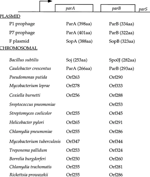
Two speculative models to account for the rapid movement of the origin regions Plasmid and chromosomally encoded ParA and ParB homologues
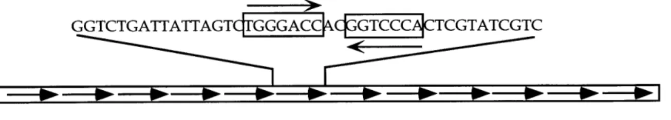
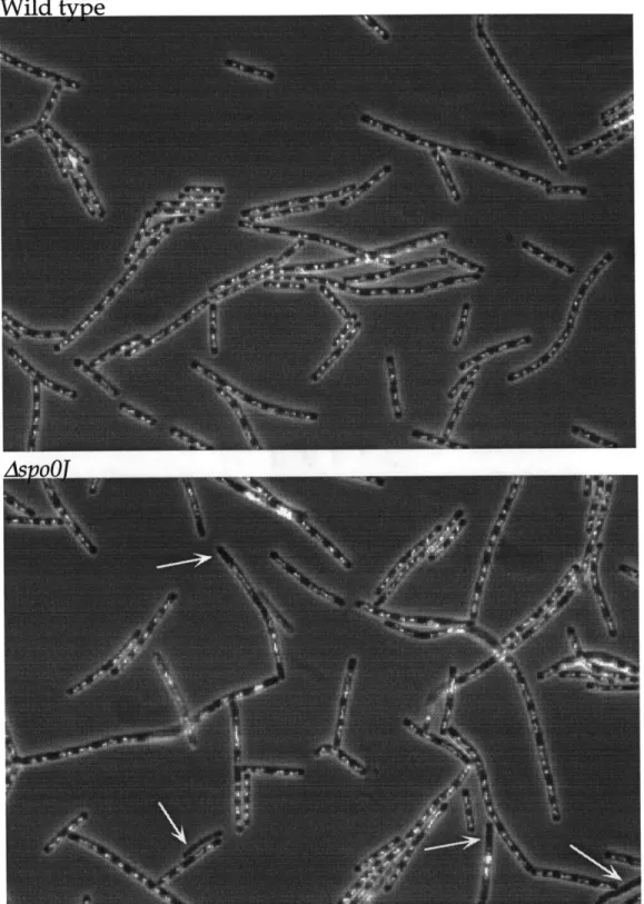
par sites from P1 prophage and F plasmid DAPI strained wild type and spoOJ cells Localization of SpoOJ during growth and sporulation
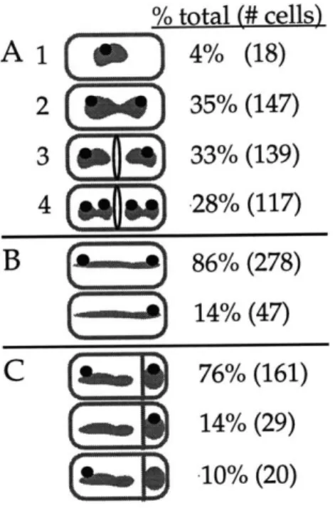
Analysis of SpoOJ localization
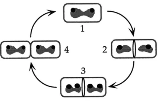
Model for chromosome segregation and SpoOJ localization during the cell cycle In vivo association of SpoOJ with DNA from near the spoOj gene
In vivo identification of the SpoOJ binding site located in the spoOj gene
13 16 21 23 26 34 44 51 53 59 93 95 101 115 117
Figure 3-3 Figure 3-4 Figure 3-5 Figure 3-6 Figure 3-7 Figure 4-1 Figure 4-2 Figure 4-3 Figure Al-1 Figure A1-2 Figure A1-3 Figure A1-4 Appendix2 Figure A2
Site-specific binding of SpoOJ to DNA in vitro
Multiple SpoOJ binding sites in the B. subtilis chromosome
Increased occupancy of the SpoOJ binding site at 15' in the mutant with six sites inactivated
The SpoOJ binding site can stabilize a plasmid
SpoOJ may be involved in pairing newly replicated origin regions
Comparison of LacI-GFP marked origin localization in wild type, spo0J, and AparS5; ter::parS16 strains
Construction of the parS 16 array and plasmid stability assay with the parS16 array
Localization of Spo0J-GFP in AparS or AparS6; ter::parS16 strains
Gel Shift Spo0J-parS / Spo0J-parS Interaction Assay
Ligation measured Spo0J-parS / Spo0J-parS interaction assays
Spo0J-DNA/ Spo0J-DNA interaction shows low preference for parS+ DNA Effect of Soj on Spo0J-parS
/
Spo0J-parSinteractions
Sequence of the spoOJ93 allele and localization of Spo0J93-GFP Chapter 4 Appendixi 120 124 128 132 139 160 165 170 216 217 218 219 224
List of Tables
Chapter 3 Table 3-1 Copy number comparison of plasmids with and without a SpoOJ binding site
Table 3-2 Possible Spo0J/ParB binding sites in other bacteria that are similar to the B. subtilis
consensus
Table 3-3 B. subtilis strains used Table 3-4 Plasmids used
Chapter 4 Table 4-1 Characterization of strains with lacO/LacI-GFP marked origins
Table 4-2 Characterization of Spo0J-GFP in DCL616 and DCL631 (AparS6; ter::parS16) cells Table 4-3 Anucleate cell production in cells carrying
parS16 at the terminus or the origin Table 4-4 B. subtilis strains used
131 134 148 149 159 169 174 180
Chapter 1 Introduction
The proper inheritance of chromosomes is essential for all organisms. In both eukaryotic and prokaryotic organisms, elegant systems have evolved to ensure the orderly duplication and partitioning of the genetic material.
Chromosome partitioning encompasses multiple steps in order to set up and move chromosomes to each of the new daughter cells prior to cell division. Improper partitioning of the chromosomes is costly. In bacteria, defects in partitioning leads to the production of chromosomeless (anucleate) cells, a certain selective disadvantage. In yeast, mutations in genes required for
chromosome partitioning can be lethal. In humans, Downs syndrome and some types of cancer are associated with improper chromosome partitioning.
All cells accomplish several fundamental tasks in order to partition their chromosomes (reviewed briefly in this chapter. See text below for references). For example, the chromosomes need to be recognized by the partitioning apparatus. This can be accomplished by a cis-acting DNA sequence, the centromere, and proteins that recognize this sequence. Also, the duplicated chromosomes need to be oriented so that they move to opposite poles.
Centrosomes, kinetochores, and sister-chromatid cohesion proteins contribute to this task in eukaryotic cells. In addition, the duplicated chromosomes must be
organized. Sister chromatid cohesion proteins and chromosome condensation proteins are important for this process in eukaryotes, and homologous proteins
probably act in prokaryotes. Finally, the chromosomes need to be moved. In eukaryotic cells, microtubules and motor proteins play key roles in the
underway, and may be due to mitotic-like motor proteins or the action of DNA polymerase.
This thesis focuses on experiments addressing the function of spoDj, which is required for normal chromosome partitioning in the Gram positive bacterium Bacillus subtilis. Before describing the specific experiments on spo0J, I will review aspects of both eukaryotic and prokaryotic chromosome partitioning. In
eukaryotes, partitioning of the chromosomes occurs during the M (mitotic) phase of the cell cycle. I will review the eukaryotic cell cycle with special emphasis on factors required for proper chromosome partitioning, and the mechanisms used to set up the mitotic apparatus that drives chromosome partitioning. Next, I will review many of the key experiments that have led to our current understanding of bacterial chromosome partitioning. Special emphasis will be placed upon the partitioning of the origin regions in bacteria, a central subject in this thesis. I will go in depth with studies of proteins related to SpoOJ from plasmid partition systems in prokaryotes.
Compared with the study of chromosome partitioning in eukaryotes, the study in prokaryotes is still in its infancy, but growing rapidly. As we begin to
learn more about prokaryotic chromosome partitioning, a broad question is whether any of the mechanism involved in this process are similar to
mechanisms in eukaryotic mitosis. Lessons and ideas from eukaryotic mitosis can help to frame models for bacterial chromosome partitioning, since the fundamental tasks that need to be accomplished are common to all organisms. However, the precise mechanisms and proteins that accomplish these tasks are
different in prokaryotes and eukaryotes, although there are exceptions. For example, the SMC proteins have been conserved from bacteria to human, and their functions in chromosome partitioning probably overlap. Throughout this thesis, I will make comparisons between eukaryotic and prokaryotic
chromosome partitioning. Perhaps the only thing that is clear is that
chromosome partitioning in both prokaryotes and eukaryotes is intricate, yet elegant.
OVERVIEW OF EUKARYOTIC MITOSIS
There are four phases in the eukaryotic cell cycle: from the first Gap phase of the cell cycle, called G1, the cell moves to S phase, where DNA replication occurs. This is followed by another gap phase, G2, followed by the mitotic phase, M, where the chromosomes are separated and the cell division occurs. Following the M phase, the two new daughter cells enter the G1 phase. Interphase is a combination of the G1, S, and G2 phases. M phase is further divided into four broad phases; these phases are, in order, prophase, metaphase, anaphase, and telophase. Many of the events, described below, to construct the mitotic apparatus occur during prophase and the beginning of metaphase. By the end of metaphase, the chromosomes are aligned between the centrosomes
(described below). During anaphase, the chromosomes move apart, and during telophase, the cell divides. A key difference between eukaryotes and
partitioning of the chromosome occurs while replication is still proceeding (described below).
The specialized structure that drives chromosome partitioning in
eukaryotes is the mitotic apparatus (figure 1-1). The mitotic apparatus contains the bipolar spindle, which is football shaped and is composed of microtubules. The microtubules emanate from the two centrosomes, located at the poles. The duplicated and condensed chromosomes (sister chromatids) attach to the microtubules from opposite poles. The sister chromatids are held together, in part, by sister chromatid cohesion proteins. The microtubules are attached to the kinetochore, a nucleoprotein structure that forms on the centromere. The
centromere is the specialized DNA region that is required in cis for proper partitioning of the chromosome. During the first step of anaphase, called anaphase A, chromosomes move towards the stationary centrosomes.
Microtubule depolymerization at the kinetochores and motor proteins drive the sister chromatids away from each other. During anaphase B, the centrosomes move farther apart, further separating the chromosomes.
All of the components listed above play important roles in the series of
steps necessary to partition chromosomes. What follows is a brief review of the properties of these factors and some of what we know of the various stages of mitosis.
sister chromatids
SI
chromosome arm microtubule
kinetochore microtubule
. . pole-to-pole microtubule
cen osome astral microtubule
eto r~e
sister chromatid cohesion protein
Figure 1-1. Metaphase configuration of the mitotic apparatus.
Microtubules emanate from the centrosome. Microtubules attach to the chromosomes both along the arms and at the kinetochore. The kinetochore is a specialized protein structure that assembles on the centromere. Pole-to-pole microtubules arise from the opposite centrosomes and interdigitate (also called anti-parallel microtubules). Astral microtubules emanating from the centrosome can attach to the cell cortex. The duplicated
chromosomes, or sister chromatids, are paired along their length and at the centromeres by sister chromatid cohesion proteins.
Centromeres and kinetochores
A crucial component in eukaryotic mitosis is the centromere. The centromere is a DNA sequence required in cis to promote proper chromosome transmission. The centromere is a highly specialized, transcriptionally silent region of the chromosome where the assembly of the kinetochore occurs. In higher eukaryotes, the kinetochore is visualized as a relatively flat, triple-layered proteinaceous structure and sister kinetochores face opposite poles. The
centromere and kinetochore serve multiple important function in mitosis. Microtubules emanating from the centrosomes bind to the kinetochore. Some motor proteins act at the kinetochore to generate force for chromosome
movement both away and towards the poles. Checkpoint proteins, which function to delay the onset of anaphase if the mitotic spindle is not properly set up, localize to the kinetochore and monitor microtubule attachment and/or tension (Chen et al. 1996; Taylor and McKeon 1997). Some sister chromatid cohesion proteins act at the centromere and may also function to constrain the kinetochore region so the kinetochores face in opposite directions (described below) (Bickel and Orr-Weaver 1996; Kerrebrock et al. 1995; Saitoh et al. 1997).
The centromere and kinetochore proteins have been best characterized in
S. cerevisiae (reviewed in (Hyman and Sorger 1995)). The budding yeast
centromere is -125 bp and is composed of three regions, CDEI, CDEII, and CDEIII (figure 1-2) (Cottarel et al. 1989). Protein complexes that bind to CDEI
Lechner and Carbon 1991). CDEII and CDEIII are essential for centromere function, and CDEI is important but not essential.
The centromeres of other eukaryotes are more complex. The centromeres from the three S. pombe chromosomes are ~40-100 kb in length (Chikashige et al. 1989; Clarke et al. 1986) The Drosophila melanogaster centromere is ~420 kb in length, and the human centromere is several megabases in length (figure 1-2) (Murphy and Karpen 1995 ; Pluta et al. 1995 review; Sun et al. 1997). The S. pombe and human centromeres contain large repeat elements, on the order of ~5 kbp. In humans, these large repeats are composed of smaller repeats of a -200 bp A-T rich region termed alphoid DNA (Harrington et al. 1997; Heller et al. 1996).
Currently, it is not known precisely what sequence, such as that in
budding yeast, defines a centromere in S. pombe, D. melanogaster, or humans. In fact, in higher eukaryotes, a primary DNA sequence appears to be neither
necessary nor sufficient for centromere function. Centromeres and kinetochores of D. melanogaster and humans are able to form de novo under certain
circumstances on previously acentric DNA (du Sart et al. 1997; Williams et al. 1998). Although the specifics are unclear, it appears that an epigenetic
mechanism involving heterochromatin is important for this transformation (Karpen and Allshire 1997; Williams, et al. 1998).
The kinetochore components, and their specific functions, are just being described. These proteins mediate several functions, including microtubule binding, signaling to checkpoints, and connecting the chromosome to motor
A. S. cerevisiae centromere (-125 bp)
A/T rich
CDEI CDEII CDEIII
B. S. pombe centromere (~40-100 kb) B L K C. D. melanogaster centromere (~420 kb) transposons
E
AATAT satellite 2 AAGAG satelliteFigure 1-2. Centromeres of S. cerevisiae, S. pombe, and D. melanogaster.
A. The -125 bp S. cerevisiae centromere sequence is composed of CDEI, CDEII, and
CDEIII. CDEI and CDEIII both contain inverted repeats (denoted by arrows). The ~90 bp CDEII region contains -90% A/T base pairs. B. The -40-100 kb S. pombe centromeres from the three S. pombe chromosomes contain multiple, different, large repeat elements. Shown is a schematic of the centromere from chromosome 2 (adapted from Clarke et al.,
1993). Here, the B, J, K, and L regions (a single repeat is -2-7 kb) are repeated on either side of the central core (-4-7 kb). Note that the central core is flanked by a large inverted repeat (noted by arrows). C. The D. melanogaster centromere was defined to -420 kb contained within centric heterochromatin. The D. melanogaster centromere contains two large regions of repeated satellite DNA sequence ( 0 and 0 ). Eight transposable elements are also found in the centromere. (Adapted from Sun et al., 1997)
proteins. In S. cerevisiae and S. pombe, several genes isolated in a screen for mutants defective in chromosome transmission are kinetochore components
(Hoyt et al. 1990; Meeks-Wagner et al. 1986; Spencer et al. 1990; Strunnikov et al.
1995; Takahashi et al. 1994). As expected, some of these proteins are DNA
binding proteins that specifically recognize the budding yeast CEN DNA (Espelin, et al. 1997; Sorger et al. 1994). Some S. cerevisiae kinetochore components, such as Cse4p and Mif2p, appear to be homologues to the mammalian kinetochore proteins CENP-A and CENP-C, respectively (Brown
1995; Meluh and Koshland 1995; Meluh et al. 1998; Stoler et al. 1995).
A common requirement in all eukaryotic organisms is that the kinetochore
must form once and only once on a chromosome (reviewed in (Wiens and Sorger
1998)). A functionally dicentric chromosome will be broken if it is attached to
opposite poles. In S. cerevisiae, formation of a single kinetochore is controlled, in part, by the specific CEN sequence which is present only once per chromosome. However, in D. melanogaster and humans, there is an added level of complexity, since de novo formation of a kinetochore must be suppressed.
Sister chromatid cohesion and chromosome condensation factors
Proper sister chromatid cohesion and chromosome condensation are required for efficient chromosome separation. Sister chromatids are attached to each other both along the chromosome arms and at their centromere. Sister
chromatid cohesion proteins presumably function between the two sisters as a molecular "glue."
Sister chromatid cohesion serves multiple functions (reviewed in (Bickel and Orr-Weaver 1996; Biggins and Murray 1998; Miyazaki and Orr-Weaver 1994)). First, through cohesion of identical molecules, i.e. sister chromatids, the cell ensures that they are ultimately segregated apart. In this light, cohesion
functions to both let the cell know that two identical molecules need to be segregated, and functions to organize these molecules in a way that they can be segregated from one another. Rather than being two independent entities that need to be partitioned, the sisters are first grouped together prior to partitioning. Next, sister chromatid cohesion proteins may aid in orienting the kinetochore by constraining the centromeric regions so that the kinetochores face opposite directions. Outward facing orientation is important since kinetochores capture microtubules, and each sister kinetochore needs to be attached to microtubules from opposite poles. During meiosis I, a situation arises where the sisters need to face the same pole and homologues face opposite poles. Hence, additional factors may regulate the orientation of kinetochores during meiosis. A third possible function of cohesion proteins is to ensure that the chromatin from the two sisters do not become entangled as they emerge from the replication fork. Cohesion proteins may serve as a wall to keep the DNA from the two sisters organized and separate even while being held together. Another function of sister chromatid cohesion proteins is to counteract the forces on the sister
to anaphase. Finally, the regulated dissolution of sister chromatid cohesion is required at the metaphase-anaphase transition (described below).
In budding yeast, sister chromatid cohesion is established during S phase, and there is speculation that cohesion is coupled with the movement of the replication forks and with chromosome condensation (Skibbens et al. 1999; Toth et al. 1999; Uhlmann and Nasmyth 1998). The S. cerevisiae sccl gene (also known as mcdl) was discovered in a genetic screen for sister chromatid cohesion
mutants (Guacci et al. 1997; Guacci et al. 1993; Michaelis et al. 1997) and Scclp is synthesized and functions during S phase (Uhlmann and Nasmyth 1998).
Timing of cohesion is important; if Scclp is synthesized after replication, sister chromatids remain separated. It has been proposed that the strict timing of sister chromatid cohesion is important to prevent sister DNA from mixing with one
another.
Chromosome condensation occurs during prophase, and in most eukaryotic cells can lead to an approximately 20-100 fold compaction of the chromosomes compared to chromosomes in interphase cells. Without
chromosome condensation, the DNA may not be moved efficiently from the division septum. S. pombe mutants defective in chromosome condensation genes exhibit a "cut" phenotype, where the DNA is guillotined by the division septum (Saka et al. 1994).
Chromosome condensation has been studied in several other systems, including S. cerevisiae, X. laevis, and C. elegans. Members of the SMC (structural maintenance of chromosomes) family of proteins play a central role in
chromosome condensation and in sister chromatid cohesion (reviewed in (Hirano 1999; Koshland and Strunnikov 1996)). SMC proteins are large polypeptides (1000-1500 a.a.) composed of an N-terminal globular ATPase domain, two coiled-coiled dimerization domains separated by a hinge, and a C-terminal DNA binding domain (figure 1-3). These proteins are conserved from bacteria to human. Most eukaryotic organisms contain multiple SMC proteins that form SMC heterodimers. Eukaryotic SMC proteins are usually found in large complexes with other proteins. In Xenopus, the 13S condensin complex is composed of the SMC proteins XCAP-C and XCAP-E and other proteins and
functions in chromosome condensation (Hirano et al. 1997). In contrast, the 14S cohesin complex from Xenopus , composed of XSMC1 and XSMC-3 and other proteins, is involved in sister chromatid cohesion. XRAD21, a homologue of S. cerevisiae Scclp (described above), is part of the 14S cohesin complex (Losada et al. 1998).
A link between chromosome cohesion and chromosome condensation was
revealed when sccl mutants (described above) were found to be defective not only in cohesion but also in condensation (Guacci et al. 1997). This led to a model where Scclp establishes cohesion following S phase, and during
prometaphase recruits the condensin complex for chromosome condensation. This model, however, does not appear to be accurate in Xenopus, since the
A. SMC dimer
flexible hinge region
coiled-coil domain
B. Models for SMC in:
Sister chromatid cohesion Chromosome condensation
Figure 1-3. SMC proteins involved in chromosome condensation.
both sister chromatid cohesion and
A. An anti-parallel SMC homodimer, based on the model of Melby et al., 1998, studying the B. subtilis SMC protein. SMC dimers have a flexible hinge region and show ATP-dependent activity in vitro. SMC proteins are conserved from bacteria to humans and have been found to function in many aspects of chromosome organization. B. In yeast and Xenopus,
different SMC proteins function in sister chromatid cohesion and chromosome condensation and function as SMC heterodimers. Simple models for SMC function in sister chromatid cohesion and chromosome condensation are shown. During sister chromatid cohesion, DNA from both sister chromatids can be crosslinked by a single SMC dimer. During chromosome condensation, the DNA is compacted as the flexible hinge allows the two ends to come close toghether. (part B from Hirano, 1998)
cohesin and condensin complexes can act separately (Losada, et al. 1998). Nonetheless, the involvement of SMC in different aspects of chromosome organization has helped in understanding the role of SMC in bacteria.
Microtubules
Microtubules are polymers that are made up of subunits of alpha and beta tubulin heterodimers. Microtubules have inherent polarity. The plus end of the microtubule is defined as the end where both assembly and disassembly of the microtubule preferentially occurs. In mitosis, the minus ends are attached to the centrosome, which is also known as the microtubule organizing center. Gamma-tubulin, a third distinct tubulin family member, localizes to the centrosomes and is required for microtubule nucleation (Moritz et al. 1995; Oakley and Oakley 1989; Stearns et al. 1991; Zheng et al. 1995).
Microtubule assembly occurs with the addition of alpha and beta tubulin heterodimers, and disassembly comes with their removal (figure 1-4).
Microtubules display "dynamic instability," a term to describe both the slow growth and rapid depolymerization (called "catastrophe") of individual microtubules (Mitchison and Kirschner 1984). Polymerization and
depolymerization of the microtubule is regulated by GTP hydrolysis. In a microtubule, most of the beta subunits have hydrolyzed GTP and are GDP bound. GTP hydrolysis favors depolymerization. The reason why microtubules can grow is because the tip of the microtubule has a GTP cap, where GTP bound
GTP cap sheet structure; plus nd
000
Eo)
o
0
0
00
minus endgrowing tube catastrophe
microtubule closure
0
alpha-beta tubulin heterodimer Figure 1-4. Microtubule growth and deploymerization.Microtubule growth is stabilized by a GTP cap, which forms a sheet structure at the top of the tube. If the sheet closes into a tube, this event is thought to lead to a conformational change in the alpha--beta tubulin in the sheet. This conformational change in the heterodimer leads to hydrolysis
of GTP to GDP on the beta tubulin subunit. This leads to catastrophe, where the micotubule rapidly depolymerizes (catastrophe). The built up energy of GTP hydrolysis in the microtubule lattice drives this rapid depolymerization. (Adapted from Hyman and Karsenti, 1996).
to the beta tubulin subunit has not been hydrolyzed (figure 1-4) (Carlier 1989; Drechsel and Kirschner 1994). The GTP cap prevents depolymerization. At the GTP cap, the tube form is opened up into a sheet structure (Chretien et al. 1995; Mandelkow et al. 1991). If this sheet closes up and forms a tube, the current thought is that this leads to a conformational change in the tubulin subunits that triggers GTP hydrolysis. Following tube closure, the free energy of GTP
hydrolysis that has built up into the microtubule lattice leads to catastrophe. Microtubules emanating from the centrosome are divided into four
different categories (figure 1-1). Kinetochore microtubules attach to kinetochores during prophase and early metaphase and are responsible for most of the
anaphase A movement. Chromosome arm microtubules attach along the length of the chromosome and are responsible for the movement toward the metaphase plate, called congression (described below). Both interdigitating pole-to-pole
and astral microtubules are required for centrosome separation (described below). Microtubules, together with motor proteins, are responsible for
essentially all the movement in setting up the mitotic apparatus and partitioning chromosomes.
Mitotic motor proteins
Motor proteins move cargo along tracks of microtubules in an ATP-dependent manner (reviewed in (Afshar et al. 1995; Hyman and Karsenti 1996)). Motor proteins move directionally. Some motor proteins, such as kinesin, are
plus end directed. Plus end directed motor proteins carry cargo towards the plus end of a microtubule. Other motor proteins, such as dynein, are minus end directed. Many motors important in mitosis are kinesin-related proteins and are either plus end or minus end directed. Many motor proteins are elongated structures that function as parallel homodimers. Typically, the globular motor "head" domain moves along the microtubules. Dimerization is mediated by a long coiled-coil domain, and the "tail" domain binds to the cargo. Motor proteins sometimes are found as homomultimers of homodimers, and these multimeric motor proteins may be involved in aspects of microtubule bundling.
There are many motor proteins that are important in mitosis. These motors are involved in moving the centrosomes apart, in chromosome movement, and in setting up the football shape of the mitotic spindle.
Centrosomes separate towards opposite poles during S phase to set up the mitotic spindle. Centrosomes also move apart during anaphase B to aid in
chromosome partitioning. Centrosome movement is powered, in part, by plus end directed motors (figure 1-5). Members of the BimC family of kinesin-related motor proteins are required for centrosome separation during prophase and during anaphase B (Enos and Morris 1990; Hoyt et al. 1992; Kashina et al. 1996; Roof et al. 1992; Saunders et al. 1995).
How do plus-end directed motors move centrosomes apart? These motors move along interdigitating pole-to-pole microtubules that arise when microtubules from opposite centrosomes overlap one another (figure 1-5). The BimC proteins tether to one microtubule, and use the motor domain to move to
pole-to-pole microtubule
motor direction
motor direction +
ma ++U
@ centrosome i plus end directed microtubule motor Figure 1-5. Centrosome separation by motors on pole-to-pole
microtubules.
A plus end directed motor on interdigitating pole-to-pole microtubules can
separate the centrosomes. One possible mechanism is that a motor protein could bind to a microtubule from one pole, and use the motor domain to move to the plus end of a microtubule from an opposite pole.
the plus end of the overlapping microtubule. Centrosome movement is also driven by the minus end directed motor protein dynein (Saunders, et al. 1995; Vaisberg et al. 1993). Dynein tethered to the plasma membrane could pull on the astral microtubules, moving centrosomes further apart.
Microtubules attach to the kinetochores and the chromosome arms
Chromosomes are attached to microtubules both at the kinetochore and along the chromosome arms. Both kinetochore and arm attachments are important for the chromosome movement, called congression, to set up the metaphase plate (Fuller 1995). The metaphase plate refers to the alignment of all the sister chromatids midway between the two centrosomes.
Attachment of the microtubule to the kinetochore occurs at random through a "search and capture" mechanism. Microtubules rapidly grow and shrink (termed "dynamic instability") from the centrosomes during
prometaphase. The goal of the microtubule is to attach to a free kinetochore of a sister chromatid. When the side of a microtubule is captured by a chromosome, that microtubule is stabilized (Merdes and De Mey 1990; Rieder and Alexander
1990).
In S. cerevisiae, only one microtubule is attached to the kinetochore, but in higher eukaryotes, multiple microtubules attach to the kinetochore. Following the capture of the first microtubule by the kinetochore, the mono-oriented
probably by both microtubule depolymerization and minus end directed motors (Rieder and Alexander 1990). More microtubules are the thought to attach "end-on" (as opposed to the side of the microtubule) to the captured kinetochore, and together these microtubules form a microtubule filament.
Attachment of microtubules to the chromosome arms is also important for chromosome alignment along the metaphase plate and for the football shape of the spindle apparatus. Microtubule capture by the chromosome arms also occurs
by stabilization of microtubules emanating from the centrosomes. It has been
proposed that microtubule associated proteins (MAPs) and/or motor proteins can stabilize microtubules along the chromosome arms (Afshar, et al. 1995; Mandelkow and Mandelkow 1995; Vernos et al. 1995). The ability of non-kinetochore chromatin factors to stabilize microtubules is well documented. A bipolar spindle can form in a Xenopus extract around beads coated with
chromatin in the absence of centrosomes (Heald et al. 1996). In vivo, meiotic spindle formation in Xenopus or female Drosophila, and mitotic spindle formation in plants also occurs in the absence of centrosomes and nucleates around
chromatin (Gard 1992; Theurkauf and Hawley 1992).
Congression
Once microtubules have attached to the kinetochore during
prometaphase, the mono-oriented sister chromatids oscillate both towards and away from the poles. Minus end directed motor proteins and microtubule
depolymerization at the kinetochore govern movement towards the pole. Plus end directed motors and microtubule polymerization, acting both at the
kinetochore and the chromosome arms, push the chromosome away from the pole (Hyman and Mitchison 1991; Rieder et al. 1986; Vernos, et al. 1995; Afshar et al, 1995).
The pushing forces acting at the chromosome arms are called the "polar winds" forces and act to push the mono-oriented sister chromatids away from the poles (Skibbens et al. 1993). The pushing force was revealed when the chromosome arm of a mono-oriented sister chromatid was severed, using micromanipulation techniques, to separate it from the kinetochore. The chromosomal fragment with the kinetochore moved towards the pole, but the severed chromosome arm was pushed away from the pole due to the "polar wind" force (Rieder, et al. 1986). Two plus end directed kinesin-related motors, Nod from D. melangaster and Xlkpl from Xenopus, contribute to this pushing
force (Afshar, et al. 1995; Vernos, et al. 1995). Inhibition of these motors results in improper chromosome positioning.
Equilibrium of tension is the basis for proper chromosome alignment at metaphase, and perhaps is a signal to initiate anaphase (described below)
(Nicklas 1997; Skibbens, et al. 1993). In order to congress to the metaphase plate, the remaining unattached kinetochore must attach to a microtubule from the opposite pole. Once attached, a sufficient pulling force from the opposite pole is obtained to drive the sister chromatids to the metaphase plate. At the metaphase plate, the pushing forces on the arms and pulling forces at kinetochore are in
equilibrium. In addition, sister chromatid cohesion proteins function at the kinetochore and along the chromosome arms and act as "protein glue" to counteract the pulling forces towards both poles. Capture of microtubules and equilibrium of tension is so important to the cell that if any error occurs, a
checkpoint is activated to keep the cells in mitosis until the proper attachments can be made (Hoyt et al. 1991; Li and Murray 1991). These checkpoint proteins, called Mads and Bubs, are conserved from yeast to humans (Li and Benezra 1996; Taylor and McKeon 1997).
Initiation of anaphase
Equilibrium of tension may be a signal that leads to initiation of anaphase (Li and Nicklas 1995; Nicklas 1997; Rieder et al. 1994). A mono-oriented
kinetochore from a praying mantid spermatocyte delays the onset of anaphase. Using micromanipulation techniques, Li and Nicklas pulled on the mono-oriented kinetochore towards the opposite pole, and the cell was able to enter anaphase (Li and Nicklas 1995). This and other experiments led to the
hypothesis that the cell senses tension before it initiates anaphase. It is not known how this tension is sensed, but other experiments implicate a putative phosphoprotein of unknown identity as a possible tension sensing factor
(Nicklas et al. 1995). Kinetochore proteins may also be putative sensing tension sensing proteins. Checkpoints also appear to be regulated, in part, by these
tension sensing factors. The studies of the spindle assembly checkpoint mad and bub genes may provide a tool for identifying these tension sensing proteins.
Once tension equilibrium has been achieved on all chromosomes, anaphase initiates. Proteolysis plays a crucial role in the onset of anaphase
(Holloway et al. 1993). The anaphase promoting complex (APC) is a
multisubunit ubiquitin-ligase that attaches ubiquitin to proteins containing the destruction box (Glotzer et al. 1991; Irniger et al. 1995; King et al. 1995; Sudakin et al. 1995). Proteins that are ubiquitinated are proteolyzed by the proteasome
(Ciechanover 1994). Degradation of factors, which may include sister chromatid cohesion proteins and/or anaphase inhibitors, allows the poleward forces at kinetochores to move the chromosomes apart. This has been best characterized
in the lower eukaryotes S. cerevisiae and S. pombe. In S. cerevisiae, the Pdslp protein acts as an inhibitor of anaphase (Cohen-Fix et al. 1996; Funabiki et al.
1996; Yamamoto et al. 1996). Pds1p binds to and sequesters Esp1p, an anaphase
promoter. Pdslp is a target of the APC, and its degradation by the APC
presumably frees Espip to promote dissociation of the sister chromatid cohesion proteins, including Scc1p (Ciosk et al. 1998). It is not known exactly how Espip promotes dissolution of the sister chromatid cohesion proteins.
Once sister chromatid cohesion is dissolved, the sister chromatids move towards opposite poles. Microtubules attached to kinetochores rapidly
depolymerize. In vitro, depolymerization of microtubules themselves provides enough force to move chromosomes apart in an ATP-independent matter
case is to simply keep the chromosome attached to the shrinking microtubule (Lombillo, et al. 1995). However, in vivo, minus end directed motor proteins, including dynein are associated with kinetochores and could drive movement (Pfarr et al. 1990; Steuer et al. 1990). However, genetic analysis of dynein mutants in S. cerevisiae reveal that dynein does not appear to be involved in
anaphase A movement, but is involved in anaphase B movement (Eshel et al.
1993; Saunders, et al. 1995). Most likely, both microtubule depolymerization and
motor function are important for chromosome movement in vivo.
OVERVIEW OF CHROMOSOME PARTITIONING IN BACTERIA
In prokaryotic cells, recent advances have helped to uncover the series of events that lead to proper duplication and partitioning of the chromosomes to the daughter cells. However, the proteins that coordinate the chromosome partitioning process in prokaryotes are just beginning to be understood.
Bacillus subtilis is a Gram-positive rod-shaped soil bacterium. Both B. subtilis, and the intensely studied Gram-negative rod-shaped bacterium,
Escherichia coli, contain a single, circular chromosome. Replication initiates from a single origin, oriC, and proceeds bidirectionally. The proper transmission of genetic material is dependent on a process for partitioning the duplicated chromosomes to either half of the cell prior to cell division. The fidelity of chromosome partitioning in both E. coli and B. subtilis is high; anucleate cells
accumulate to less than 0.03% percent during vegetative growth (Hiraga et al.
1989; Ireton et al. 1994).
Many of the advances made in the past several years in prokaryotic
chromosome partitioning have come with the advent of cell biological techniques to visualize different regions of the chromosome and proteins involved in
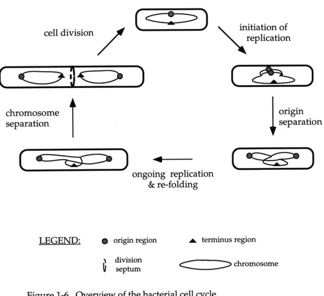
chromosome partitioning. Experiments using these techniques have painted a picture of the bacterial cell cycle that appears simple, although we are still at an early stage in describing the partitioning process. An overview of our current understanding of the bacterial cell cycle is shown in Figure 1-6 and is described briefly below.
Initiation of replication at oriC presumably begins at midcell, where the replicative DNA polymerase localizes (Lemon and Grossman 1998) (Figure 1-6). Soon after duplication of the origin regions, the sister origins move apart to positions near the cell quarters, where they remain for the bulk of the cell cycle (chapter 2,4) (Glaser et al. 1997; Gordon et al. 1997; Lin et al. 1997; Mohl and Gober 1997; Niki and Hiraga 1998; Webb et al. 1997). Replication ends at the terminus of the chromosome, which is diametrically opposed to the oriC on the physical map. In contrast to the localization of oriC, the terminus region of the chromosome is located near the center of the cell through the bulk of the cell cycle (Gordon, et al. 1997; Niki and Hiraga 1998; Webb, et al. 1997). Unlike in eukaryotic cells, partitioning of the bacterial chromosomes occurs concurrently with DNA replication. Following the completion of DNA replication, site specific recombinases resolve chromosomal dimers that may arise by single
initiation of replication cell division
//
CI4
D
chromosome separation origin separation ongoing replication & re-foldingC
Q
D
LEGEND: p origin region A terminus region
1 division
septum chromosome
Figure 1-6. Overview of the bacterial cell cycle.
Starting with the cell at the top of the figure: The origin region, the site where DNA replication initiates, is shown as a filled gray circle (0). The replicative polymerase, PolC, localizes at midcell throughout most of the cell cycle and presumably initiation of replication also occurs at midcell. Soon after duplication of the origin region, the sister origins rapidly move apart. The origin regions localize near the cell quarter positions through the bulk of the cell cycle. In contrast, the terminus region (A), localizes near the center of the cell through most of the cell cycle. While replication
continues, the bulk of the nucleoid mass is partitioned towards either cell half. Finally, in the last step of chromosome separation, topoisomerases
and recombinases are involved in resolving chromosomal catenanes or chromosomal dimers, respectively. In B. subtilis and E. coli, during exponential growth, the cell divides at midcell to produce two equally sized daughter cells. Adapted from (Lemon and Grossman, 1998).
C
)
I
MEMENIM.,)
crossover events (Blakely et al. 1991; Blakely et al. 1993). Following replication, the sister chromosomes are catenated and must be decatenated by
topoisomerases (Adams et al. 1992; Kato et al. 1992; Luttinger et al. 1991). The distinct localization pattern of the origin region indicates that a mechanism exists to both move and tether the origin regions near the cell poles, and that this is an early step in the bacterial chromosome partitioning process. It is currently unknown how the origin region and the bulk of the chromosome are moved, but the movement might be due to motor proteins or by the extrusion of replicated DNA by DNA polymerase (described below). Proteins from the ParA and ParB family are important for both plasmid and chromosome partitioning.
SMC proteins probably function in chromosome organization. The properties of
these proteins and the experiments that led to our current understanding of the bacterial chromosome partitioning process are reviewed below.
The origin region
Inheritance of genetic material is dependent not only on the duplication of chromosomes, but also on its partitioning to future daughter cells. This problem was addressed in part by the replicon model, first published by Jacob, Brenner, and Cuzin in 1963 (Jacob et al. 1963). Concerning chromosome replication, the authors proposed that an initiator protein activated DNA replication from a specific region of the chromosome, the "replicator" (or the origin, later called oriC). The identification of the origin of replication and the identification and characterization of proteins involved in the initiation of replication supported
these ideas (reviewed in (Kornberg and Baker 1992)). The structure of the origin region and proteins involved in replication are for the most part conserved throughout the prokaryotic kingdom (Ogasawara and Yoshikawa 1992; Salazar et al. 1996).
Concerning partitioning, Jacob, Brenner, and Cuzin proposed that the origin region was attached to the cell membrane at the center of the cell, for two reasons. First, attachment to the cell surface could allow for a direct connection between cell growth and DNA replication. Presumably, DNA replication would proceed in response to some signal related to the cell cycle such as cell size. Secondly, attachment to the center could be a mechanism by which the duplicated chromosomes are partitioned. The authors proposed that growth along the long axis of the cell by preferential insertion of new cell membrane material into the center of the cell would lead to separation of the chromosomes (Jacob, et al. 1963).
The partitioning mechanism proposed by the replicon model was explored in several experiments. It is now clear that insertion of new cell membrane material occurs randomly throughout the cell, inconsistent with cell growth being the driving force proposed in the replicon model (Woldringh et al.
1990; Nanninga et al. 1990). However, we now know that the origin region is associated with the membrane, in part by proteins involved in DNA replication (reviewed below). Not unexpectedly, the replicon model is too simple to account for all we now understand about bacterial chromosome partitioning. However,
the replicon model provided a framework to address aspects of origin localization and origin movement.
Localization of bacterial chromosomal origin and terminus
The cellular localization of the B. subtilis and E. coli origin and terminus regions was an advance in the understanding of the bacterial chromosome partitioning process. These localization studies show that for most of the cell
cycle, the localization of the origin and terminus is defined, not random. The duplicated origin regions are usually located near the cell quarters and the terminus is usually positioned near the center of the cell.
Localization of the origins near the poles was inferred from studies of chromosome orientation during sporulation in B. subtilis. Sporulation is a postexponential phase developmental pathway that leads to the production of dormant resistant endospores in response to starvation and crowding (reviewed in (Grossman 1995; Stragier and Losick 1996). During sporulation, the cell divides near a cell pole rather than at midcell, generating a large mother cell and a smaller forespore cell. This specialized asymmetric division initially traps part of the chromosome in the small compartment near the pole, and the remainder of the chromosome is later translocated through (Wu and Errington 1994). Using a genetic mutation that blocked the cells prior to DNA translocation in
combination with gene expression studies, it was demonstrated that the origin proximal 30% of the genome is preferentially trapped in the smaller forespore
the cell poles in sporulating cells (Sun et al. 1991; Wu and Errington 1994; Wu et al. 1995).
Confirmation of this idea, not only in sporulating cells but also in vegetatively growing cells, came with the visualization of the origin and terminus regions in B. subtilis (Webb et al, 1997). The origin and terminus regions were also visualized in the nonsporulating bacterium E. coli (Gordon et al, 1997; Niki and Hiraga, 1998). Localization was accomplished either by using FISH (fluorescence in situ hybridization) or by inserting multiple tandemly repeated lac operator sequences at a desired chromosomal location and
visualizing those regions with a GFP-LacI fusion protein (Gordon, et al. 1997; Niki and Hiraga 1998; Webb et al, 1997). In addition, the localization of
Spo0J/ParB, a protein that binds to sequences in the origin region (Lin and
Grossman 1998), provided independent evidence for the localization of the origin in B. subtilis (see below and chapter 2, 4) (Glaser, et al. 1997; Lin, et al. 1997) and in Caulobacter crescentus; (Mohl and Gober 1997).
In B. subtilis, these results show that in exponentially growing cells, the duplicated origin regions are usually localized near the cell quarters while the terminus is localized at midcell (Webb et al, 1997, Lin et al, 1997, Glaser et al,
1997). During sporulation in B. subtilis, the duplicated origins appear much closer to the poles (Webb et al, 1997). In E. coli, the duplicated origin regions
appear near the poles (-10-15% the length of the cell) and the terminus is at midcell (Niki and Hiraga, 1997; Gordon et al, 1997). In C. crescentus, the duplicated origins localize at the poles (Mohl and Gober, 1997). Further
expansion of this work in B. subtilis shows that regions intermediate to the origin and terminus on the physical map typically localize to intermediate positions between the origin and terminus in the cell (Teleman et al. 1998).
Are the origins always near the cell quarters in B. subtilis? Or, does duplication and separation of the origins occur elsewhere in the cell, such as the center of the cell as proposed by Jacob, Brenner and Cuzin (Jacob, et al. 1963)? The answer to this question is not completely clear. Time-lapse microscopy of B. subtilis and E. coli cells with the lacO-marked origin regions was performed to address this. Origin duplication and separation usually occurs at midcell in B. subtilis (after origin movement from near the poles to midcell) (Webb et al. 1998). In E. coli, origin duplication usually occurs near the cell pole, and subsquently one sister origin moves to the opposite pole (Gordon, et al. 1997). Visualizing origin localization in germinating spores of B. subtilis (germination of spores occurs synchronously) supports a model for midcell duplication (Lewis and Errington 1997). Finally, the replicative DNA polymerase localizes in the center of the cell for most of the cell cycle in B. subtilis, favoring a model for duplication of the origins at midcell (Lemon and Grossman 1998). Although B. subtilis and E. coli may use different mechanisms, the question of where in the cell origin
duplication occurs requires more rigorous experiments.
The separation of the origin regions is an active process that cannot be attributed simply to the attachment of the origin regions to the cell membrane and longitudinal growth (Webb et al. 1998; Gordon et al. 1997). Time-lapse microscopy visualizing the origin regions in live cells indicates that the origin
movement is abrupt, with a average maximal velocity of 170 nm/min in B. subtilis (Webb, et al. 1998). Similar observation were made in E. coli (Gordon, et
al. 1997). The velocity of origin movement is within the range of movement that can be attributed to motor proteins or DNA polymerase (Webb et al. 1998). Treatment of the cells with drugs that inhibit cell wall growth or septum formation did not inhibit movement (Gordon et al. 1997; Webb et al. 1998). Together these observations led to the idea that an active mechanism was involved in separation of the origin regions. Two proposals are that the
separation of the origin regions and/or the entire nucleoid could be carried out by DNA polymerase (Lemon and Grossman 1998) or by mitotic-like motor proteins (Glaser et al. 1997; Gordon et al. 1997; Lin et al. 1997; Webb et al. 1997).
Membrane association of the origin and terminus regions and replication forks
The attachment of the origin to the membrane occurs in both E. coli and B. subtilis, but how membrane attachment relates to chromosome partitioning has not been demonstrated conclusively (for review, see (Firshein 1989)). Origin membrane attachment was demonstrated by a number of membrane
fractionation techniques (Hendrickson et al. 1982; Laffan and Firshein 1987; Sueoka and Quinn 1968; Winston and Sueoka 1980). In B. subtilis, the terminus is also associated with the membrane (Beeson and Sueoka 1979; Sargent and
important for origin membrane attachment have been characterized, and in both cases these proteins play roles in DNA replication.
In E. coli, origin attachment is detectable in an outermembrane preparation (which may contain innermembrane components) (Hendrickson, et al. 1982) (E.
coli is has an outermembrane lipid bilayer, a peptidoglycan layer, and an
innermembrane lipid bilayer). The attachment of origin DNA is dependent on its methylation state. E. coli DNA is methylated at a specific site by the DNA
adenine methyltransferase (Dam). It was observed that hemimethylated (methylated on only one strand) origin DNA binds preferentially to an
outermembrane preparation (Ogden et al. 1988). Subsequently, the SeqA protein was identified as a factor important for binding hemimethylated origin DNA (Lu et al. 1994; von Freiesleben et al. 1994).
The binding of hemimethylated DNA to SeqA has mainly been studied for its role in sequestering the initiation of replication. Initiation of DNA replication in E. coli usually occurs on fully methylated DNA. Following passage of the replication forks, the origin region DNA becomes hemimethylated. The SeqA protein presumably binds to the hemimethylated DNA and functions to prevent reinitiation. In vitro, SeqA binds nonspecifically to hemimethylated DNA, calling into question whether origin binding (and attachment) is specific to SeqA (Slater et al. 1995). Nonetheless, an obvious question is if the membrane attachment by SeqA, or some factor in combination with SeqA, also functions in partitioning of the origin regions and/or of the bulk chromosome. Two results suggest that SeqA could function in partitioning of the bulk chromosome. seqA mutants
appear to have some chromosome partitioning defect (~1.6% anucleate cells), and the migration of the SeqA protein from midcell to the quarter sites late in the cell cycle suggests it may play some role in partitioning (Bahloul et al. 1996; Hiraga et al. 1998; Onogi et al. 1999). However, this migration of SeqA protein is
independent of oriC, leading Hiraga et al to suggest that SeqA is important for bulk nucleoid partitioning (Hiraga et al. 1998; Onogi et al. 1999).
Origin-membrane association has been best characterized in B. subtilis (reviewed in (Firshein 1989; Firshein and Kim 1997)). Dam methylation does not occur in B. subtilis. In B. subtilis, origin membrane attachment and the initiation of replication are dependent on the gene product of dnaB (B. subtilis DnaB is distinct from E. coli DnaB, the replicative helicase) (Hoshino et al. 1987; Sueoka et al. 1988; Winston and Sueoka 1980). DnaB is a 472 aa protein with two putative ATP binding domains and an N-terminal transmembrane domain (Hoshino, et al. 1987). It is likely that DnaB interacts with origin DNA, directly or indirectly. Preliminary localization studies have shown that DnaB appears to localize as distinct foci in the cell and can localize in the absence of DNA (K. Lemon and AD Grossman, unpublished data). One idea is that DnaB may serve as a membrane anchor for the origin regions and the region of DNA that DnaB contacts could be analogous to a eukaryotic centromere. However, no studies on DnaB have separated its role in DNA replication from a possible role in chromosome partitioning.
Finally, replication of the entire chromosome is also thought to occur at the membrane, although this has been less well characterized (reviewed in
(Firshein 1989)). Initiation factors, such as B. subtilis DnaB and the conserved DnaA protein (Yung and Kornberg 1988) are associated with membranes. Also, DNA Pol III (the replicative polymerase) activity can be detected in purified membrane fractions of B. subtilis and Pneumococcus (Benjamin et al. 1982;
Firshein and Gelman 1981). Replication can proceed in vitro from these purified complexes without the addition of other proteins and is inhibited by
hydroxyphenylazouracil, a specific inhibitor of DNA PolIII.
Localization of the replicative DNA polymerase
The replicative polymerase was localized in B. subtilis by use of a GFP fusion to the C-terminus of PolC. polC encodes the gene for the alpha subunit of the PolIl core. The polC-gfp fusion is functional, and PolC-GFP is found to be stationary and localizes in the center of the cell (Lemon and Grossman 1998). Localization of PolC-GFP is dependent on ongoing rounds of DNA replication (Lemon and Grossman 1998). These result support a "factory" model of DNA replication, in which the DNA is threaded through the stationary DNA
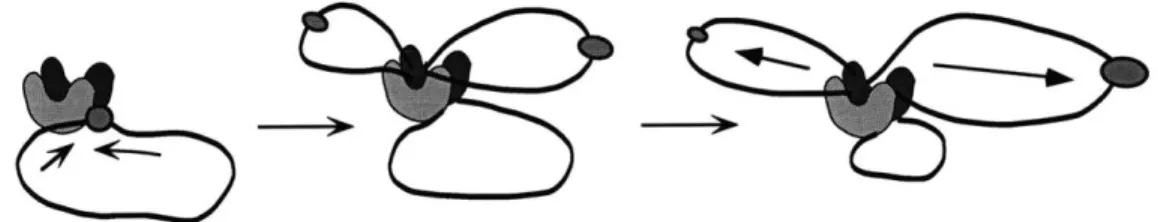
polymerase rather than the polymerase moving along the DNA (Lemon and Grossman 1998). One possibility is that the actions of DNA polymerase may contribute to or be sufficient for the movement of the chromosomes to either half of the cell. In the "extrusion-capture" model, the newly replicated DNA strands are extruded from the stationary polymerase and the origin regions are captured by a protein complex that binds both the origin DNA and regions near the cell poles (figure 1-7) (Lemon and Grossman 1998).
A. Extrusion of DNA by a stationary DNA polymerase
B. Movement of DNA by motor proteins along a polarized track
/
(~3)
origin region motor protein sr trackFigure 1-7. Two speculative models to account for the rapid movement of the origin regions.
A. In the extrusion-capture model, the motive force for origin movement is
provided by DNA polymerase. DNA polymerase is stationary in the cell. As the chromosome is replicated, the sister chromosomes are pushed away from midcell by the force of polymerization and the origins are captured
by an origin anchor near the cell poles. For simplicity, the replisomes are
shown as giant U-shaped blobs. (adapted from Lemon and Grossman,
1998). B. Mitotic-like motor proteins may recognize the origin regions and
move them along polarized tracks towards opposite poles, analogous to the function of eukaryotic mitotic motor proteins. (Adapted from Hiraga,
1992).
:MMm or PC
>
77 7
A second possibility is that origin movement and/or chromosome
movement is accomplished by the actions of motor proteins (Glaser, et al. 1997; Lin, et al. 1997; Webb, et al. 1998; Webb, et al. 1997), analogous to the motor proteins that move chromosomes during eukaryotic mitosis (figure 1-7). Proteins similar in sequence to kinesins or dyneins have not been identified in genomes of several completely sequenced prokaryotes. It is possible that a novel motor protein functions in bacterial chromosome partitioning. Finally, by structural homology, the best candidates for possible mitotic motor proteins are members of the SMC family, which are conserved from bacteria to human. However, SMC proteins appear to be involved in chromosome organization, as described below.
MukB and SMC proteins
The mukB gene was originally identified in E. coli in a genetic screen for mutations that led to an increase in the percentage of anucleate ("mukaku" in Japanese) cells (Hiraga, et al. 1989; Niki et al. 1991). The mukB gene is in an
operon with two other genes, mukE and mukF, which are also required for proper chromosome partitioning, although the precise functions of these gene products are unknown (Yamanaka et al. 1996). MukE and MukF are not found in B.
subtilis. The MukB protein encodes a large 1534 amino acid protein with globular N and C terminal regions separated by two central coiled-coil domains that mediate homodimerization (Niki et al. 1992; Niki, et al. 1991 ).
MukB shares limited amino acid similarity but good structural similarity (see below) with the 1186 amino acid SMC protein of B. subtilis (Britton et al. 1998; Moriya et al. 1998; Oguro et al. 1996). B. subtilis SMC also has a structure, composed of two globular domains separated by two elongated coiled-coil domains (figure 1-3) (Melby et al, 1998). Currently, all but two completely sequenced bacterial genomes contain an SMC homologue, and the two that do not have SMC do contain MukB. Both proteins contain a nucleotide binding domain and the C-terminus has been implicated in DNA binding (Akhmedov et al. 1998; Niki, et al. 1991; Oguro, et al. 1996). B. subtilis SMC is capable of making homodimers (Melby et al. 1998), and null mutations in smc and mukB cause similar phenotypes (figure 1-3 part A) (Britton et al. 1998; Hirano and Hirano 1998; Moriya et al. 1998; Oguro, et al. 1996). Deletion of smc or mukB leads to the production of - 10% or ~ 5% anucleate cells in a growing culture, respectively
(mukB experiments were done at 22'C in enriched media, smc experiments done at 30'C in minimal media) (Britton et al. 1998; Niki et al. 1991). In addition, both smc and mukB null cells are temperature sensitive for growth (Britton et al. 1998; Moriya et al. 1998; Niki et al. 1991). B. subtilis SMC and E. coli MukB probably play similar roles in chromosome partitioning.
The evidence suggests that MukB and SMC probably function in some aspect of chromosome organization. However, the structural similarity (both proteins have been visualized by electron microscopy) of these proteins to mitotic motor proteins initially led to the speculation that these proteins may