HAL Id: hal-00009072
https://hal.archives-ouvertes.fr/hal-00009072v2
Submitted on 8 Nov 2005
HAL is a multi-disciplinary open access
archive for the deposit and dissemination of
sci-entific research documents, whether they are
pub-lished or not. The documents may come from
teaching and research institutions in France or
abroad, or from public or private research centers.
L’archive ouverte pluridisciplinaire HAL, est
destinée au dépôt et à la diffusion de documents
scientifiques de niveau recherche, publiés ou non,
émanant des établissements d’enseignement et de
recherche français ou étrangers, des laboratoires
publics ou privés.
Autogenous modulation of the Bacillus subtilis
sacB-levB-yveA levansucrase operon by levB transcript.
Jean-Pierre Daguer, Thomas Geissmann, Marie-Françoise Petit-Glatron, Régis
Chambert
To cite this version:
Jean-Pierre Daguer, Thomas Geissmann, Marie-Françoise Petit-Glatron, Régis Chambert.
Autoge-nous modulation of the Bacillus subtilis sacB-levB-yveA levansucrase operon by levB transcript..
Mi-crobiology, Microbiology Society, 2004, 150, pp.3669-3679. �10.1099/mic0.27366-0�. �hal-00009072v2�
This version of our manuscript contains the modeling of the dynamics of expression of the
levansucrase operon which was not included in the final version that was published in Microbiology (2004) 150 3669-3679 DOI 10.1099/mic.0.27366-0.
Bacillus subtilis: characterisation and role of autogenous modulation of
sacB-levB-yveA levansucrase operon by levB transcript
Jean-Pierre Daguer, Thomas Geissmann, Marie-Françoise Petit-Glatron and Régis Chambert*
Institut Jacques Monod, Laboratoire Génétique et Membranes, CNRS/ Universités Paris 6 et Paris 7
Tour 43, 2 place Jussieu, 75251 Paris Cedex 05, France
*Corresponding author:
Régis Chambert , Tel: + 33 1 44 27 47 19. Fax: +33 1 44 27 59 94.
e-mail:
chambert@ccr.jussieu.fr
SUMMARY
Silencing of levB, the second structural gene of the tricistronic levansucrase operon encoding the endolevanase LevB, decreases the level of levansucrase expression. Conversely, independent expression of levB greatly stimulates operon expression. This autogenous effect is mediated by the levB transcript, which carries an internal sequence (5’-AAAGCAGGCAA-3’) involved in the enhancing effect. In vitro, the levB transcript displays an affinity to the N-terminal fragment of SacY, the regulatory protein that prevents transcription termination of levansucrase operon, (KD = 0.2 µM). Simulation of the dynamics of operon
expression showed that this positive feedback loop increases the capacity of Bacillus subtilis to produce the three proteins encoded by the operon when bacteria are grown in the presence of high concentrations of sucrose. Under such conditions, extracellular levan synthesized by the fructosyl polymerase activity of levansucrase can be degraded mainly into levanbiose by the action of LevB. Levanbiose is neither taken up nor metabolized by the bacteria. This work modifies the present view of the status of levansucrase in B.
subtilis physiology.
INTRODUCTION
The levansucrase tricistronic operon of Bacillus subtilis consists of an upstream cis-acting control region, the sacR locus (Aymerich et al., 1986), and three genes sacB, levB and yveA, the transcription of which is simultaneously induced by sucrose (Pereira et al., 2001b).
Levansucrase, encoded by sacB, is a secreted enzyme whose in vivo and in vitro catalytic activities are well characterized (Dedonder, 1966; Chambert et al., 1974). The enzyme acts mainly as a sucrose hydrolase when the
concentration of sucrose is low (< 10 mM). At higher concentrations of sucrose, levansucrase catalyses the formation of high molecular mass fructan of the levan type by the addition of fructosyl residues from sucrose. The enzyme is able to hydrolyse levan into fructose, but its exolevanase activity is arrested at the 2
→
1 branch points of the polymer (Rapoport & Dedonder, 1963). Only 30 % of available fructose is released by the prolonged action of the enzyme on the polymer.The protein encoded by levB is a peripheric membrane protein remaining anchored to the cytoplasmic membrane and displays an endolevanase activity, which has been preliminarily characterized (Pereira et al., 2001b). YveA, the third protein, might function as a permease, as predicted by its similarity to proteins of known function (Kunst et al., 1997). Its numerous predicted transmembrane segments suggest that YveA is a membrane intrinsic protein.
Northern blotting analyses with specific probes showed that, under exponential phase of growth and in the presence of 50 mM sucrose, the yield of the full length tricistronic transcript sacB-levB-yveA was lower than that of the bicistronic sacB-levB, whose yield is itself about 10 % of the monocistronic sacB mRNA (Pereira et al., 2001b). This results from partial arrests of the RNA polymerase at the internal terminator structures located between sacB and levB, and levB and yveA. Considerable efforts have been made in the last three decades (Lepesant et al., 1972; Steinmetz et al., 1985; Tortosa & Le Coq, 1995, 1997; Idelson & Amster-Choder, 1998; Declerck et al., 2002) to identify the mechanism underlying sacB expression and to situate within the carbohydrate catabolism network of B. subtilis the role and regulation of this gene involved in the metabolism of sucrose. All the molecular genetic investigations were carried out on the
assumption that the monocistronic sacB locus encodes only levansucrase. Within this context, it was difficult to find a satisfying explanation concerning the physiological function of this enzyme, because B. subtilis possesses a higher efficient pathway for sucrose metabolism constituted of a PTS dependent permease specific for sucrose and an intracellular sucrase (Lepesant et al., 1972). Therefore, we considered stimulating to reopen the debate from the finding that LevB is a part of a functional unit composed of the three proteins encoded by the operon.
We anticipated that the expression of the two additional proteins of the operon might play a role in the function and regulation of the operon expression by means of the transport or metabolism of sucrose or its derivatives. In order to study this hypothesis, we first carefully characterized the catalytic activity of LevB and we investigated the contribution made by the expression of levB and yveA to the regulation of operon expression. The silencing of these two operon distal genes led to a decrease in sacB expression by a factor of two. Independent expression of yveA had no effect on sacB expression. In contrast, overexpression of levB greatly increased the level of SacB synthesis. Surprisingly, this enhancing effect was not related to the catalytic activity of levB. We found however that the levB transcript carries a short internal sequence identical to a motif of eleven nucleotides present in the leader region of the operon. We therefore explored the possibility that an interaction existed between the levB transcript and the components of the transcription antitermination system that controls expression of the operon.
METHODS
Bacterial strains and media. The strains and plasmids
used are listed in Table 1. All the strains constructed were obtained by transformation with replicative or integrative plasmids of strain GM96100, a derivative of the degU32(Hy) Bacillus subtilis mutant (Leloup et al.,1997). Bacteria were grown at 37 °C in minimal medium (Chambert & Petit-Glatron, 1984) supplemented with 1 % (w/v) glucose. One optical density (OD) unit at 600 nm of cell suspension (
≈
108 bacteria) corresponds to approximately 100 µg ml-1 protein (Chambert & Petit-Glatron, 1984). Escherichia coli XL1-Blue strain and its transformants were grown in TerB rich medium (Sambrook et al., 1989) containing 150 mg ampicillin ml-1.Plasmid and strain constructions. All the DNA
fragments were amplified by PCR with primers including restriction sites, as indicated in Table 2, from the chromosomal DNA of strain QB112 isolated as described by Leloup et al. (1997). The amplified blunt-ended fragments were inserted into the pCR(+) vector at the SrfI site, after appropriate treatment, according to the supplier’s recommendations (Stratagene). The resulting plasmids were used to transform E. coli XL1-Blue.
Plasmids purified from E. coli transformants exhibiting fragments of the expected size after digestion by various endonucleases were selected and the complete sequence of the fragments inserted was controlled using appropriate primers.
Construction of plasmid pGMK80. This integrative plasmid was constructed in order to introduce, by double crossing over, DNA fragments into the sacR-sacB chromosomal site and was obtained as follows: pGMK50 (Petit–Glatron & Chambert, 1992) was digested by EcoRV and re-ligated. Plasmid pGMKD50, from which the EcoRV fragment had been deleted, was selected. A 1 kb H1 fragment corresponding to the chromosomal sequence upstream from sacR-sacB was amplified by PCR from genomic DNA of strain QB112 using oligonucleotides H1-fw and H1-rev as primers containing the restriction sites AvaI and BamHI, respectively, and inserted into pCR(+) vector as described above, resulting in plasmid pGMC20. H1 fragment was purified from this plasmid after digestion by AvaI and BamHI and ligated into pGMKD50, digested with the same enzymes. An appropriate plasmid was selected and named pGMK80. Construction of plasmid derivatives of pWH1520. The structural genes levB or yveA were amplified by PCR using oligonucleotides levB-fw and levB-rev1 or yveA-fw and yveA-rev (Table 2) containing the restriction sites SpeI and KpnI, respectively. The amplification products were cloned into pCR(+) vector. The resulting plasmids pGMC21 and pGMC22 were digested with SpeI and KpnI. The DNA fragments levB (1.6 kb) or yveA (1.6 kb) were ligated into pWH1520 (Rygus et al., 1991) digested with the same enzymes giving plasmids pWHlevB or pWHyveA.
Construction of plasmid pWHlevBmut. Plasmid pGMC21 containing the levB gene sequence was used as a template to amplify two PCR DNA fragments using oligonucleotides levBmut and KS and levB-rev2 and T3 (Table 2). The two PCR products of approximately 1650 bp and 50 bp were then mixed and reamplified with oligonucleotides KS and T3. The resulting PCR product was cloned into pCR(+), giving pGMC23 which was sequenced using appropriate oligonucleotides. The mutated levB gene was SpeI-KpnI digested and cloned into pWH1520. The plasmid was named pWHlevBmut. Construction of strain GM2101: The levansucrase structural gene sacB was amplified using oligonucleotides LS-fw and LS-rev containing the restriction sites AatII and XhoI (Table 2) and cloned into the pCR(+) vector as described above. The 1.4 kb DNA fragment obtained by AatII and XhoI digestion was purified and ligated into plasmid pGMC9, digested with the same enzymes, which contains the sacR locus cloned as a BamHI/AatII fragment (Leloup et al., 1999). The plasmid obtained, pGMC24, was digested with BamHI and EcoRV and the corresponding fragment was inserted into pGMK80 digested with the same enzymes. The resulting plasmid was used to transform E. coli XL1-Blue. The correct sequence of the inserted fragment was verified from purified plasmids and levansucrase activity was assayed in
the cell extracts of the transformants. An appropriate plasmid (pGMK81) was chosen to transform strain GM96100.
Transformants were selected on LB plates for both their resistance to kanamycin (10 mg ml-1) and their sensitivity to spectinomycin (100 mg ml-1) and chloramphenicol (3 mg ml-1). One of the transformants exhibiting sucrose inducible expression of levansucrase was chosen and named GM2101.
Construction of strain GM2102. The endolevanase structural gene levB sequence was amplified by PCR as described by Pereira et al. (2001b). The amplification product was cloned in pCR(+) vector.The resulting plasmid was digested with AatII and EcoRV. The 1.6 Kb fragment containing the levB gene was ligated into pGMC9. The transcriptional fusion sacR-levB was then purified by BamHI EcoRV digestion and ligated into pGMK80 resulting in plasmid pGMK82 which was used to transform GM96100. Transformants were selected on LB plates for both their resistance to kanamycin (10 mg ml-1) and their sensitivity to spectinomycin (100 mg ml-1) and chloramphenicol (3 mg ml-1). One of the transformants exhibiting sucrose inducible expression of LevB was chosen and named GM2102.
Construction of strains GM2201,GM2202, GM2203, GM2204. These strains were obtained by transformation of strain GM2101 with the replicative plasmid pWH1520 (GM2201) and its derivatives pWHlevB (GM2202), pWHyveA (GM2203) and pWHlevBmut (GM2204).
Levansucrase assay. Levansucrase activity was estimated
by measuring the initial rate of the fructosyl exchange reaction (Chambert et al., 1974). A reaction mixture (20 µl) containing 0.2 M uniformly labeled [14C]glucose and 0.1 M sucrose in 0.05 M phosphate buffer pH 6 was incubated at 30 °C for 10 min. The reaction was initiated by the addition of 5 µl of culture supernatant. Aliquots of 8 µl were removed at intervals and 14C labeled sugars were quantitatively analyzed by paper chromatography. One unit of enzyme activity (EU) defined as the amount of enzyme exchanging 1 µmole glucose min-1 corresponds to 2 mg of levansucrase..
LevB assay. Uniformly labelled [14C]levan was prepared by the action of immobilized levansucrase on [14C]sucrose and used as a substrate (Chambert & Petit-Glatron, 1993). LevB was assayed on the membrane fraction obtained as previously described by Pereira et al. (2001b).
RNA techniques. Total RNA extraction, Northern blotting
and mRNA half-life determinations were done as described by Pereira et al. (2001a, 2001b). We confirmed transcription of levB or yveA in strains GM2202 or GM2203 grown in minimal medium upon xylose induction by Northern blotting using, as probes, the levB and yveA genes purified from plasmid pGMC21 and pGMC22, respectively, by SpeI/KpnI digestion. Probes were radiolabelled with [a-33P] ATP by random priming using Amersham DNA Megaprime Labelling System.
In vitro transcription. The DNA template (pWHlevB or
pWHlevBmut) for in vitro transcription was generated by PCR with forward primer levB-T7 containing the T7 promoter sequence and reverse primer levB-332rev. RNA was then produced by transcription in vitro with T7 polymerase (T7 Megashortscript kit, Ambion). Transcripts were de-phosphorylated with alkaline phosphatase and radioactively labelled at the 5' end with [g-32P]ATP and T4 polynucleotide kinase (Kinasemax labelling kit, Ambion). The radioactively labelled RNA was purified on a denaturing 8 % (w/v) polyacrylamide / 8 M urea gel and eluted in 0.5 M ammonium acetate, 1 mM EDTA and 0.1 % (w/v) SDS. The transcripts were collected by ethanol precipitation and suspended in 10 mM Tris/-HCl, pH 8.5.
RESULTS
Catalytic activity of LevB. In order to increase LevB
synthesis, which production is very low in the context of the levansucrase operon (Pereira et al., 2001b), we constructed strain GM2102 in which levB expression was under the control of the sacR leader region of the operon as described in Methods. Under these conditions, LevB was overproduced which made it possible to analyse, in vitro, the catalytic specificity of this enzyme in its membrane associated form.
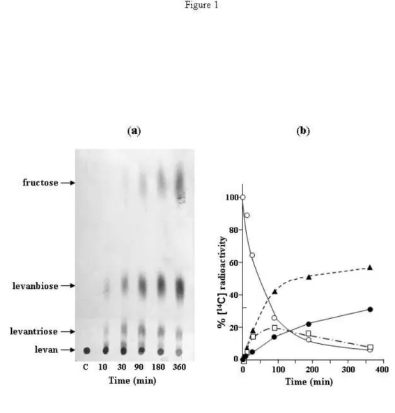
We first identified the products released by LevB acting on levan. Results showed (Fig. 1a) that the main products, identified by chromatographic migration and subsequent analysis of acid hydrolysed products, were fructose, levanbiose (difructose) and levantriose (trifructoside). After incubation for 6 h (Fig. 1b), these compounds represented 32, 55 and 7.4 % of available fructose, respectively. After a longer incubation time, levanbiose reached 62 % and remained stable. In addition, we observed that LevB is devoid of any catalytic activity on sucrose, the only known inducer of the levansucrase operon. LevB is unable to use this sugar either as a fructosyl donor or as a fructosyl acceptor (results not shown).
Silencing of both levB and yveA gene leads to a decrease in sacB expression. Silencing of the levB and yveA genes
was carried out by disrupting the operon sacB-levB-yveA. For this purpose a transcriptional fusion sacR-sacB-KmR was inserted by double crossing-over into the chromosome of strain GM96100 deleted of the sacR-sacB region (Leloup et al., 1997) (Fig. 2a). In the resulting strain, GM2101, introduction of the KmR cassette prevented the expression of levB and yveA, the last two genes of the operon. This silencing did not affect the growth rate of the cells. Endolevanase LevB activity was not detected in membrane fractions and the differential rate of levansucrase synthesis induced by sucrose was approximately 3 % of total protein compared to strain QB112 in which levansucrase production represented 6.5 % of total protein after full sucrose induction (Fig. 2b). This result suggests that the products of levB or yveA
expression participate in an auto-activation mechanism of levansucrase operon expression.
Independent expression of levB greatly increases levansucrase production whereas expression of yveA has no effect. In order to determine which of the two
candidate genes affected levansucrase production, we cloned each gene under the control of an inducible promoter xylA in plasmid pWH1520 (Rygus et al., 1991). Strain GM2101 was transformed as indicated in Methods with plasmids pWH1520, pWHlevB and pWHyveA and the corresponding tetracycline resistant strains GM2201, GM2202 and GM2203 were grown in minimal medium. Transcription of levB or yveA upon xylose induction in strain GM2202 or GM2203 was confirmed by Northern blotting (not shown). Levansucrase synthesis subsequent to sucrose addition was measured in the presence of various concentrations of xylose (Fig. 3). When xylose was used within a range of 0 – 2 % (w/v) xylose, a four fold increase in levansucrase synthesis was obtained in strain GM2202. Production of levansucrase corresponded to 13 % of total cellular proteins under optimum conditions of induction. Pulse-chase experiments carried out as described by Chambert & Petit-Glatron (1988) indicated that the kinetics of levansucrase was not modified by LevB overproduction. A similar experiment was carried out with yveA. The results obtained indicate that independent expression of this gene has no effect on the production of levansucrase. Given the results, we concluded that SacB synthesis depends on levB gene expression. We therefore question whether the enhancing effect of levB expression is exerted at the transcriptional or translational level.
Expression of levB increases the yield of sacB transcription but not mRNA stability or translation efficiency of sacB. Northern blotting analysis of sacB
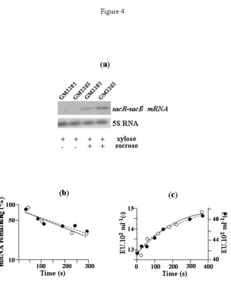
transcripts was carried out with the sacR probe (Pereira et al., 2001b) in strains GM2201 and GM2202. The results indicated a three fold increase in the steady state level of sacB transcripts in strain GM2202 which overexpressed levB compared to strain GM2201 (Fig. 4a). We analysed the kinetics of sacB mRNA decay in the two strains by Northern blotting after inhibition of transcription initiation by rifampicin. Quantification of the labelled bands on the Northern blot gave similar values for the two strains, 120 s ± 30 (GM2201) and 126 s ± 25 (GM2202) (Fig. 4b). Quantification of the increase in the amount of levansucrase synthesized was also performed after addition of rifampicin (Fig. 4c) to determine the functional mRNA stability. The half-life values deduced from the curves were 105 s ± 20 for strain GM2201 and 115 s ± 18 for strain GM2202. The two methods resulted in the estimation that the half-lives of sacB mRNA were similar in the two strains. Moreover, the ratio of the total amount of levansucrase synthesized in each strain (Fig. 4c), after inhibition of the transcription initiation was the same as the ratio of the steady state sacB mRNA quantified from Northern blotting analyses (Fig. 4a). It can be concluded
that the increase in levansucrase production due to levB expression is exerted at the transcriptional level.
The enhancing effect of levB expression on sacB transcription is not related to the catalytic activity of LevB. Control of sacB gene expression has been
thoroughly investigated during the last two decades (Aymerich et al., 1986; Crutz et al., 1990). All the results obtained support the conclusion that sucrose induction of the sacB gene occurs via an antitermination mechanism involving the sacX/Y regulatory operon of B. subtilis. We therefore explored the hypothesis that the enhancing effect of the independent levB expression is mediated by the products of the catalytic activity of LevB, which might be a better inducer than sucrose.
We have shown above that LevB acts on levan only and has no catalytic action on sucrose, the inducer of sacB expression. Previous work showed that levansucrase is able to catalyse levan synthesis only when the sucrose concentration is higher than 10 mM (Chambert & Gonzy-Treboul, 1976). It was therefore interesting to test whether the transcription enhancing effect of levB expression could be observed in the absence of levan synthesis. The sucrose induction profiles of levansucrase production by strains GM2201 and GM2202 were compared with that of strain QB112 (Fig. 5). First we observed that the presence of sucrose was required to induce levansucrase expression in the three strains. However, the response curves of SacB production to the inducer are quite different. One of the main features concerned inducer concentrations required to reach full induction. The concentration was lower than 20 mM for strain GM2201 and GM2202, whereas it was equal or higher than 50 mM for the reference strain QB112. This point will be clarified by the simulation approach proposed below. Secondly, the enhancing effect of levB expression (strain GM2202) occurs at 0-10 mM sucrose, concentrations at which levan, the substrate of LevB, is not synthesized. This result suggested that the enhancing effect of levB expression is not dependent on the catalytic activity of LevB. To confirm this, we added to the cell suspension a mixture of levanbiose plus fructose obtained in vitro by digestion of levan by LevB (see legend to Fig. 1). No effect on levansucrase production was observed (results not shown).
It can be concluded that there is no relation between the enzyme activity of LevB and the enhancing effect of levB expression on levansucrase production. We therefore propose the unconventional hypothesis that the levB transcript can act as a transcriptional activator.
The levB transcript carries a sequence motif involved in the enhancing effect. Sequence comparison of the
non-coding sacR operon leader region and the three genes of the levansucrase operon showed that the levB gene shares an identical sequence of 11 nucleotides with sacR (Fig. 6). This motif is included in the 29 nucleotides of the ribonucleic anti-terminator (RAT) sequence folded into a stem loop structure essential for an efficient interaction with SacY, the anti-terminator protein (Declerck et al.,
2002). To question whether this motif plays a role in the enhancing effect of levB expression, we substituted by site-directed mutagenesis codons synonymous to those included in the motif. The codons AAA, GCA, GGC were replaced by AAG, GCG, GGG which modify the sequence without modifying the amino acid sequence of the protein. Strain GM2204 carrying the mutated levB gene under the control of xylA promoter was grown in the absence or in the presence of 1 % xylose. Levansucrase production subsequent to sucrose addition is similar under both conditions. This result indicated that the mutations introduced in the sequence motif of levB, identical to the RAT, impair its capacity to improve levansucrase production when compared with strain GM2202.
In vitro the transcript levB displays an affinity to SacY,
the antitermination protein of the operon. We tested the
ability of the levB transcript to bind SacY, the anti-termination protein of the operon. We used similar experimental conditions to those used by Manival et al. (1997) to demonstrate specific binding of SacY (1-55) to the RAT sequence of the leader region.
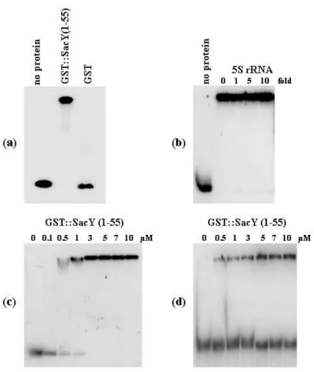
A fragment of 144 nucleotides of levB mRNA containing the motif AAAGCAGGCAA was generated by in vitro transcription as described in Methods and subjected to gel mobility shift experiments. One major shift was detected when the levB fragment was incubated with GST::SacY(1-55) fusion protein (Fig. 7a). The intensity of the shifted band was not affected by the presence of increasing amounts of 5S rRNA indicating that SacY(1-55) specifically binds the levB mRNA fragment (Fig. 7b). The affinity constant of SacY(1-55) to levB mRNA was evaluated from gel shifts repeated with various amounts of GST::SacY(1-55) fusion protein (Fig. 7c). The binding pattern was quantified and gave an estimate of 0.2 µM for the dissociation constant. When the mutated levB mRNA fragment was used in the same experiment, the binding pattern was greatly modified (Fig. 7d) and in this case the affinity constant of SacY(1-55) to levB mRNAwas estimated to 5 µM.
Modelling of the dynamics of the induction of levansucrase operon. The expression of the sacB gene has
been shown to be regulated by transcription antitermination involving the binding of SacY to the transcript (Aymerich & Steinmetz, 1992; Declerck et al., 2002). This mechanism is characterized by a constitutive transcription of the operon leader region. When sucrose (the inducer) is absent, the transcript of the leader region folds into a stable terminator which serves as a transcription pause signal. SacY prevents termination allowing readthrough transcription by stabilizing an antiterminator structure. The active state of SacY depends on the activity of SacX, a sucrose permease which is possibly involved in catalyzing the reversible phosphorylation of SacY (Idelson & Amster-Choder, 1998). This mechanism is not contested by our finding that sacB is the proximal gene of a sucrose inducible tricistronic operon including levB and yveA. However, the results presented above suggest an additional
circuit of control. levB transcript exerts, via its interaction with SacY, a positive feedback modulation of transcription of the levansucrase operon mainly under conditions of high inducer concentrations. Using quantitative modelling, we attempted to simulate the dynamics of the response of this system to various signal values.
We propose that functional SacY can exist under the free form SacYf or associated with levB transcript, SacYa. This association could stabilize the active dimeric form of SacY (Manival et al., 1997). We postulate that the two forms display a similar affinity to the antiterminator site. However, the delay time for the subsequent destabilization of the terminator hairpin structure could be shorter in the presence of SacYa. It results that the increase in the transcription frequency of the downstream coding region increases the yield of the transcript of each gene of the operon.
If SacYT is the total cellular concentration of functional SacY at any inducer concentration, the following equations can be established:
(SacY
T) = (SacY
f) + (SacY
a)
]
transcript
[
K
K
)
SacY
(
)
SacY
(
D D T flevB
+
=
[1]]
transcript
[
K
]
transcript
[
)
SacY
(
)
SacY
(
D T alevB
levB
+
=
where KD is the dissociation constant of the SacYa complex.
Since SacB production is well documented, we focused our attention on the dynamics of sacB transcript accumulation in cells, after the addition of sucrose. We can model this event by writing the phenomenological dynamical equation
) transcript ( k ) transcript , SacY ( f dt ] transcript [ d d T levB sacB sacB ! =
The first term corresponds to a positive effect of functional SacY and levB transcript on the rate of sacB transcript synthesis. The second term corresponds to the decay rate of sacB transcripts. The function f cannot be obtained from experiments since it is difficult to accurately quantitate the cellular level of functional SacYT (Idelson & Amster-Choder, 1998). Therefore, we propose to express the rate of sacB transcript synthesis with respect to the external inducer concentration using data obtained with the mutants constructed in this work.
We observed that the response curves of SacB production to the inducer for the strains GM2201 and GM2202 were hyperbolic (Fig. 8). These findings suggest that the cellular steady state level of sacB transcript depends on sucrose (I) according to the following equation:
I
K
I
]
transcript
[
]
transcript
[
I M st st+
= sacB
sacB
From the induction pattern of each strain, a similar value for the inducer concentration leading to
M st st
[
transcript
]
2
1
]
transcript
[
sacB
=
sacB
wasevaluated, KI = 8 mM. Since strain GM2201 does not synthesize levB transcript, it results that, in this strain, functional SacY is active in its SacYf form only. Conversely, under conditions of overproduction of levB transcript it can be assumed that functional SacY is active in its SacYa form in GM2202.
We previously correlated the rate of SacB production in B. subtilis with the steady state yield of sacB mRNA (Petit-Glatron & Chambert, 1981). Combination of this data with the decay rate of the entity, kd = 0.35 min-1 enabled us to evaluate the rate of sacB mRNA synthesis in the cells of each strain at any inducer concentration. The maximum rates were estimated to be 0.14 µM min-1 and 0.6 µM min-1 for strains GM2201 and GM2202, respectively.
Therefore, the dynamics of sacB mRNA synthesis subsequent to sucrose addition in such bacteria suspension are represented by the following equations
Strain GM2201
)
mRNA
(
k
I
K
I
14
.
0
dt
)
mRNA
(
d
d IsacB
sacB
!
+
=
[2a] StrainGM2202)
mRNA
(
k
I
K
I
6
.
0
dt
)
mRNA
(
d
d IsacB
sacB
!
+
=
[2b]These equations were established by postulating that the rate of SacY transition from the inactive to the active state is an instantaneous process compared to the transcription process at any sucrose concentration.
The reference strain degU32(Hy) produces both the monocistronic sacB mRNA and the bicistronic sacB-levB mRNA (Pereira et al., 2001b). This latter molecule, which carries the transcriptional enhancing motif, is not produced to the same extent as that of the monocistronic sacB mRNA depending on the yield of the readthrough of the internal terminator structure located between sacB and levB. In addition, we have experimentally evaluated (results not shown) that the apparent rate constant of the decay reaction of the bicistronic mRNA is similar to that of the monocistronic mRNA. The transition from SacY to SacYa is modulated by the bicistronic mRNA according to [1]. Therefore if x stands for sacB mRNA, one can describe the dynamics of accumulation of this transcript in the reference strain by the rate equation:
x
k
]
x
.
c
K
x
.
c
.
6
.
0
x
.
c
K
K
14
.
0
[
I
K
I
dt
dx
d D D D I!
+
+
+
+
=
[3]Where c is the yield of the readthrough of the internal terminator structure.
We numerically solved the model equations (Fig. 8) to study the effects of the positive feedback loop on the
temporal approach of sacB mRNA to its steady state value when bacteria are in the presence of various inducer concentrations. We first compared (Fig. 8a, b) the dynamics of a system without a feedback loop with those of a system displaying a loop. The value of c was 0.1, similar to that experimentally evaluated by Pereira et al. (2001b). The results obtained show that the enhancing effect of levB transcript leads to an increase in both the steady state level of sacB mRNA and the lag period needed to reach this concentration. The magnitude of such effects increases as the inducer concentration increases. These results are in good agreement with the experimental data shown in Fig. 5 and previous results (Petit-Glatron & Chambert, 1981) that emphasized on the surprisingly large and unexplained induction lag period of levansucrase production in the presence of high sucrose concentrations. In addition, they suggest that the positive feedback loop plays a role mainly under these conditions. The simulation shows (Fig. 8c) that, if in vivo a modulation of readthrough of the internal terminator sacB-levB occurs, the feedback loop can greatly increase the capacity of B. subtilis to produce the proteins encoded by the operon.
Differences in uptake and metabolization of fructose and levanbiose by B. subtilis
When sucrose concentration is high, B. subtilis accumulates levan in its microenvironment via the polymerase activity of extracellular levansucrase. We show here that the fructosyl polymer is degraded into fructose and levanbiose by the catalytic activity of LevB located on the cell surface. The question arises whether both sugars are used as substrates in the carbon and energy metabolism of the microorganism. We therefore compared the fate of each 14C-labelled sugar after its addition to the growing cell suspension (Fig. 9). The results indicated that fructose was rapidly taken up and metabolized by the microorganism. In contrast, levanbiose was not transported within the cells and remained unmodified in the culture supernatant. The same surprising result was obtained when cells are grown in the presence of unlabelled fructose or sucrose that had been added in order to induce sugar transport systems. The same result was obtained whatever the B. subtilis strain tested: QB112, GM2201, GM2202 or GM2101. Moreover, we observed that levanbiose was not modified by SacB secreted by strains QB112 or GM2202. We can conclude that levanbiose is not used as a source of energy for B. subtilis under these conditions. We discuss below possible other roles which can be played by this small molecule.
DISCUSSION
The results presented in this work lead us to propose that the expression of levansucrase operon is modulated by a positive autogenous mechanism. This feedback loop requires transcripts of levB, the second gene of the operon. Autogenous regulation of operon expression is a mechanism common to a number of systems in both
prokaryotes and eukaryotes. Until recently it was accepted (Goldberger, 1974; Serfling, 1989) that this mechanism involved proteins specified by a given structural gene of the operon acting as a regulatory macromolecule. But during the last decade it has been demonstrated that RNA molecules can also serve as transcriptional enhancers and repressors (Henkin & Yanofsky, 2002). Riboswitches in the paradigms of genetic regulation in eukaryotes and prokaryotes are presently being subjected to intense investigation (Hesselberth & Ellington, 2002; Le Hir et al., 2003; Nudler & Mironov, 2004). The modern RNA world has recently undergone a resurgence of interest resulting from the discovery of the wide distribution and utility of miRNAs and siRNAS, the small RNA regulators of gene expression (Hesselberth & Ellington, 2002 ).
The expression of an operon occurring via an antitermination mechanism requires antiterminator protein interaction with a very short RNA sequence. Therefore it is reasonable to expect that the cellular regulatory networks responsible for the integration of such operon behaviour into a set of metabolic reactions can use short sequence signals to mediate crucial regulatory decisions.
It was tempting to investigate whether the expression level of other operons involved in degradation of carbohydrates could be coordinated by the RNA sequence motif found in the levB transcript. We therefore tested the manner in which the 11 nucleotide sequence that plays the key role, was distributed on the B. subtilis chromosome. This sequence was found 13 times. Only four loci contain this sequence which lies within the leader region of sacB-levB-yveA, sacPA, bglPH operons and bglS gene (Yang et al., 2002). Such a result could be fortuituous, resulting from the evolution of a common ancestor or could provide preliminary information concerning the organization of the complex regulatory network underlying the coordination of the synthesis of the different enzymes involved in carbohydrate metabolism in B. subtilis.
The finding that the feedback loop, mediated by the levB transcript, modulates the levansucrase operon provides information concerning the physiological function of the operon. The loop increases sacB and levB production when B. subtilis is grown in the presence of high sucrose concentrations. Under such conditions, the catalytic activities of extracellular levansucrase on the disaccharide release mainly glucose and levan into the external medium. The former metabolite is readily taken up and metabolized by the bacteria. Levan, which cannot be transported into the cells, can be degraded into levanbiose by LevB. Seemingly, B. subtilis is not equipped for the uptake of this sugar which as a consequence accumulates in the cell’s environment.
What can its physiological role be if it is not used as a source of carbone or energy? We propose as a working hypothesis that it is a signalling molecule. B. subtilis is a soil bacterium, found in the rhizosphere of plants, which, in its natural environment, competes with other inhabitants of the same niche. The survival of this bacterium requires the production and diffusion of small molecules which might be sensed either by B. subtilis or by other soil
microorganisms or by plants. It is now accepted that sugars (hexoses, disaccharides such as sucrose and trehalose) act as signalling molecules and play a central part in the control of plant metabolism, growth and development (Rollan et al., 2002; Smeekens, 2000). One could expect levanbiose to participate in interactions between plants and B. subtilis.
If this hypothesis turns out to be correct, it will change our vision of the status of the levansucrase in B. subtilis physiology. As previously noted, the contribution of levansucrase to sucrose metabolism is negligible in the wild type strain (Lepesant et al., 1976). The most efficient pathway for sucrose metabolism, which involves a PTS dependent permease specific for sucrose encoded by sacP and an intracellular sucrase encoded by sacA, is fully induced in the presence of low sucrose concentrations (within the range of 1 mM) (Débarbouillé et al., 1991). Therefore, the crucial role of levansucrase operon induction by higher sucrose concentrations would be to synthesize levanbiose from sucrose via the synthesis of levan subsequently degraded. In this hypothesis levan is regarded as a source of levanbiose rather than a reserve of fructose. We are currently exploring the postulate that levanbiose is a signalling molecule.
Finally, modelling of operon expression showed that the destabilisation of the internal terminator structure located between sacB and levB had a positive effect on the yield of operon transcription. If such a mechanism exists in the cell, it would allow B. subtilis to fit in with environmental conditions that require an overproduction of the enzymes encoded by the operon without any increase in the functional level of SacX and SacY.
ACKNOWLEDGEMENTS
We are grateful to members of the European Bacillus Secretion Group for valuable discussions during this work. We are also grateful to Antonia Kropfinger for revision of the English text. This work was supported by a grant from the European Commission (Biotech programme, QLK3-CT-1999-00413).
REFERENCES
Aymerich, S. & Steinmetz, M. (1992). Specificity
determinants and structural features in the RNA target of the bacterial antiterminator proteins of the BglG/SacY family. Proc Natl Acad Sci U S A 89, 10410-10414.
Aymerich, S., Gonzy-Tréboul, G. & Steinmetz, M. (1986). 5'-noncoding region sacR is the target of all
identified regulation affecting the levansucrase gene in Bacillus subtilis. J Bacteriol 166, 993-998.
Chambert, R. & Gonzy-Tréboul, G. (1976).
Levansucrase of Bacillus subtilis: kinetic and thermodynamic aspects of transfructosylation processes. Eur J Biochem 62, 55-64.
Chambert, R. & Petit-Glatron, M.F. (1984).
subtilis: examination of the phenotype of a sacUh strain. J Gen Microbiol 130, 3143-3152.
Chambert, R. & Petit-Glatron, M.F. (1988). Secretion
mechanism of Bacillus subtilis levansucrase: characterization of the second step. J Gen Microbiol 134, 1205-1214.
Chambert, R. & Petit-Glatron, M.F. (1993).
Immobilisation of levansucrase on calcium phosphate gel strongly increases its polymerase activity. Carbohydr Res
244, 129-136.
Chambert, R., Tréboul, G. & Dedonder, R. (1974).
Kinetic studies of levansucrase of Bacillus subtilis. Eur J Biochem 41, 285-300.
Chambert, R., Rain-Guion, M.C. & Petit-Glatron, M.F. (1992). Readthrough of the Bacillus subtilis levansucrase
stop codon produces an extended enzyme displaying a higher polymerase activity. Biochim Biophys Acta 1132, 145-153.
Crutz, A.M., Steinmetz, M., Aymerich, S., Richter, R. & Le Coq, D. (1990). Induction of levansucrase in
Bacillus subtilis: an antitermination mechanism negatively controlled by the phosphotransferase system. J Bacteriol
172, 1043-1050.
Débarbouillé, M., Martin-Verstraete, I., Arnaud, M., Klier, A. & Rapoport, G. (1991). Positive and negative
regulation controlling expression of the sac genes in Bacillus subtilis. Res Microbiol 142, 757-64.
Declerck, N., Minh, N.L., Yang, Y., Bloch, V., Kochoyan, M. & Aymerich S. (2002). RNA recognition
by transcriptional antiterminators of the BglG/SacY family: mapping of SacY RNA binding site. J Mol Biol
319,1035-1048.
Dedonder, R. (1966). Levansucrase from Bacillus subtilis.
Meth Enzymol 8, 500-506.
Goldberger, R. (1974). Autogenous regulation of gene
expression. Science 183, 810-816.
Henkin, T.M. & Yanofsky, C. (2002). Regulation by
transcription attenuation in bacteria: how RNA provides instructions for transcription termination/antitermination decisions. Bioessays 24, 700-707.
Hesselberth, J.R. & Ellington, A.D. (2002). A
(ribo)switch in the paradigms of genetic regulation. Nature structural biology 9, 891-893.
Idelson, M. & Amster-Choder, O. (1998). SacY, a
transcriptional antiterminator from Bacillus subtilis, is regulated by phosphorylation in vivo. J Bacteriol 180, 660-666.
Kunst, F., Ogasawara, N., Moszer, I. & 148 other authors (1997). The complete genome sequence of the
gram-positive bacterium Bacillus subtilis. Nature 390, 249-256.
Le Hir, H., Nott, A. & Moore, M.J. (2003). How introns
influence and enhance eukaryotic gene expression. Trends Biochem Sci 28, 215-220.
Leloup, L., Haddaoui, E., Chambert, R. & Petit-Glatron, M.F. (1997). Characterization of the rate-limiting
step of the secretion of Bacillus subtilis alpha-amylase overproduced during the exponential phase of growth. Microbiology 143, 3295-3303.
Leloup, L., Le Saux, J., Petit-Glatron, M.F. & Chambert, R. (1999). Kinetics of the secretion of Bacillus
subtilis levanase overproduced during the exponential phase of growth. Microbiology 145, 613-619.
Lepesant, J.A., Kunst, F., Lepesant-Kejzlarova, J. & Dedonder, R. (1972). Chromosomal location of mutations
affecting sucrose metabolism in Bacillus subtilis Marburg. Mol Gen Genet 118, 135-160.
Lepesant, J.A., Kunst, F., Pascal, M., Lepesant-Kejzlarova, J. , Steinmetz, M. & Dedonder, R. (1976).
Specific and pleiotropic regulatory mechanism in the sucrose system of Bacillus subtilis. In Microbiology – 1976, pp. 58-69. Edited by D. Schlessinger. Washington, DC: American Society for Microbiology.
Manival, X., Yang, Y., Strub, M.P., Kochoyan, M., Steinmetz, M. & Aymerich, S. (1997). From genetic to
structural characterization of a new class of RNA-binding domain within the SacY/BglG family of antiterminator proteins. EMBO J 16, 5019-5029.
Nudler, E. & Mironov, A.S. (2004). The riboswitch
control of bacterial metabolism. Trends Biochem Sci 29, 11-17.
Pereira, Y., Chambert, R., Leloup, L., Daguer, J. P. & Petit-Glatron, M.F. (2001a). Transcripts of the genes
sacB, amyE, sacC and csn expressed in Bacillus subtilis under the control of the 5´ untranslated sacR region display different stabilities that can be modulated. Microbiology
147, 1331-1341.
Pereira, Y., Petit-Glatron, M.F. & Chambert, R. (2001b). yveB, Encoding endolevanase LevB, is part of the
sacB-yveB-yveA levansucrase tricistronic operon in Bacillus subtilis. Microbiology 147, 3413-3419.
Petit-Glatron, M.F. & Chambert, R. (1981).
Levansucrase of Bacillus subtilis: Conclusive evidence that its production and export are unrelated to fatty-acid synthesis but modulated by membrane-modifying agents. Eur J Biochem 119, 603-611.
Petit-Glatron, M.F. & Chambert, R. (1992). Peptide
carrier potentiality of Bacillus subtilis levansucrase. J Gen Microbiol 138, 1089-1095.
Rapoport, G. & Dedonder, R. (1963). La lévane
saccharase de Bacillus subtilis. II. Hydrolyse et transfert à partir des lévanes. Bull Soc Chim Biol 45, 493-513.
Rolland, F., Moore, B. & Sheen., J. (2002). Sugar
sensing and signaling in plants. Plant Cell 14, 185-205.
Rygus, T., Scheler, A., Allmansberger, R. & Hillen, W. (1991). Molecular cloning, structure, promoters and
regulatory elements for transcription of the Bacillus megaterium encoded regulon for xylose utilization. Arch Microbiol 155, 535-542.
Sambrook, J., Fritsch, E.F. & Maniatis, T. (1989).
Molecular Cloning: a Laboratory Manual, 2nd edn. Cold Spring Harbor, NY: Cold Spring Harbor Laboratory.
Serfling, E. (1989). Autoregulation--a common property of
eukaryotic transcription factors? Trends Genet 5, 131-133.
Smeekens, S. (2000). Sugar-induced signal transcduction
in plants. Annu Rev Physiol Plant Mol Biol 51,49-81.
Steinmetz, M., Le Coq, D., Aymerich, S., Gonzy-Tréboul, G. & Gay, P.(1985). The DNA sequence of the
gene for the secreted Bacillus subtilis enzyme levansucrase and its genetic control sites. Mol Gen Genet 200, 220-228.
Tortosa, P. & Le Coq, D. (1995). A ribonucleic
antiterminator sequence (RAT) and a distant palindrome are both involved in sucrose induction of the Bacillus subtilis sacXY regulatory operon. Microbiology 141, 2921-2927.
Tortosa, P., Aymerich, S., Lindner, C., Saier, M.H. Jr, Reizer, J. & Le Coq, D. (1997). Multiple phosphorylation
of SacY, a Bacillus subtilis transcriptional antiterminator negatively controlled by the phosphotransferase system. J Biol Chem 272, 17230-17237.
Yang, Y., Declerck, N., Manival, X., Aymerich, S. & Kochoyan, M. (2002). Solution structure of the LicT-RNA
antitermination complex: CAT clamping RAT. EMBO J
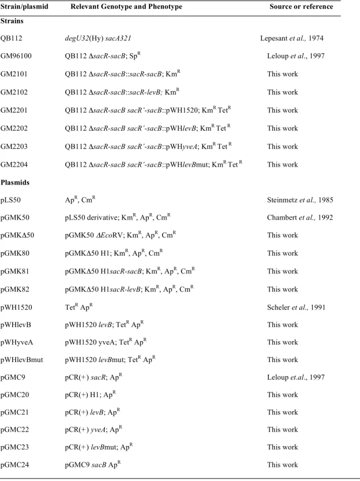
Table 1. Strains and Plasmids
Strain/plasmid Relevant Genotype and Phenotype Source or reference Strains
QB112 degU32(Hy) sacA321 Lepesant et al., 1974
GM96100 QB112 ΔsacR-sacB; SpR Leloup et al., 1997
GM2101 QB112 ΔsacR-sacB::sacR-sacB; KmR This work
GM2102 QB112 ΔsacR-sacB::sacR-levB; KmR This work
GM2201 QB112 ΔsacR-sacB sacR’-sacB::pWH1520; KmR TetR This work
GM2202 QB112 ΔsacR-sacB sacR’-sacB::pWHlevB; KmR Tet R This work
GM2203 QB112 ΔsacR-sacB sacR’-sacB::pWHyveA; KmR Tet R This work
GM2204 QB112 ΔsacR-sacB sacR’-sacB::pWHlevBmut; KmR Tet R This work
Plasmids
pLS50 ApR, CmR Steinmetz et al., 1985
pGMK50 pLS50 derivative; KmR, ApR, CmR Chambert et al., 1992
pGMKΔ50 pGMK50 ΔEcoRV; KmR, ApR, CmR This work
pGMK80 pGMKΔ50 H1; KmR, ApR, CmR This work
pGMK81 pGMKΔ50 H1sacR-sacB; KmR, ApR, CmR This work
pGMK82 pGMKΔ50 H1sacR-levB; KmR, ApR, CmR This work
pWH1520 TetR ApR Scheler et al., 1991
pWHlevB pWH1520 levB; TetR ApR This work
pWHyveA pWH1520 yveA; TetR ApR This work
pWHlevBmut pWH1520 levBmut; TetR ApR This work
pGMC9 pCR(+) sacR; ApR Leloup et.al., 1997
pGMC20 pCR(+) H1; ApR This work
pGMC21 pCR(+) levB; ApR This work
pGMC22 pCR(+) yveA; ApR This work
pGMC23 pCR(+) levBmut; ApR This work
Table 2. Oligonucleotides used in this study
Oligonucleotides (5’ – 3’)
Restriction site at the 5’ end
LS-fw: GGAGACGTCAACGATGAACATCA AatII
LS-rev: CCGCTCGAGGGAATACGGTTAGCCATTTGCCTGC XhoI
yveA-fw: GGACTAGTGCGGTATTCTCTGTTACATATTGG SpeI
yveA-rev: GGGGTACCGGCATGAGGAACACCTCC KpnI
levB-fw: GGACTAGTGCAAAAGAAAATGCCGCCGATATCC SpeI
levB-rev1: GGGGTACCCAATATGTAACAGAGAATACCGC KpnI
levB-rev2: TAGCCATTTCCCCGCCTTTATATAGTTCATAT
levBmuta: AACTATATAAAGGCGGGGAAATGGCTAACC
H1-fw: TCCCCCGGGCCATCCTCCGCTGCTGTGGCTG AvaI
H1-rev: GATGGGTTAAAAAGGATCCCTAACTGAAGGA BamHI
5S RNA probe: ACTACCATCGGCGCTGAAGA
levB-T7b: tgtaatacgactcactataggTGAATCCCATATGAACTA
levB-332rev: CCGGTAGTCCGGCTTCTG
a
Modified nucleotides are indicated in bold
Figure 1. Levan degradation by LevB.
The reaction mixtures (60 µl) contained 20 mg ml
-1[
14C]-levan in 0.1 M potassium phosphate, pH 6.
Reactions were initiated by the addition of a suspension of membranes isolated from strain GM2102
grown in the presence of 50 mM sucrose. At the indicated intervals, samples (10 µl) were removed.
14C-labelled sugars were identified by paper chromatography (a) and quantitatively estimated (b).
(○ levan), (□ levantriose), (▲ levanbiose), (● fructose).
Control (C) was achieved by incubation of labelled levan for 360 min in the presence of a membrane
suspension isolated from strain GM2102 grown in the absence of sucrose.
Figure 2.
(a) Schematic representation of the double crossing-over insertion of sacR-sacB fusion into the B. subtilis chromosome. (b) Levansucrase production by B. subtilis QB112 (●) and its derivative strain GM2101 (○).
The arrow indicates the addition of 50 mM sucrose to exponentially growing cells, at an OD600 of 0.2. Levansucrase was assayed by measuring the initial rate of the fructosyl exchange reaction (Chambert & Petit-Glatron, 1984).
Figure 3. Production of levansucrase in strains GM2201, GM2202 and GM2203
.(a) Cell suspensions of each strain grown in minimal medium were divided into equal portions at OD600 = 0.2 in flasks
containing sucrose (50 mM) and various concentrations of xylose. During exponential growth, samples of the suspensions were withdrawn at intervals.
Levansucrase production by strains GM2201 (○), GM2202 (●) and GM2203 (■) was estimated from the differential rate of levansucrase synthesis at each xylose concentration (Chambert & Petit-Glatron, 1984).
(b) SDS-PAGE analysis of the supernatant (40 µl, at OD600 = 1) of strain GM2202 grown in minimal medium in the presence of 50 mM sucrose and different concentrations of xylose. The arrow corresponds to levansucrase (50 kDa).
Figure 4. Steady state and stability analyses of sacB transcripts in strains GM2201 and 2202.
(a) Strains GM2201 and GM2202 were grown in minimal medium in the presence (+) or absence (-) of 50 mM sucrose and in the presence of 1 % (w/v) xylose added to the cultures at OD600 = 0.2. Samples of the cultures were withdrawn at OD600 = 1.5, immediately frozen in liquid nitrogen and then treated as described in Methods. RNA preparations (10 µg) were analyzed by Northern blotting. Hybridization was done with a [33P]labelled sacR probe (Pereira et al., 2001b).
(b) Stability of sacB mRNA in strains GM2201 and GM2202. Samples were withdrawn at intervals after the addition of rifampicin from cultures grown in the presence of 50 mM sucrose and 1 % xylose, treated and analyzed as described in Methods. Decay curves of sacB mRNA stability in strains GM2201 (○) and GM2202 (●) were estimated from the quantification of the Northern blot experiment. The sacB mRNA half-lives were determined using Sigma plot software. (c) Functional sacB mRNA decay was estimated by levansucrase production subsequent to rifampicin addition in strains GM2201 (○) and GM2202 (●) grown as indicated in (b).
Figure 5.
(a) Induction pattern of SacB production in strains GM2201 (○), GM2202 (●) and QB112 (□).
Cells were grown in minimal medium in the presence of sucrose at various concentrations and 1 % xylose. The differential rate of levansucrase synthesis was evaluated at each sucrose concentration as described (Chambert & Petit-Glatron, 1984).
(b) Northern blotting analysis of levB and sacB-levB transcripts in strains GM2202 and QB112.
Cells of strain GM2202 were grown in minimal medium supplemented with 50 mM sucrose in the absence (lane 1) or presence (lane 2) of 1 % xylose as indicated. Cells of strain QB112 were grown in minimal medium supplemented with 1 % xylose in the absence (lane 3) or presence (lane 4) of 50 mM sucrose. Samples (10 µg) of each RNA preparation were analyzed by Northern blotting.
Hybridization was done with the [33P]labelled levB probe, as described in Methods. Migration of the 23 S (2928 nt) and 16 S (1553 nt) is indicated by arrowheads on the right.
Figure 6.
(a) sacB transcript. The 5’ untranslated region of the transcript is shown (the start transcription is indicated) with the potential secondary structure of the Ribonucleic AntiTerminator (RAT) and the Rho independent terminator alternative structure, marked by an arrow. The start translation codon of sacB is underlined.
(b) Sequence of the 5’ coding region of levB transcript. The motif of eleven nucleotides homologous to that present in the RAT is indicated in bold.
(c) Sequence of the 5’ coding region of levB transcript in pWHlevmut. In b) and c), the start translation codon of levB is underlined.
Figure 7. Gel mobility shift assay
.For all binding reactions, the reaction mixture contained 0.1 pmol of labelled transcript (prepared as described in Methods using as DNA template pWHlevB in a, b,and c or pWHlevBmut in d), 1 µg of yeast RNA, 10 µg of BSA and 1 U of RNase inhibitor (rRNasin, Promega) in 9 µl of 1 x binding buffer (50 mM Tris-HCl, pH 7.5; 250 mM NH4Cl;1 mM EDTA; 5 % (w/v) glycerol; 0.1 % (v/v) Triton-X100). Samples were prepared by addition of (a) 1 µM of purified GST::SacY(1-55) or GST, (b) 1 µl of 5 S rRNA of various concentrations prior to the addition of 1 µM SacY (the molar excess of 5 S rRNA is indicated at the top of the lane), (c and d) 1 µl of purified GST::SacY(1-55) of various concentrations. The samples were incubated at 25 °C for 30 min and analyzed on a 5 % native polyacrylamide gel run in 1 x TBE at 4 °C.
Figure 9. Uptake of fructose and levanbiose by B. subtilis QB112 strain.
Cells were grown in YT medium (Sambrook et al., 1989). [14C]levanbiose (0.4 mM) or [14C]fructose (0.4 mM) were added to 2 ml of cell suspension at OD600 = 0.4. Aliquots (0.5 ml) were removed at intervals as indicated and centrifuged. Cell pellets were resuspended in 0.5 ml of 0.05 M sodium phosphate pH 7.0 in the presence of lysozyme (100 µg ml-1). The same volume of cell supernatant and lyzed cell suspension were submitted to paper chromatography analysis with n-butanol/acetic acid/water (4/1/1 volume by volume) as developing solvent.
Distribution of radioactivity between cells and cell supernatants: cells growing in the presence of [14C]levanbiose (a) or [14C]fructose (b). S and P indicate supernatant and pellet, respectively
Quantitative evaluation of labelled sugar remaining in culture supernatant (c): [14C]levanbiose (□); [14C]fructose (■); culture growth (○).