Supplementary material
Broth versus surface-grown cells: Differential regulation of RsmY/Z small RNAs by HptB in Pseudomonas aeruginosa by the Gac/HptB system
Fabrice Jean-Pierre1, Julien Tremblay1,2 & Eric Déziel1*
1
INRS-Institut Armand-Frappier, 531 Boul. Des Prairies, Laval, Québec, Canada
2
National Research Council Canada, Montréal, Canada
Supporting experimental procedures Congo Red assay
Tryptone (1%) agar plates were supplemented with 80 µg ml-1 Congo Red and 20 µg ml-1 Coomassie brilliant blue solidified with 0.5% agar (Bacto) were inoculated with 5 µl of bacterial suspension diluted to OD600 = 0.05 and incubated at room temperature for 7 days. Experiment
was done using three replicates on two different days. Shown is typical EPS production phenotype.
β-galactosidase assays
Activity of lacZ fusion reporters was tested for β-galactosidase activity with o-nitrophenyl-β-D-galactopyranoside (ONPG, Thermo Fisher Scientific, Nepean, ON, Canada) as substrate (Miller, 1972). Each experiment was performed using three biological replicates. Overnight TSB cultures were diluted at a starting OD600 of 0.05 in M9DCAA and incubated at 34°C. Results
were obtained for five sampling points during bacterial growth over 8 hours.
After 12 hours incubation at 34°C the cells located at the extremity of three separate tendrils of a swarming strain were collected. The tendrils were resuspended in 100 µl of 1X PBS and vortexed thoroughly. The OD600 was measured using a Nanodrop ND-1000 and adjusted to an
OD600= 0.1. β-galactosidase activity was determined as described above.
Static biofilm formation
A 500 µl bacterial suspension volume at OD600 = 0.05 was cultivated statically in a 5 mL
polystyrene tube at 34°C during various sampling times in M9DCAA medium. For analysis, the tube was vigorously rinsed by milli-Q water then incubated 10 minutes with 1 mL of 1% crystal violet at room temperature then rinsed again with milli-Q water. The colorized biofilm ring was then solubilized with 4 mL of 95% ethanol then vortexed with glass beads. OD595 was measured
using a spectrophotometer. The experiment were performed using three technical replicates at least three times.
Creation of a ΔhptBgacA mutant by lambda-red recombinase
The mutated gacA gene was amplified from the gacA::MaR2xT7 ID34781 PA14 non-redundant set with the primers listed in Table S1. The ΔhptB mutant containing the pUCP18-RedS was grown in TSB supplemented with 300 µg ml-1 carbenicillin to an OD600 = 0.5 at 37°C. Then, the
plasmid was induced for 3 hrs with 0.2% L-arabinose. After induction, the cells were washed four times with 1 ml of 10% sucrose and concentrated to a final volume of 100 µl. A concentration of 5 µg the gel-purified gacA::Mar2xT7 gene amplification was electroporated and incubated in 1 ml TSB for 2 hrs at 37°C. Transformants were selected by plating electroporated cells on TSB agar plates supplemented with 300 µg ml-1 and incubated at 37°C until colonies were visible. The selected colonies were further verified by PCR for correct insertion using the PA14_gacA_For_FJP + FJP_PA14_gacA_Rev and PA3345_Left_FWD_HindIII + PA3345_Right_REV_SmaI primer sets.
Supporting figures
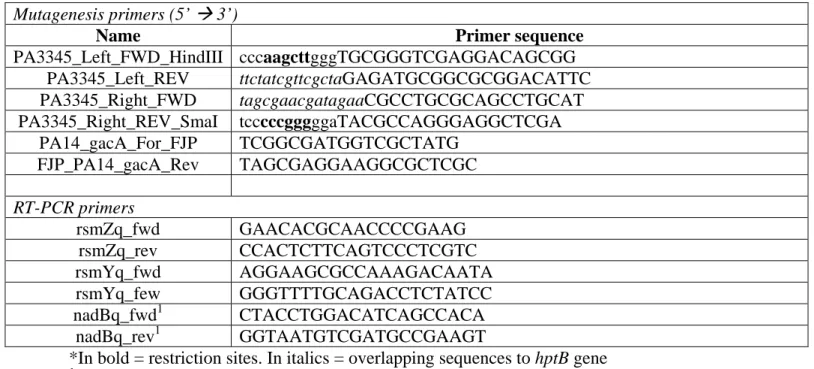
Table S1 Primers used in this study Mutagenesis primers (5’ 3’)
Name Primer sequence
PA3345_Left_FWD_HindIII cccaagcttgggTGCGGGTCGAGGACAGCGG PA3345_Left_REV ttctatcgttcgctaGAGATGCGGCGCGGACATTC PA3345_Right_FWD tagcgaacgatagaaCGCCTGCGCAGCCTGCAT PA3345_Right_REV_SmaI tcccccgggggaTACGCCAGGGAGGCTCGA PA14_gacA_For_FJP TCGGCGATGGTCGCTATG FJP_PA14_gacA_Rev TAGCGAGGAAGGCGCTCGC RT-PCR primers rsmZq_fwd GAACACGCAACCCCGAAG rsmZq_rev CCACTCTTCAGTCCCTCGTC rsmYq_fwd AGGAAGCGCCAAAGACAATA rsmYq_few GGGTTTTGCAGACCTCTATCC nadBq_fwd1 CTACCTGGACATCAGCCACA nadBq_rev1 GGTAATGTCGATGCCGAAGT
*In bold = restriction sites. In italics = overlapping sequences to hptB gene
1
Table S2 Strains used in this study
Strains/Plasmids ED # Phenotype/Genotype Reference
Strains
PA14 14 UCBPP-PA14 wild-type strain (Rahme et al., 1995)
ΔhptB 1214 Markerless hptB deletion This study
mvaT- 289 MrT7 transposition insertion mutant, GmR, ID34492
(Liberati et al., 2006) mvaU- 806 MrT7 transposition insertion mutant, GmR,
ID42058
(Liberati et al., 2006) rsmA- 282 MrT7 transposition insertion mutant, GmR,
ID44507
(Liberati et al., 2006) PA3346- 1260 MrT7 transposition insertion mutant, GmR,
ID31270
(Liberati et al., 2006) PA3347- 1261 MrT7 transposition insertion mutant, GmR,
ID39911
(Liberati et al., 2006) bswR- 2681 MrT7 transposition insertion mutant, GmR,
ID24728
(Liberati et al., 2006) Plasmids
pUCP18-RedS Arabinose-inducible recombination plasmid, CbR
(Lesic and Rahme, 2008) pCTX-rsmY-lacZ Self-proficient integration vector with lacZ
reporter for rsmY transcriptional expression
(Brencic and Lory, 2009) pCTX-rsmZ-lacZ Self-proficient integration vector with lacZ
reporter for rsmZ transcriptional expression
Supporting figures
Figure S1: Growth curve of the PA14 and ΔhptB strain in M9DCAA at 34°C. Data represents the average of three technical replicates. Error bars represent the standard deviation. Experiment was repeated at least twice.
Figure S2: Endpoint pictures of the ΔrsmY/Z mutants time-lapse analysis at different time points.
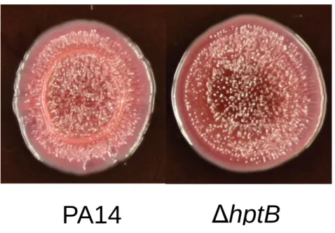
Figure S3: Exopolysaccharide fixation determined by Congo Red assay.
Figure S4: Biofilm formation assay of the PA14 and ΔhptB strains. Data represents the average of three replicates. Error bars represent the standard deviation. Experiment was repeated at least twice.
ΔhptB
PA14
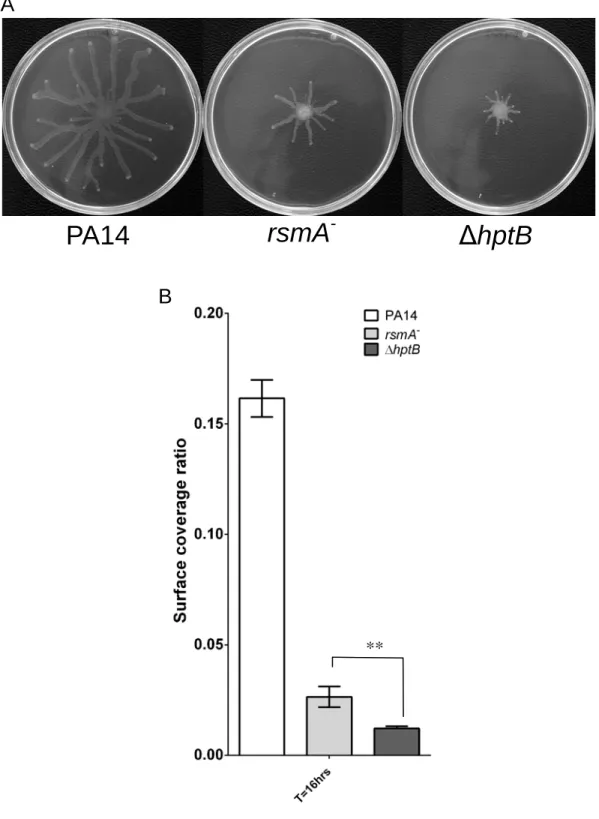
Figure S5: Various swarming phenotype. (A) Swarming motility of the PA14, rsmA- and ΔhptB strains. (B) Surface coverage of the PA14, rsmA-and ΔhptB strains. Data represents the average of three technical replicates. Error bars represent the standard deviation of the three technical replicates. Experiment was repeated at least twice. Statistical Student’s t-test analysis was based on two independent experiments (**, p < 0.01).
**
PA14
rsmA
-ΔhptB
A
Figure S6: Swarming phenotypes of the PA14, mvaT- and mvaU- strains.
Fig S7: Time-course of rsmY-lacZ and rsmZ-lacZ in various genetic backgrounds grown in M9DCAA broth over 8 hours. Data represents the average of three biological replicates. Error bars represent the standard deviation.
rsmY-lacZ
Fig S8: Time-course of rsmY-lacZ and rsmZ-lacZ in various genetic backgrounds grown as swarming colonies on M9DCAA. Data represents the average of three biological replicates. Error bars represent the standard deviation. Statistical Student’s t-test analysis was performed with ns = not significant.
rsmY-lacZ
rsmZ-lacZ
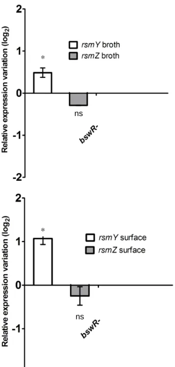
ns ns ns nsFigure S9: qRT-PCR on the ΔbswR mutant grown in M9DCAA broth and swarming conditions. Data represents the average of three biological replicates. Error bars represent the standard deviation of three biological replicates. Statistical Student’s t-test analysis was performed on two independent experiments with *, p < 0.05, ns = not significant.
References *
ns
*
Brencic, A., and Lory, S. (2009). Determination of the regulon and identification of novel mRNA targets of Pseudomonas aeruginosa RsmA. Mol Microbiol 72, 612-632. doi: 10.1111/j.1365-2958.2009.06670.x.
Brencic, A., Mcfarland, K.A., Mcmanus, H.R., Castang, S., Mogno, I., Dove, S.L., and Lory, S. (2009). The GacS/GacA signal transduction system of Pseudomonas aeruginosa acts exclusively through its control over the transcription of the RsmY and RsmZ regulatory small RNAs. Mol Microbiol 73, 434-445. doi: 10.1111/j.1365-2958.2009.06782.x.
Lesic, B., and Rahme, L.G. (2008). Use of the lambda Red recombinase system to rapidly generate mutants in Pseudomonas aeruginosa. BMC Mol Biol 9, 20. doi: 10.1186/1471-2199-9-20.
Liberati, N.T., Urbach, J.M., Miyata, S., Lee, D.G., Drenkard, E., Wu, G., Villanueva, J., Wei, T., and Ausubel, F.M. (2006). An ordered, nonredundant library of Pseudomonas aeruginosa strain PA14 transposon insertion mutants. Proc Natl Acad Sci U S A 103, 2833-2838. doi: 10.1073/pnas.0511100103.
Miller, J.H. (1972). Experiments in molecular genetics. Cold Spring Harbor, N.Y.: Cold Spring Harbor Laboratory.
Rahme, L.G., Stevens, E.J., Wolfort, S.F., Shao, J., Tompkins, R.G., and Ausubel, F.M. (1995). Common virulence factors for bacterial pathogenicity in plants and animals. Science 268, 1899-1902.
Tremblay, J., and Deziel, E. (2010). Gene expression in Pseudomonas aeruginosa swarming motility. BMC Genomics 11, 587. doi: 10.1186/1471-2164-11-587.