HAL Id: insu-03025855
https://hal-insu.archives-ouvertes.fr/insu-03025855
Submitted on 15 Dec 2020
HAL is a multi-disciplinary open access
archive for the deposit and dissemination of
sci-entific research documents, whether they are
pub-lished or not. The documents may come from
teaching and research institutions in France or
abroad, or from public or private research centers.
L’archive ouverte pluridisciplinaire HAL, est
destinée au dépôt et à la diffusion de documents
scientifiques de niveau recherche, publiés ou non,
émanant des établissements d’enseignement et de
recherche français ou étrangers, des laboratoires
publics ou privés.
eolian sediments in Gale crater, Mars: A review after six
Earth years of exploration with Curiosity
Elizabeth B. Rampe, David F. Blake, T. F. Bristow, Douglas W. Ming, D.T.
Vaniman, R. V. Morris, C. N. Achilles, S. J. Chipera, S. M. Morrison, V. M.
Tu, et al.
To cite this version:
Elizabeth B. Rampe, David F. Blake, T. F. Bristow, Douglas W. Ming, D.T. Vaniman, et al..
Min-eralogy and geochemistry of sedimentary rocks and eolian sediments in Gale crater, Mars: A review
after six Earth years of exploration with Curiosity. Chemie der Erde / Geochemistry, Elsevier, 2020,
80 (2), pp.125605. �10.1016/j.chemer.2020.125605�. �insu-03025855�
Contents lists available atScienceDirect
Geochemistry
journal homepage:www.elsevier.com/locate/chemer
Invited Review
Mineralogy and geochemistry of sedimentary rocks and eolian sediments in
Gale crater, Mars: A review after six Earth years of exploration with Curiosity
E.B. Rampe
a,e,*
, D.F. Blake
b,e, T.F. Bristow
b,e, D.W. Ming
a,e, D.T. Vaniman
c,e, R.V. Morris
a,e,
C.N. Achilles
d,e, S.J. Chipera
c,e, S.M. Morrison
e,f, V.M. Tu
e,g, A.S. Yen
e,h, N. Castle
e,i,
G.W. Downs
e,j, R.T. Downs
e,j, J.P. Grotzinger
e,k, R.M. Hazen
e,f, A.H. Treiman
e,i,
T.S. Peretyazhko
e,g, D.J. Des Marais
b,e, R.C. Walroth
b,e, P.I Craig
c,e, J.A. Crisp
e,h, B. Lafuente
e,l,
J.M. Morookian
e,h, P.C. Sarrazin
e,l, M.T. Thorpe
a,e, J.C. Bridges
e,m, L.A. Edgar
e,n, C.M. Fedo
e,o,
C. Freissinet
e,p, R. Gellert
e,q, P.R. Mahaffy
d,e, H.E. Newsom
e,r, J.R. Johnson
e,s, L.C. Kah
e,o,
K.L. Siebach
e,t, J. Schieber
e,u, V.Z. Sun
e,h, A.R. Vasavada
e,h, D. Wellington
e,v, R.C. Wiens
e,w, the
MSL Science Team
aNASA Johnson Space Center, Houston, TX, USA bNASA Ames Research Center, Moffett Field, CA, USA cPlanetary Science Institute, Tucson, AZ, USA dNASA Goddard Space Flight Center, Greenbelt, MD, USA eChesapeake Energy, Oklahoma City, OK, USA
fGeophysical Laboratory, Carnegie Institution for Science, Washington, DC, USA gJacobs Technology at NASA Johnson Space Center, Houston, TX, USA hJet Propulsion Laboratory, California Institute of Technology, Pasadena, CA, USA iThe Lunar and Planetary Institute, Houston, TX, USA
jUniversity of Arizona, Tucson, AZ, USA
kCalifornia Institute of Technology, Pasadena, CA, USA lSETI Institute, Mountain View, CA, USA
mUniversity of Leicester, Leicester, LE1 7RH, UK nUSGS Astrogeology Science Center, Flagstaff, AZ USA oUniversity of Tennessee, Knoxville, TN, USA
pLaboratoire Atmosphères, Milieux, Observations Spatiales, Guyancourt, France qUniversity of Guelph, Ontario, Canada
rUniversity of New Mexico, Albuquerque, NM, USA
sJohns Hopkins University Applied Physics Laboratory, MD, USA tRice University, Houston, TX, USA
uUniversity of Indiana, Bloomington, IN, USA vArizona State University, Tempe, AZ, USA
wLos Alamos National Laboratory, Los Alamos, NM, USA
A R T I C L E I N F O
Handling Editor: Carita Augustsson
Keywords:
Mars Mineralogy CheMin
Mars Science Laboratory
A B S T R A C T
The Mars Science Laboratory Curiosity rover arrived at Mars in August 2012 with a primary goal of character-izing the habitability of ancient and modern environments. Curiosity was sent to Gale crater to study a sequence of ∼3.5 Ga old sedimentary rocks that, based on orbital visible and near- to short-wave infrared reflectance spectra, contain secondary minerals that suggest deposition and/or alteration in liquid water. The sedimentary sequence in the lower slopes of Mount Sharp in Gale crater preserves a dramatic shift on early Mars from a relatively warm and wet climate to a cold and dry climate, based on a transition from smectite-bearing strata to sulfate-bearing strata. The rover is equipped with instruments to examine the sedimentology and identify compositional changes in the stratigraphy. The Chemistry and Mineralogy (CheMin) instrument is one of two internal laboratories on Curiosity and includes a transmission X-ray diffractometer (XRD) and X-ray fluorescence (XRF) spectrometer. CheMin measures loose sediment samples scooped from the surface and drilled rock powders, and the XRD provides quantitative mineralogy to a detection limit of ∼1 wt.% for crystalline phases.
https://doi.org/10.1016/j.chemer.2020.125605
Received 27 May 2019; Received in revised form 23 December 2019; Accepted 18 January 2020
⁎Corresponding author.
E-mail address:elizabeth.b.rampe@nasa.gov(E.B. Rampe).
Curiosity has traversed > 20 km since landing and has primarily been exploring an ancient lake environment fed
by streams and groundwater. Of the 19 drilled rock samples analyzed by CheMin as of sol 2300 (January 2019), 15 are from fluvio-lacustrine deposits that comprise the Bradbury and Murray formations. Most of these samples were drilled from units that did not have a clear mineralogical signature from orbit. Results from CheMin demonstrate an astounding diversity in the mineralogy of these rocks that signifies geochemical variations in source rocks, transportation mechanisms, and depositional and diagenetic fluids. Most detrital igneous minerals are basaltic, but the discovery in a few samples of abundant silicate minerals that usually crystallize from evolved magmas on Earth remains enigmatic. Trioctahedral smectite and magnetite at the base of the section may have formed from low-salinity pore waters with a circumneutral pH in lake sediments. A transition to dioctahedral smectite, hematite, and Ca-sulfate going up section suggests a change to more saline and oxidative aqueous conditions in the lake waters themselves and/or in diagenetic fluids. Perhaps one of the biggest mys-teries revealed by CheMin is the high abundance of X-ray amorphous materials (15–73 wt.%) in all samples drilled or scooped to date. CheMin has analyzed three modern eolian sands, which have helped constrain se-diment transport and mineral segregation across the active Bagnold Dune Field. Ancient eolian sandstones drilled from the Stimson formation differ from modern eolian sands in that they contain abundant magnetite but no olivine, suggesting that diagenetic processes led to the alteration of olivine to release Fe(II) and precipitate magnetite. Fracture-associated halos in the Stimson and the Murray formations are evidence for complex aqu-eous processes long after the streams and lakes vanished from Gale crater. The sedimentology and composition of the rocks analyzed by Curiosity demonstrate that habitable environments persisted intermittently on the surface or in the subsurface of Gale crater for perhaps more than a billion years.
1. Gale crater and the mission goals of the Mars Science Laboratory
The Mars Science Laboratory (MSL) rover Curiosity landed in Aeolis Palus on the northwestern plains of Gale crater, Mars in August 2012 and has been actively exploring its surface for more than six Earth years. Gale crater is an impact basin ∼155 km in diameter that formed ∼3.8 Ga before present during the late Noachian epoch (Thomson et al., 2011) near the Mars equator, on the dichotomy boundary be-tween the southern cratered highlands and northern lowlands (Fig. 1A). The floor of Gale crater is 4.5 km below the Mars datum (Mars average elevation), and Gale existed as a deep depression in the Mars crust (Fig. 1B) at a time when planet-wide morphological evidence suggests that water flowed on its surface (e.g.,Craddock and Maxwell, 1993; Howard et al., 2005;Irwin et al., 2005). Therefore, prior to landing it was recognized that there was a high likelihood that water was present in the crater during an early “wet and warm” (at least relatively speaking) period of Mars’ geologic history, at a time when evidence of the first oceans and signs of early life are found on Earth.
The science goals of the Mars Science Laboratory mission are to: 1) assess Mars’ biological potential, 2) characterize the geology of the landing region, 3) explore the past habitability of Mars, and 4) char-acterize the broad spectrum of surface radiation (Grotzinger et al., 2012). Since landing over six Earth years ago, the MSL Science Team has addressed each of the above goals while investigating processes that were active over much of Mars’ history, from the formation of the crust to modern eolian and atmospheric processes.
The principal goal of the MSL mission is to find evidence of “ha-bitability” on Mars. In understanding terrestrial habitability and how life originated and radiated on the early Earth, Mars serves as a valu-able surrogate; nearly all of Earth’s rocks of this age have been sub-ducted and destroyed as a result of plate tectonics and the remaining rocks from early Earth have been heated to metamorphic temperatures such that the original compositions have been altered (e.g.,Fedo, 2000; Mloszewska et al., 2012). Mars never experienced extensive plate tec-tonics (e.g.,Breuer and Spohn, 2003), and there is little evidence that its surface sediments have ever been deeply buried or extensively he-ated. The opportunity exists on Mars, and especially in Gale crater, therefore, to characterize these ancient sediments that are in much the same condition today as they were when deposited, elucidating the habitability potential of their depositional environments and perhaps even identifying evidence of early life.
Gale is among a class of craters that, after they were formed, were partially or fully filled with sediment, and over eons of time eroded,
exposing original features and any remaining sediments. Recent gravimetric analyses using Curiosity’s accelerometers, however, suggest the crater was never completely filled with sediment (Lewis et al., 2019). A key distinguishing feature of Gale is the presence of a ∼5 km-high central mound of layered sedimentary rock named Aeolis Mons (informally known as Mount Sharp, named for the American geomor-phologist Dr. Robert Sharp;Fig. 1B). Visible and near- to short-wave infrared reflectance spectra collected from orbit reveal that the lower-most slopes of Mount Sharp contain a variety of minerals that are in-dicative of water-rock interactions, and that these mineral assemblages change as a function of stratigraphic position in a succession of flat-lying, laterally extensive units (Fig. 1C;Milliken et al., 2010;Fraeman et al., 2013,2016). Phyllosilicate (i.e., layered silicate minerals com-posed of stacked sheets of silica tetrahedra and Mg-, Fe(II,III)-, and Al-O (OH) octahedra, including the groups smectite, vermiculite, illite, kaolinite, serpentine, micas, and chlorite, commonly called clay mi-nerals) spectral signatures are observed in some stratigraphic units near the base of Mount Sharp, and sulfate-bearing minerals become more prevalent in younger, stratigraphically higher sedimentary units (Milliken et al., 2010). This mineralogical transition suggests that the conditions under which the sediments were deposited changed through time. The broad mineral stratigraphy with sulfate-bearing units over-lying phyllosilicate-bearing units has been recognized in similarly aged deposits planet-wide (e.g., Wiseman et al., 2010; Grotzinger and Milliken, 2012), and this mineralogical succession may mark the be-ginning of the transition from a relatively wet and warm early Mars to a very dry and cold modern Mars (e.g.,Bibring et al., 2006; Milliken et al., 2010). The lowest visible units of Mount Sharp are ∼3.6–3.8 Ga old, based on crater counting ages and superposition relationships (Thomson et al., 2011). The presence of minerals in these strata that either form in water or as a consequence of aqueous alteration suggests that they preserve evidence of ancient aqueous environments. In ad-dition to phyllosilicate and sulfate mineral spectral signatures, the lower slope of Mount Sharp displays a prominent, local ridge (in-formally known as Vera Rubin ridge, named for the American astron-omer) characterized by strong orbital spectral signatures of the Fe(III) oxide mineral hematite (Fraeman et al., 2013,2016). The upper portion of Mount Sharp is comprised of dust-mantled cross-bedded units that unconformably overlie portions of sulfate-bearing materials and are likely eolian deposits (e.g.,Anderson and Bell, 2010).
The main goal of this paper is to provide an overview of MSL’s major mineralogical and geochemical discoveries with a focus on the miner-alogical results from the Chemistry and Mineralogy (CheMin) instru-ment. We start by describing the capabilities of Curiosity’s science
payload and methods utilized to perform the mineralogical and chemical investigations. We then summarize the geologic and geo-morphologic units and materials encountered by Curiosity during its traverse. This introduction sets the stage for a more in-depth descrip-tion of the mineralogy of samples, organized according to their age and depositional environment. These mineralogic details, in concert with geologic context and geochemical measurements, provide insights into the diversity of igneous materials on Mars, modern eolian processes, and the nature of ancient aqueous conditions in Gale crater, leading to the identification of ancient habitable martian environments. Finally, we discuss MSL’s most recent investigation of Vera Rubin ridge and look ahead to what the rover may encounter as it climbs Mount Sharp.
2. Curiosity’s science payload
Curiosity carries a suite of scientific instruments to characterize surface and near-surface rocks and sediments (Fig. 2). The elemental composition of rocks and loose sediments is measured remotely (∼1–7 m distance) using the ChemCam Laser Induced Breakdown Spectro-scopy instrument (LIBS;Maurice et al., 2012,2016;Wiens et al., 2012, 2015a, 2015b). After more than two years of nominal operations on Mars, ChemCam lost autofocus functionality for LIBS and Remote Micro-Imager (RMI) operation on sol 801. The instrument team
redesigned the flight software to regain full operational capacity and improve the instrument’s ability to focus on infinity, which allowed for long-distance RMI mosaics of the upper parts of Mount Sharp and other distant localities (Le Mouélic et al., 2015;Peret et al., 2016). ChemCam can also passively measure spectra in the visible and near-infrared wavelength range. These spectra are useful in identifying Fe-bearing minerals present in rocks and loose sediments (Johnson et al., 2015, 2016, 2017a) and they complement the multispectral imaging cap-ability of Mastcam (Bell et al., 2017; Wellington et al., 2017a). ChemCam has the ability to select autonomously targets using the Autonomous Exploration for Gathering Increased Science (AEGIS) system (Francis et al., 2017). AEGIS selects geological targets in images from the rover’s navigation cameras and measures them with ChemCam without input from ChemCam scientists and engineers on Earth. The system has markedly increased the pace of data collection with ChemCam since its implementation in May 2016, in particular im-proving the statistics on baseline bedrock compositions along the rover traverse.
The Alpha Particle X-ray Spectrometer (APXS), located on the arm of the rover, provides quantitative elemental data from ∼2 cm dia-meter areas of target rocks and soils (i.e., loose, unconsolidated surface materials) through the detection of characteristic X-rays emitted by incident alpha particles and X-rays from 244Cm sources (i.e., using
Fig. 1. A) Location of Gale crater on Mars Orbital Laser Altimeter (MOLA) elevation map (image credit: NASA/JPL/GSFC); B) Composite of Gale crater and Mount
Sharp showing Curiosity’s landing ellipse in the northwest plains in black (image credit: NASA/JPL-Caltech/ESA/DLR/FU Berlin/MSSS); and C) HiRISE mosaic of the lower slopes of Mount Sharp and the secondary mineral units identified from orbital visible and near- to short-wave infrared spectroscopy (image credit: NASA/JPL-Caltech/MSSS/JHUAPL/Brown University).
particle induced X-ray emission spectroscopy and X-ray fluorescence spectroscopy;Gellert et al., 2006;Campbell et al., 2012). Minerals and X-ray amorphous materials in scooped soil or drilled rock samples are identified and quantified by the CheMin instrument using X-ray dif-fraction (Blake et al., 2012). Evolved gas analyses from the Sample Analysis at Mars (SAM) instrument of these same samples can inform mineralogy as well as detect and identify organic molecules (Mahaffy et al., 2012). Geomorphological, structural, and sedimentological fea-tures are assessed with images of landscapes, outcrops, and rock and sediment textures obtained by the science cameras on the rover: Mastcam (Malin et al., 2017), the Mars Hand Lens Imager (MAHLI, Edgett et al., 2012), the Mars Descent Imager (MARDI, Malin et al., 2017), and the RMI on ChemCam (Maurice et al., 2012). Instruments that interrogate the modern environment include the Dynamic Albedo of Neutrons (DAN) instrument, which uses neutron spectrometry to quantify H abundances in the upper ∼1 m of the surface as a proxy for water or hydrated minerals (Litvak et al., 2008); the Rover Environ-mental Monitoring Station (REMS), which measures wind speed/di-rection, pressure, relative humidity, air and ground temperatures, and ultraviolet radiation (Gómez-Elvira et al., 2012); and the background solar and cosmic radiation detector (RAD;Hassler et al., 2012), which measures ambient particle and radiation fluxes.
Samples provided to the two laboratory instruments inside the rover (CheMin and SAM) are collected, processed and delivered by the Sample Acquisition, Sample Processing, and Handling (SA/SPaH) and Collection and Handling for In-Situ Martian Rock Analysis (CHIMRA) systems (Anderson et al., 2012). Samples can be obtained in two ways: 1) loose soil samples are scooped from the upper few cm of the surface and sieved to < 1 mm and < 150 μm grain sizes. Aliquots of the < 150 μm material are delivered to CheMin and SAM for analysis; SAM can also process the < 1 mm sieved fraction. 2) Powdered rock samples are obtained with a percussion drill. The drill bit is ∼1 cm in diameter and can penetrate to a depth of ∼6 cm. For most of the mission to date, the powdered material that reached SA/SPaH and CHIMRA traveled up the drill stem and was sourced from the lower ∼4 cm of drilled material. The samples were sieved, and aliquots were portioned and then deliv-ered to the two laboratory instruments inside the rover.
An anomaly in the drill feed mechanism occurred on sol 1536 that precluded its use. As a result, beginning with the sample “Duluth,” rocks were drilled by a new method called “feed-extended drilling,” in which the drill remains extended to its full length and is pressed into
the surface by Curiosity’s arm (essentially used like a hand drill). With feed-extended drilling, the sample can no longer pass through CHIMRA to be sieved and portioned. Drilled rock samples are now delivered to CheMin and SAM by hovering the drill bit over the instruments’ inlets and reversing the drill rotation to allow sample material to exit the drill bit and drop into the inlets. Engineers at the Jet Propulsion Laboratory (JPL) used the testbed arm and drill to constrain drill and delivery parameters so that CheMin and SAM receive sample volumes similar to those delivered during prior drill operations. The grain size of the powder produced by the drill is typically < < 150 μm (Anderson et al., 2012), and any large fragments that could potentially clog the CheMin inlet are kept from entering the instrument by a 1 mm screen in the funnel. Whether scooped or drilled, once the analyses are completed, the remaining material in the drill stem or CHIMRA is dumped onto the martian surface and the APXS instrument is used to obtain a quantita-tive elemental analysis of the bulk sample and any sieved size fractions. Dump piles are typically > 2 cm in diameter and the alignment of the centers of MAHLI images with the APXS field of view is precise within a few mm such that the dumped material fills the APXS field of view and the underlying surface does not contribute significantly to the APXS-measured chemistry (e.g.,VanBommel et al., 2016).
Data collected by the instruments aboard Curiosity can be used to characterize the elemental, molecular, isotopic, and mineralogical compositions of samples collected from the upper ∼2−6 cm of the surface, and the abundance of H in the upper meter of the surface. With this information, the MSL Science Team can infer ancient martian ha-bitability, albeit based on an Earth-centric understanding of the re-quirements of microbial life (e.g.,Hoehler, 2007).
3. The CheMin instrument and methods for quantifying mineralogy
Here, we provide a brief overview of the CheMin instrument and the methods used for calibrating the data, identifying mineralogy, quanti-fying mineral and X-ray amorphous abundances, and calculating crystal chemistry from refined unit-cell parameters. Blake et al. (2012) de-scribe the instrument, its data products, and calibration, andMorrison et al. (2018a,b) discuss the methods for calculating crystal chemistry from unit-cell parameters.
The CheMin X-ray Diffraction (XRD) / X-ray Fluorescence (XRF) instrument produces XRD patterns of scooped soil or drilled rock
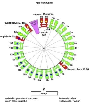
powder. Samples have been either sieved to < 150 μm or a comparable powder has been delivered directly; a ∼75 mm3aliquot of material (this volume was more tightly constrained when the drill and CHIMRA operated nominally) is delivered to the CheMin funnel, accessed through a port on the rover deck. Sieved soils and drill powders are delivered through CheMin’s inlet funnel to one of the instrument’s 27 reusable sample cells. Sample cells are arrayed in pairs on the perimeter of a sample wheel (Fig. 3). The cells are designed to be used more than once. Individual cells are filled and analyzed at the top of the wheel, then rotated 180° and emptied into a sump at the bottom of the in-strument. Each dual-cell sample holder is shaped like a tuning fork, with a sample cell attached to each arm. The cells hold the sample in a 7 mm diameter, 170 μm thick volume between two ∼7 μm-thick polymer (Mylar or Kapton) windows. A piezoelectric actuator drives the sample holder at its natural resonance and the resulting vibration causes a convective flow of sample material, randomizing grain orientations and minimizing preferred orientation effects. The instrument utilizes a transmission geometry with a microfocus Co X-ray source collimated to a 70 μm diameter X-ray beam that passes through the center of the sample cell (Fig. 4). An X-ray energy-sensitive charge-coupled device (CCD) collects two-dimensional (2D) XRD images over 3–38 hours of analysis. The CCD has a 600 × 582 pixel data collection area, where pixels are 40 × 40 μm, creating an angular resolution of ∼0.35°2θ. This angular resolution allows the refinement of plagioclase and olivine unit-cell parameters such that An and Fo values are constrained to a 1σ error of ∼2-10 (Morrison et al., 2018b). On average, samples are measured for a total of 22.5 h over three separate nights. The CCD detector is operated in single-photon counting mode and can be used to measure the amount of charge generated by each photon (and hence its energy). Diffracted CoKα X-ray photons (i.e., λ =1.79027 Å) are identified by their energy and are summed circumferentially to yield a 2D energy-discriminated CoKα diffraction pattern (Fig. 4). All detected photons are summed into a histogram that represents an XRF spectrum of the sample. Parts of the XRF system were descoped prior to launch such that the XRF data are qualitative, so quantitative compositional data from APXS and/or ChemCam are used to complement miner-alogical data from CheMin. The 2D XRD images are converted to 1D diffraction patterns (Fig. 5) for analysis using a modification of the GSE_ADA software (Dera et al., 2013). Initial calibration of the pattern is made with reference to beryl-quartz standards contained in two of five sealed standards on the CheMin sample wheel.
CheMin’s mineral detection limit is as low as ∼1 wt.%, depending on the crystallinity of the phase and the presence or absence of over-lapping peaks (Castle and Treiman, 2019). Quantitative mineral abundances and unit-cell parameters of the major phases (e.g. > ∼5 wt.%) are determined through Rietveld refinement of 1-dimensional XRD patterns. The Rietveld refinement technique (e.g.,Rietveld, 1969; Bish and Howard, 1988;Post and Bish, 1989) is used to fit the peak positions, intensities, and breadths by adjusting the scale factors, unit-cell parameters, and full-width half-maxima of peaks generated from crystallographic information files (CIFs). The CheMin team commonly uses the Jade™ software by Materials Data Inc. to perform these re-finements. Mathematical relationships between unit-cell parameters and crystal chemistry are used to determine compositions of major phases (feldspar, pyroxene, olivine, spinel, and jarosite) in each sample (Morrison et al., 2018a). Tolerance variations in the machining of the sample cell assemblies cause individual cells to be offset from their ideal diffraction position by -25 to -113 μm relative to an ideal sample cell-to-CCD distance of 18.5302 mm. To correct for this positioning offset and the resulting shift in 2-theta peak positions, an internal ca-libration method was developed that is based on the refined cell parameters of plagioclase feldspar, present as a major phase in all but one of the samples measured by CheMin (Morrison et al., 2018b).
Abundances of X-ray amorphous, poorly crystalline, and para-crystalline materials (e.g., volcanic glasses, opaline silica, smectite) are estimated using a modified version of the full pattern fitting program
FULLPAT (Chipera and Bish, 2002, 2013). FULLPAT uses a least-squares minimization to optimize the fit between measured CheMin patterns and patterns of individual minerals and X-ray amorphous phases measured on CheMin test-bed instruments. X-ray amorphous materials and phyllosilicates in the FULLPAT library include basaltic and rhyolitic glasses, opal-A, allophane, Fe-allophane, ferrihydrite, il-lite, saponite, montmorillonite, and nontronite.
The composition of X-ray amorphous materials is estimated from mass balance calculations using mineral abundances and the crystal chemistry of major phases calculated from unit-cell parameters derived from Rietveld refinements, the estimated abundance of X-ray amor-phous materials from FULLPAT, and the bulk chemical composition measured by APXS of the drill fines that best represent the sample measured by CheMin (e.g., Blake et al., 2013; Morris et al., 2013; Dehouck et al., 2014;Morrison et al., 2018b). When the drill was op-erating nominally, APXS data of fines sieved to < 150 μm were used in the calculations. In the current drill configuration, APXS data of the bulk sample dumped from the drill stem are used. Subtracting the calculated composition of all crystalline phases from the APXS bulk composition yields the composition of the X-ray amorphous component. If the abundance of the amorphous component is underestimated by FULLPAT, its calculated composition may yield negative values for some elemental oxides, which is geochemically impossible. In such cases, the amorphous abundance determined by FULLPAT is increased until the values for all elemental oxides are zero or positive. Note that minor and trace elements in minerals and elements that are present in crystalline phases below CheMin’s detection limit (∼1wt.%) are in-corporated into the calculated amorphous composition, but may not actually reside in that component of the sample. As a result, some elemental oxides, including TiO2, Cr2O3, MnO, and P2O5, may be un-realistically concentrated in the amorphous component.
The experimental data records (EDRs, i.e., the raw data) and re-duced data records (RDRs, i.e., the processed data) for CheMin dif-fraction and fluorescence data are available on the Planetary Data System. Data and results for individual samples and sample descriptions
are also compiled on an Open Data Repository (https://odr.io/CheMin), as are companion data from APXS and open-access downloadable pdfs of all publications authored by the CheMin team.
4. An overview of Curiosity’s traverse in Gale crater
Curiosity’s tenure on Mars is measured in “sols” or martian days (a sol is roughly 39 min longer than an Earth day, and a martian sidereal year is ∼668 sols or slightly less than two Earth years in length). As of sol 2300, Curiosity has driven over 20 km laterally and gained ∼400 m of elevation (Fig. 6). Because the sediments composing Mount Sharp are roughly flat lying, this corresponds to ∼400 m of stratigraphic section (Fig. 7). Geologic materials encountered by Curiosity during its traverse are almost exclusively sedimentary, and the geochemical compositions of the sedimentary units are largely consistent with low-temperature (e.g., depositional, authigenic, and diagenetic) alteration of rock and mineral fragments derived from a basaltic protolith (McLennan et al., 2014;Siebach et al., 2017;Bedford et al., 2019). However, many ex-amples of float (rocks transported away from their original source re-gion) having primary igneous textures have been identified (Sautter et al., 2014;Cousin et al., 2017a) and their compositions from APXS and ChemCam suggest magmatic diversity in and around Gale crater (Stolper et al., 2013;Sautter et al., 2014,2015;Cousin et al., 2017a). Some of this float is present as pebbles in the conglomerate facies in the Bradbury group (Williams et al., 2013;Mangold et al., 2016;Fig. 8A shows the Hottah conglomerate where Curiosity landed, exposed by the sky crane’s landing rockets). The northwest crater rim shows incision from fluvial processes, indicating the nearby crater rim was a likely source of the pebbles (Williams et al., 2013). Some of the igneous float
was probably directly emplaced onto the Gale surface as impact ejecta (Yingst et al., 2013).
Many of the igneous samples are notably feldspar-rich, mainly ex-trusive trachybasalts/trachyandesites, with some feldspathic cumulates where feldspar phenocrysts (primarily plagioclase) compose up to 80% of the rock. These igneous rocks are thought to have resulted from low pressure fractional crystallization of basaltic melt of the Adirondack Class (from the Mars Exploration Rovers) (Edwards et al., 2017). The similarity of the basaltic end member to much of the Gale sediment bulk compositions suggests that this type of Fe-rich olivine tholeiite is the dominant constituent of the Bradbury group (Edwards et al., 2017; Bedford et al., 2019). However, intriguingly, potassium and sanidine-rich sedimentary units in the Bradbury group (Anderson et al., 2015;Le Deit et al., 2016;Mangold et al., 2016;Treiman et al., 2016) and pos-sible alkaline igneous float (Stolper et al., 2013) show that alkaline igneous rocks were also a significant part of the Gale catchment (Siebach et al., 2017;Bedford et al., 2019) and were present over a larger portion of the southern highlands (Sautter et al., 2015).
The physical sedimentology of the portion of Gale crater traversed by Curiosity has been carefully documented by the MSL Sedimentology / Stratigraphy working group, who pored over thousands of images and observations to assemble the stratigraphic column shown inFig. 7. This figure does not represent a vertical section, but is more akin to a log of the lithologies encountered along the traverse. The mineralogical and geochemical data obtained by Curiosity should be viewed and inter-preted in the context of this stratigraphic record.
On the plains of Gale crater (i.e., Aeolis Palus), before climbing the slopes of lower Mount Sharp, Curiosity investigated primarily fluvial and deltaic deposits (Fig. 8) that compose the Bradbury group (e.g., Grotzinger et al., 2015). The observation of coarse-grained sedimentary deposits, including conglomerate, grain-supported sandstone, and pebbly sandstone with common cross-stratification is consistent with deposition in a fluvial environment (e.g., Williams et al., 2013; Vasavada et al., 2014;Grotzinger et al., 2015;Edgar et al., 2018a). The cobble-sized clasts in the conglomerate identified near the landing site (Fig. 8A) suggest that the ancient rivers that transported these sedi-ments were up to 0.9 m deep with an average velocity of 0.20–0.75 m/s (Williams et al., 2013).
The lower slopes of Mount Sharp, located southwest of the landing site, were the ultimate goal for Curiosity. Gale crater is what is termed a “go-to” site in the planetary science community, meaning that the principal science target of the MSL mission was actually outside its 6 × 20 km landing ellipse. However, it was apparent upon landing that a region of high scientific interest where there was a contact between three geomorphological terrain types was nearby. Therefore, from the landing site Curiosity drove east to investigate the Yellowknife Bay formation prior to driving southwest toward the lower slopes of Mount Sharp. At Yellowknife Bay, Curiosity investigated both sandstone and mudstone facies (e.g.,Grotzinger et al., 2014;Edgar et al., 2018a). The Sheepbed mudstone in the Yellowknife Bay formation (Fig. 8B) is in-terpreted to represent an ancient lake (Grotzinger et al., 2014) and is located at the lowest elevation (and stratigraphic level) investigated by Curiosity. If we assume that the strata in Gale crater are flat lying and laterally extensive, the Sheepbed mudstone is the oldest deposit that Curiosity investigated. Two samples were drilled from the Sheepbed mudstone in Yellowknife Bay, named “John Klein” and “Cumberland.” These samples were drilled a few meters apart laterally and ∼10 cm apart vertically, allowing the mission to investigate local-scale miner-alogical and geochemical variations in an area with significant evidence for early-stage diagenesis (Grotzinger et al., 2014;Léveillé et al., 2014; Siebach et al., 2014;Stack et al., 2014).
Sandstone beds that dip gently to the south (i.e., toward Mount Sharp) were identified in multiple locations along the 8 km traverse across Aeolis Palus from Yellowknife Bay and are consistent with an-cient fluvio-deltaic deposits (Fig. 8C,Grotzinger et al., 2015). Deltas form when rivers flow into standing bodies of water and deposit
Fig. 4. Illustration of the basic components of the CheMin instrument and the
path of the X-rays. FromBlake et al. (2012).
Fig. 5. CheMin 1D XRD pattern of the “Marimba” (MB) sample from the
Karasburg member of the Murray formation on Mount Sharp. “Marimba” con-tains the highest proportion of phyllosilicate of any sample drilled to date (28 wt.%). The (001) and (02l) phyllosilicate peaks are labeled. The X-ray amor-phous background is outlined in black.
sediment that was transported by the rivers. The dip of distinct clino-forms (i.e., layered deltaic bodies) indicates that the rivers in the region of Curiosity’s traverse flowed from north to south, possibly emanating from ice/snow melt in the northern crater rim (Grotzinger et al., 2015). The sample named “Windjana” was drilled from reworked deltaic and eolian sediments in the Kimberley formation (Rice et al., 2017).
Curiosity first reached the units that make up lower Mount Sharp in September 2014. This marked the beginning of the MSL Science Team’s investigation of the Murray formation, which has continued up to the present time. The Murray formation has been divided into several dis-tinct stratigraphic members based on minor changes in facies associa-tions (Fig. 7). The outcrops that compose the lowermost portion of the Murray formation (the Pahrump Hills member) are dominated by la-minated mudstone with mm-to-cm-scale lamination (Fig. 9A,B). Lami-nated mudstone is common throughout the Murray formation and is consistent with deposition in lakes (e.g.,Fedo et al., 2018;Stack et al., 2018), where thick laminations suggest near-shore deposition in shallow water and thin laminations suggest offshore deposition in deep water (Grotzinger et al., 2015). Sedimentary rock in the Pahrump Hills member exhibits exceptional preservation of original depositional fab-rics as well as products of early and late diagenesis. Above the Pahrump Hills member, the Murray formation contains a combination of lami-nated mudstone, meter-scale trough cross-bedding, ripple cross-lami-nated sandstone (Fig. 9C), and dm-scale cross-stratified sandstone (Fedo et al., 2018). Cross-stratification suggests deposition in higher-energy environments, and the cross-stratified units observed in the Murray
formation are interpreted to have been deposited in both subaqueous (i.e., fluvial) and subaerial (i.e., eolian) environments. Uncommon de-siccation cracks (Fig. 9D) suggest drying of sediments and intermittent exposure during lake lowstands (Stein et al., 2018). Isolated observa-tions of NaCl-rich targets (Thomas et al., 2019), rare boron detections (Gasda et al., 2017), and a Mg-sulfate- and Ca-sulfate-rich interval from ChemCam (Rapin et al., 2019) also hint at evaporation. No chloride-rich strata have been found, however, suggesting that desiccation was relatively infrequent when these sediments were deposited.
Twelve samples have been drilled from the Murray formation (Table 1,Fig. 7): “Confidence Hills,” “Mojave2,” “Telegraph Peak,” and “Buckskin” from the Pahrump Hills member, “Oudam” from the Hart-mann’s Valley member, “Marimba” and “Quela” from the Karasburg member, “Sebina” from the Sutton Island member, “Duluth” from the Blunts Point member, “Stoer” from the Pettegrove Point member, and “Highfield” and “Rock Hall” from the Jura member.
The deposits observed in the Bradbury group and in the Murray formation indicate that Gale crater was the site of a long-lived fluvio-laucustrine environment at ∼3.5 Ga (Fig. 10; e.g.,Grotzinger et al., 2015;Fedo et al., 2018). Rivers and streams originating from the crater rim flowed into lakes on the crater floor. The paucity of mud cracks suggests lake waters were long-lived. Based on sediment deposition rates on Earth, the 400+ m of primarily lacustrine stratigraphy that Curiosity has investigated so far represents a few hundreds of thousands to a few millions of years of time (Grotzinger et al., 2015). After de-position, fluids moved through the sediments both before and after they
Fig. 6. Curiosity’s traverse through Sol 2300 with major waypoints listed. Locations and target names along the traverse are unofficial designations and are selected
based on themes for each map quadrant. Each quadrant’s theme is a location of geologic interest on Earth. For example, there are locations and target names selected from localities in the Northwest Territories in Canada, the Kimberley province in Australia, Namibia, and Scotland. Image credit: NASA/JPL-Caltech/Univ. of AZ/ MSSS/USGS.
were lithified (e.g.,Martin et al., 2017), suggesting a prolonged history of aqueous diagenesis in surface and subsurface environments at Gale crater, as will be explored in detail later.
In addition to ancient fluvio-lacustrine deposits, Curiosity has in-vestigated modern and ancient eolian deposits. Curiosity sampled a re-cently inactive eolian bedform called “Rocknest” (Fig. 11A) close to the landing site (e.g.,Blake et al., 2013). Curiosity also executed a two-phase sampling campaign in an active eolian basaltic dune field in early 2016 and early 2017 (e.g.,Bridges and Ehlmann, 2018;Lapotre and Rampe, 2018). Curiosity’s investigation of the Bagnold Dune Field was the first in-situ study of active wind-blown sediments on another pla-netary surface. Two samples were collected to study sediment transport and sorting across the dune field. The “Gobabeb” sample was collected on sol 1225 from a ripple crest on the Namib Dune, a barchanoidal dune on the northern margin and trailing edge of the dune field (Fig. 11B). The “Ogunquit Beach” sample was collected on sol 1650 from a ripple trough of the Mount Desert Island sand patch near the southern edge of the dune field (Fig. 11C).
The Stimson formation, interpreted as an ancient lithified eolian dune field (Banham et al., 2018), is part of the Mount Sharp group and
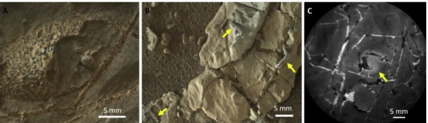
unconformably overlies the Murray formation (Fig. 12). An un-conformity occurs when there is a cessation of sediment deposition, usually with erosion of preexisting sediment prior to continued sedi-ment influx. On Earth, unconformities often occur as a result of tectonic uplift or sea level retreat. Since there is no tectonic uplift in Gale crater and no evidence for lake water breaching the crater rim, this must re-present a major change in the climate and depositional regime, in-cluding complete drying out of the lake and erosion and removal of a significant portion of the Murray and Bradbury formation sedimentary rocks that had formed in Gale crater. The hiatus in time between the deposition of the Murray and Stimson formations has not been quan-tified, but it was likely significant because the stratigraphic boundary is abrupt and transitional deposits in relatively wet to relatively dry conditions are not preserved (Banham et al., 2018). Light-toned frac-ture-associated halos in the Stimson and nearby Murray suggest leaching by late-stage fluids after burial (Frydenvang et al., 2017;Yen et al., 2017; Hausrath et al., 2018). Two of these fracture-associated halos were studied in detail. Samples were drilled from both in the light-toned fracture-associated halos (“Greenhorn” and “Lubango”) and in the nearby parent sandstone (“Big Sky” and “Okoruso”) to constrain the geochemical processes that formed the halos.Fig. 13 shows an example of a fracture-associated halo observed prior to sampling at a location called Teakettle Junction.
5. Mineralogy of samples measured by CheMin
The characterization of the mineralogy and geochemistry of samples from a variety of different depositional environments and time periods provides insight into past and present geologic processes on Mars. All samples measured to date contain igneous minerals, secondary phases, and X-ray amorphous materials. Here we summarize the abundances of minerals and amorphous components, crystal chemistry of major phases from refined unit-cell parameters, and calculated composition of amorphous components from all samples analyzed through sol 2300, the limit of reportable analyses at the time of this writing. Results are grouped according to their depositional environment: Fluvio-lacustrine, ancient eolian, and modern eolian.
5.1. Fluvio-lacustrine samples
Most of the rock samples drilled to date were obtained from ancient fluvio-lacustrine deposits (Table 1). Of the 19 rock samples analyzed by CheMin, 13 are mudstones interpreted to be lacustrine deposits and two are fine-grained sandstones interpreted to be reworked fluvial/eolian deposits. The mineralogy of fluvio-lacustrine rocks can tell us about the composition of rocks in the source regions of the sediments, the weathering history of the sediments, and the fluid chemistry of syn- and post-depositional aqueous environments. CheMin has recorded en-ormous mineralogical variability between fluvio-lacustrine samples that suggests differences in igneous sources, paleoclimatic conditions, and geochemistry of aqueous environments. The mineral abundances of fluvio-lacustrine samples are presented inTable 2, the crystal chemistry of the major minerals inTables 3 and 4, the abundances of phyllosili-cates and X-ray amorphous materials inTable 5, and the calculated composition of the X-ray amorphous materials inTable 6. These results have been reported previously byVaniman et al. (2014);Treiman et al. (2016);Rampe et al. (2017);Yen et al. (2017);Bristow et al. (2018); Morrison et al. (2018b), Achilles et al. (submitted), andRampe et al. (2020). Here, we provide an overview of the minerals identified in fluvio-lacustrine samples and summarize the mineralogy of each unit moving up section.
The mineralogy of fluvio-lacustrine samples is variable within the stratigraphic section. Samples contain varying abundances of plagio-clase, felsic igneous minerals, mafic igneous minerals, Fe-oxide mi-nerals, phyllosilicates, sulfate mimi-nerals, the phosphate fluorapatite, halite, and X-ray amorphous materials (Fig. 14). Felsic minerals include
Fig. 7. Stratigraphic column showing the sedimentary units that Curiosity has
investigated through January 2019. Note that the Vera Rubin ridge topography does not follow elevation. The western leg of Curiosity’s traverse up the Vera Rubin ridge showed lower elevations for the Pettegrove Point and Jura mem-bers than the eastern leg of Curiosity’s traverse down and back up the Ridge. Credit: Sed/strat group of the MSL Science Team.
alkali feldspar (variety sanidine), tridymite, cristobalite, and trace amounts of quartz. Mafic igneous minerals identified include pyroxene and olivine. The CheMin team has reported on pyroxene compositions previously (Blake et al., 2013;Bish et al., 2013;Vaniman et al., 2014; Treiman et al., 2016;Rampe et al., 2017;Bristow et al., 2018;Morrison et al., 2018b;Rampe et al., 2018). Overlapping peaks of pyroxenes in CheMin X-ray diffraction patterns and the low angular resolution of the instrument, however, preclude confident identification of pyroxene phases or crystal chemistry. We report the published pyroxene subtype compositions for several samples inTable 3, but caution that there is a great deal of uncertainty associated with them and they should not be used for detailed petrologic interpretations.
The most common Fe-oxide minerals include hematite and magne-tite, but the Fe-oxyhydroxide mineral, akaganeite [β-FeO(OH,Cl)], has been identified in the Bradbury group on the plains of Gale crater (Vaniman et al., 2014;Treiman et al., 2016) and very recently from the Pettegrove Point and Jura members of the Murray formation (Morris et al., 2019;Rampe et al., 2020). For samples that contain at least minor amounts of magnetite, the refined magnetite unit-cell parameters are smaller than those of stoichiometric magnetite, suggesting partial oxi-dation of the Fe(II) toward the Fe(III) defect-spinel maghemite. Alter-natively, substitution of smaller cations such as Cr3+, Mg2+, or Al3+ could account for a smaller unit cell, although it is not clear whether sufficient amounts of these cations are present (Morrison et al., 2018a, b).
Phyllosilicates are readily identified in CheMin diffraction data by the presence and position of (001) peaks (Fig. 15), which are a measure of interlayer spacing along the c-axis of the crystal. Most patterns show a d(001) of ∼10 Å, which is consistent with a collapsed (i.e., dehy-drated) swelling clay mineral (e.g., smectite) or illite. One sample from Yellowknife Bay, named “Cumberland,” has a d(001) up to ∼13.5 Å that may indicate intercalated metal-hydroxyl groups in the smectite
interlayer site (Bristow et al., 2015). The low abundance of potassium in most samples suggests little, if any, illite is present (Bristow et al., 2015, 2018). The detection of collapsed smectite confirms the hy-pothesis that smectite on the martian surface is dehydrated based on orbital spectral data (e.g., Wray et al., 2009; Che et al., 2011). The position of the (02l) diffraction band, which is a measure of the b-axis of the crystal (Fig. 15), suggests a change from trioctahedral smectite to smectite with more dioctahedral character moving up section (Bristow et al., 2015,2018). SAM evolved gas data show complementary evi-dence for changes in smectite structure and composition within the stratigraphic section based on the temperature of structural H2O release (Ming et al., 2014;Bristow et al., 2018). Phyllosilicates in the samples collected from the Hartmann’s Valley member and the members that compose Vera Rubin ridge have d(001) peaks at ∼9.6 Å, consistent with a completely collapsed smectite or Fe-pyrophyllite (Bristow et al., 2018;Rampe et al., 2020).
The Ca-sulfate minerals anhydrite, bassanite, and gypsum are common (Vaniman et al., 2014,2018), especially up section, and jar-osite is present in minor to trace amounts in many of the samples col-lected from the Murray formation on the lower slopes of Mount Sharp (Rampe et al., 2017; Bristow et al., 2018; Achilles et al., submitted, Rampe et al., 2020). Minor amounts of fluorapatite were identified in some samples from the Pahrump Hills member at the base of the Murray formation, and trace amounts of halite were identified in one sample from the Karasburg member in the Murray formation (“Quela”). X-ray amorphous materials are abundant (∼20–60 wt.%) in all fluvio-lacustrine samples. X-ray amorphous materials are identified in CheMin diffraction patterns from the presence of a broad hump in the background centered between ∼22–26°2θ and scattering at low angles (Fig. 5). The identity of the X-ray amorphous materials is difficult to ascertain from XRD data alone because they generally do not have distinct XRD patterns. The relative position of the X-ray amorphous
Fig. 8. Images of outcrops on the plains of Gale crater. A) “Link” conglomerate near Curiosity’s landing site (sol 27, sequence mcam00129). B) Sheepbed mudstone at
hump can be used to discern relative SiO2abundances in amorphous silicates, where high-SiO2amorphous silicates (e.g., opal-A, rhyolitic glass) have a hump centered at lower angles, whereas amorphous sili-cates with lower SiO2 (e.g., basaltic glass) have a hump centered at higher angles. This trend should not be used, however, to characterize the amorphous component when multiple amorphous materials are present in the same sample, as is the case for samples from Gale crater. XRD patterns of amorphous sulfate, for example, look very similar to amorphous silicates (Morris et al., 2015).
Calculations of the composition of the X-ray amorphous component using CheMin mineral abundances and APXS bulk chemistry of the
post-sieve or bulk powder dump piles demonstrate a large variation in compositions between samples (Table 6). There are many sources of error for the calculated amorphous component (e.g., abundance of crystalline phases, abundance of amorphous material, calculated crystal chemistry of major phases, assumed composition of phyllosilicates), so the calculated amorphous component likely represents the amorphous composition plus the residual error of the calculated composition based on the model mineralogy and mineral chemistry determined by Riet-veld refinement. The aliquot of sample delivered to CheMin comes from the same material as the post-sieve or bulk powder dump piles, so the bulk APXS compositions of these dump piles are likely similar to those
Fig. 9. Images of outcrops from the Murray formation. Thickly (A) and thinly (B) laminated mudstone deposits from the Pahrump Hills (sol 712, mcam03030; sol
792, mcam03445). C) Ripple cross-lamination in the Sutton Island member (sol 1477, mcam07413). D) Desiccation cracks in the “Old Soaker” target (sol 1555, mcam07981). Image credits: NASA/JPL-Caltech/MSSS.
of the samples inside CheMin. Therefore, differences between the composition of the CheMin-analyzed sample and the APXS-analyzed sample are unlikely to be significant sources of error.
Despite some sources of error for these calculations, the results can be used to examine relative elemental enrichments and depletions be-tween samples (e.g., Dehouck et al., 2014; Rampe et al., 2017; Yen et al., 2017). The amorphous component in all samples contains SO3, FeOT, and SiO2, suggesting amorphous sulfates, nanophase Fe-oxides, and silicates are present in all samples. The amorphous component in many samples contains abundant FeOT(e.g., “Windjana,” “Marimba,” and “Duluth”), suggesting the presence of nanophase Fe-oxides and/or amorphous Fe-sulfates. The amorphous component in some samples is relatively enriched in SiO2(e.g., “Buckskin” and “Oudam”), suggesting the presence of opal-A and/or rhyolitic glass. Aluminum abundances
are relatively low in all samples, suggesting amorphous aluminosili-cates like allophane and maskelynite do not make up a significant portion of the amorphous component. None of the calculated compo-sitions of amorphous material is consistent with pure basaltic glass. Although some basaltic glass could be present, low amounts of SiO2, Al2O3, MgO, and/or CaO in all samples preclude abundant basaltic glass. This finding is consistent with Mössbauer data collected by the Mars Exploration Rovers in Gusev crater and Meridiani Planum, which demonstrate that Fe is not present in basaltic glass at either landing site (Morris et al., 2006,2008).
The dominant oxides present in the amorphous component in each sample could provide further information about aqueous conditions. High concentrations of FeOTsuggest the presence of an amorphous Fe phase, like ferrihydrite. Ferrihydrite-like materials form by rapid hy-drolysis of Fe3+in solution (e.g., Schwertmann and Cornell, 2000). Aqueous Fe3+can be mobilized at low pH, or it could form by rapid oxidation and hydrolysis of Fe2+in solution that was sourced from the alteration of mafic igneous minerals. Elevated FeOT and SO3 in the amorphous component could indicate the presence of amorphous Fe-sulfate, which would implicate acidic conditions. Abundant SiO2in the amorphous component suggests the presence of opaline silica. There are no diffraction properties to refine the identification of opal-A, but in some samples, opal-CT has been identified and based on characteristic diffraction peaks in this paracrystalline material (Table 5). Opaline si-lica can form in a variety of environments, and is an expected alteration product of basaltic sediments because Ca-rich plagioclase and mafic igneous silicates are more susceptible to dissolution than felsic minerals (e.g.,McLennan, 2003). The presence of abundant amorphous silica may signify aqueous alteration and dissolution of basaltic minerals or it may be a primary volcanic glass in the case of the “Buckskin” sample. The abundance of X-ray amorphous materials in the ancient rock samples is especially perplexing because amorphous materials in rocks on Earth mature into crystalline phases over time. Opaline silica, for example, transforms to quartz through a dissolution-precipitation re-action in early diagenetic environments (e.g.,Kastner et al., 1977). On Earth, amorphous silica is not usually found in rocks over a few million years old and is absent in rocks over ∼145 Ma old (e.g.,Tosca and Knoll, 2009). Compositionally complex amorphous materials, similar to those identified in Gale crater, have been identified in modern sub-glacial sediments formed from basaltic andesite and andesite sources in the Three Sisters Volcanic Complex in Oregon (Smith et al., 2018). These materials are enriched in SiO2(∼50−60 wt.%), Al2O3(∼15 wt.
Table 1
Information about samples analyzed by CheMin.
Sample
(abbreviation) Sol(s)Collected Elevation (m) DepositionalEnvironment Rocknest (RN) 61, 66, 69, 74,
93 −4516.9 Modern eolian(inactive)
John Klein (JK) 182 −4519.5 Lacustrine
Cumberland (CB) 279 −4519.5 Lacustrine
Windjana (WJ) 621 −4481.5 Reworked eolian and
fluvial Confidence Hills
(CH) 759 −4460.3 Lacustrine
Mojave2 (MJ) 882 −4459.4 Lacustrine
Telegraph Peak (TP) 908 −4453.2 Lacustrine
Buckskin (BK) 1060 −4446.8 Lacustrine
Big Sky (BS) 1119 −4434.7 Ancient eolian
Greenhorn (GH) 1137 −4434.5 Ancient eolian (halo) Gobabeb (GB) 1224 −4423.8 Modern eolian (active) Lubango (LB) 1320 −4429.0 Ancient eolian (halo)
Okoruso (OK) 1332 −4429.3 Ancient eolian
Oudam (OU) 1361 −4435.5 Reworked eolian and
fluvial
Marimba (MB) 1422 −4410.4 Lacustrine
Quela (QL) 1464 −4379.7 Lacustrine
Sebina (SB) 1495 −4360.8 Lacustrine
Ogunquit Beach (OG) 1651 −4300.0 Modern eolian (active)
Duluth (DU) 2057 −4192.5 Lacustrine
Stoer (ST) 2136 −4169.9 Lacustrine
Highfield (HF) 2223 −4147.0 Lacustrine
Rock Hall (RH) 2261 −4143.8 Lacustrine
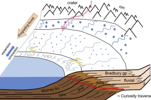
Fig. 10. Schematic showing the depositional
environ-ments preserved by the Bradbury Group and Murray formation. The Bradbury group is dominated by fluvio-deltaic deposits as rivers and streams emanating from the rim delivered sediments across the plains and into lakes on the crater floor. The Murray formation is dominated by lacustrine deposits. Desiccation features and salts would have precipitated along the lake margin. Curiosity’s traverse is drawn in red, showing the beginning of the traverse in fluvio-deltaic deposits then transitioning to lacustrine and lake-margin de-posits. Credit: C. Fedo. (For interpretation of the re-ferences to colour in this figure legend, the reader is referred to the web version of this article.).
%), and FeOT(∼15 wt.%) and have lesser amounts of CaO, MgO, Na2O, and K2O. Transmission electron microscopy of the sediments indicates that much of the amorphous component is secondary in nature, sug-gesting it formed from water-rock interactions at relatively low tem-peratures. Although these materials have more Al2O3and less SO3than amorphous materials in Gale crater, the formation of compositionally heterogeneous amorphous materials in cold and icy environments may explain some portion of the amorphous component in Gale crater. Their preservation for perhaps billions of years, however, suggests ground-water was intermittent and was not present in sufficient volumes to cause the maturation of amorphous materials to crystalline phases.
5.2. Modern eolian samples
The mineralogy of modern eolian sediments can be used to evaluate the extent of sorting and grain segregation in a single bedform and across dune fields, the relative contribution of underlying bedrock to the sediment, and the composition of modern sediment sources. The in-situ measurements by CheMin also allow us to ground truth orbital remote sensing measurements and clarify differences between the in-situ and orbital measurements. CheMin has analyzed three modern eolian sediments. “Rocknest” was scooped from an inactive sand shadow near the landing site, whereas “Gobabeb” and “Ogunquit
Fig. 11. Curiosity’s sampling sites of modern eolian materials. A) Curiosity selfie taken by MAHLI showing scoops from “Rocknest” (RN) sand shadow (sol 84).
Curiosity’s selfies are a mosaic of dozens of MAHLI images. B) Front Hazcam image showing wheel scuff and scoops of “Gobabeb” (GB) from Namib Dune (sol 1229,
fhaz00323). C) Meter-scale ripples at Mount Desert Island sand patch, from which “Ogunquit Beach” (OG) was scooped (sol 1647, mcam08526). Images credit: NASA/JPL-Caltech/MSSS.
Fig. 12. Unconformable contact between the Murray and Stimson formations at the Marias Pass location at the top of the Pahrump Hills member (sol 992,
Beach” were scooped from different locations in the active Bagnold Dune Field. “Gobabeb” was scooped from a barchanoid dune named Namib Dune near the trailing edge of the dune field and “Ogunquit Beach” from a ripple field downwind of Namib Dune. All three samples have similar mineralogy and are dominated by basaltic igneous mi-nerals and X-ray amorphous materials with minor amounts of Fe-oxide minerals (magnetite and hematite are present in all samples and ilme-nite is present in “Rocknest”), anhydrite, and quartz (Table 7;Fig. 16). Mineralogical results from the modern eolian samples are reported by Blake et al. (2013);Bish et al. (2013);Achilles et al. (2017);Morrison et al. (2018b), andRampe et al. (2018).
The basaltic igneous minerals present in each sample include pla-gioclase, pyroxene, and olivine. Plagioclase constitutes 38–47 wt.% of the crystalline fraction, and the calculated plagioclase compositions (Table 8) from the refined unit-cell parameters for each sample are An49(4), An63(5), and An48(5) for Rocknest, Gobabeb, and Ogunquit Beach, respectively. Each sample has ∼26–33 wt.% pyroxene. The
presence of both augite and pigeonite were reported previously (Bish et al., 2013;Blake et al., 2013;Achilles et al., 2017;Morrison et al., 2018b;Rampe et al., 2018) but, again, we caution that the low angular resolution of the CheMin instrument and overlapping pyroxene peaks reduces our confidence in these identifications and their calculated crystal compositions from refined unit-cell parameters. Olivine is pre-sent in abundances of 18–26 wt.% and the olivine composition is the same between all samples within a 1σ error (Fo54-60).
The major constituents of the calculated amorphous components are SiO2, FeOT, Al2O3, and SO3(Table 9). Thus, like the amorphous mate-rials in fluvio-lacustrine samples, they are not consistent with any one single X-ray amorphous material. When compared to an Adirondack basalt composition in Gusev crater, basaltic glass is inconsistent with the calculated amorphous compositions. The Rocknest amorphous component is too depleted in SiO2, the Gobabeb amorphous component is too enriched in SiO2, and the amorphous components from all modern eolian samples are too depleted in Al2O3, MgO, and CaO and
Fig. 13. Mastcam mosaic of a fracture-associated halo observed at Teakettle Junction (sol 747, mcam03215). Image credit: NASA/JPL-Caltech/MSSS.
Table 2
Mineral abundances (in wt.%, renormalized to 100% crystalline) from CheMin data of fluvio-lacustrine samples. Plag = plagioclase, Kspar = potassium feldspar, Px = pyroxene, Oliv = olivine, Qtz = quartz, Crist = cristobalite, Trid = tridymite, Magn = magnetite, Hem = hematite, Akag = akaganeite, F-ap = fluorapatite, Anhy = anhydrite, Bass = bassanite, Gyps = gypsum, Jaro = jarosite, Hal = halite. Uncertainties in parentheses are 1σ. Abundances and errors have been rounded to the nearest whole number. The symbol “–” indicates that phase is at or below the detection limit of CheMin.
JK1 CB1 WJ1 CH1 MJ1 TP1 BK1 OU2 MB2 QL2 SB2 DU3 ST3 HF3 RH3 Plag 44(1) 43(1) 6(1) 38(2) 55(2) 44(2) 43(2) 52(1) 44(3) 44(3) 38(2) 56(1) 45(2) 47(2) 38(4) Kspar 4(1) 5(1) 26(1) 9(1) – 8(2) 8(1) – 8(2) 7(1) 4(1) 7(1) 3(3) 3(1) – Px 26(2) 31(2) 42(3) 26(7) 16(2) 13(3) – 8(2) 2(2) 7(2) 7(4) 10(3) 6(2) 10(3) 17(2) Oliv 8(0) 3(1) 7(2) 2(1) 1(1) 2(1) – – – – – – – – – Qtz – – – 1(1) 2(0) 1(0) – 2(0) 2(1) 1(1) 1(0) 2(1) 1(1) 1(1) – Crist – – – – – 12(2) 6(0) – – – – – – – – Trid – – – – – – 34(1) – – – – – – – – Magn 9(1) 11(0) 16(1) 6(1) 7(1) 13(1) 7(0) – – – – 2(1) 1(1) 1(1) – Hem 1(0) 1(0) 2(1) 13(1) 7(1) 2(0) – 26(1) 20(1) 23(2) 24(2) 13(2) 28(1) 20(1) 5(0) Akag 1(0) 2(1) 1(1) – – – – – – – – – 2(1) – 11(1) F-ap – – – 2(1) 4(1) 3(0) – – – – – – – – 3(1) Anhy 4(0) 1(0) 1(0) – – – 2(0) 6(1) 12(1) 9(1) 17(1) 3(1) 6(1) 8(1) 21(3) Bass 3(0) 2(0) 1(0) – – – – – 4(1) 6(0) 4(0) 7(1) 1(0) 3(1) – Gyps – – – – – – – 6(0) 7(1) 1(1) 4(0) – 4(1) 5(1) – Jaro – – – 2(1) 8(2) 2(2) – – 2(1) 1(1) 3(1) – 2(1) – 4(1) Hal – – – – – – – – – 1(0) – – – – –
1FromMorrison et al. (2018b). 2From Achilles et al. (submitted). 3FromRampe et al. (2020).
are too enriched in SO3. This composition suggests that multiple X-ray amorphous materials constitute the X-ray amorphous component, in-cluding volcanic and/or impact glass, nanophase Fe-oxides, and amorphous sulfates. There are clear differences in the compositions of
the amorphous components in active eolian samples (“Gobabeb” and “Ogunquit Beach”) and in the inactive eolian sample (“Rocknest”). The amorphous component of the active samples is more enriched in SiO2, whereas the amorphous component of the inactive sample is more en-riched in SO3and Cl. The composition of dust on Curiosity’s observation tray as measured by APXS is enriched in SO3 and Cl (Berger et al.,
2016), so this difference in composition between the active and inactive eolian samples is a result of higher dust contents in the inactive sedi-ments (Achilles et al., 2017).
5.3. Ancient eolian samples
Curiosity drilled two pairs of samples from the Stimson formation, a lithified eolian dune field that unconformably overlies the Murray formation. These samples were selected because of their association with light-toned alteration halos parallel to fractures in the rock (Fig. 13). One sample of each pair was collected from parent rock and the second from within the alteration halo: “Big Sky” (parent rock) was drilled 2 m from “Greenhorn” (alteration halo), and “Okoruso” (parent rock) was drilled 3 m from “Lubango” (alteration halo), and the “Okoruso”/“Lubango” pair was collected ∼650 m southwest of “Big Sky”/“Greenhorn.” APXS and ChemCam measurements were also made in parent rock and in the alteration halos to identify geochemical trends associated with the processes that formed the halos. The mineralogical and geochemical trends observed by Curiosity are reported in detail by Yen et al. (2017).
CheMin analyses of the parent eolian sandstone samples (“Big Sky” and “Okoruso”) show that they are dominated by igneous minerals, magnetite, and X-ray amorphous materials (Table 10). The parent sandstone contains abundant plagioclase feldspar and pyroxene (mod-eled as pigeonite and orthopyroxene; Table 11) and lacks olivine. Magnetite constitutes ∼15 wt.% of the crystalline component, and it may have formed diagenetically from the dissolution of olivine and partial oxidation of Fe(II)-bearing fluids (Yen et al., 2017; Hausrath et al., 2018). The bulk sample comprises ∼20 to 35 wt.% X-ray amorphous materials. The X-ray amorphous component is depleted in SiO2relative to a basaltic composition and contains abundant FeOTand MgO (Table 12), which is also consistent with alteration of olivine. The amorphous component also contains SO3, suggesting nanophase Fe-oxides and amorphous sulfates are present in the parent sandstone. These amorphous materials, in addition to the magnetite, are likely cementing agents. Minor amounts of sanidine, hematite, quartz, fluor-apatite, and Ca-sulfate are also present in the parent sandstone.
The alteration halos have a distinctly different mineralogy and geochemical composition from the parent sandstone. The samples drilled from halos show a significant decrease in igneous mineral abundance (particularly for pyroxene), an enrichment in Ca- and mixed-cation sulfates, and a three-fold increase in X-ray amorphous materials. The calculated composition of the X-ray amorphous
Table 3
Crystal chemistry of major igneous phases in fluvio-lacustrine samples calculated from refined unit-cell parameters. Uncertainties in parentheses are 1σ.
Plag K-spar Augite Pigeonite OPX Olivine
JK1 An 43(4) Or53(18) En60(8)Fs35(9)Wo5(4) En31(3)Fs69 Fo60(12) CB1 An 35(5) Or77(19) En58(9)Fs37(10)Wo6(5) En27(5)Fs73 Fo60(21) WJ1 An 28(45) Or87(5) En50(7)Fs9(9)Wo42(6) En70(12)Fs28(13)Wo3(5) Fo72(8) CH1 An 40(4) Or82(11) En57(19)Fs43(19)Wo0(6) MJ1 An42(3) En60(18)Fs41(19)Wo0(6) TP1 An 37(3) Or69(11) En58(23)Fs43(24)Wo0(7) BK1 An 41(3) Or77(14) OU2 An 40(7) MB2 An 39(5) QL2 An 39(6) SB2 An 42(6)
1FromMorrison et al. (2018b). 2FromAchilles et al. (2020).
Table 4
Magnetite unit-cell parameters and crystal chemistry for fluvio-lacustrine samples1. Uncertainties in parentheses are 1σ.
Sample a cell length (in Å)2 Chemical Formula
JK 8.372(2) Fe2.82(5)□0.18O4 CB 8.369(2) Fe2.81(5)□0.19O4 WJ 8.373(1) Fe2.83(5)□0.17O4 CH 8.365(3) Fe2.79(5)□0.21O4 MJ 8.357(2) Fe2.76(5)□0.24O4 TP 8.355(1) Fe2.75(5)□0.25O4 BK 8.359(1) Fe2.77(5)□0.23O4
1FromMorrison et al. (2018b).
2The a cell length for stoichiometric magnetite is 8.3969 Å.
Table 5
X-ray amorphous and phyllosilicate abundances in fluvio-lacustrine samples from FULLPAT analyses*. Uncertainties in parentheses are 1σ. The symbol “–” indicates that material was not detected in FULLPAT models.
Sample Phyllosilicate Amorphous Opal-CT
JK1 22(2) 28 – CB1 18(2) 31 – WJ2 10(2) 15 – CH3 8(2) 39(15) – MJ3 5(1) 53 – TP3 – 27(15) 11(2) BK4 – 54 6(1) OU5,6 3(1) 44(11) 7(1) MB5,6 28(3) 40(10) – QL5,6 16(2) 52(13) – SB5,6 19(2) 51(13) – DU7 15(4) 37 – ST7 10(3) 38 – HF7 5(1) 49 4(1) RH7 13(3) 34(8) –
* Abundances of amorphous phases for JK, CB, WJ, MJ, BK, DU, ST, and HF were constrained by CheMin crystalline abundances and APXS bulk chemistry, because the FULLPAT-derived values resulted in compositions with negative oxide values.
1FromVaniman et al. (2014). 2FromTreiman et al. (2016). 3FromRampe et al. (2017). 4FromMorris et al. (2016). 5FromBristow et al. (2018). 6From Achilles et al. (submitted). 7FromMorris et al. (2019).