HAL Id: hal-02072762
https://hal.archives-ouvertes.fr/hal-02072762
Submitted on 19 Mar 2019
HAL is a multi-disciplinary open access archive for the deposit and dissemination of sci-entific research documents, whether they are pub-lished or not. The documents may come from teaching and research institutions in France or abroad, or from public or private research centers.
L’archive ouverte pluridisciplinaire HAL, est destinée au dépôt et à la diffusion de documents scientifiques de niveau recherche, publiés ou non, émanant des établissements d’enseignement et de recherche français ou étrangers, des laboratoires publics ou privés.
mechanisms sponge larvae use to become adult sponges?
Ilya Borisenko, Olga Podgornaya, Alexander Ereskovsky
To cite this version:
Ilya Borisenko, Olga Podgornaya, Alexander Ereskovsky. From traveler to homebody: Which sig-naling mechanisms sponge larvae use to become adult sponges?. Advances in Protein Chemistry and Structural Biology, 116, pp.421-449, 2019, �10.1016/bs.apcsb.2019.02.002�. �hal-02072762�
UNCORRECTED
PROOF
Which signaling mechanisms
sponge larvae use to become adult
sponges?
Ilya Borisenkoa,⁎, Olga I. Podgornayaa, b, c,
Alexander V. Ereskovskya, d
aSt. Petersburg State University, St. Petersburg, Russia bFar Eastern Federal University, Vladivostok, Russia cInstitute of Cytology RAS, St. Petersburg, Russia
dMediterranean Institute of Marine and Terrestrial Biodiversity and Ecology (IMBE), Aix Marseille
University, CNRS, IRD, Avignon Université, Marseille, France
⁎Corresponding author: Email address: ilja.borisenko@gmail.com (I. Borisenko)
Contents
1. Introduction 2
2. Embryonic development 3
3. Morphology of the metamorphosis 6
4. The fate of ciliated cells during metamorphosis of H. dujardini 8
5. A journey from the outside inward: How ciliated cells become detached 12
6. Signaling pathways in sponges 15
7. Conclusions and perspective 24
Acknowledgments 24
References 25
Further Reading 31
Abstract
Cell-to-cell signaling is responsible for regulation of many developmental processes such as proliferation, cell migration, survival, cell fate specification and axis pattern-ing. In this article we discussed the role of signaling in the metamorphosis of sponges with a focus on epithelial–mesenchymal transition (EMT) accompanying this event. Sponges (Porifera) are an ancient lineage of morphologically simple animals occu-pying a basal position on the tree of life. The study of these animals is necessary for understanding the origin of multicellularity and the evolution of developmental processes. Development of sponges is quite diverse. It finishes with the metamorpho-sis of a free-swimming larva into a young settled sponge. The outer surface of sponge larvae consists of a ciliated epithelial sheath, which ensures locomotion, while their internal structure varies from genus to genus. The fate of larval ciliated cells is the
ISSN 1876-1623, https://doi.org/10.1016/bs.apcsb.2019.02.002
Advances in Protein Chemistry & Structural Biology, Volume xx © 2019.
UNCORRECTED
PROOF
most intriguing aspect of metamorphosis. In this review we discuss the fate of larval ciliated cells, the processes going on in cells during metamorphosis at the molecular level and the regulation of this process. The review is based on information about sev-eral sponge species with a focus on Halisarca dujardini, Sycon ciliatum and
Amphime-don queenslandica. In our model sponge, H. dujardini, ciliated cells leave the larval
epithelium during metamorphosis and migrate to the internal cell mass as amoeboid cells to be differentiated into choanocytes of the juvenile sponge. Ciliated cells un-dergo EMT and internalize within minutes. As EMT involves the disappearance of ad-herens junctions and as cadherin, the main adad-herens junction protein, was identified in the transcriptome of several sponges, we suppose that EMT is regulated through cad-herin-containing adherens junctions between ciliated cells. We failed to identify the master genes of EMT in the H. dujardini transcriptome, possibly because transcription was absent in the sequenced stages. They may be revealed by a search in the genome. The master genes themselves are controlled by various signaling pathways. Sponges have all the six signaling pathways conserved in Metazoa: Wnt, TGF-beta, Hedgehog, Notch, FGF and NO-dependent pathways. Summarizing the new data about intercel-lular communication in sponges, we can put forward two main questions regarding metamorphosis: (1) Which of the signaling pathways and in what hierarchical order are involved in metamorphosis? (2) How is the organization of a young sponge related to that of the larva or, in other words, is there a heredity of axes between the larva and the adult sponge?
Sponges (Porifera), which were among the first multicellular ani-mals, are a sister group to all other Metazoa (Feuda et al., 2017). They are an ancient phyletic lineage of morphologically simple animals that di-verged from other metazoans at least 700 Mya, well before the Cambrian explosion (Simion et al., 2017). Sponges occupy a key phylogenetic posi-tion, and the study of their molecular organization is a powerful tool for elucidating the first steps of the evolution of multicellularity. There is di-rect evidence of the origin of Metazoans from protists, and the first multi-cellular animals are sponges (Sebé-Pedrós, Degnan, & Ruiz-Trillo, 2017). Although sponges are quite unlike other metazoans anatomically, their embryonic and larval development is surprisingly similar to that of higher animals. For instance, sponges express virtually all the morphogenetic movements observed during embryonic development of Eumetazoa (Ereskovsky, Renard, & Borchiellini, 2013). Yet there are relatively few omics data on sponges, and it is difficult to perform a meaningful compar-ative analysis with bilaterians.
UNCORRECTED
PROOF
The phylum Porifera is divided into four classes. The Calcarea are sponges with a calcareous skeleton. The Hexactinellida are syncytial glass sponges, whose skeleton consists of silicone dioxide. The Demospongiae are “common” sponges, and the Homoscleromorpha comprises sponge species possessing a basement membrane and metazoan-like cell junc-tions. Morphogenesis during embryonic development is highly diverse in different orders. Larvae, the only motile stage in the life cycle of sponges, are as diverse as the developmental types, with nine larval morphotypes being distinguished (Ereskovsky, 2010).
Our model organism, Halisarca dujardini Johnston, 1842 (Demospon-giae) is a small sponge encrusting stones or algae in the sublittoral of bo-real and Arctic seas (Fig. 1A). The embryonic development and meta-morphosis of H. dujardini is well documented (Chen, 1976; Ereskovsky & Gonobobleva, 2000; Lévi, 1956; Metschnikoff, 1879; Schulze, 1877) (Fig. 1B and C). Transcriptomes of adults and larvae as well as a draft of genome sequenced with Illumina technology are available (Borisenko, Adamski, Ereskovsky, & Adamska, 2016). Although metamorphosis of numerous sponges was described at morphological level, the only study of this process with cell-type specific protein markers was performed on H.
dujardini (Mukhina, Kumeiko, Podgornaya, & Efremova, 2006). Keeping
in mind all information available on H. dujardini including data on mor-phology, transcriptome, and protein markers of larval cell types, we tried to analyze and compare the known facts about molecular mechanisms of sponge larva metamorphosis. In this review we focused on the signaling mechanisms involved in metamorphosis since the major morphogenetic events such as changes of the cell shape, rearrangement and migration of cells and changes in the gene expression profiles should be related to cell-to-cell communication.
Three model species of sponges were considered in the review:
Hal-isarca dujardini, Sycon ciliatum and Amphimedon queenslandica.
Devel-opmental mode is remarkably different in each of these species.
H. dujardini is a viviparous species. Embryos develop inside
tem-porary embryonic capsules formed from dedifferentiated choanocytes (Korotkova & Apalkova, 1975) (Fig. 1B and C). Cleavage is complete, equal and polyaxial (Fig. 1C(b–c)) (Ereskovsky, 2010). A single-layered
UNCORRECTED
PROOF
Fig. 1. (A and В). Sponge Halisarca dujardini from the White Sea. (A) Top—adult sponge on brown algae Fucus vesiculosus. Sponges have a canal system, through which they pump water. Water enters the sponge through pores, flows along the canals to choanocyte chambers and exits through large openings (oscula). Oscula are marked with arrowheads.
Bottom—Rhagon, a young sponge just after metamorphosis. It has only a few choanocyte chambers and one osculum. (B) A sponge with developing larvae (a group of larvae is outlined with a dotted line). The aquiferous system in the central and the basal part of the sponge is destroyed, and normal tissue organization remains only at the periphery. ac, aquiferous canal, chc, choanocyte chambers, m, mesohyl. (C) Developmental stages of sponges with described developmental signalings from classes Demospongiae (Halisarca and Amphimedon) and Calcarea (Sycon; a scheme). a, e, j: egg; b, c: polyaxial cleavage; d: coeloblastula-like larva; f: incurvational cleavage; g, h: stomoblastula and its excurvation; j: amphiblastula larva; k: unequal and asynchronous cleavage; l: layers formation by de-lamination in morula; m: parenchymella larva. Panel (C): Modified from Ereskovsky, A.
UNCORRECTED
PROOF
coeloblastula with a small cavity is formed, and outer cells become columnar in shape and ciliated.
The larvae start to move a few days before leaving the parent sponge. The walls of the embryonic capsule soon fuse with those of the aquifer-ous canal, and larvae escape through the osculum. Released larvae con-tinue to swim freely by moving forward and rotating clockwise around the anterio-posterior axis. The larva of H. dujardini is rounded and measures about 120–130 μm in diameter. It is evenly ciliated, except at the poste-rior pole, which is ciliated less densely (Fig. 3B). Its external cells are connected with belt desmosome-like junctions (zonula adhaerens) in their apical parts (Gonobobleva, 2007; Gonobobleva & Ereskovsky, 2004a, 2004b).
The larvae of S. ciliatum develop in the mesohyl between the choanocyte and the pinacocyte layer (Fig. 1C(e–h); Franzen, 1988; Leys & Eerkes-Medrano, 2005). Early cleavage is equal and highly stereotypic. The first four blastomeres form a rhomboid arranged in the plane defined by the choanocyte and the pinacocyte layers. Since the third cleavage the mitotic spindle turns to divide daughter cells laterally and in the di-rection of choanoderm (Fig. 1C(f–g)). This results in the formation of a cup-shaped stomoblastula embryo with an opening directed to the choan-oderm. By the end of cleavage and differentiation, the embryo is com-posed of three cell types: large, granular, non-ciliated macromeres adja-cent to the choanocytes; smaller and more numerous micromeres, which have cilia pointing into the embryonic cavity; and four cruciform cells. The embryo then undergoes excurvation (inversion) through stomoblas-tula opening (Fig. 1C(h)). As a result of this process, during which the embryo is turned inside out, the cilia are positioned on the outer larval surface and the larva is moved to the choanocyte chamber. During meta-morphosis, the larva settles on the anterior pole; within minutes the cili-ated cells of the anterior half undergo epithelial–mesenchymal transition (EMT) and form the inner cell mass. In contrast, the macromeres maintain their epithelial organization, completely enclose the micromeres, and be-come the pinacocytes of the forming juvenile.
The embryos of A. queenslandica also develop in brooding cham-bers in follicular capsules (Fig. 1C(j); Adamska et al., 2007; Leys & Degnan, 2002). At the early stages of cleavage, chaotically arranged mi-cromeres and mami-cromeres of different size are formed (Fig. 1C(k)). By the end of cleavage, the morula (solid blastula) already contains micro- and
UNCORRECTED
PROOF
macromeres, amoeboid cells, sclerocytes, ciliated cells and pigment cells (Fig. 1C(l)). All these cells are mixed until positional signals guide them to their definitive place in the larvae. Cleavage is followed by the for-mation of cell layers due to differential cell movements in the embryo. The main results of these movements are the formation of an outer cili-ated cell layer by cilicili-ated cells and an accumulation of pigment cells and spicules, referred to as the pigment spot, at the posterior pole of the em-bryo (Fig. 1C(m)). The outer ciliated cells will provide locomotion of the larva, while the pigment spot will transform into a pigment ring, a cir-cular arrangement of pigmented cells with a photosensory function. By this stage the embryo elongates along the anterio-posterior axis, the pig-ment ring marking the posterior pole. Many of the developpig-mental genes expressed in the embryo are closely related to the pigment ring.
Metamorphosis is a process in the postembryonic period during which the larva develops into a juvenile. Fundamental morphological and physiological changes occurring during metamorphosis involve a reorga-nization of the basic body plan.
The most intriguing aspect of metamorphosis is the mechanism of morphogenetic movement, which results in the formation of the main definitive structures. Delage proposed the hypothesis of the “inversion of the germ layers” in sponges late in the 19th century (Delage, 1892). He described an inward migration of superficial ciliated cells during metamorphosis in some Demospongiae larvae and suggested that these cells formed the choanoderm.“Inversion” meant that external larval cells formed internal gut-like structures of an adult sponge. Delage's observa-tions have been confirmed and disproved several times. The fate of cili-ated cells and the formation of definitive tissues in sponges is still debcili-ated (Efremova, 1997; Ereskovsky, 2010; Ereskovsky & Dondua, 2006; Leys, 2004; Leys & Ereskovsky, 2006; Sogabe, Nakanishi, & Degnan, 2016). However, it has been demonstrated that the fate of ciliated cells depends on the larval organization (reviewed in Ereskovsky, 2010). This means that no single interpretation of their fate during metamorphosis can be pro-posed.
The larvae of H. dujardini attach to the substrate with their anterior pole and flatten along the anterio-posterior axis. The ciliated cells of the
UNCORRECTED
PROOF
posterior pole form the exopinacoderm (Fig. 2A–C). Those of the ante-rior hemisphere flatten parallel to the substrate (Fig. 2B and C). An in-ner conglomerate, a mass of ciliated cells of the anterior pole, the internal cells and the ciliated cells of the internal larval cavity, is formed below the exopinacoderm (Fig. 2B). By the end of metamorphosis, the cells of this conglomerate differentiate into choanocytes, endopinacocytes, basopina-cocytes and mesohyl cells (Gonobobleva & Ereskovsky, 2004a). Calcium signaling plays an important role in initiation of metamorphosis in another demosponge, Amphimedon queenslandica (Nakanishi, Stoupin, Degnan, & Degnan, 2015).
Cytological markers have been successfully used to describe the dif-ferentiation of some cells of sponge larvae (Amano & Hori, 1993, 1996, 2001; Ereskovsky, Konjukov, & Willenz, 2007; Ereskovsky, Tokina, Bézac, & Boury-Esnault, 2007; Gaino, Baldacconi, & Corriero, 2007; Usher & Ereskovsky, 2005). Vital fluorescent non-specific tracer CMFDA was used to label larval peripheral cells (Leys & Degnan, 2002). Elegant experiments combining EdU-labeling of newly synthesized DNA with lipophilic tracer CM-DiI were performed on A. queenslandica, the first demosponge whose genome was sequenced (Sogabe et al., 2016). The authors followed the fate of ciliated cells to adult choanocytes and showed that they transformed into mesenchymal pluripotent stem cells resembling archaeocytes. Some of these EdU-labeled cells differenti-ated in choanocytes, but other, unlabeled cells were also involved in the formation of choanocyte chambers. These results indicate that mul-tiple lineages of larval cells usually contribute to the formation of in-dividual choanocyte chambers during metamorphosis, con
Fig. 2. Schematic drawing of the main stages of metamorphosis in Halisarca
dujar-dini. (A) First stage, immediately after the attachment of the disphaerula and the
destruc-tion of the internal cavity (inc); cells of larval posterior pole (pci) begin to flatten to form exopinacoderm. (B) Pupa with developing exopinacoderm. (C) Early rhagon, which has a single choanocyte chamber (cc) and the exopinacoderm (exp), but lacks the ba-sopinacoderm. From Gonobobleva, E., & Ereskovsky, A. (2004a). Metamorphosis of the
larva of Halisarca dujardini (Demospongiae, Halisarcida). Bulletin de l’Institut Royal Des Sciences Naturelles de Belgique—Bulletin van Het Koninklijk Belgisch Instituut Voor Natuurwetenschappen, 74, 101–115, reproduced with permission of l'Institut Royal des Sciences Naturelles de Belgique.
UNCORRECTED
PROOF
tradicting previous reports that in other sponge species choanocyte cham-bers form clonally (Nakanishi et al., 2015).
Molecular markers are now available for sclerocytes (cells produc-ing the mineral skeleton) of Ephydatia fluviatilis and Sycon ciliatum (Funayama, Nakatsukasa, Kuraku, et al., 2005; Voigt et al., 2017) and archaeocytes of E. fluviatilis (Funayama, 2010). Annexin was shown to be a specific choanocyte marker, and archaeocytes were found to be the precursors of choanocytes during asexual reproduction of the freshwater sponge E. fluviatilis (Funayama, Nakatsukasa, Hayashi, & Agata, 2005). However, few of these molecules seem to be expressed in a lineage-re-stricted manner: homologs of piwi and annexin are present in H.
du-jardini, but they are not specifically expressed in, respectively,
archaeo-cytes and choanoarchaeo-cytes (GenBank accession numbers MH006695 for
pi-wiA, MH107849 for piwiB and MH107850 for annexin). In general, the
range of the available markers is very limited. This means that sponge cell lineages, especially those involved in development and reproduction, are poorly understood.
The metamorphosis of H. dujardini was described using light and electron microscopy (Ereskovsky, Konjukov, & Willenz, 2007; Ereskovsky, Tokina, et al., 2007; Gonobobleva & Ereskovsky, 2004a, 2004b; Lévi, 1956). However, given considerable changes in the mor-phology of ciliated cells during metamorphosis, molecular markers are obviously required to trace their fate precisely. Intercellular junctions in sponges are calcium-dependent, and sponge tissues dissociate to cells in calcium-free media. A suspension of isolated sponge cells can be eas-ily obtained by placing tissues in artificial seawater without Ca2 +
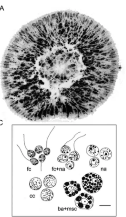
sup-plied with a chelator such as EDTA (Ereskovsky, Konyukov, & Tokina, 2010; Ereskovsky, Konjukov, & Willenz, 2007; Ereskovsky, Tokina, et al., 2007; Mukhina et al., 2006; Mukhina, Kumeiko, Podgornaya, & Efremova, 2007). The larvae of H. dujardini were dissociated, and the cell suspension was overlaid onto discontinuous density gradient made with Percoll solution and centrifuged. The gradient consisted of layers from 10% to 45% Percoll in calcium-free seawater, producing five fractions numbered from top to bottom of a tube (Fig. 3).
4. THE FATE OF CILIATED CELLS DURING
METAMORPHOSIS OF H. DUJARDINI
UNCORRECTED
PROOF
Fig. 3. Larvae of H. dujardini, their cell composition and the ability of cells to
aggre-gate. (A) Semi-thin longitudinal section of the free-swimming disphaerula larva. ba: blas-tomere-like amoebocytes; ci: cilia; cil: ciliated cells; ich: internal ciliated cells cavity, msc: maternal spherulous cells; na: nucleolated amoebocytes; pci: posterior flagellated cells; ap, anterior pole; pp, posterior pole. Scale bar: 20μm. (B) SEM of disphaerula larva. ci, cilia; pci, posterior flagellated cells; ap, anterior pole; pp, posterior pole. Scale bar: 20μm. (C) Scheme of larval cell fractions of H. dujardini obtained by Percoll density gradient cen-trifugation. Scale bar: 5μm.
The separation of larval cell types (Fig. 3C) made it possible to de-termine the polypeptide composition of the fractions by SDS-PAGE. The protein composition of fraction I (ciliated cells) differed dramatically from the rest. There were four major bands in the range of 42–68 kDa. One lane with a molecular weight of 42 kDa probably corresponded to actin. Another double lane of ~ 55 kDa corresponded to α and β tubu-lin isoforms. A third band of unknown nature with an apparent molec-ular weight (Mr) of 68 kDa (antigen S, p68) was chosen for polyclonal antibody production. Anti-p68 (S antigen) antibodies produced in rabbit were shown to be specific to larval ciliated cells, and were used in double immunofluorescence together with commercial anti-tubulin antibodies to trace the fate of ciliated cells during metamorphosis.
UNCORRECTED
PROOF
Double staining of larvae with both antibodies showed that protein p68 (S) was present in granules in the body of ciliated cells but was absent in the cilium. Tubulin was present in the cilium, the basal body and, to a lesser extent, in the cortical cytoskeleton structures (Fig. 4A and D(a)). Thus, anti-S and anti-tubulin antibodies stained different compartments in ciliated cells.
Cilia visualized with anti-tubulin antibody were seen all over the lar-val surface except at the posterior pole, which was bare and lacked any specific fluorescence (Fig. 4A(a)). Such larvae settled on the substrate and flattened rapidly. In the narrow transparent monolayer of lateral cil-iated cells attached to the substrate, the cilia appeared to be submerged into the cytoplasm, i.e., were internalized (Fig. 4A(b–c)). Both anti-S and anti-T staining were confined to the internal cell mass at the middle sec-tions of metamorphosing larvae (Fig. 4B). By 3 days post-settlement most of the ciliated cells joined the newly formed choanocyte chambers (Fig. 4C). S-specific labeling was seen in choanocytes but not in pinacocytes or amoebocytes.
At the morphological level choanocytes differ from ciliated cells by a prominent collar around cilia. S antigen (p68) was concentrated in the api-cal part of the choanocyte and visualized as rings or curves outlining the apical border (Fig. 4D(c)). Hence, ciliated cells from the anterio-lateral surface of the larva internalized during metamorphosis, and then the cells of the internal cell mass with the same immunoreactivity (S/tubulin-posi-tive) participated in the formation of choanocyte chambers in the juvenile. Some of the larval ciliated cells appear to be the source of internal cells in juvenile H. dujardini. A very unusual polypeptide composition of the ciliated cells, with few prominent major proteins, could be a sign of their terminal differentiation, but no evidence of exfoliation of cili-ated cells or their phagocytosis by any other cells during the short time of metamorphosis has yet been found (Gonobobleva & Ereskovsky, 2004b). Alternatively, a unique protein composition may be associated with the preparation of ciliated cells for a precipitous change of their fate. The S protein may be meant for choanocytes, which later become able to syn-thesize their own protein. The localization of p68 (S) must be checked by electron microscopy and its identity, by mass-spectrometry or Edman degradation procedure. The transcriptome of the adult sponge is available (Borisenko et al., 2016), and since the adult sponge can produce S anti
UNCORRECTED
PROOF
Fig. 4. Free-swimming and metamorphosing larva of H. dujardini double-stained with
anti-tubulin and anti-p68 antibodies. Sequential steps of larval settlement (A), metamor-phosis (B and C), transformation of ciliated cell (D and E) and adult sponge (F). (A) Lar-val settlement. Laser confocal scanning microscopy of whole mount larvae. a: Free-swim-ming larvae before settlement; top optical section and midsection (inset). Here and else-where green staining indicates tubulin, and red staining indicates S antigen. Arrow points to cilia. b: Larvae 40 min after contact with substrate (glass). c: Peripheral part of flattened larvae. Colors and designations are the same as above. Internalized cilia on c are indi-cated by arrow. (B) Larvae 6 h after contact with substrate. Ciliated cells are submerged into the larva. Scale bar for all images: 20μm. (C) Young sponge 3 days after settlement, immunostained paraffin section. chc–choanocyte chamber formed de novo. Note the ab-sence of S antigen staining in young pinacoderm. Scale bar 10μm. (D) Transformation of ciliated cell (a) by inward migration (b) into the choanocyte chamber (c) of a defini-tive sponge. Scale bar 2μm for a, b and 20 μm for c. (E) Scheme based on original image (D). a: Ciliated larval cells, b: ciliated cells inside larva, c: choanocyte of young sponge, d: choanocyte of definitive sponge. (F) Adult sponge with non-motile larva (correspond to Fig. 2B). a: DIC microscopy; b: immunofluorescence with anti-S. Pinacocytes are stained but note the absence of staining in cells of the larva before the release (white arrow). Scale bar: 20μm.
gen (Fig. 4(F)), we may be fairly sure that the S transcript can be found. Once this has been done, we may be able to trace specific cell-type tran-scription. By now we have proved its specificity for ciliated cells and ju-venile choanocytes at the protein level.
UNCORRECTED
PROOF
Transdifferentiation of cells is common in sponges. For instance, it occurs during regeneration and the formation of primmorphs (Borisenko, Adamska, Tokina, & Ereskovsky, 2015; Ereskovsky et al., 2015; Lavrov, Bolshakov, Tokina, & Ereskovsky, 2018; Lavrov & Kosevich, 2016). Light and electron microscopic data demonstrate that choanoblasts can develop in parenchymella from ciliated cells and archaeocytes in some genera (Mycale, Hamigera) (Borojevič & Lévi, 1965; Boury-Esnault, 1976), only from archaeocytes in some other taxa (Baikalospongia,
Mi-crociona) (Efremova & Efremov, 1979; Kaltenbach, Kuhns, Simpson, &
Burger, 1999), or from an internal population of archaeocytes and lar-val choanocytes (Spongillidae) (Weissenfels, 1989). A recent study of an-other demosponge, A. queenslandica, has shown that multiple larval cell types contribute to choanocyte chambers of the juvenile (Sogabe et al., 2016). So, while the fate of ciliated cells has been elucidated in H.
dujar-dini, it has to be studied in other species with the use of a board range of
markers.
Our approach could be extended to the search for new protein mark-ers with the separation of adult cell types and powerful techniques such as 2D-DIGE. Different cell types of the adult sponge clearly possessed spe-cific proteins (Fig. 5, arrows). Proteins in these spots could be identified after excision by mass-spectrometry and compared with the assembled transcriptome of the adult sponge. Then the specific synthesis could be traced by the whole-mount RNA–RNA in situ hybridization (Borisenko et al., 2016).
Antero-lateral ciliated cells leave the epithelium of the larva and migrate to the internal cell mass as amoeboid cells to be differentiated into choanocytes and/or other cell types of the juvenile sponge. Intercel-lular junctions have been found between larval ciliated cells, choanocytes and pinacocytes (Amano & Hori, 1996; Bergquist & Green, 1977; Boury-Esnault, Ereskovsky, Bézac, & Tokina, 2005; Evans, 1977; Ereskovsky et al., 2009; Ereskovsky & Tokina, 2004; Gonobobleva & Ereskovsky, 2004b; Gonobobleva & Maldonado, 2009). In our opinion, this indicates the presence of epithelial–mesenchymal transitions (EMT):
5. A JOURNEY FROM THE OUTSIDE INWARD:
HOW CILIATED CELLS BECOME DETACHED
UNCORRECTED
PROOF
Fig. 5. Difference gel electrophoresis (DIGE; according Alban et al., 2003) revealed
cell-specific protein markers. Cell suspension of an adult sponge was separated by cen-trifugation in density gradient, and proteins were extracted from choanocyte- and ar-chaeocyte-enriched fractions with urea-containing solution. Each protein mixture was co-valently labeled with reactive cyanine dye: choanocyte proteins were labeled with Cy3 (green), and archaeocyte proteins, with Cy5 (red). Then protein mixtures were pooled and underwent 2D electrophoresis by isoelectrofocusing and SDS-PAGE. After scan-ning the gel with lasers of different wavelength, we could recognize proteins specific for choanocytes (green), for amoebocytes (red), or common proteins (yellow).
ciliated cells break free from junctions and leave the polarized sheath to become amoeboid mesenchymal-like cells of the internal mass.
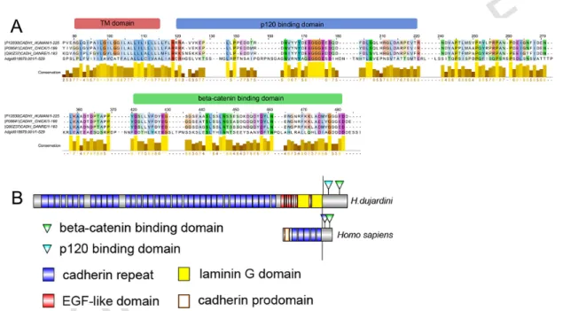
The loss of E-cadherin-mediated adherens junctions is the key event of EMT (Huang, Guilford, & Thiery, 2012). There are numerous proteins with cadherin repeats in their extracellular parts, but unlike true cadherins they have neither delta- nor beta-catenin binding domains in the cytoplas-mic part. Proteins containing cadherin repeats have long been known, but the presence of true epithelial cadherin remained a mystery. The presence of E-cadherin has been shown for sponges from all classes (Belahbib et al., 2018). We identified one cadherin homolog in H. dujardini transcrip-tome, which contained multiple cadherin repeats, a transmembrane helix and a cytoplasmic tail with domains for binding intercellular components of junction, delta-catenin (p120 protein) and beta-catenin. The sequence also contains seven EGF-like domains and two laminin domains, which is characteristic of invertebrate cadherin (Fig. 6). So, H. dujardini, similarly to other sponges (Belahbib et al., 2017), also has true cadherin, which is the main component of adherens junctions serving as a cell-to-cell lock in epithelial sheath.
UNCORRECTED
PROOF
Fig. 6. Comparison of E-cadherin structure in sponge H. dujardini and bilateria. (A) Alignment of the cytoplasmic parts of E-cadherins in
hu-mans, chick, zebrafish and H. dujardini. Conserved domains responsible for interaction with delta-catenin (p120) and beta-catenin, present in the intracellular part of E-cadherin molecule in H. dujardini, anchor adherens junctions to the cytoskeleton. (B) Domain organization of E-cadherins in humans and sponges. Invertebrates cadherins are characterized by multiplication of conservative cadherin repeats and the presence of laminin G-like domains, while lacking the prodomain, a short N-terminal sequence releasing with proteolytic maturation. Sequence from H. dujardini was identified in transcriptome with local BLAST (option tblastn) and domains identified with SMART and InterPro 70.0 (EMBL-EBI); GenBank accession number is MH107848.
UNCORRECTED
PROOF
The repression of E-cadherin expression is followed by the disap-pearance of adherens junctions after the start of EMT (Huang et al., 2012). EMT is controlled by the master genes snai1/snai2, twist and ZEB (Lamouille, Xu, & Derynck, 2014). Their expression induces EMT in many processes associated with development, regeneration and diseases. Snai and ZEB are zinc finger transcription factors. Snai has a SNAG do-main and sites for binding LATS2, KPNB1, NOTCH1 and PARP1 pro-teins; ZEB possesses an atypical homeobox and\or Glu-rich C-end. There are many transcripts for zinc finger DNA-binding proteins in H.
dujar-dini transcriptome but none of them has the characters typical of Snai and
ZEB. A similar situation observed for Twist. It is a basic helix-loop-helix (bHLH) transcription factor (TF), and H. dujardini has a lot of bHLH TFs, but they have only the DNA-binding domain in common with Twist. We failed to identify the master genes of EMT in H. dujardini transcriptome, possibly because transcription was absent during the sequenced staged. They may be revealed by a search in the genome.
Master genes themselves are controlled by various signaling pathways, mainly TGF-beta, FGF and Wnt. Finally, the process of internalization of flagellated cells during metamorphosis is under control of intercellular signals.
Cell-to-cell communications regulate many processes during devel-opment and adulthood of metazoans. During embryogenesis intercellular signals are the third main informational input to developmental process after maternal factors and zygotic genome. Key processes such as prolifer-ation, cell migration and survival, cell fate specification and axis pattern-ing are managed by cell-to-cell signalpattern-ing. These processes are also reg-ulated through intercellular communication. Six signaling pathways are conserved across Metazoa: Wnt, TGF-beta, Hedgehog, Notch, FGF and NO-dependent. Here we summarize facts and assumptions about each of these pathways in sponges.
Wnt signaling is the best studied pathway in sponges due its
proba-ble role in axial patterning, especially in early branching Metazoa. Rep-resentatives of all the four poriferan classes are studied for Wnt ligands. Syncytial sponges, Hexactinellida, have no Wnt ligands although they have all the other components of this pathway (Schenkelaars et al., 2017;
UNCORRECTED
PROOF
Windsor Reid et al., 2018). Distribution of ligands and receptors is quite different even between genera. For instance, A. queenslandica (Demo-spongiae) has two Wnt ligands, three Frizzled receptors, a co-receptor LRP5/6, a secondary messenger Disheveled, beta-catenin, cytoplasmic beta-catenin destruction complex (GSK3, APC, Axin), nuclear factors TCF and Groucho (Adamska et al., 2010). Freshwater sponge Ephydatia
muelleri has three ligands and three Frizzled receptors (Windsor Reid et
al., 2018). In our model demosponge, H. dujardini, there are ten ligands and five receptors (Borisenko et al., 2015). Cytoplasmic scaffold proteins Axin and APC have similar features in both species. The beta-catenin binding site not found in Axins, and most of the conserved domains ex-cept armadillo repeats are not found in APCs. Biochemical studies on poriferan beta-catenin demonstrate that intermolecular interactions dur-ing cytoplasmic signal transduction can be mediated by highly diverged sequences (Nichols, Dirks, Pearse, & King, 2006; Schippers & Nichols, 2018). This may be a reason why such domains cannot be recognized by ordinary bioinformatic methods.
The champion in Wnts complexity, a calcisponge Sycon ciliatum, con-tains 21 ligands. In addition to ligands, four receptors and all core intra-cellular components except Axin are described in S. ciliatum (Leininger et al., 2014). Sponges from the genus Oscarella (Homoscleromorpha) have from 3–8 ligands (Lapébie et al., 2009; Leininger et al., 2014). The pres-ence of other pathway components has not been ascertained.
Phylogenetic analysis cannot place poriferan Wnt in any of the estab-lished subfamilies of metazoan ligands (Adamska et al., 2010; Lapébie et al., 2009; Leininger et al., 2014). Phylogeny inference made with sponge sequences only demonstrates the presence of Porifera-specific subfamilies (Windsor Reid et al., 2018). In demosponges Wnts form three subfamilies (named PorWntA-C), in homoscleromorphs, two families (WntI-II), an in calcisponges, 21 families (CWntA-U). Notably, the nine of the ten H.
du-jardini ligands cannot be classified within any of these families.
Wnt ligands are expressed in embryos, larvae and adult sponges. The expression of some Wnt ligands was shown in adult and larvae of H.
du-jardini (Fig. 7A and B). Two ligands, HdWntD and HdWntE, were
ex-pressed in the oscular tube, while HdWntG transcripts were observed in exopinacocytes. In addition, a set of Wnt is expressed in the posterior
UNCORRECTED
PROOF
Fig. 7. Summarized data about axial-related expression of wnt and TGF-beta in three
model sponges from Demospongiae (A–C) and Calcarea (D and F) classes. Wnt ligands are red and TGF-beta is blue; brightness depends on expression intensity. For embryo and larvae anterior end is oriented at top and posterior at bottom. (A and B) Main expres-sion patterns of HdWnt is in oscular tube at adult H. dujardini, and posterior half of larva. Some wnt's are expressed in basal part of sponge, and in exopinacoderm in“salt and pep-per” pattern as well (data not shown). Expression patterns of TGF-beta are unknown in
Halisarca yet. (C) expression of AqWnt and AqTGFb in A. queenslandica embryo at the
pigment spot formation stage. (D) Adult Sycon ciliatum sponge demonstrate oscular ex-pression of wnt and TGF-beta ligands; two wnt's expresses in proximal part of choanocyte chambers as well. (E) Only TGF-beta transcript presented at stomoblastula stage, in abo-ral macromeres; wnt expression is not detected. (F) expression of both ligands at posterior pole of larva, with broader pattern of wnt. Panels (A) and (B): Modified from Borisenko,
I., Adamski, M., Ereskovsky, A., & Adamska, M. (2016). Surprisingly rich repertoire of Wnt genes in the demosponge Halisarca dujardini. BMC Evolutionary Biology, 16, 123; Kozin, V. V., Borisenko, I. E., & Kostyuchenko, R. P. (2019). Establishment of the Axial Polarity and Cell Fate in Metazoa via Canonical Wnt Signaling: New Insights from Sponges and Annelids. Biological Bulletin, 46(1), 12; Panel (C): Modified from Adamska, M., Degnan, S. M., Green, K. M., Adamski, M., Craigie, A., Larroux, C., & Degnan, B. M. (2007). Wnt and TGF-β Expression in the Sponge Amphimedon queenslandica and the Origin of Metazoan Embryonic Patterning. Plos One, 2(10), e1031; Panel (D)–(F): Modified from Leininger, S., Adamski, M., Bergum, B., Guder, C., Liu, J., Laplante, M., et al. (2014). Developmental gene expression provides clues to relationships between sponge and eumetazoan body plans. Nature Communications, 5. https://doi.org/10.1038/ ncomms4905.
hemisphere of the larvae: HdWntL at the posterior pole, HdWntK from the equator to the latest lateral flagellated cells, and HdWntJ in a belt-like pattern at the equator (Borisenko et al., 2015). In A. queenslandica
UNCORRECTED
PROOF
AqWntA is first expressed in micromeres throughout the embryo in a
dif-fuse manner. During the development these cells migrate into the inner part of the embryo, then to the outer cell layer, and eventually turn up at the further posterior pole of the larva (Fig. 7C; Adamska et al., 2007, 2010).
Unlike Amphimedon, in S. ciliatum most Wnt expressed in oocytes and early cleavage stages, though transcripts disappeared by the end of em-bryonic development and before larvae formation. Expression domains of three Wnt are associated with the posterior half of larvae (Fig. 7E–F). Two out of five Frizzled receptors expressed in micromeres, at the anterior pole of the embryo. Such combinatorial expression of different ligands and re-ceptors allow diversifying results from use of limited set of signaling mol-ecules. Another set of Wnt is expressed in staggered domains in the os-culum of the adult sponge, and in the opening of the choanocyte chamber (Leininger et al., 2014). In S. ciliatum the choanocyte chamber is the mod-ule of axis determined by water flow.
Expression of Wnt in ostia, incurrent opening of aquiferous canals, is described in adult Oscarella lobularis (Homoscleromorpha). This sponge has a true epithelium, and ostia form by epithelial invagination of the ex-opinacoderm. Localization of Wnt expression is characteristic of epithe-lial appendage formation in eumetazoans such as Hydra or Nematostella tentacles and wings in Drosophila (Lapébie et al., 2009).
The data on the diversity of Wnt pathway components, their phy-logeny and expression allow us to make two important conclusions. (1) Sponges acquired their Wnt ligands independently of Cnidaria + Bilate-ria. It is still unclear whether the small number of ligands (two or three) in some species is a consequence of secondary loss or an ancestral condi-tion. Lineage-specific expansion has definitely occurred in Calcarea. (2) Wnt pathway is likely to be involved in axes specification in Porifera. The sets of Wnt ligands expressed in a pole-dependent manner (the pos-terior half of the larva, the osculum, the opening of the choanocyte cham-ber). An opposite viewpoint is based on the study of Wnt expression in juvenile E. muelleri (Windsor Reid et al., 2018). It demonstrated the ex-pression of Wnt ligands in different cell types not restricted to any region of the sponge. Although no expression of any Wnt in the osculum was shown, pharmacological alteration of this pathway increased the num-ber of oscula upon activation and decreased upon inhibition (Windsor & Leys, 2010; Windsor Reid et al., 2018). So, we tend to think that in
UNCORRECTED
PROOF
sponges Wnt is implicated in axial patterning rather than cell specifica-tion, as the first metazoan mechanism of anterio-posterior determination.
The second key player in axial patterning is the transforming growth factor (TGF-beta) signaling pathway. Core components of this pathway are extracellular ligands, transmembrane receptors and transcription fac-tors SMADs. The superfamily of TGF-beta ligands encompasses many families of proteins, the main families being TGF-beta sensu stricto, BMPs, Nodals and Activins. Receptors are single pass serine-threonine kinases of two classes, TGFbRI or TGFbRII. Upon binding of the lig-and, type II receptors activate type I receptors, and they phosphorylate SMADs, which leads to transcriptional activation. The patterning of the dorso-ventral axis during the development of Bilateria starts with the ex-pression of BMP and Chordin (a soluble inhibitor of TGF-beta ligands) at the opposite sides of the embryo. At later stages TGF-beta signaling is involved in numerous developmental processes, from germ layer specifi-cation to the development of organs and tissues (Kitisin et al., 2007). This pathway is a novelty of Metazoa since neither ligand nor receptor mole-cules are found outside the animal kingdom (Srivastava et al., 2010).
A. queenslandica genome contains eight TGF-beta genes, of which
two belong to TGF-beta sensu stricto and six fall into divergent clades such as GDF or DVR (Srivastava et al., 2010). Phylogenetic analysis of these diverged genes group suggests an independent expansion of lig-ands. There are ligand receptors of both types. The expression of one of the ligands is described in the development. Similarly to one of the Wnt genes, this TGF-beta is expressed in some micromeres from early cleav-age stcleav-ages. It is dynamically expressed throughout the outer cell layer of the embryo, and then in a concentric pattern around the posterior pole and at the anterior pole of embryo (Fig. 7C). In contrast to bilaterians and an-thozoans, where TGF-beta patterns the axis perpendicular to the one pat-terned by Wnt signals, in A. queenslandica Wnt and TGF-beta are ex-pressed along the same embryonic axis (Adamska et al., 2007).
A calcisponge S. ciliatum has 22 TGF-beta genes, one of which falls into TGF-beta sensu stricto family and remaining sequences cannot be ascribed to any defined eumetazoan families (Leininger et al., 2014). Several ligands are expressed uniformly in oocytes and during cleavage, while the expression of four TGFs is restricted to the macromeres located at the posterior pole of the embryo. The most prominent region of
TGF-UNCORRECTED
PROOF
beta expression in the adult sponge is the apical region: seven ligands are
expressed at a significantly higher level in the apical part of sponge bear-ing the osculum (Fig. 7D). Thus, the expression of TGFs is similar to that of Wnt in S. ciliatum, being predominantly posterior in the larvae and api-cal in the adults. SMAD genes are expressed either in the same cells or in the cells adjacent to those expressing TGF-beta ligands. In contrast to lig-ands, downstream components of pathway are expressed more uniformly along the apico-basal axis of the adult sponge.
A search in H. dujardini transcriptome revealed eight TGF-beta lig-ands. All of them have a structure typical of TGF-beta: a signaling pep-tide at the N-terminal end, a large latency-associated peppep-tide, the site of protease furin cleavage, and the C-terminal signal peptide with cys-teines in conserved positions. Phylogenetic analysis revealed some simi-larities with other sponges: three sequences fall into the TGF-beta family
sensu stricto, while the other five ligands do not belong to any
eumeta-zoan families (Borisenko et al., in press). Interestingly, TGF-beta s.s. lig-ands have only been identified in deuterostomes and ctenophores, with no members found in the genome screens of C. elegans, Drosophila or
Nematostella (Matus et al., 2006; Pang, Ryan, Baxevanis, & Martindale,
2011; Srivastava et al., 2010).
Many developmental processes in Bilateria involve Hedgehog
path-way in a classical morphogen mode, acting in a concentration-dependent
manner (Jiang & Hui, 2008). The Hedgehog ligand, a secreted protein, un-dergoes autocatalytic cleavage following acetylation and binds to Patched receptor on the cell surface. Hedgehog-competent signal cells express a zinc finger transcription factor Cubitus interruptus (Ci), which undergoes cleavage and releases a peptide (CiR) translocating in the nucleus and act-ing as a transcriptional repressor. The bindact-ing of Hedgehog to Patched al-lows the suppression of Ci cleavage; a full-size Ci works as a transcrip-tional activator. Thus, the level and the set of expressed target genes de-pend on the ligand concentration. In this way Hedgehog participates in many patterning processes such as limb formation in vertebrates and the establishment of segment polarity in Drosophila. However, in some cases this pathway acts as short-range signaling, e.g., in case of the induction of the neural floor plate by the notochord in vertebrates.
Hedgehog genes have not been identified outside cnidarians and bila-terians but a Hedgling homolog was found in a choanoflagellate
UNCORRECTED
PROOF
genes has been analyzed in Nematostella vectensis (Matus, Magie, Pang, Martindale, & Thomsen, 2008). One of them is expressed in the pha-ryngeal ectoderm, while the other one is expressed in the endoderm; the receptor Patched is expressed only in the endoderm. This pattern sug-gests that both short- and long-range Hedgehog-mediated pathways may function in Cnidaria. Neither the ligand nor any other key components of this pathway were found in the ctenophore Mnemiopsis leidyi (Ryan et al., 2013). As at least some components of this pathway are present in choanoflagellates, its loss in Ctenophora seems to have been was sec-ondary and independent (Adamska, 2015).
A typical Hedgehog protein contains an N-terminal Hedge domain and a C-terminal Hog domain. Hog domain separates due to autocatalytic cleavage. Both these domains are found in A. queenslandica genome but in different genes. The key domain, Hedge, is a part of a large trans-membrane protein Hedgling also containing the von Willebrand domain, cadherin repeats, immunoglobulin and EGF domains. Hedgling expresses during late embryonic development and is associated with the formation of the sensory pigment ring at the posterior pole of the embryo. It is un-known whether Hedgling proteins undergo modifications and/or cleavage. If they do not, this transmembrane protein could only signal across ad-jacent cells through direct contact of cell surfaces, in contrast to the true Hedgehog. The genome of A. queenslandica contains several other com-ponents of the Hedgehog pathway but not the receptor Patched (Richards & Degnan, 2009; Srivastava et al., 2010). It is un clear whether Hedgling transmits intracellular signals through the canonical Hedgehog pathway or through an unidentified mechanism (Adamska et al., 2007).
Growth factors family encompasses fibroblast growth factors (FGF), platelet-derived growth factors (PDGF), vascular endothelial growth fac-tors (VEGF) and epidermal growth facfac-tors (EGF). All of these proteins were described as growth factors for defined cell types and act through the same type of receptors, although ligands do not demonstrate similarity of sequences. FGF bind to receptor tyrosine kinase that initiates a series of phosphorylation events, the tyrosine kinase cascade, resulting in acti-vation of transcription factors. Unlike other signalings, there is no exact transcription factor activated by FGF pathway, and the output of the path-way activation depends on the combination of ligand, receptor and cellu-lar content.
UNCORRECTED
PROOF
FGF signaling is involved in various developmental processes in Bi-lateria, from axes specification to tissue induction and organ patterning (Thisse & Thisse, 2005). Another conserved role of FGF is boundary for-mation as shown in Hydra (Sudhop et al., 2004). Nevertheless, a true FGF protein is the novelty of Bilateria + Cnidaria: no FGF ligand was found in Trichoplax, A. queenslandica, Mnemiopsis or Monosiga genomes al-though FGF-like proteins with a similar domain organization were iden-tified in many Metazoa including sponges (King et al., 2008; Ryan et al., 2013; Srivastava et al., 2008, 2010). Receptor tyrosine kinases are broadly distributed from Protista to Bilateria, and some of them display a weak similarity to the intracellular portion of the FGF receptors (Manning, Young, Miller, & Zhai, 2008). The intracellular signal transduction pro-teins are all present in Monosiga. This demonstrates a gradual assembly of the pathway, from signal transduction cascade utilized by single cell eu-karyotes, through addition of receptors allowing more sophisticated sens-ing of the neighborsens-ing cells in the colony, to the emergence of a highly effective morphogen in the ancestor of Cnidaria + Bilateria (Adamska, 2015).
All signaling events mentioned above are mediated by secreted solu-ble proteins. In the Delta/Notch signaling pathway both the ligand (Delta or Serrate) and the receptor (Notch) are transmembrane proteins, and the signaling event means close contact of two neighbor cells. Upon binding of the ligand to the receptor, several proteolytic modifications occur at the ligand-receptor complex resulting in the production of Notch intracellular domain, which is translocated to the nucleus and acts as a transcription activator. This pathway appears to be the evolutionarily the oldest one: all the participants are identified in Monosiga genome except ligands. In choanoflagellates Delta/Notch might be involved in sensing environmen-tal stimuli rather than intercellular signaling (Richards & Degnan, 2009).
Delta/Notch signaling is implicated in the development of many or-gans in Bilateria, and is crucial for segmentation in Vertebrata (Andersson, Sandberg, & Lendahl, 2011). In Cnidaria it is involved in the boundary formation during budding and affects oogenesis and the dif-ferentiation of nematoblasts and neurons (Käsbauer et al., 2007; Marlow, Roettinger, Boekhout, & Martindale, 2012; Matveev, Shaposhnikova, & Podgornaya, 2007; Münder et al., 2010). In sponges, Delta/Notch path-way components and their developmental expression
UNCORRECTED
PROOF
are described in A. queenslandica (Gazave et al., 2009; Richards & Degnan, 2012; Richards et al., 2008). Five different Delta ligands, one Notch receptor and all core components were identified. The expression of Notch and Delta genes indicates that they are involved in specification of putative sensory cells, with Notch expression being broad and the five
Delta genes displaying dynamic, unique patterns. The domain
composi-tion of the identified genes, as well as their expression patterns, indicate that the pathway operates in a manner similar to that in Cnidaria + Bilate-ria, and is involved in evolutionarily conserved processes such as cell-type specification and differentiation.
Nitric oxide (NO) pathway is another signaling mechanism usually
mentioned in the context of blood vessel physiology or differentiation of endothelial cells. NO is an ancient signal molecule working via the activation of its primary cytoplasmic receptor, soluble guanylyl cyclase, or by direct covalent modification of target proteins such as S-nitrosyla-tion and nitraS-nitrosyla-tion. Data about its participaS-nitrosyla-tion in early development are just beginning to accumulate although the role of NO in metamorphosis has been described for many phyla of invertebrates (Annona et al., 2017; Tomankova, Abaffy, & Sindelka, 2017).
Sponges have an almost full repertoire of neuronal genes needed to form functional neural scaffolds (Francis et al., 2017; Sakarya et al., 2007). Nevertheless, they have no neuronal structures. This is especially strange in case of sponge larvae, which have no neurons but are photosen-sitive and can coordinate ciliary beating for directional driving. It was ele-gantly demonstrated that NO is an activator of larva metamorphosis in A.
queenslandica (Ueda et al., 2016). An inhibition of the NO pathway
sup-presses metamorphosis whereas an addition of endogenous NO activates it. In this case NO pathway works upstream of (or together with) ERK (extracellular signal-regulated kinase) pathway, one of the many receptor tyrosine kinase cascades, and the expression of NO synthase increases in localized cell populations when the larva becomes competent.
We can see that metazoans have an almost full toolkit for body pat-terning and the establishment of different cell types. Until new data about intercellular communication in sponges have been obtained, we can put forward two main questions regarding metamorphosis: (1) which of the signaling pathways and in what hierarchical order are involved in meta-morphosis? (2) How is the body axis determined after metamorphosis and is it the same axis as in the larva?
UNCORRECTED
PROOF
During metamorphosis sponge larvae lose ciliary cells and this ability to locomotion. The similarity of larval ciliated cells with the choanocytes of the adult sponge gave rise to the classical hypothesis of the“inversion of the germ layers” in sponges (Delage, 1892). Now that we have evidence from different sponges, we know that the fate of ciliated cells is species-specific. At the same time, ciliated cells often leave their place in the epithelial-like sheath and migrate inside during metamorpho-sis, i.e., undergo EMT. Here we tried to show the connection between this process and the signaling pathways present in sponges through mol-ecules involved in intercellular junctions. While proteins managing EMT were not found in transcriptomes of adults or larvae of H. dujardini, the this process in sponges remains mysterious. At the same time, sponges have most of the components of the six main signaling pathways. There is evidence that in sponges Wnt and TGF-beta signaling pathways are im-plicated in axial patterning rather than cell specification, and these path-ways were the first metazoan mechanism of anterio-posterior determina-tion. The link between the axis of larvae and adult sponge looks highly likely as many signaling molecules are expressed at the posterior larval pole and in the osculum of the adult sponge. However, it is still unclear whether expression of signaling molecules in adult sponge marks the axis heredity from the larva, or it is de novo established after metamorphosis. The developmental regulation pathways may be traced with the help of transcriptomes of sponge development stages, which are available only for a couple of species. The application of traditional techniques to simple animals at the key root position of Metazoa is a necessary step for under-standing developmental events at the molecular level.
ACKNOWLEDGMENTS
The study was financially supported by the Russian Science Foundation (grants 17-14-01089 in part of bioinformatics analysis), by the Russian Foundation for the Ba-sic Research (grant 18-34-00398 in part of TGF-beta study) and the“Molecular and Cell Biology” program of the Presidium of the Russian Academy of Sciences (grant 01.2.01457147). We gratefully appreciate the help provided at the White Sea Biologi-cal Station of the ZoologiBiologi-cal Institute of the Russian Academy of Sciences and at Be-lomorskaya Educational and Research Station of the St Petersburg State University. We thank the Research Resource Center for Molecular and Cell Technologies of St Peters
UNCORRECTED
PROOF
burg State University for technical support, and Natalia Lentsman for improving the English.
REFERENCES
Adamska, M., 2015. Developmental signalling and emergence of animal multicellularity. In: Ruiz-Trillo, I., Nedelcu, A.M. (Eds.), Evolutionary transitions to multicellular life. Vol. 2, Springer Netherlands, Dordrecht, pp. 425–450.
Adamska, M., Degnan, S.M., Green, K.M., Adamski, M., Craigie, A., Larroux, C., et al., 2007. Wnt and TGF-β expression in the sponge Amphimedon queenslandica and the origin of metazoan embryonic patterning. PLoS One 2 (10), e1031.
Adamska, M., Larroux, C., Adamski, M., Green, K., Lovas, E., Koop, D., et al., 2010. Structure and expression of conserved Wnt pathway components in the demosponge Amphimedon queenslandica: Wnt pathway components in Amphimedon queens-landica. Evolution & Development 12 (5), 494–518.
Alban, A., Olu, D.S., Lennart, B., Christian, A., Erik, S., Steve, L., et al., 2003. A novel experimental design for comparative two-dimensional gel analysis: Two-dimensional difference gel electrophoresis incorporating a pooled internal standard. Proteomics 3 (1), 36–44.
Amano, S., Hori, I., 1993. Metamorphosis of calcareous sponges II. Cell rearrangement and differentiation in metamorphosis. Invertebrate Reproduction and Development 24 (1), 13–26.
Amano, S., Hori, I., 1996. Transdifferentiation of larval flagellated cells to choanocytes in the metamorphosis of the demosponge Haliclona permollis. The Biological Bulletin 190, 161–172.
Amano, S., Hori, I., 2001. Metamorphosis of coeloblastula performed by multipotential larval flagellated cells in the calcareous sponge Leucosolenia laxa. The Biological Bulletin 200 (1), 20–32.
Andersson, E.R., Sandberg, R., Lendahl, U., 2011. Notch signaling: Simplicity in design, versatility in function. Development (Cambridge, England) 138 (17), 3593–3612. Annona, G., Caccavale, F., Pascual-Anaya, J., Kuratani, S., De Luca, P., Palumbo, A., et
al., 2017. Nitric oxide regulates mouth development in amphioxus. Scientific Reports 7 (1), 8432.
Belahbib, H., Renard, E., Santini, S., Jourda, C., Claverie, J.-M., Borchiellini, C., et al., 2018. New genomic data and analyses challenge the traditional vision of animal ep-ithelium evolution. BMC Genomics 19 (1), 393.
Bergquist, P.R., Green, C.R., 1977. An ultrastructural study of settlement and metamor-phosis in sponge larvae. Cahiers de Biologie Marine 18, 289–302.
Borisenko, I.E., Adamska, M., Tokina, D.B., Ereskovsky, A.V., 2015. Transdifferentia-tion is a driving force of regeneraTransdifferentia-tion in Halisarca dujardini (Demospongiae, Porifera). PeerJ 3, e1211.
Borisenko, I., Adamski, M., Ereskovsky, A., Adamska, M., 2016. Surprisingly rich reper-toire of Wnt genes in the demosponge Halisarca dujardini. BMC Evolutionary Biol-ogy 16, 123.
Borojevič, R., Lévi, C., 1965. Morphogenese experimentale d'une Eponge a partir de cel-lules de la larvt nageante dissociee. Zeitschrift Fur Zellforschung Und Mikroskopische Anatomie 68, 57–69.
UNCORRECTED
PROOF
Boury-Esnault, N., 1976. Ultrastructure de la larve parenchymella d'Hamigera hamigera (Schmidt) (Demosponge, Poecilosclerida). Origine des cellules grises. Cahiers de Bi-ologie Marine 17, 9–20.
Boury-Esnault, N., Ereskovsky, A., Bézac, C., Tokina, D., 2005. Larval development in the Homoscleromorpha (Porifera, Demospongiae). Invertebrate Biology 122 (3), 187–202.
Chen, W.T., 1976. Reproduction and speciation in Halisarca. In: Harrison, F.W., Cowden, R.R. (Eds.), Aspects of sponge biology. Academic Press, New York, San Francisco, London, pp. 113–139.
Delage, Y., 1892. Embryogenese des eponges silicieuses. Arch. Z. Exp. Gen. 10, 345–498. Efremova, S.M., 1997. Once more on the position among Metazoa—gastrulation and ger-minal layers of sponges. In: Ereskovsky, A.V., Keupp, H., Kohring, R. (Eds.), Modern problems of Poriferan biology. Freie Universitat, Berlin, pp. 7–15.
Efremova, S.M., Efremov, V.I., 1979. Proliferation cellulaire chez la larve nageante de l’ éponge d'eau douce: Baikalospongia bacillifera (Dybowski). In: Lévi, C., Boury-Es-nault, N. (Eds.), Biologie des Spongiaires. Vol. 291, Colloq Int CNRS, Paris, pp. 59–65.
Ereskovsky, A.V., 2010. The comparative embryology of sponges. Springer, Dordrecht. Ereskovsky, A.V., Borchiellini, C., Gazave, E., Ivanisevic, J., Lapébie, P., Perez, T., et al.,
2009. The Homoscleromorph sponge Oscarella lobularis, a promising sponge model in evolutionary and developmental biology. BioEssays 31 (1), 89–97.
Ereskovsky, A.V., Borisenko, I.E., Lapébie, P., Gazave, E., Tokina, D.B., Borchiellini, C., 2015. Oscarella lobularis (Homoscleromorpha, Porifera) regeneration: Epithelial mor-phogenesis and metaplasia. PLoS One 10 (8), e0134566.
Ereskovsky, A.V., Dondua, A.K., 2006. The problem of germ layers in sponges (Porifera) and some issues concerning early metazoan evolution. Zoologischer Anzeiger–A Journal of Comparative Zoology 245 (2), 65–76.
Ereskovsky, A.V., Gonobobleva, E.L., 2000. New data on embryonic development of Halisarca dujardini Johnston, 1842 (Demospongiae, Halisarcida). Zoosystema 22 (2), 355–368.
Ereskovsky, A.V., Konjukov, P., Willenz, P., 2007. Experimental metamorphosis of Halis-arca dujardini larvae (Demospongiae, Halisarcida): Evidence of flagellated cell totipo-tentiality. Journal of Morphology 268 (6), 529–536.
Ereskovsky, A.V., Konyukov, P.Y., Tokina, D.B., 2010. Morphogenesis accompanying larval metamorphosis in Plakina trilopha (Porifera, Homoscleromorpha). Zoomor-phology 129 (1), 21–31.
Ereskovsky, A.V., Renard, E., Borchiellini, C., 2013. Cellular and molecular processes leading to embryo formation in sponges: Evidences for high conservation of processes throughout animal evolution. Development Genes and Evolution 223 (1–2), 5–22. Ereskovsky, A., Tokina, D., 2004. Morphology and fine structure of the swimming larvae
of Ircinia oros (Porifera, Demospongiae, Dictyoceratida). Invertebrate Reproduction and Development 45 (2), 137–150.
Ereskovsky, A.V., Tokina, D.B., Bézac, C., Boury-Esnault, N., 2007. Metamorphosis of cinctoblastula larvae (Homoscleromorpha, porifera). Journal of Morphology 268 (6), 518–528.
UNCORRECTED
PROOF
Evans, C.W., 1977. The ultrastructure or larvae from the marine sponge Halichondria moorei Bergquist (Porifera, Demospongiae). Cahiers de Biologie Marine 18 (4), 427–433.
Feuda, R., Dohrmann, M., Pett, W., Philippe, H., Rota-Stabelli, O., Lartillot, N., 2017. Im-proved modeling of compositional heterogeneity supports sponges as sister to all other animals. Current Biology 27 (24), 3864–3870.e4.
Francis, W.R., Eitel, M., Vargas, S., Adamski, M., Haddock, S.H., Krebs, S., 2017. The genome of the contractile demosponge Tethya wilhelma and the evolution of meta-zoan neural signalling pathways. BioRxiv https://doi.org/10.1101/120998.
Franzen, W., 1988. Oogenesis and larval development of Scypha ciliata (Porifera, Cal-carea). Zoomorphology 107 (6), 349–357.
Funayama, N., 2010. The stem cell system in demosponges: Insights into the origin of so-matic stem cells: Stem cell system in demosponges. Development, Growth & Differ-entiation 52 (1), 1–14.
Funayama, N., Nakatsukasa, M., Hayashi, T., Agata, K., 2005. Isolation of the choanocyte in the fresh water sponge, Ephydatia fluviatilis and its lineage marker, Ef annexin. De-velopment, Growth & Differentiation 47 (4), 243–253.
Funayama, N., Nakatsukasa, M., Kuraku, S., Takechi, K., Dohi, M., Iwabe, N., 2005. Iso-lation of Ef silicatein and Ef lectin as molecular markers Sclerocytes and cells in-volved in innate immunity in the freshwater sponge Ephydatia fluviatilis. Zoological Science 22 (10), 1113–1122.
Gaino, E., Baldacconi, R., Corriero, G., 2007. Post-larval development of the commer-cial sponge Spongia officinalis L. (Porifera, Demospongiae). Tissue & Cell 39 (5), 325–334.
Gazave, E., Lapébie, P., Richards, G.S., Brunet, F., Ereskovsky, A.V., Degnan, B.M., 2009. Origin and evolution of the notch signalling pathway: An overview from eu-karyotic genomes. BMC Evolutionary Biology 9, 249.
Gonobobleva, E.L., 2007. Basal apparatus formation in external flagellated cells of Hal-isarca dujardini larvae (Demospongiae: Halisarcida) in the course of embryonic de-velopment. In: Porifera research: Biodiversity, innovation and sustainability. Rio de Janeiro: Museu nacional. pp. 345–351.
Gonobobleva, E., Ereskovsky, A., 2004. Metamorphosis of the larva of Halisarca dujardini (Demospongiae, Halisarcida). Bulletin de l'Institut Royal Des Sciences Naturelles de Belgique–Bulletin van Het Koninklijk Belgisch Instituut Voor Natuurwetenschappen 74, 101–115.
Gonobobleva, E., Ereskovsky, A., 2004. Polymorphism in free-swimming larvae of Halis-arca dujardini (Demospongiae, Halisarcida). Bollettino dei Musei e degli istituti Bio-logici dell'Università di Genova 68, 349–356.
Gonobobleva, E., Maldonado, M., 2009. Choanocyte ultrastructure in Halisarca dujardini (Demospongiae, Halisarcida). Journal of Morphology 270 (5), 615–627.
Huang, R.Y.-J., Guilford, P., Thiery, J.P., 2012. Early events in cell adhesion and po-larity during epithelial-mesenchymal transition. Journal of Cell Science 125 (19), 4417–4422.
Jiang, J., Hui, C.-C., 2008. Hedgehog signaling in development and cancer. Developmen-tal Cell 15 (6), 801–812.
Kaltenbach, J.C., Kuhns, W.J., Simpson, T.L., Burger, M.M., 1999. Intense concanavalin a staining and apoptosis of peripheral flagellated cells in larvae of the marine sponge
UNCORRECTED
PROOF
Microciona prolifera: Significance in relation to morphogenesis. Biological Bulletin 197 (2), 271–273.
Käsbauer, T., Towb, P., Alexandrova, O., David, C.N., Dall'armi, E., Staudigl, A., 2007. The notch signaling pathway in the cnidarian Hydra. Developmental Biology 303 (1), 376–390.
King, N., Westbrook, M.J., Young, S.L., Kuo, A., Abedin, M., Chapman, J., 2008. The genome of the choanoflagellate Monosiga brevicollis and the origin of metazoans. Na-ture 451 (7180), 783–788.
Kitisin, K., Saha, T., Blake, T., Golestaneh, N., Deng, M., Kim, C., et al., 2007. Tgf-Beta signaling in development. Science's STKE: Signal Transduction Knowledge Environ-ment 2007 (399), cm1.
Korotkova, G.P., Apalkova, L.V., 1975. Oogenesis of the Barents Sea sponge Halisarca dujardini. In: Tokin, I.B. (Ed.), Comparative and experimental morphology of the sea organisms. pp. 9–26, (in Russian) Apatity.
Lamouille, S., Xu, J., Derynck, R., 2014. Molecular mechanisms of epithelial –mesenchy-mal transition. Nature Reviews Molecular Cell Biology 15 (3), 178–196.
Lapébie, P., Gazave, E., Ereskovsky, A., Derelle, R., Bézac, C., Renard, E., 2009. WNT/β-catenin signalling and epithelial patterning in the Homoscleromorph sponge Oscarella. PLoS One 4 (6), e5823.
Lavrov, A.I., Bolshakov, F.V., Tokina, D.B., Ereskovsky, A.V., 2018. Sewing up the wounds: The epithelial morphogenesis as a central mechanism of calcaronean sponge regeneration. Journal of Experimental Zoology. Part B, Molecular and Developmental Evolution 330, 351–371.
Lavrov, A.I., Kosevich, I.A., 2016. Sponge cell reaggregation: Cellular structure and mor-phogenetic potencies of multicellular aggregates. Journal of Experimental Zoology Part A: Ecological Genetics and Physiology 325 (2), 158–177.
Leininger, S., Adamski, M., Bergum, B., Guder, C., Liu, J., Laplante, M., 2014. Develop-mental gene expression provides clues to relationships between sponge and eumeta-zoan body plans. Nature Communications 5, https://doi.org/10.1038/ncomms4905. Lévi, C., 1956. Étude des halisarca de roscoff: Embryologie et systématique des
démo-sponges. 7, Travaux de La Station Biologique Roscoff N. S., 3–181.
Leys, S.P., 2004. Gastrulation in sponges. In: Stern, C. (Ed.), Gastrulation. Cold Spring Harbor Laboratory Press, Cold Spring Harbor, New York, pp. 23–31.
Leys, S.P., Degnan, B.M., 2002. Embryogenesis and metamorphosis in a haploscle-rid demosponge: Gastrulation and transdifferentiation of larval ciliated cells to choanocytes. Invertebrate Biology 121 (3), 171–189.
Leys, S.P., Eerkes-Medrano, D., 2005. Gastrulation in calcareous sponges: In search of Haeckel's Gastraea. Integrative and Comparative Biology 45 (2), 342–351.
Leys, S.P., Ereskovsky, A.V., 2006. Embryogenesis and larval differentiation in sponges. Canadian Journal of Zoology 84 (2), 262–287.
Manning, G., Young, S.L., Miller, W.T., Zhai, Y., 2008. The protist, Monosiga brevicol-lis, has a tyrosine kinase signaling network more elaborate and diverse than found in any known metazoan. Proceedings of the National Academy of Sciences of the United States of America 105 (28), 9674–9679.
Marlow, H., Roettinger, E., Boekhout, M., Martindale, M.Q., 2012. Functional roles of notch signaling in the cnidarian Nematostella vectensis. Developmental Biology 362 (2), 295–308.