Chondrocyte Gene Expression
by Paula M. Ragan B.S., Mechanical Engineering, Tufts University, 1992 M.S., Biomedical Engineering, Boston University, 1994Submitted to the M.I.T.-Harvard Division of Health Sciences and Technology
in Partial Fulfillment of the Requirements for the Degree of
Doctor of Philosophy
at the
Massachusetts Institute of Technology May 1999
@
Massachusetts Institute of TechnologySCHERING PLO"GH
MASSACHUSETTS INSTITUTE OF TECHNOLOGYLIBRARIES
Signature of Author M. Certified byI.T.-Harv rd Division of Health Sciences and Technology May 1999
Thesis Supervisor
Accepted by
\Chair, Department Committee
-The Effect of Mechanical Compression on
Chondrocyte Gene Expression
by
Paula M. Ragan
Submitted to the M.I.T.-Harvard Division of Health Sciences and Technology in partial fulfillment of the requirements for the Degree of Doctor of Philosophy, May 1999.
Abstract
Chondrocytes are the cells responsible for the growth, maintenance, and repair of articular cartilage. Recent research has shown that chondrocytes are very sensitive to mechanical forces in their environment, and they can alter
their synthesis of extracellular matrix (ECM) molecules to meet the functional demands of loading in vivo. Presently, the mechanisms by which mechanical forces cause changes in gene expression or other intracellular pathways that regulate chondrocyte synthesis of matrix molecules are not known. The goal of this thesis is to investigate the influences of mechanical compression chon-drocyte gene expression (through messenger RNA analyses) and the resulting down-stream synthesis of matrix macromolecules.
In previous studies, static mechanical compression has been shown to decrease chondrocyte synthesis of aggrecan and type II collagen. However, there have only been limited studies examining the effects of static loading on chondrocyte gene expression and aggrecan and type II collagen. We have shown that static compression can cause a down-regulation in expression of both aggrecan and type II collagen that is dependent on the duration and magnitude of the compression. In addition, our studies suggest that static compression does not act to accelerate the decay of existing mRNA molecules, rather it acts to inhibit new transcription of these mRNA molecules. Finally, our studies have shown that inhibition of macromolecular synthesis occurs earlier after the application of static compression then corresponding changes in expression. This suggests that the total decrease in chondrocyte production of aggrecan and type II collagen is only in part due to down-regulation in the expression levels of these molecules.
chondrocyte gene expression of aggrecan and type II collagen. Previous reports have shown that dynamic stimulation can cause significant increases in pro-duction of aggrecan and type II collagen molecules. Our series of experiments have shown that dynamic loading can result in increased expression of aggre-can and type II collagen. Interestingly in some cases, we have seen an increase in macromolecular synthesis of some ECM molecules without a corresponding upregulation of mRNA expression. This phenomena appears to be related to the bulk levels of the surrounding extracellular matrix.
Together our data show that chondrocytes are influenced by mechanical compression at the gene-expression level. In trends similar to those previously reported for macromolecular protein synthesis, static compression appears to down-regulate matrix molecule mRNA expression and dynamic loading can upregulate ECM-specific messages. However, it still remains to be determined what intracellular mechanisms are responsible for these changes and what initiates the cascade that ultimately leads to alterations in extracellular matrix production.
Acknowledgments
This is the best part of my thesis: saying 'thanks' to all of the people who helped me so much over my Ph.D. years. To everyone who reads this, pull up a chair and grab a cold beverage, as this will take some time.
Firstly, I must thank my advisor, Alan Grodzinsky for giving me the op-portunity to work with him in his lab. I've learned a great deal about research, and more importantly, about life getting to work with Al. I appreciated his ever-present optimism and insights, and know I'll carry many lab memories with me in the years to come.
I'd also like to thank my thesis committee Drs. Spector, Lauffenburger,
and Housman for their help and guidance. It was a pleasure and honor to work with such exceptional scientists. The SmithKline group, Dr. Mike Lark, Dr. Alison Badger, and Mike Cook got the 'RNA' ball rolling and helped launch my thesis forward. Not to mention, they're great people to work with. The Variagenics folks, Cassie Gibson and Drs. Jim Basilion, Ling Shen, Andrea Schievella, and Christine DeMaria were generous teachers, especially for an engineer working with molecular biology. I really enjoyed working with Dr. John Sandy, aggrecan king. He was an excellent mentor in my understanding of aggrecan Western blot analysis and it's applications in cartilage research.
The folks of HST cannot go unnoticed. Dr. Gray, Dr. Gehrke, and Dr. Mark helped guide me in my career path. I really appreciate their honesty and their willingness to listen. I hope I can return the favor to any future students
I might encounter.
My years in the lab would have not been as great without the wonderful
people that made the lab "the lab". Firstly, I'd like to thank Vicki Chin. Her dedication and hard work were essential to my thesis work and I am proud to have gotten to work with her. I'd like to thank Han-hwa Hung and Linda Bragman. They are the corner stones of the lab, keeping it moving on all levels. Not to mention, they added a lot of fun and sunshine. I'd also like to thank Andy Loening, my scuba partner and friend. His scientific nature always pushed me to think harder, and not to mention, he was just plain fun to be around. Bodo, Emerson, Fen, Cyndi, Moonsoo, Parth, and Marc were always helpful and added color. People make the place, and everyone made the lab special.
My roommates, Michelle, Danielle, Catie and Sooz made my home away
from lab an even more wonderful place to be. I appreciate their support, warmth, and laughter. I especially thank Brandon (the spring in my step)
been more blessed being surrounded by such good people.
In life, there are always a set of people who make the difference, no matter what the situation. It's these people I'd like to thank now. To Sol Eisenberg, "ditto" to my master's thesis acknowledgements. I now know I was right 5 years ago when I realized what a tremendous advisor he is. His friendship has been invaluable. To Ernie Cravalho, I give my thanks for being so supportive and encouraging. He took the time with me when I really needed it and always made me feel like I could do anything if I set my mind to it. To Steve Treppo (super fantastic super duper Steve!), I owe the best thing that came out of my MIT experience: my friendship with him. Steve, Sandra and Markus have been a second family to me (especially when it comes to having me over for dinner!). To put it simply Steve, "You are my friend!".
Saving the best for last: I owe everything I have to my family. My mom and dad have been unyielding in their support, encouragement, and love. Their lives have set examples for me that are truly inspiring. My sister, Terry, has given me great advice, encouragement, and a positive attitude, always lifting my spirit. I'm lucky to have such a friend in her. My brother, Chris, has always been a strong example of what it means to be steady. His humor has made me laugh many times when I needed it. I look forward to more time fly fishing with him. Mom, Dad, Terry, and Chris, I love you all very much.
This research was funded by NIH grants AR45779, AR33236, SmithK-line Beecham, and Genzyme Tissue Repair.
Contents
Acknowledgments 4
I Introduction 17
1.1 Explant Model Systems and Chondrocyte Cellular
Pathw ays . . .... .. .. .. . .. . . .. . . . .. .. . . . . . 18
1.2 Mechanical Forces on Isolated Chondrocytes ... 20
II Down-Regulation of Chondrocyte Aggrecan and Type II Col-lagen Gene Expression Correlates With Increases in Static Com-pression Magnitude and Duration 23 2.1 Sum m ary ... . 23
2.2 Introduction ... . 24
2.3 Methods ... ... 26
2.4 Results ... ... 29
2.5 D iscussion . .. .... .. .. . ... .. . . . . .. .. . . . . 31
III Chondrocyte Extracellular Matrix Synthesis and Turnover in Alginate Culture Is Influenced by Mechanical Compression 39 3.1 Introduction ... . 39
3.2 Methods ... ... 40
3.2.1 Chondrocyte isolation and alginate gel culture ... 40
3.2.2 Evolution of cell and matrix constituents in the alginate disk system ... ... 42
3.2.3 Compression and radiolabeling protocols ... 43
3.2.4 Biochemical analysis of rates of incorporation, GAG con-tent, total collagen, and DNA content: ... 44
3.2.5 Static Compression and Aggrecan Turnover ... 44
3.3 Results ... ... 47
3.3.1 Evolution of cell and matrix constituents in the alginate disk system ... 47
3.3.2 Effects of Static and Dynamic Compression on 3 5S-sulfate Incorporation ... 49
3.3.3 Effects of Compression on Aggrecan Turnover . . . . 50
3.4 D iscussion . ... ... .. ... .... . 52
IV Dynamic Loading Upregulation of mRNA Expression is a Function of the Surrounding Extracellular Matrix 59 4.1 Introduction ... . 59
4.2.2 Experiments with Isolated Chondrocyte Culture in
Algi-n ate: . . . . 62
4.3 R esults . . . . 64
4.3.1 Effect of Dynamic Loading Chondrocyte mRNA Levels in Tissue Explants . . . *. ... 64
4.3.2 Effect of Dynamic Loading on Isolated Chondrocytes Sus-pended in Alginate Disks . . . . 68
4.4 D iscussion . . . . 70
V mRNA Stability is Unaltered during Static Mechanical Com-pression 72 5.1 Introduction ... . 72
5.2 Methods ... ... 73
5.2.1 Effects of Blocked Transcription on Down-Stream Protein Synthesis: . . . .. .. . . . . 73
5.2.2 mRNA Stability in Isolated Chondrocytes Cultured in Al-ginate Disks: ... ... 74
5.3 Results ... ... 75
5.4 D iscussion . .. ... . . . .. . . . .. . . . . .. . . . 78
A Alginate Material Properties are Stable in Long-Term Culture 83 A. 1 Introduction ... . 83
A.2 Methods ... ... 83
A.3 Results and Discussion ... 85
B Human Chondrocytes In Native Tissue, and as Primaries and Dedifferentiated Cells 90 B. 1 Introduction ... . 90
B.2 Methods ... ... 90
B.2.1 Human Chondrocytes in Native Tissue ... 90
B.2.2 Primary Human Chondrocytes in Alginate Disks ... 91
B.2.3 Dedifferentiated Human Chondrocytes in Alginate Slabs or Cylinders ... . 92
B.3 Results and Discussion ... 92
B.3.1 Chondrocytes Maintained in Native Tissue ... 92
B.3.2 Human Primary Cells Cultured in Alginate Disks: A Time Course Study ... 93
B.3.3 Dedifferentiated Human Chondrocytes Cultured in Alginate 99 B.4 Conclusions ... ... 104
C Response of a Cartilagenous Tissue Construct to Dynamic Compression 107 C. 1 Introduction ... ... 107
C.2 M ethods . ... ... ... .. . 108 C.3 Results and Discussion . . . 108
List of Figures
1.1 Chpndrocytes synthesize and maintain the macromolecules
which form the bulk extracellular matrix (ECM) of articular car-tilage. Aggrecan and type II collagen, as well as other macro-molecules, cytokines, and degradative enzymes can influence cell metabolism. Mechanical compression can influence the synthesis of these components, and can mediate chondrocyte metabolism of the E C M . . . . 18
2.1 Schematic of the cartilage disk explant protocol. Larger 12-mm plugs are cored from the medial and lateral femoropatellar grove. Four 3-mm disks are punched from the 1-mm by 12-mm diameter cores. Groups are matched for disk depth and location. . . . 26 2.2 Expression of aggrecan and type II collagen mRNA in response
to static compression as assessed by Northern blot. Expression of
A) aggrecan G1 domain; B) collagen type Ila; and C) GAPDH for
normalization was evaluated at the compressed thickness shown. Lanes 1 through 4 show increasing amplitudes of static uncon-fined compression (1.15-mm (FS) to 0.5-mm); compression dura-tion was 24 hours. . . . . 32 2.3 Aggrecan and type II collagen gene expression and
biosynthe-sis in response to graded static compression of cartilage disks.
A) mRNA levels for aggrecan E and collagen type Ila El were
quantified and normalized to GAPDH as a function of increasing amounts of static unconfined compression. Values are reported as mean +/- SD (n=3) normalized to the 1.15-mm (free-swell) value.
* p < 0.05 vs 1.00-mm. B) 3 5S-sulfate E and 3H-proline 1I incorpo-ration into macromolecules as a function of increasing amounts of static unconfined compression. Values are reported as mean
+/- SD (n=10) normalized to the 1.15-mm (free swell) value. * p
< 0.05 vs 1.00-mm; * p < 0.0001 vs all other thicknesses. The
correlation coefficients, r, relating mRNA expression to radiola-bel incorporation rates were calculated for aggrecan and type IIA collagen. The values are: raggrecan = 0.90; rcoIlagenIIA = 0.89. . . . . 33
2.4 Northern blot analysis for one of three similar experiments to examine kinetics of mRNA change due to 50% static compression using bovine probes for: A) aggrecan G1 domain; B) collagen type Ia; C) iNOS; and D) GAPDH for normalization. Lanes 1 through 4 show increasing duration of compression for disks held
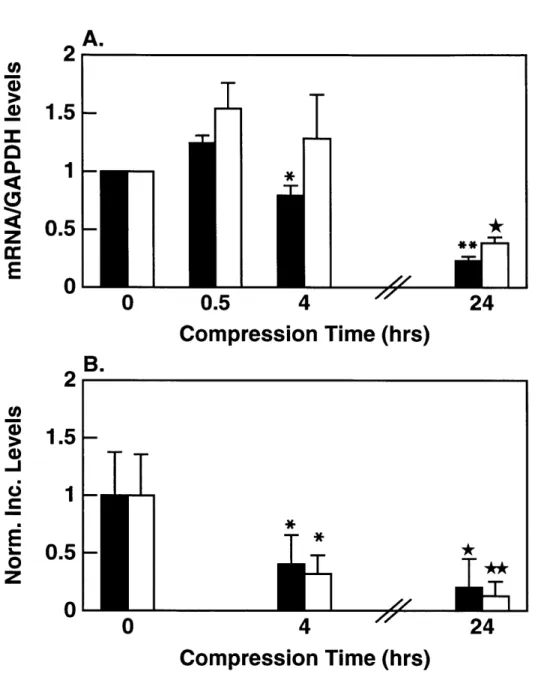
2.5 Aggrecan and type II collagen gene expression and biosynthesis
in response to increased duration of static compression to 0.5-mm thickness. A) mRNA levels for aggrecan G1 El and collagen type Ha E were quantified and normalized to GAPDH as a function of increased duration of compression. Values are reported as mean +/- SD (n=3) normalized to the 0 hour (control) value. *
p < 0.005 vs 0.5 hours; ** p < 0.005 vs 0.5 and 4 hrs; * p < 0.05 and 4 hrs. B) 35S-sulfate EL and 3H-proline l incorporation
into macromolecules as a function of increasing compression time. Values are reported as mean +/- SD (n=10) normalized to the 24
hour free-swell (control) value. * p < 0.001 vs 0 hrs, * p < 0.01 vs 0 and 4 hrs, * * p < 0.01 vs 0 and 4 hrs. . . . . 35 3.1 Alginate casting frame. A) Exploded view showing the steel
cast-ing frame (thickness = 1.5 mm) sandwiched between Whatman 2
filter paper supported by an 80 pm polyester mesh. B) The mesh, filter paper and frame are clamped together and the alginate with cells is injected into the 1.5 mm space between the two pieces of filter paper. The casting frame with alginate is then submerged into a bath of 102 mM CaCl2, 0.15 M NaCl for 10 minutes to
polymerize the alginate. . . . . 41
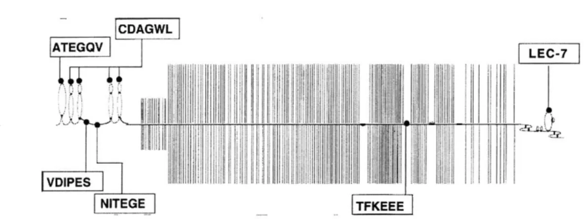
3.2 Schematic diagram showing the epitope sites used for analysis
of the aggrecan core protein. ATEGQV and CDAGWL (HAL) are used to examine the presence of the G1 region. VDIPES and NITGE are sites of aggrecanase activity in the interglobular domain. TFKEEE is used to assess aggrecanase activity in the CS-region of the core protein. LEC-7 detects the presences of the
G 3 dom ain. . . . . 47
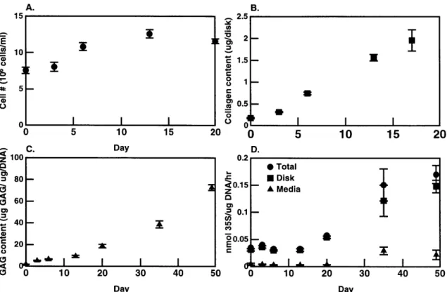
3.3 Cell number and matrix constituents as a function of time in
cul-ture (n=4/time point, avg +/- SD). A) Cell number was calculated
from DNA content using 7.9 X 10⁻¹² g/cell [42] on days 0, 3, 6,
13, and 20. B) Total collagen content in one 6-mm alginate disk
(n=4/time point) was measured from total hydroxyproline content [14] on days 0, 3, 6, 13, and 17. C) Total GAG was measured us-ing DMMB reactivity methods on days 0, 3, 6, 13, 20, 35 and 49. GAG content (pg GAG) was normalized to DNA content (pg
DNA). Corrections for the alginate background were made [131. D) Macromolecular 3 5S-sulfate incorporated into the disk and re-leased into the media was measured on days 0, 3, 6, 13, 20, 35, and 49. Incorporation rates were normalized to DNA content. . . 48
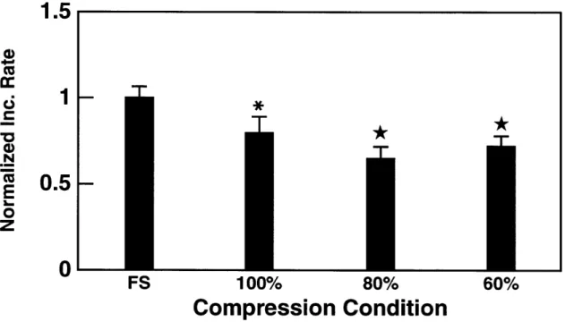
3.4 The effect of static compression on chondrocyte proteoglycan syn-thesis. 3
1S-sulfate incorporation into alginate disks decreased with increasing static compression. Maximal inhibition of macro-molecular synthesis occured at compression levels between 60% and 80% of the disks original thickness (* p < 0.05 vs FS; * p <
0.01 vs FS). . . . . 49
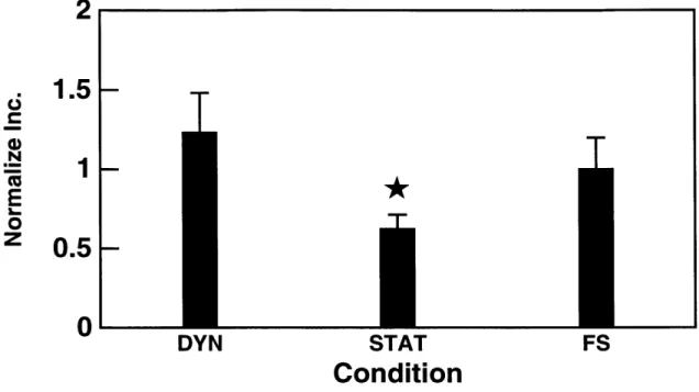
3.5 The effect of dynamic mechanical compression on chondrocytes
grown in alginate disks. A sinusoidal dynamic compression (0.5 Hz, 4% dynamic amplitude) superimposed on a 80%static com-pression increased 3 5S-sulfate incorporation in alginate disks by
1.9 times than in control disks subjected to 80% static
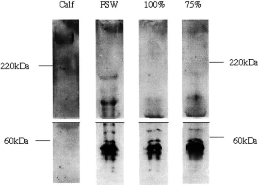
compres-sion alone (* p < 0.05 vs DYN and FS). . . . . 50 3.6 Western analysis of media samples from alginate disks cultured
for 40 hours at 75% of original disk thickness, 100% of original thickness, and under free swelling conditions. Media was pooled from 12 wells of the compression chamber or culture dish for each condition. Samples were analyzed with anti-CDAGWL (H) and anti-KEEE (E). Release of full length (400 kDa) aggrecan core protein and core protein fragments (300 kDA and 220 kDA) consistent with CS-domain aggrecanase activity are evident in all three conditions. . . . . 51 3.7 Western analysis of the cell-associated matrix of chondrocytes
cul-tured in alginate disks for 40 hours at 75% of original disk thick-ness, 100% of original thickthick-ness, and under free swelling condi-tions. Disks (n=12) were pooled, dissolved in a calcium chelating agent, and cells were pelleted via centrifugation. Samples were analyzed with anti-CDAGWL (H), anti-ATEGQV (A), anti-KEEE
(E), an 8-A-4 monoclonal antibody for link protein (L), and LEC-7 (3) for the G3 domain. Full length aggrecan core protein (400 kDa), a link protein doublet band (60 kDA) and core protein frag-ments consistent with normal chondrocyte catabolism (300 kDa) were evident for all conditions. There was no reactivity with the anti-NITEGE or anti-VDIPEN epitopes in all conditions. . . . . . 53
3.8 Western analysis of the further-removed matrix of chondrocytes
cultured in alginate disks for 40 hours at 75% of original disk thickness, 100% of original thickness, and under free swelling conditions. Disks (n=12) were pooled, dissolved in a calcium chelating agent, and the further-removed matrix was separated from cells via centrifugation. The supernatants were subject to analysis with anti-CDAGWL, anti-NITEGE and anti-KEEE. The FRM contained only small G1 fragments (detected with
anti-CDAGWL) that result from proteolysis of the interglobular
do-m ain . . . . . 54 4.1 Expression of aggrecan and type II collagen mRNA in response to
static (S) and dynamic (D) compression as assessed by Northern blot. Expression of mRNA for A) aggrecan G1 domain; B) collagen type Ila; C) GAPDH and D) EF-1a for normalization was evalu-ated for each condition. The blots shown are characteristic of the three repeats of the experimental protocol. Dynamic
compres-sion of 0.01 Hz, 4% dynamic strain amplitude was superimposed on a static compression to 1.0-mm (cut thickness); compression duration was for 12 hours. . . . . 65
4.2 Aggrecan and type II collagen gene expression and biosynthesis in response to 12 hours of dynamic loading of cartilage disks. A) mRNA levels for aggrecan LI and collagen type IIA LI were quan-tified and normalized to GAPDH. Values are reported as mean
+/-SD (n=3) normalized to the static (STAT) control value. B)
35S-sulfate L and 3H-proline
LI incorporation into macromolecules. Values are reported as mean +/- SD (n=20-24) normalized to the
static control. * p < 0.05 vs static control for 3H-proline
incorpo-ration . . . . . 66
4.3 Expression of aggrecan and type II collagen mRNA in response to static (S) and dynamic (D) compression as assessed by North-ern blot. Expression of A) aggrecan G1 domain; B) collagen type Ila; and C) EF-la for normalization was evaluated for each con-dition. Dynamic loading of 0.1 Hz, 4% dynamic amplitude was superimposed on a static compression to 1.0-mm (cut thickness); compression duration was for 48 hours. Lanes 1 through 4 repre-sent 2 repeats of the loading protocol, with static (S) and dynamic
4.4 Aggrecan and type II collagen gene expression and biosynthesis in response to 48 hours of dynamic loading of cartilage disks.
A) mRNA levels for aggrecan D and collagen type Ia E were quantified and normalized to EF-1a. Values are reported as mean
+/- SD (n=3) normalized to static (STAT) control value. B) 35S_
sulfate l and 3H-proline l incorporation into macromolecules. Values are reported as mean +/- SD (n=18) normalized to the
static control. * p < 0.001 vs static control. . . . . 67
4.5 Expression of aggrecan and type II collagen mRNA in response to static (S) and dynamic (D) compression in chondrocytes cultured in alginate disks as assessed by Northern blot. Expression of
A) aggrecan G1 domain; B) collagen type Ila; and C) GAPDH
for normalization was evaluated for each condition. Dynamic loading of 0.1 Hz, 4% dynamic amplitude was superimposed on a
90% static compression; compression duration was for 20 hours.
Lanes 1 through 6 represent 3 repeats of the loading protocol, with dynamic (D) and static (S) conditions alternating lanes. . . 68
4.6 Aggrecan and type II collagen gene expression of chondrocytes in alginate in response to 20 hours of dynamic loading. mRNA levels for aggrecan El and collagen type Ia l were quantified and normalized to GAPDH. Values are reported as mean +/- SD (n=3)
normalized to static (STAT) control value. In all cases, aggrecan mRNA was greater in the dynamic condition compared to static controls. . . . . 69 5.1 The biosynthetic response of chondrocytes in native explants to
two dosage levels of actinomycin D. 1-mm by 3-mm diameter cartilage disks were cultured for 1, 4, 9, 12 and 24 hours in 0, 10pg/ml and 50pg/ml. Disks were radiolabeld for 1 hour prior to being removed from culture with 35S-sulfate (10[Ci/ml) and 3
H-proline (20piCi/ml). Disks (n=4/condition) were then removed and washed 4 by 15 minutes in PBS to remove unincorporated label. Disks matched for joint depth and location were used for each time point group. Values are normalized to the control (0 [g/ml) for each time point. A) kinetics of 3 5S-sulfate incorporation; B) kinetics of 3H-proline incorporation. . . . . 76
5.2 The biosynthetic response of chondrocytes in native explants
sub-jected to mechanical compression and dosed with actinomycin
D. 1-mm by 3-mm diameter cartilage disks were cultured for 1,
2, 5, 11 and 24 hours in either 0 pg or 10 p/g of actinomycin
D with or with a superimposed 75% static compression. Disks
were radiolabeld for 1 hour prior to being removed from cul-ture with 35S-sulfate (l0pCi/ml) and 3H-proline (20Ci/ml). Disks
(n=4/condition) were then removed and washed in PBS to remove unincorporated label. Disks matched for joint depth and location were used for each time point group. Values are normalized to the
control (0 pg/ml Free Swelling) for each time point. A) kinetics of 3 5S-sulfate incorporation; B) kinetics of 3H-proline incorporation. 77
5.3 Northern blot analysis of chondrocytes cultured in alginate disks
and dosed with 10 pg of actinomycin D for 1, 2, 4, 8, 12 14 and
18 hours. After time in free swelling culture, total RNA was
extracted using the Qiagen RNeasy protocol for animal cells. Ap-proximately 2 pg of RNA was run in each lane. Expression of A) both aggrecan (9.0 kb) and GAPDH (1.4kb); B) collagen type IIa; and C) EF-1 was evaluated at each time point. D) 28S (ethid-ium bromide) was used for normalization. Each lane represents increasing times in drug-treated culture. . . . . 78 5.4 Kinetics of changes in mRNA levels after transcription-inhibition
via actinomycin D. Values are normalized to 28S. Increasing sta-bility is seen with A) aggrecan and B) collagen type Ha. C) Elon-gation factor-la remained unchanged. D) GAPDH decreased to less than half of its starting levels. . . . . 79
A. 1 Equilibrium stress-strain behavior for alginate disks from day 0
of culture. The dynamic stiffness and streaming potential were measured in the linear region of the data (30% static strain). . . 86 A.2 Equilibrium confined compression modulus HA of alginate disks
versus time in culture. These were calculated from the slope of the stress-strain curve in the linear region of the data for each sam ple. . . . . 87 A.3 Amplitude of the streaming potential for frequencies ranging
from 0.02 to 1.0 Hz, at four time point during a 7 week culture of alginate. Displacement amplitude was 45pm. . . . . 87 A.4 Amplitude of the dynamic stiffness of alginate at frequencies
be-tween 0.02 and 1.0 Hz at days 0, 15, 29, 56 in culture. Displace-ment amplitude was 45pm. . . . . 88
A.5 Dedifferentiated human chondrocytes (2nd passage) were cast in
alginate slabs and cultured for up to 56 days. At days 0, 15, 29, and 56, the mechanical integrity of newly synthesized matrix was assessed. No significant differences in equilibrium modulus, HA,
and streaming potential were evident between alginate samples with and without cells (data not shown). Significant differences
in dynamic stiffness were evident at all frequencies (p < 0.005). 89
B. 1 Total GAG content of human cartilage explants maintained in culture for up to eleven days. Glycosaminoglycan content was assess using the DMMB dye method. . . . . 94 B.2 Aggrecan synthesis (as measured by macromolecular sulfate
in-corporation) as a function of time in culture. . . . . 95
B.3 Total protein synthesis (as measure by macromolecular proline incorporation) as a function of time in culture. . . . . 96
B.4 Cartilage disks (1-mm by 3-mm diameter) were cored from human cartilage and statically compressed to either 1.0-mm (control) or to 0.5-mm (50% compression). Compression lasted either 12 or 24 hours, with 35S-sulfate incorporation assessed during the last 12 hours of culture. . . . . 97
B.5 Cartilage disks (1-mm by 3-mm diameter) were cored from human cartilage and statically compressed to either 1.0-mm (control) or to 0.5-mm (50% compression). Compression lasted either 12 or 24 hours, with 3H-proline incorporation assessed during the last
12 hours of culture. . . . . 98
B.6 Cell density of primary human chondrocytes in alginate disks versus time in culture. Cell number was calculated from DNA content using 7.9 X 10¹² g/cell [42]. . . . . 99
B.7 Total GAG content accumulated by primary human chondrocytes
in alginate versus time. GAG content was assessed using a mod-ified DMMB method that accounted for the background charge of the alginate. . . . 100 B.8 Macromolecular 31S-sulfate incorporation into the alginate disk
and released into the media versus time in culture. The multi-screen 35S-sulfate incorporation assay [52] was used in analysis. 101 B.9 Cell density of dedifferentiated human chondrocytes in alginate
disks versus time in culture. Chondrocytes were initially seeded at 1 X 10⁵ or 1 X 10⁶ cells/ml. Cell number was calculated from
DNA content using 7.9 X 10⁻¹² g/cell [42]. . . . 102
B. 10 Macromolecular 35S-sulfate incorporation into the alginate disk
and released into the media versus time in culture. The multi-screen 3 5S-sulfate incorporation assay [52] was used in analysis. 103
B. 11 Cell density of dedifferentiated human chondrocytes in alginate disks versus time in culture. Chondrocytes were initially seeded at 6 X 10' cells/ml. Cell number was calculated from DNA content using 7.9 X 10⁻¹ g/cell [42]. . . . 104 B. 12 3 5S-sulfate incorporation by dedifferentiated human chondrocytes
into the alginate disk and released into the media versus time in culture. The multiscreen 31S-sulfate incorporation assay [52] was used in analysis. . . . 105
C.1 3 5S-sulfate incorporation of chondrocytes grown in ARC method subjected to dynamic, static and free swelling conditions. The top figure shown radiolabel incorporation into the cartilage-like disk. The bottom figure shows macromolecular radiolabel released into the media during culture . . . . .. ... 110 C.2 Top: Total 31S-sulfate incorporation of chondrocytes grown in
ARC method subjected to dynamic, static and free swelling con-ditions. Bottom: Total GAG normalized to DNA content of the cartilage-like disks for each compression condition. . . . 111
C.3 35S-sulfate incorporation of chondrocytes grown in ARC method subjected to dynamic, static and free swelling conditions. The top figure shown radiolabel incorporation into the cartilage-like disk. The bottom figure shows macromolecular radiolabel released into
the media during culture. This is a repeat of the first series of experim ents. . . . 112
C.4 Top: Total 35S-sulfate incorporation of chondrocytes grown in ARC method subjected to dynamic, static and free swelling con-ditions. Bottom: Total GAG normalized to DNA content of the cartilage-like disks for each compression condition. . . . 113
Chapter I
Introduction
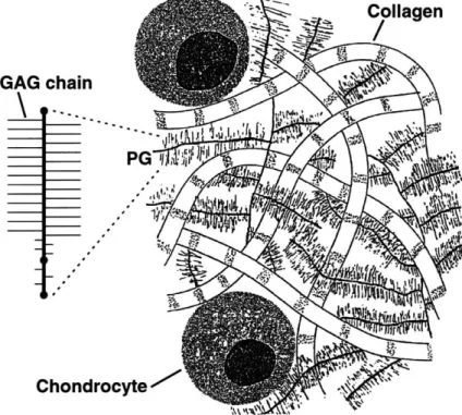
Articular cartilage covers the bony surfaces in synovial joints and functions to with-stand mechanical loads and reduce friction and wear. It is avascular, aneural, alym-phatic and highly hydrated. This connective tissue is primarily comprised of extra-cellular matrix (ECM) molecules, which include proteoglycans (PGs), glycoproteins, and collagens. Aggrecan, the proteoglycan found most abundantly in articular carti-lage, has a large core protein (400 kDa) to which glycosaminoglycan (GAG) chains are attached. The negatively charged GAG chains provide cartilage's resistance to compressive forces normally occurring during joint loading. Type II collagen is the dominant collagen type in articular cartilage. Type II collagen molecules are ho-motrimers of a single type of alpha chain. Molecules of types II, IX, and XI together form the heteropolymeric fibrils, which account for the most of the tensile strength of the tissue. Aggrecan and the type II collagen network, along with other matrix molecules, form the functional matrix of cartilage that withstands static and dynamic loads associated with physiologic conditions.
Articular cartilage is synthesized and maintained by a specialized cell popula-tion known as chondrocytes 1.1. Recent research has shown that chondrocytes are very sensitive to mechanical forces in their environment, and they can alter their syn-thesis of extracellular matrix (ECM) molecules to meet the functional demands of in vivo loading [68, 43]. Presently it is not known how mechanical forces cause changes in the gene expression or other intracellular pathways that regulate chondrocyte syn-thesis of ECM molecules. One difficulty impeding such investigations has been the technical challenges associated with isolating critical intracellular molecules, such as message RNA (mRNA) for analysis of gene expression [1] due to the sparse cell pop-ulation of chondrocytes in cartilage. Yet another challenge has been the inability to isolate living chondrocytes immediately following exposure to physiologic loading
conditions for analysis of other signal transduction marker molecules. Some progress GAG chain Collagen
PG
Iy / *( 11%)'4 cytetFigure 1.1: Chpndrocytes synthesize and maintain the macromolecules which form the bulk extracellular matrix (ECM) of articular cartilage. Aggrecan and type II collagen, as well as other macromolecules, cytokines, and degradative enzymes can influence cell metabolism. Mechanical compression can influence the synthesis of these components, and can mediate chondrocyte metabolism of the ECM.
has been made to elucidate the influence of mechanical forces on chondrocyte gene expression and intracellular pathways using various explant or isolated cell culture systems. The following sections describe previous research that has focused on such investigations.
1.1
Explant Model Systems and Chondrocyte Cellular
Pathways
Tissue explant systems have been used extensively to elucidate intracellular path-ways for chondrocyte synthesis of extracellular matrix molecules [30, 53] and how
sensitive to media, pH, potassium and sulfate concentrations and to the concentra-tions of serum and various growth factors [53, 24, 8]. Mechanical forces also affect chondrocyte metabolism. Increased levels of static compression has been shown to decrease synthesis of aggrecan and total protein either as measured by 35S-sulfate and 3H-proline radiolabel incorporations [24, 68, 411, whereas application of dynamic
compression at frequencies 0.01 to 1 Hz and 1-5% dynamic amplitude have been shown to increase synthesis of aggrecan and total protein [68, 43].
Others investigations have focused more specifically on the interplay between mechanical compression and aggrecan synthesis. The aggrecan core protein con-tains three globular domains, G1, G2 and G3, and intervening regions to which gly-cosaminoglycan (GAG) chains and oligosaccharides are attached [82]. Synthesis of aggrecan involves RNA-dependent synthesis of the core protein on ribosomes of the rough endoplasmic reticulum (ER) followed by post-translational events in which
GAG chains and oligosaccharides are enzymatically added to the polypeptide core.
Previous experiments have shown that static compression inhibits the synthesis of new core protein, but does not seem to alter the processing rate of existing core protein and addition of GAG chains [41] These results suggest that mechanical compression can influence aggrecan synthesis at the transcription level or prior to translation of the core protein.
Additional studies in which aggrecan RNA transcription was inhibited using actinomycin D (which blocks total RNA transcription) in bovine cartilage explants showed an exponential decay in aggrecan synthesis measured by 35S-sulfate incorpo-ration [54]. The presence of benzyl
#-xyloside
(when added to actinomycin D dosed media) delayed the inhibition of synthesis by approximately 10 hours, but ultimately aggrecan synthesis decreased at the same rate as non-#-zyloside treated controls [54]. Therefore, the decrease in aggrecan synthesis could be a result of inhibition of mRNA encoding for the aggrecan core protein or mRNA encoding for other enzymes neces-sary for GAG synthesis, such xylosyl transferase or glycosyl transferases [41].Ultimately, mechanisms by which mechanical compression affects chondrocyte aggrecan and type II collagen gene expression in cartilage explants still remains to be understood. However recent study of gene expression in explant cultures of bovine tendons implicated the importance of mechanical compression and expression of ag-grecan and type II collagen messages [65]. Investigators harvested bovine fetal and adult deep flexor tendons (DFT) which have regions which are exposed to predom-inantly tensile and compressive forces. The goal of the study was to determine if any differences in gene expression existed between adult and fetal tissue and in the compressed versus stretched regions of the tendon [65]. Using isolated total RNA and Northern blot techniques with probes for aggrecan, type II collagen and GAPDH, Robbins and Vogel showed that aggrecan and type II collagen mRNA levels were at least 50 times larger in the compressed regions of the adult DFT compared to re-gions loaded in tension in the same adult tendon, and also compared to both tension and compression regions in fetal tissue. This strongly supports the hypothesis that mechanical forces can alter cell phenotypic expression.
Most recently, there have been a reports of the influence of mechanical com-pression on aggrecan mRNA levels in bovine cartilage explants [78, 81, 63]. Static compression applied for times greater than 12 hours caused a 50% decrease in normal-ized values of aggrecan mRNA [81, 63]and in type II collagen mRNA as compared to uncompressed controls [63]. In addition, normalized 3
1S-sulfate and 3H-proline incorporation as markers for aggrecan and type II collagen synthesis, respectively, decreased in compressed tissue versus controls with trends similar to those found in mRNA levels [63]. However, more rigorous studies are necessary to elucidate how mechanical forces affect stability and synthesis of the mRNA inside the chondrocytes.
1.2 Mechanical Forces on Isolated Chondrocytes
chon-molecules that rapidly degrade. Therefore, researchers have turned to initially isolat-ing chondrocytes from native tissue, platisolat-ing them in high density ( 2 X 10' cells/cm2)
monolayer cultures, and then subjecting the cells to various mechanical forces, such as fluid shear [75] hydrostatic [80, 76] or tensile forces [38]. While these systems are useful models, there have been reports of altered gene expression or protein synthesis for chondrocytes cultured in such conditions as compared to chondrocytes suspended in agarose gels or in native tissue [45, 36]. Specifically, Hering et al reported drastic decreases in type II collagen, link protein, and aggrecan gene expression during the first 5 days of monolayer culture, and also show an increase in mRNA for type I collagen and decorin which more characteristic of fibroblastic cell lines [36] .
Isolated chondrocytes seeded in three-dimensional matrices have been reported to maintain chondrocyte morphology and phenotype that closely resembles that of chondrocytes in native tissue [29, 56, 45, 11, 32, 48, 64]. When seeded in gel matri-ces such as agarose and alginate, cells retain their rounded shape but undergo some mitoses, as demonstrated by a 50-100% increase in cell number over the initial three to four weeks of culture [45, 11, 64]. Typically by week 4 of culture, chondrocyte synthesis rates of proteoglycans and collagens approach those of chondrocytes in na-tive cartilage, and total GAG and collagen content increase over the course of the culture confirming matrix synthesis [11, 64]. More detailed studies of chondrocytes in alginate have shown production of aggrecan as part of cell-associated matrix pro-duction [56]. These studies demonstrate the effectiveness of chondrocyte suspension cultures in retaining metabolic and morphologic states that more closely resemble that of chondrocytes in articular cartilage.
Chondrocyte/agarose cultures have been used to investigate the influence of mechanical compression on isolated cells during culture [20, 11, 48]. Using the agarose system, Buschmann and colleagues have shown that chondrocyte 3"S-sulfate and 3 H-proline incorporation rates decrease with increasing amounts of static compression. Compressive effects were more pronounced and similar to native cartilage at day 41
compared to day 2 in culture. They also demonstrated that dynamic compression at frequencies and amplitudes similar to previous cartilage explant experiments stimu-lated chondroctye production of proteoglycans and collagen; again more pronounced effects were evident at later times (day 25 versus day 4) in culture [11]. Lee and Bader demonstrated stimulatory effects of dynamic loading on 3H-thymidine uptake,
suggesting that chondrocyte mitosis is upregulated by dynamic compression frequen-cies ranging between 0.3 Hz to 3 Hz
[48].
Together, these studies have shown that both static and dynamic loads can affect isolated chondrocyte metabolism and extra-cellular matrix synthesis similar to effects demonstrated with cartilage explants. The advantages of the agarose system relate to maintaining chondrocyte morphology; however cells maintained in agarose, like those within cartilage, cannot be rapidly released from the gel matrix for intracellular analyses. Other models systems for isolated chondrocyte culture are necessary for further investigation of the affects of mechanical forces on intracellular pathways of ECM molecule synthesis.Chapter II
Down-Regulation of Chondrocyte Aggrecan and
Type II Collagen Gene Expression Correlates
With Increases in Static Compression Magnitude
and Duration
2.1 Summary
The goal of this study was to examine the simultaneous effects of mechanical com-pression of chondrocytes on messenger RNA (mRNA) excom-pression and macromolecular synthesis of aggrecan and type II collagen. Bovine cartilage explants were exposed to different magnitudes and durations of applied mechanical compression and lev-els of aggrecan and type Ila collagen mRNA normalized to GAPDH were measured and quantified by Northern blot. Synthesis of aggrecan and type II collagen pro-tein was measured using radiolabel incorporation of "S-sulfate and 3H-proline into
macromolecules.
Results showed a dose-dependent decrease in mRNA levels for aggrecan and type II collagen with increasing compression relative to physiological cut thickness applied for 24 h. Radiolabel incorporation into glycosaminoglycans (GAGs) and collagen also decreased with increasing compression in a dose-related manner similar to the changes seen in mRNA expression. The modulation of both aggrecan and type II collagen mRNA and protein synthesis were dependent on the duration of the compression. Aggrecan and type II collagen mRNA expression increased during the initial 0.5 hours of static compression, but by 4 to 24 hours after application of compression, total mRNA levels had significantly decreased. The synthesis of aggrecan and collagen protein decreased more rapidly than the rate of decrease in
mRNA levels after the application of a step compression. Together, these results suggest that mechanical compression rapidly alters chondrocyte aggrecan and type II collagen gene expression rapidly upon application of load. However, our results indicate that the observed decreases in biosynthesis may not be related solely to changes in mRNA expression. The mechanisms by which mechanical forces affect different segments of the biosynthetic pathways remain to be determined.
2.2
Introduction
Articular cartilage functions to withstand compressive load and allow fluid joint ar-ticulation. Like other biological tissues, cartilage can adapt its structure to meet the functional demands of its surrounding environment. For example, moderate cyclic loading has been observed to stimulate proteoglycan synthesis and accumulation in vivo [34]. In contrast, reduced joint loading or immobilization results in more rapid degradation and loss of extracellular matrix (ECM) and decreased synthesis of ECM macromolecules [5, 67]. These changes in cartilage matrix turnover may ultimately result in loss of articular cartilage, causing joint pain, decreased mobilization, and significant morbidity.
The functional mechanical properties of cartilage depend on the composition and quality of the ECM. Cartilage contains aggregating and non-aggregating proteo-glycans, collagen fibrils and non-collagenous proteins. These molecules are distributed in defined patterns throughout the cartilage matrix and interact to efficiently main-tain water balance and prevent collapse of the tissue. These ECM components are synthesized, deposited, and maintained by articular chondrocytes. Recent studies have shown that chondrocytes are very sensitive to mechanical forces and that they respond by altering the synthesis of ECM molecules, such as aggrecan, type II colla-gen, link protein, and hyaluronan [24, 68, 41, 28]. Additionally, studies have shown that mechanical compression can lead to the restructuring of intracellular organelles
the synthesis of the ECM [26].
Mechanical forces can also lead to changes in chondrocyte gene expression. In recent studies [80, 76, 38], the effect of various mechanical forces on gene expression were investigated using chondrocytes in monolayer culture. For example, static and intermittent hydrostatic pressure increase the expression of transforming growth fac-tor 3-1 (TGF-31) as well as aggrecan and type II collagen mRNA in high-density monolayer cultures [80, 76]. Additionally, constant fluid shear forces stimulate ex-pression of mRNA for tissue inhibitor of metalloproteinase-1 (TIMP-1) in isolated human chondrocytes grown in monolayer [75]. Dynamic mechanical forces have also been shown to influence matrix gene expression. When isolated bovine and human chondrocytes are cyclically stretched on flexible membranes, aggrecan and type II col-lagen mRNA expression are increased [38]. Isolated chondrocyte systems are useful models for investigating chondrocyte response to mechanical load. However, extrap-olating information obtained from isolated cells in monolayer culture to chondroctyes maintained in their native tissue is difficult due to the complex physicochemical in-teractions that exist between the chondrocyte and the ECM in vivo.
Presently, there is limited information regarding the effects of compression on chondrocyte gene expression within native articular cartilage. One study reported that static loads of 0.1 MPa applied for 1 hour can transiently increase levels of chondrocyte aggrecan mRNA [81]. In addition, aggrecan mRNA levels are found to be independent of the magnitudes of applied stress during 24 hours of static compression
[81]. In contrast, many previous studies have reported a dose-dependent reduction of
aggrecan synthesis (as measured by 31S-sulfate incorporation) with increased amounts static compression [73, 24, 68, 43, 28]. The goal of the present study was to examine the influence of static mechanical compression on chondrocyte synthesis of aggrecan and type II collagen both at the protein and mRNA levels. Our data suggest that mechanical compression can modulate both chondrocyte gene expression, as well as post transcriptional biosynthesis.
2.3
Methods
Explant and Culture: Cylindrical disks of cartilage were prepared as previously
described by Sah et al (Fig. 2.1)[68]. Briefly, the saddle sections of 1-2 week old calves were obtained from a local abattoir within 4 hours after slaughter (Research '87, Boston, MA). Four to five cylindrical cores of cartilage and underlying bone (9.5-mm in diameter and 20-mm deep) were drilled from each facet (medial and lateral) of the femoropatellar groove. During the process, the cartilage was kept moist using sterile phosphate buffered saline (PBS) supplemented with antibiotics (100 U/ml penicillin,
100 pg/ml streptomycin, and 0.025 gm/ml amphotericin B).
Cartilag e-one 'xCyHinders 1mm Thick 2 3mm Diameter CARTILAGE DISKS ~eeee G D ee e 5 CALF ARTICULAR CARTILAGE femoropatellar groove
Figure 2.1: Schematic of the cartilage disk explant protocol. Larger 12-mm plugs are cored from the medial and lateral femoropatellar grove. Four 3-mm disks are punched from the 1-mm by 12-mm diameter cores. Groups are matched for disk depth and location.
Each cartilage-bone core was then inserted into the sample holder of a sledge microtome (Model 860, American Optical, Buffalo, NY). After removing the superfi-cial layer of cartilage to obtain a level surface, two 1-mm thick plane-parallel cartilage slices were dissected and placed in 1-ml PBS with antibiotics. From these slices (9.5 mm diameter, 1-mm thick), four 3-mm disks were obtained using a sterile dermal punch (Miltex Instruments, Lake Success, NY) giving a total of 76 disks from each joint surface. The disks were incubated in medium (Dulbecco Modified Eagle Medium
amino acids, antibiotics as described previously, 1 mM sodium pyruvate, an addi-tional 0.4 mM proline and 20 pg/ml ascorbate) at 370 C in 5% CO2. The total time
from slaughter to incubation was < 8 hours. Disks were cultured for four days and
the medium was changed daily.
Static Compression: Dose Dependence: Using specially designed
polysul-phone chambers described previously [68], cartilage disks were uniaxially compressed in a radially unconfined manner.
Cartilage disks were maintained between fluid-impermeable platens at their free swelling thickness of 1.15-mm or compressed to 1.0 mm (cut thickness), 0.7-mm, and 0.5-mm. Each disk was bathed in > 100 volumes of medium and maintained in an incubator at 370 C and 5% CO2 for 24 hours. Cartilage disks in each condition were
matched for joint position and tissue depth, as previous studies have reported joint location dependent variations in chondrocyte response to mechanical compression
[68].
After compression for 24 hours, disks ( 300 mg: 32 disks/condition) were flash-frozen in liquid nitrogen within 1 minute of removal from the compression chambers, and stored at -800 C prior to RNA isolation for Northern blot analysis. Total RNA was isolated from the cartilage plugs by a modified method of Adams et al. [1].
All reagents were nuclease-free. Frozen cartilage was powdered in a Spex Freezer
Mill (SPEX, Metuchen, NJ) and immediately homogenized in 4M guanidinium isoth-iocyanate solution, 0.3% 0-mercaptoethanol (Gibco BRL, Gaithersburg, MD). The extracts were clarified by centrifugation at 1,500 X g for 10 minutes at 40 C and the supernatant was removed. The extraction of the pellet was repeated and the super-natants were pooled and brought to 1% Triton X-100 and 1.5 M sodium acetate, pH
6.0. The supernatants were then extracted with an equal volume of chlorform/phenol
(1:4) on ice for 10 minutes. The extraction and separation of the aqueous phase was repeated twice in equal volumes of chloroform/isoamyl alcohol (1:1). The RNA in the aqueous phase was precipitated with equal volumes of isopropanol on ice for 5
minutes followed by centrifugation at 15,000 X g for 30 minutes. The RNA pellet was subsequently purified using the Qiagen RNeasy kit according to the manufacturer's instructions. RNA was quantified spectrophotometrically and structural integrity was confirmed by electrophoresis. Equivalent amounts of RNA (5 jg) from each condi-tion were loaded onto agarose gels and mRNA expression was analyzed on Northern blots using 3 2P-labeled probes for bovine collagen Ila, collagen Ilb, the GI region of aggrecan [70], and GAPDH. Northern blots were scanned and quantified using phosphorimaging. Aggrecan GI and collagen Ila levels were normalized to that of
GAPDH. [65, 9]. Other normalization standards including total DNA, total RNA
and elongation factor-la (EF-la) were measured. mRNA levels at each compression were normalized to the those at 1.15 mm (FS) and are reported as the mean from three separate experiments (n=3) +/- SD.
To measure matrix biosynthesis, 4-6 disks per condition were dual-radiolabeled with 10 pCi/ml 31S-sulfate and 20 pCi/ml 3H-proline during the 24-hour compression period. Previous control studies using this same newborn calf explant culture system showed that more than 98% of 3
1S-sulfate incorporation was into macromolecules, predominantly aggrecan, in both compressed and free swelling (FS) control disks
[68, 69]. Furthermore, in free swelling controls, over a 24 hour label period, 90% of
the 3H-proline incorporation was into macromolecules, 83% of which was 3H-collagen
[69]. After the 24 hours of compression, disks were washed four times for 15 minutes
each in PBS with 0.8-mM sodium sulfate and 1 mM proline to remove free label, lyophilized, and then digested overnight with papain. Digests were analyzed for DNA content using the Hoechst 33258-dye assay [42], total sulfated glycosaminoglycan
(GAG) content by the DMMB dye [16], and 3H proline and 3
1S-sulfate incorporation
by scintillation counting [68]. Incorporation levels of 3
1S-sulfate and 3H-proline were normalized to that in free swelling control disks. Values are reported as the mean of 4-6 disks from 2 experiments (n=10) +/- SD.
Static Compression: Kinetics of Response Using the same chambers
described above, cartilage disks were statically compressed to 0.5-mm thickness and maintained in culture for 0 (uncompressed controls), 0.5, 4 and 24 hours. Matched cartilage disks were used in each condition as described above. After culture, disks
( 300 mg: 32 disk/condition) were flash-frozen in liquid nitrogen and RNA was isolated
for Northern blot analysis. Northern blots were examined for mRNA expression of aggrecan, types Ila anb IIb collagen, and inducible nitric oxide synthase (iNOS) [21]. Signals were standardized using GAPDH, and data were normalized to uncompressed controls. Values are reported as the mean of three separate experiments (n=3)
+/-SD. In addition, 4-6 disks per condition were radiolabeled with 10 pjCi/ml 3 5S-sulfate and 20 puCi/ml 3H-proline during 4 and 24 hours of static compression to 0.5-mm. An additional 4-6 plugs were maintained under free swelling conditions for 24 hours. This condition was used as the "0-hour" control for evaluation of the kinetics of radiolabel incorporation after application of static compression. After culture, these disks were washed to remove free label, lyophilized, and then digested with papain. Digests were analyzed for DNA content, GAG content, and radiolabel as above. Radiolabel incorporation values were normalized to those of 0-hour control disks. Values are reported as the mean of 4-6 plugs from 2 experiments (n=10) +/- SD.
2.4 Results
Dose Dependence of Aggrecan and Type II Collagen Expression and Synthesis: Cartilage disks were compressed up to 50% of the cut thickness and
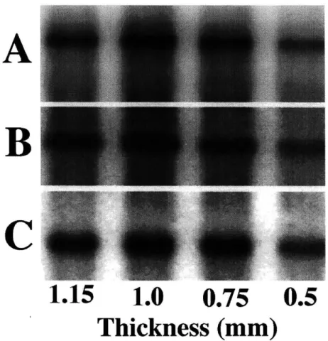
compared to a free-swelling control. Aggrecan GI (9.0 kb), collagen Ila (4.4 kb), and GAPDH (1.4 kb) signals were evident in all lanes on Northern blot analysis for the four compression conditions (Fig. 2.2). Normalized aggrecan mRNA expression decreased significantly (p < 0.05 )with increasing amounts of static compression from
cut thickness (1.0 mm) to 0.5-mm (Fig. 2.3A). Collagen Ila mRNA expression showed similar trends, although compressed values were not statistically different from the
1.15-mm control (Fig. 2.3A). Collagen lIb mRNA levels showed identical results to collagen Ila levels (data not shown). Maximum normalized aggrecan (1.34 +/- 0.18) and collagen Ila (1.01 +/- 0.13) expression occurred at cut thickness (1.0 mm) (Fig. 2.3A). These same dose-related trends were also observed when aggrecan and collagen
Ila mRNA levels were normalized to total DNA, total RNA, or EF-1a mRNA levels on a specimen-by-specimen basis (data not shown). To determine if these changes in gene expression were reflected in biosynthesis of aggrecan and type II collagen, parallel cultures were compressed and dual-labeled with 35S-sulfate and 3H-proline to
quantify proteoglycan or protein synthesis, respectively. Normalized values for 35S_
sulfate incorporation and 3H-proline incorporation decreased significantly (p < 0.05)
with increasing compression from cut thickness (1.0 mm) to 0.5-mm thickness with maximum levels occurring at cut thickness (Fig. 2B). Total GAG and DNA values were not statistically different between groups.
Kinetics of Changes in Aggrecan and Type II Collagen: To
deter-mine the kinetics of the reduction in gene expression observed at 0.5 mm-thickness, disks were compressed for times ranging between 30 minutes and 24 hours. Aggre-can G1 (Fig. 2.4A), collagen Ila (Fig. 2.4B), collagen Ilb (data not shown) and
GAPDH (Fig. 2.4D) mRNA signals were evident in all lanes for the different
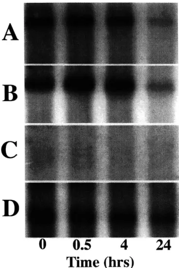
du-rations of compression; collagen Ila and Ilb showed identical trends. In contrast, no expression of chondrocyte inducible nitric oxide synthase (iNOS) mRNA was seen under any condition (Fig. 2.4C). Within the first 30 minutes of compression, there was an increase in aggrecan (1.24 +/-0.07) and collagen Ila (1.54 +/-0.22) mRNA compared to 0 hour controls. However, after the initial 30 minutes, total aggrecan and collagen Ila normalized to GAPDH showed statistically significant decreases (p
< 0.05) with increasing amounts of time under static compression (Fig. 2.5A). By
24 hours of compression, total aggrecan GI and collagen Ila mRNA levels decreased to approximately 35% of 0-hour values. In contrast, we have previously shown that IL-1 stimulates chondrocyte iNOS mRNA expression and iNOS production [4] in
ex-planted bovine tissue, whereas in our similar culture system with applied mechanical load, no increased expression of iNOS mRNA was observed under conditions where aggrecan and type II collagen expression was reduced.
Normalized values for 31S-sulfate and 3H-proline incorporation also decreased
significantly (p < 0.01) with increasing compression time (Fig. 2.5B). Due to the
lim-itations of equilibration of the radiolabel precursors, a 0.5 hour time point could not be measured for incorporation studies. However at later times, there was a monotonic decrease in the incorporation of 35S-sulfate and 3H-proline into ECM macromolecules.
This time-related decrease was also observed for aggrecan and collagen Ila mRNA (Fig. 2.5A). The data presented in these graphs represent averages of data from 3 separate experiments. However, the trends evident in the averages were present
within individual experiments and are not artifacts of data analysis (data not shown). Finally, total GAG and DNA values were not statistically different between groups.
2.5 Discussion
The major findings during application of a 24 hour static compression were that aggre-can and collagen Ila mRNA levels decreased with increasing compression relative to the levels at physiological cut thickness (Fig. 2.3A). Concomitantly, 3 5S-sulfate and
3H-proline incorporation also decreased in a dose-dependent manner with increasing
compression up to 50% of cut thickness (Fig. 2.3B). Previous control studies us-ing this same newborn calf explant culture system showed that more than 98% of
35S-sulfate incorporation was into macromolecules, predominantly aggrecan, in both
compressed and free-swelling control disks [68, 69]. Furthermore, in free swelling con-trols, over a 24 hour label period, 90% of 3H-proline incorporation was into
macro-molecules, 83% of which was 3H-collagen [69]. Thus, a 24-hour static compression
caused decreases in both aggrecan and collagen gene expression and protein synthe-sis. Previous studies [41, 43] showed that such compression to 50% did not cause cellular damage, and that the decreases seen in proteoglycan and protein synthesis
A
B
C
1.15
1.0
0.75
0.5
Thickness (mm)
Figure 2.2: Expression of aggrecan and type II collagen mRNA in response to static compression as assessed by Northern blot. Expression of A) aggrecan GI domain; B) collagen type Ha; and C) GAPDH for normalization was evaluated at the compressed thickness shown. Lanes 1 through 4 show increasing amplitudes of static unconfined compression (1.15-mm (FS) to 0.5-mm); compression duration was 24 hours.
2
mRNA Levels
1.5- 0.51-1.15 1.0 Thic 0.70 kness (mm) * Aggrecan G1 o Collagen Ila 0.50 2Label Incorporation
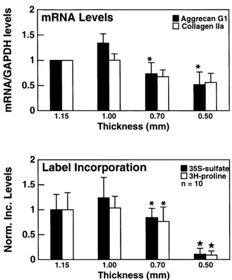
1.5- 0.5-1.15 1.00 0.70 Thickness (mm) * 35S-sulfat o 3H-proline n = 10 T* 0.50Figure 2.3: Aggrecan and type II collagen gene expression and biosynthesis in response to graded static compression of cartilage disks. A) mRNA levels for aggrecan l and collagen type Ila l were quantified and normalized to GAPDH as a function of increasing amounts of static unconfined compression. Values are reported as mean
+/- SD (n=3) normalized to the 1.15-mm (free-swell) value. * p < 0.05 vs 1.00-mm.
B) 15S-sulfate l and 3H-proline L incorporation into macromolecules as a function of increasing amounts of static unconfined compression. Values are reported as mean +/- SD (n=10) normalized to the 1.15-mm (free swell) value. * p < 0.05 vs 1.00-mm;
* p < 0.0001 vs all other thicknesses. The correlation coefficients, r, relating mRNA
expression to radiolabel incorporation rates were calculated for aggrecan and type IIA collagen. The values are: raggrecan = 0.90; rcojuagenIIA= 0.89.
U) I
E
"-C 0Z
A
C
0
0.5
4
24
Time (hrs)
Figure 2.4: Northern blot analysis for one of three similar experiments to examine kinetics of mRNA change due to 50% static compression using bovine probes for: A) aggrecan GI domain; B) collagen type Ila; C) iNOS; and D) GAPDH for normaliza-tion. Lanes 1 through 4 show increasing duration of compression for disks held at 0.5-mm thickness (0 hrs (control) to 24 hrs).