BACTERIOLOGICAL SAMPLING IN PERITONEAL DIALYSIS FLUID :
DECREASING THE NUMBER OF STERILE PERITONITIS
PRÉLÈVEMENTS À VISÉE BACTÉRIOLOGIQUE DES LIQUIDES DE DIALYSE PÉRITONÉALE. COMMENT LIMITER LE NOMBRE DE PÉRITONITES À CULTURE NÉGATIVE ?
Antoine Grillon1, Pierre-Hugues Boyer1 et Françoise Heibel2, 1Laboratoire de Bactériologie, Hôpitaux Universitaires de Strasbourg
2Service de Néphrologie, Hôpitaux Universitaires de Strasbourg
Résumé
Les péritonites sont une complication majeure et grave en termes de morbi-mortalité chez les patients traités par dialyse péritonéale. Leur diagnostic microbiologique est compliqué tant au point vu de la détection des agents étiologiques des péritonites que de l’interprétation des cultures positives.
De nombreux micro-organismes peuvent être à l’origine de cette infection ; germes « classiques » comme les staphylocoques à coagulase négative ou les entérobactéries, mais également des bactéries « atypiques », de culture ou de détection plus fastidieuse.
Pour mettre en évidence les bactéries responsables, des techniques de biologie moléculaire et de culture peuvent être mises en place. La biologie moléculaire (particulièrement la recherche universelle (ADNr 16s)) permet d’identifier d’éventuels germes atypiques, mais ne permet pas la réalisation d’un antibiogramme.
La culture des liquides de dialyse péritonéale reste donc le « gold-standard » pour le diagnostic de ces infections. Celle-ci doit être néanmoins optimisée pour abaisser son seuil de sensibilité.
Le diagnostic étiologique des péritonites chez les patients traités par dialyse péritonéale peut être difficile, mais la microbiologie moderne combinée à une discussion bactério-clinique permet l’identification du germe responsable de l’infection dans la très grande majorité des cas.
B
D
D
Mots clés : dialyse péritonéale, péritonite, bactériologie, dialysat
Abstract
Peritonitis is a major and serious complication in terms of morbidity and mortality for patients treated with peritoneal dialysis. Microbiological diagnosis is challenging in terms of both the detection of the etiological agents and in interpretation of positive cultures.
Many microorganisms can cause this infection; micro-organisms such as coagulase-negative staphylococci or Enterobacteriaceae are frequently found, but ‘atypical’ bacteria, which can be tedious to culture and identify, are also occasionally implicated.
To successfully isolate and identify responsible bacteria, molecular biology and culture techniques can be employed. Molecular biology methods (particularly the sequencing of the universal 16s rDNA gene) make it possible to identify atypical agents, but antimicrobial susceptibility testing cannot be performed using these techniques.
The culture of peritoneal dialysis fluids remains the ‘gold-standard’ for the diagnosis of peritoneal associated infection. Nevertheless, to minimize the number of negative cultures methods must be optimized to enhance their sensitivity. The etiological diagnosis of peritonitis in patients treated with peritoneal dialysis may be difficult, but modern microbiology combined with a bacterio-clinical discussion allow the identification of the microorganism responsible for the infection in the great majority of cases.
journal officiel du Registr e de D ialyse Péritonéale de Langue Française RDPLF www .rdplf.or g
Keywords : peritoneal dialysis, peritonitis, bacteriology, dialysate sampling
INTRODUCTION
Peritonitis is a major and serious complication in patients on peritoneal dialysis (PD). They are responsible for about 18% of infectious mortality and repeated peritonitis can lead to an alteration of the peritoneal membrane. In the latter case, failure of PD requires patients to be transferred to hemodialysis (1,2). Negative culture peritonitis, also known as sterile or aseptic peritonitis, is defined as the association of a turbid effluent dialysate and / or abdominal pain with the presence in the drainage fluid of more than 100 leukocytes/ml, with more than 50% polynuclear neutrophils, without microbial growth after 72 hours on culture (3). The ISPD recommendations set a maximum level of negative culture peritonitis not to exceed 20%, and a desired level of 10% (3).
The microbiological diagnosis of peritonitis in a patient on peritoneal dialysis is a challenge for the microbiologist. Multiple factors can make it difficult to both detect causative microorganisms and interpret a positive culture.
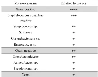
The number of bacteria present in an infected peritoneal dialysis fluid is extremely variable from one individual to another ranging between 1 and 106 CFU / mL of dialysate (4). Moreover, bacterial concentrations in the order of 1 to 10 CFU / mL have been demonstrated in asymptomatic patients with a leukocyte count of <100 mm3 in PD fluid (5). The positivity of a culture can thus be as much a reflection of a real infection as a contamination of the sample during its sampling or the analytical process (4). Added to this complexity must be the fact that the identity of many peritonitis causing organisms also belong to the commensal cutaneous, digestive or environmental flora (Table 1), thus often requiring repetitive sampling to confirm or deny their pathogenic character. The most frequently isolated organisms are Gram-positive cocci, with a predominance of coagulase-negative staphylococci, followed by the genus Streptococcus, Staphylococcus aureus, Corynebacterium sp., and Enterococcus sp (6). It should be noted that gram-positive cocci have a much lower PD fluid viability than Gram-negative bacteria, suggesting that negative-culture peritonitis is mainly related to the genus Staphylococcus. In the case of confirmed peritonitis, the number of coagulase-negative staphylococci contained in the PD fluid decreases by the fourth hour, becoming undetectable by 48 hours (7). This aspect may be explained at least in part by the presence of leukocytes and trace
antibioti in patient effluents (8). Among Gram-negative bacilli, Enterobacteriaceae predominate largely over Pseudomonas aeruginosa (6).
In addition to the so-called «classical» bacteria, certain micro-organisms responsible for peritonitis in patients treated with PD are difficult to detect and / or culture by conventional methods. Among these are tuberculous or atypical mycobacteria, and bacteria of the genera Nocardia, Actinomyces, Rhodococcus, Brucella, Campylobacter or Ureaplasma (9-16).
Faced with these various challenges, the microbiologist must find solutions adapted to each situation. Dialogue with the clinician is therefore essential for optimal patient management.
Table I Relative frequency of the main micro-organisms detected in peritonitis associated with peritoneal dialysis (from (6))
Micro-organism Relative frequency
Gram positive ++++ Staphylococcus coagulase negative +++ Streptococcus sp. ++ S. aureus + Corynebacterium sp. + Enterococcus sp. + Gram negative ++ Enterobacteriaceae ++ Acinetobacter sp. + Pseudomonas sp. + Yeast + DISCUSSION
For all the reasons mentioned above, it is necessary for the microbiologist to have sufficiently sensitive techniques to detect up to 1 CFU / mL and sufficiently reliable to eliminate, as much as possible, the risk of analytical contamination. Two approaches allow this: molecular biology and optimization of conventional bacterial culture methods.
The detection of bacterial DNA directly from PD fluid in the case of negative culture peritonitis was studied. Two approaches are proposed: pathogen-specific research via the use of targeted PCR, or so-called «universal» research (rDNA16s), a gene common to all bacteria, followed by sequencing for identification.
journal officiel du Registr e de D ialyse Péritonéale de Langue Française RDPLF www .rdplf.or g
technique, but only detects the target micro-organism. The role of this technique in PD-related peritonitis is therefore limited to tuberculous mycobacteria. The sensitivity of the 16S rDNA search technique is equivalent to that of conventional culture methods (17,18). Nevertheless, its use should be restricted to a limited number of cases for the following reasons; this approach does not allow the possibility of a follow up an antibiogram, essential to the optimization of antibiotic therapy, and is relatively long compared to culture techniques. It requires an amplification of the gene encoding 16S rRNA (1-3 days depending on the laboratory), followed by sequencing of the amplicon obtained for identification (2-5 days depending on the laboratory). In addition, this method does not allow the detection of yeasts. However, it may be useful for finding atypical micro-organisms, or when the patient has received antibiotic therapy prior to sampling (16,19,20).
The conventional culture of PD fluids remains the «gold standard» for the etiological diagnosis of PD peritonitis. Unfortunately, this approach can suffer from poor sensitivity. Direct examination of PD fluid after Gram staining, even after cytocentrifugation (a technique used for CRL), detects bacteria in less than 50% of cases (4). The sensitivity of the conventional culture, consisting of directly inoculating a portion of the PD fluid onto nutrient agar or into nutrient liquid media, has a sensitivity of the order of 50% (6). Optimization of this approach is thus essential to limit as much as possible the number of peritonitis with negative culture.
Several options have been considered to achieve this goal. The use of a large volume of DP fluid is recommended, to increase the chance of detection of microorganisms. However, the use of a large volume, generally of the order of 50 mL, followed by sedimentation (4-6 hours), or centrifugation, in order to artificially concentrate the bacteria, does not allow a real gain in sensitivity (8.21). Furthermore, the use of such techniques could delay the culture of the DP fluid, and therefore delay the determination of an antibiogram (8).
An alternative may be direct inoculation of 2 to 3 mL of PD fluid in liquid nutrient media and 1 mL onto nutrient agar media (22). In addition to the use of liquid media, the addition of phagocytic cell-lysing products has been shown to improve the sensitivity of the culture by about 10% (4). Today, the clinical microbiologist currently has technology that allows the use of a rich liquid medium containing products capable of lysing phagocytic cells and inhibiting the action of antibiotics.
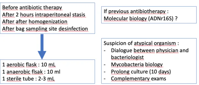
The use of blood culture flasks, an aerobic flask and an anaerobic flask, inoculated with PD liquid, increases sensitivity by 50 to 80% (6). The 2 vials are incubated for a minimum of 5 days and can be prolonged up to 10 days if necessary, bacterial growth proceeding automatically. The volume of PD fluid introduced into these flasks is important to note. A volume of 10 mL per vial allows a positivity of twice that obtained for a volume of 5 mL (23). Subcultures of positive vials are made on agar media supplemented with sheep blood and incubated aerobically and anaerobically. In the case where direct examination in the fresh state and after Gram staining reveals small Gram-negative bacilli, very mobile and curved, it is necessary to add an incubated medium in a microaerophilic atmosphere for detection of Campylobacter sp. (15). It should be noted that haemoculture flasks require rigorous antisepsis of the sampling site, in order to avoid contamination by environmental micro-organisms. The use of a sampling protocol is therefore essential to firstly reduce this risk of contamination and secondly to standardize practices (24). The recommendations for the bacteriological etiological diagnosis of peritonitis in patients treated with DP could be as follows (Figure 1): after dwell of PD fluid in the patient for 2 hours and, if possible, before antibiotic therapy is initiated, the collected fluid in the bag should be homogenized by mechanical action. Sampling of the PD fluid must be done at the level of the bag, previously rigourously disinfected. Two vials of blood cultures, aerobic and anaerobic should be inoculated with 10 mL of PD fluid. The vials must then be sent as quickly as possible to the laboratory for incubation. To this, it is necessary to add a sterile tube containing 2 to 3 ml of liquid of PD, used for leucocyte counting of the fluid, and possibly allowing the application of molecular biology techniques in case of negative culture or suspicion of atypical species. Finally, where atypical species are suspected, a clinico-biological discussion is essential in order to determine the need for additional examinations. Finally, it should not be forgotten that in some cases, negative culture peritonitis can be linked to other etiologies than bacterial ones: peritoneal inflammations with mesenteric ischemia, splenic infarction, drug causes (amphotericin B, vancomycin, dihydropyridines), excess acetaldehyde, formation of fibrin in the peritoneal cavity, presence of triglycerides, trauma to the lymphatic vessels during insertion of the catheter, etc. (25).
In conclusion, the etiological diagnosis of peritonitis in patients treated by PD may be difficult, but modern microbiology, via optimization of conventional culture
journal officiel du Registr e de D ialyse Péritonéale de Langue Française RDPLF www .rdplf.or g
and the use of molecular biology techniques, may allow identification of the causative organism of the infection
in the vast majority of cases. DISCLAIMERS No conflicts of interest
REFERENCES
1. Monteon-Ramos FJ, Correa-Rotter R, Paniagua R, Amato D, Hurtado ME, Medina JL, et al. Prevention of peritonitis with disconnect systems in CAPD: A randomized controlled trial. Kidney Int. 1998;54(6):2123–8. 2. Li PKT, Szeto CC, Piraino B, Bernardini J,
Figueiredo AE, Gupta A, et al. Peritoneal dialysis-related infections recommendations: 2010 update. Perit Dial Int. 2010;30(4):393– 423.
3. Li PKT, Szeto CC, Piraino B, de Arteaga J, Fan S, Figueiredo AE, et al. ISPD peritonitis recommendations: 2016 update on prevention and treatment. Perit Dial Int. 2016; 36:481– 508.
4. von Graevenitz a, Amsterdam D. Microbiological aspects of peritonitis associated with continuous ambulatory peritoneal dialysis. Clin Microbiol Rev.
1992;5(1):36–48.
5. Rubin J, Rogers WA, Taylor HM, Everett ED, Prowant BF, Fruto L V., et al. Peritonitis during continuous ambulatory peritoneal dialysis. Ann Intern Med. 1980;92(1):7–13.
6. Yoon SH, Choi NW, Yun SR. Detecting bacterial growth in continuous ambulatory peritoneal dialysis effluent using two culture methods. Korean J Intern Med. 2010;25(1):82– 5.
7. Sheth NK, Bartell CA, Roth DA. In vitro study of bacterial growth in continuous ambulatory peritoneal dialysis fluids. J Clin Microbiol. 1986;23(6):1096–8
8. Katzap RM, Pagnussatti VE, Figueiredo AE, Motta JG, D’Avila DO, da Costa BEP, et al. Time to positivity of bacteria cultures in peritoneal dialysis fluid: Evaluation of different laboratory techniques. Perit Dial Int. 2017;37(3):342–4.
9. Jiang SH, Senanayake S, Talaulikar GS. Peritoneal dialysis-related peritonitis due to Mycobacterium smegmatis. Perit Dial Int. 2011; 31: 215-6.
10. Song Y, Wu J, Yan H, Chen J. Peritoneal dialysis-associated nontuberculous
mycobacterium peritonitis: A systematic review of reported cases. Nephrol Dial Transplant.
journal officiel du Registr e de D ialyse Péritonéale de Langue Française RDPLF www .rdplf.or g
Figure 1. Proposal for management of bacteriological specimens for suspicion of peritonitis in patients treated with peritoneal dialysis
11. Varughese S, Bargman J. Actinomyces neuii PD peritonitis ? resolution of infection without catheter removal. Perit Dial Int. 2014; 34: 815-6.
12. Kendrick-Jones J, Ratanjee SK, Taylor SL, Marshall MR. Nocardia asteroides peritoneal dialysis-related peritonitis: A case of successful treatment and return to peritoneal dialysis. Nephrol Dial Transplant. 2008;23(8):2693–4. 13. Azzam O, Crowe A, Sajiv C, Pawar B.
Rhodococcus equi peritonitis in continuous ambulatory peritoneal dialysis: A first in Australia. BMJ Case Rep. 2015;2015.
14. Koz S, Sahin I, Kayabas U, Kuzucu C. Brucella and peritoneal dialysis related peritonitis: case report and review of literature. Clin Nephrol. 2014;82(4):283–6.
15. Ma TK wing, Lee KP, Chow KM, Pang WF, Ha Kwan BC, Leung CB, et al. Campylobacter peritonitis complicating peritoneal dialysis: A review of 12 consecutive cases. Perit Dial Int. 2013;33(2):189–94.
16. Yager JE, Ford ES, Boas ZP, Haseley LA, Cookson BT, SenGupta DJ, et al. Ureaplasma urealyticum continuous ambulatory peritoneal dialysis-associated peritonitis diagnosed by 16S rRNA gene PCR. J Clin Microbiol. 2010;48(11):4310–2.
17. Ahmadi SH, Neela V, Hamat RA, Goh BL, Syafinaz AN. Rapid detection and identification of pathogens in patients with continuous ambulatory peritoneal dialysis (CAPD) associated peritonitis by 16s rRNA gene sequencing. Trop Biomed. 2013;30(4):602–7.
Isolation of bacterial DNA followed by sequencing and differing cytokine response in peritoneal dialysis effluent help in identifying bacteria in culture negative peritonitis. Nephrology. 2018;23(2):148–54.
19. Yeo SH, Kwak JH, Kim YU, Lee JS, Kim HJ, Park KH, et al. Peritoneal dialysis-related peritonitis due to Halomonas hamiltonii A first case report. Med (United States). 2016;95(47). 20. Matsukuma Y, Sugawara K, Shimano S,
Yamada S, Tsuruya K, Kitazono T, et al. A case of bacterial peritonitis caused by Roseomonas mucosa in a patient undergoing continuous ambulatory peritoneal dialysis. CEN Case Reports. 2013;3(2):127–31.
21. Hächler H, von Graevenitz A, Vogt K, Binswanger U. Centrifugation of 50 ml of peritoneal fluid is sufficient for microbiological examination in continuous ambulatory peritoneal dialysis (CAPD) patients with peritonitis. Infection. 1986;14(3):102–4.
22. DIAGNOSIS AND MANAGEMENT OF PERITONITIS IN CONTINUOUS AMBULATORY PERITONEAL DIALYSIS. Report of a Working Party of the British Society for Antimicrobial Chemotherapy. Lancet. 1987;329(8537):845–9.
23. Chang J, Park JS, Park S, Choi B, Yoon NS, Sung H, et al. Impact of monitoring blood volume in the BD BACTECTM FX blood culture system:
Virtual volume versus actual volume. Diagn Microbiol Infect Dis. 2015;81(2):89–93. 24. Kocyigit I, Unal A, Karademir D, Bahcebasi S,
Sipahioglu MH, Tokgoz B, et al. Improvement in culture-negative peritoneal dialysis-related peritonitis: A single center’s experience. Perit Dial Int. 2012;32(4):476–8.
25. de Freitas DG, Gokal R. Sterile peritonitis in the peritoneal dialysis patient. Perit Dial Int. 2005;25:146–51. journal officiel du Registr e de D ialyse Péritonéale de Langue Française RDPLF www .rdplf.or g